Note : Les descriptions sont présentées dans la langue officielle dans laquelle elles ont été soumises.
CA 02704400 2010-04-30
WO 2009/059197 PCT/US2008/082075
07-00194-01 WO
AUTOMATED FITTING SYSTEM FOR DEEP BRAIN STIMULATION
FIELD OF THE INVENTION
The invention relates to the treatment of movement disorders, and more
particularly, to deep brain stimulation (DBS) systems and methods.
BACKGROUND OF THE INVENTION
Implantable neurostimulation systems have proven therapeutic in a wide variety
of diseases and disorders. Pacemakers and Implantable Cardiac Defibrillators
(ICDs)
have proven highly effective in the treatment of a number of cardiac
conditions (e.g.,
arrhythmias). Spinal Cord Stimulation (SCS) systems have long been accepted as
a
therapeutic modality for the treatment of chronic pain syndromes, and the
application of
tissue stimulation has begun to expand to additional applications, such as
angina
pectoris and incontinence. Further, in recent investigations, Peripheral Nerve
Stimulation (PNS) systems have demonstrated efficacy in the treatment of
chronic pain
syndromes and incontinence, and a number of additional applications are
currently
under investigation. More pertinent to the invention described herein, Deep
Brain
Stimulation (DBS) has been applied therapeutically for well over a decade for
the
treatment of neurological disorders, including Parkinson's Disease, essential
tremor,
dystonia, and epilepsy, to name but a few. Further details discussing the
treatment of
diseases using DBS are disclosed in U.S. Patent Nos. 6,845,267, 6,845,267, and
6,950,707.
Each of these implantable neurostimulation systems typically includes one or
more electrode carrying stimulation leads, which are implanted at the desired
stimulation site, and a neurostimulator implanted remotely from the
stimulation site, but
CA 02704400 2010-04-30
WO 2009/059197 PCT/US2008/082075
07-00194-01 WO
coupled either directly to the stimulation lead(s) or indirectly to the
stimulation lead(s)
via a lead extension. The neurostimulation system may further comprise a
handheld
remote control (RC) to remotely instruct the neurostimulator to generate
electrical
stimulation pulses in accordance with selected stimulation parameters. The RC
may,
itself, be programmed by a technician attending the patient, for example, by
using a
Clinician's Programmer (CP), which typically includes a general purpose
computer,
such as a laptop, with a programming software package installed thereon.
Thus, in accordance with the stimulation parameters programmed by the RC
and/or CP, electrical pulses can be delivered from the neurostimulator to the
stimulation
electrode(s) to stimulate or activate a volume of tissue in accordance with a
set of
stimulation parameters and provide the desired efficacious therapy to the
patient. The
best stimulus parameter set will typically be one that delivers stimulation
energy to the
volume of tissue that must be stimulated in order to provide the therapeutic
benefit (e.g.,
treatment of movement disorders), while minimizing the volume of non-target
tissue that
is stimulated. A typical stimulation parameter set may include the electrodes
that are
acting as anodes or cathodes, as well as the amplitude, duration, and rate of
the
stimulation pulses.
When a neurostimulation system is implanted within a patient, a fitting
procedure
is typically performed to ensure that the stimulation leads and/or electrodes
are properly
implanted in effective locations of the patient, as well as to select one or
more effective
sets of stimulation parameters for the patient. In some electrical stimulation
treatments,
the fitting procedure may be effectively directed in response to patient
feedback. For
example, in SCS for providing pain relief, patients can feel the effects of
the stimulation
2
CA 02704400 2010-04-30
WO 2009/059197 PCT/US2008/082075
07-00194-01 WO
pulses and the change in their pain status, and thus, may provide verbal
feedback as to
the efficacy of the stimulation, and thus, the proper location of the
stimulation leads
and/or electrodes and the stimulation parameters to be used in delivering the
electrical
pulses to the patient on a long-term basis.
Unlike with SCS, patients receiving DBS cannot feel the effects of
stimulation,
and the effects of the stimulation may be difficult to observe, are typically
subjective, or
otherwise may take a long time to become apparent. This makes it difficult to
set the
stimulation parameters appropriately or otherwise select stimulation
parameters that
result in optimal treatment for the patient and/or optimal use of the
stimulation
resources. Significantly, non-optimal electrode placement and stimulation
parameter
selections may result in excessive energy consumption due to stimulation that
is set at
too high an amplitude, too wide a pulse width, or too fast a frequency;
inadequate or
marginalized treatment due to stimulation that is set at too low an amplitude,
too narrow
a pulse width, or too slow a frequency; or stimulation of neighboring cell
populations that
may result in undesirable side effects. All of these issues are poorly
addressed by the
present-day DBS fitting techniques. In addition, after the DBS system has been
implanted and fitted, the patient may have to schedule another visit to the
physician in
order to adjust the stimulation parameters of the DBS system if the treatment
provided
by the implanted DBS system is no longer effective or otherwise is not
therapeutically or
operationally optimum due to, e.g., disease progression, motor re-learning, or
other
changes.
While DBS systems have been disclosed that utilize a closed-loop method that
involves sensing electrical signals within the brain of the patient and
automatically
3
CA 02704400 2010-04-30
WO 2009/059197 PCT/US2008/082075
07-00194-01 WO
adjusting the electrical stimulation delivered to a target region within the
brain of the
patient (see, e.g., U.S. Patent No. 5,683,422), such a system requires the
implantation
of an additional lead within the brain. In addition, the electrical signals
sensed within the
brain are not easily correlatable to the disorder currently experienced by the
patient.
Furthermore, such a system is not designed to be used in a fitting procedure,
including
physical adjustment of the leads and programming of the stimulation
parameters.
There, thus, remains a need for a DBS system that can be more easily fitted to
a
patient in order to optimize treatment of a patient suffering from a disease.
SUMMARY OF THE INVENTION
A method of providing therapy to a patient having a dysfunction is provided
for
purposes of better understanding the invention. In one method, the dysfunction
is a
motor dysfunction (e.g., a gait dysfunction, posture dysfunction, balance
dysfunction,
motor control dysfunction, speech dysfunction, etc.), and may be caused by
neurological disorder, such as Parkinson's Disease, essential tremor,
dystonia,
epilepsy, etc. The method comprises conveying stimulation energy from a
neurostimulator to at least one implanted electrode located within a tissue
region of the
patient, thereby changing the status of the dysfunction. The tissue region may
be
located anywhere in the patient's body, but in the preferred method, is
located in the
brain where motor dysfunctions often originate. The method further comprises
measuring a physiological end-function of the patient indicative of the
changed status of
the dysfunction, and programming at least one stimulation parameter into the
neurostimulator based on the measured physiological end-function. The measured
physiological end-function may be, e.g., a kinematic function, an electrical
muscle
4
CA 02704400 2010-04-30
WO 2009/059197 PCT/US2008/082075
07-00194-01 WO
impulse, a speech pattern, etc., and the stimulator parameter(s) may be, e.g.,
a pulse
amplitude (including the relative amplitudes of current or voltage through
electrodes of
like polarity), pulse width, pulse rate, or electrode combination. In one
method, the
physiological end-function is non-invasively measured.
One method further comprises conveying stimulation energy from the
neurostimulator to the tissue region of the patient in accordance with the
stimulation
parameter(s), thereby improving the status of the dysfunction. Another method
further
comprises quantifying the dysfunction based on the measured physiological end-
function, in which case, the stimulation parameter(s) may be programmed into
the
neurostimulator based on the quantified dysfunction. Still another method
further
comprises automatically determining the stimulation parameter(s) in response
to the
measured physiological end-function. The automatic determination of the
stimulation
parameter(s) may be performed in any one of a variety manners, e.g.,
heuristically or by
correlating the measured physiological end-function to a predetermined data
set. The
method may optionally comprise implanting the neurostimulator into the
patient.
In accordance with a second aspect of the invention, a neurostimulation system
is provided. The neurostimulation system comprises at least one electrical
terminal,
output stimulation circuitry configured for outputting stimulation energy to
the electrical
terminal(s), control circuitry configured for controlling the stimulation
energy output by
the output stimulation circuitry, monitoring circuitry configured for
measuring a
physiological end-function of a patient indicative of a changed status of a
dysfunction of
a patient, and processing circuitry configured for programming the control
circuitry with
at least one stimulation parameter based on the measured physiological end-
function.
5
CA 02704400 2010-04-30
WO 2009/059197 PCT/US2008/082075
07-00194-01 WO
The dysfunction, measured physiological end-function, and stimulation
parameter(s)
may be the same as those described above.
In one embodiment, the monitoring circuitry is configured for non-invasively
measuring the physiological end-function. In another embodiment, the
processing
circuitry is configured for programming the control circuitry with the
stimulation
parameter(s) to improve the status of the dysfunction when the output
stimulation
circuitry outputs the stimulation energy to the electrical terminal(s). In
still another
embodiment, the monitoring circuitry is configured for quantifying the
dysfunction based
on the measured physiological end-function, in which case, the processing
circuitry may
be configured for programming the stimulation parameter(s) into the control
circuitry
based on the quantified dysfunction. In still another embodiment, the
processing
circuitry is configured for automatically determining the stimulation
parameter(s) in
response to the measured physiological end-function, e.g., in the manner
discussed
above. In yet another embodiment, the system further comprises telemetry
circuitry
configured for wirelessly conveying the stimulation parameter(s) from the
processing
circuitry to the control circuitry. An optional embodiment may comprise a case
containing the electrical terminal(s), output stimulation circuitry, and
control circuitry to
form a neurostimulator, e.g., an implantable neurostimulator. The monitoring
circuitry
and the processing circuitry may be contained in one or more computers.
In accordance with a third aspect of the invention, an external programmer for
a
neurostimulator is provided. The external programmer comprises input circuitry
configured for receiving information indicative of a changed status of a
dysfunction of a
patient. The information may be, e.g., a measured physiological end-function
or a
6
CA 02704400 2010-04-30
WO 2009/059197 PCT/US2008/082075
07-00194-01 WO
quantified dysfunction, the details of which are discussed above. The
programmer
further comprises processing circuitry configured for automatically
determining at least
one programmable stimulation parameter based on the received information, and
output
circuitry configured for transmitting the programmable stimulation parameter
to the
neurostimulator. The programmable stimulation parameter(s) may be the same as
those discussed above, and the programmable stimulation parameter(s) may be
determined in the same manner described above. In one embodiment, the
processing
circuitry is configured for defining the programmable stimulation
parameter(s), such that
the status of the dysfunction is improved when stimulation energy is delivered
to the
patient in accordance with the programmable stimulation parameter(s). In
another
embodiment, the output circuitry comprises telemetry circuitry, and the input
circuitry,
processing circuitry, and output circuitry are contained in a single case.
In accordance with a fourth aspect of the invention, a method of providing
therapy to a patient having a dysfunction is provided for purposes of better
understanding the invention. In one method, the dysfunction is a motor
dysfunction
(e.g., a gait dysfunction, posture dysfunction, balance dysfunction, motor
control
dysfunction, speech dysfunction, etc.), and may be caused by neurological
disorder,
such as Parkinson's Disease, essential tremor, dystonia, epilepsy, etc. The
method
comprises placing at least one electrode adjacent to a tissue region of the
patient, and
conveying stimulation energy from the electrode(s) to the tissue region in
accordance
with at least one stimulation parameter (e.g., a pulse amplitude, pulse width,
pulse rate,
electrode combination, etc.), thereby changing the status of the dysfunction.
The tissue
region may be located anywhere in the patient's body, but in the preferred
method, is
7
CA 02704400 2010-04-30
WO 2009/059197 PCT/US2008/082075
07-00194-01 WO
located in the brain where dysfunctions often originate. The method further
comprises
measuring a physiological end-function of the patient indicative of the
changed status of
the dysfunction, and automatically adjusting the stimulation parameter(s)
based on the
measured physiological end-function. The measured physiological end-function
may
be, e.g., a kinematic function, an electrical muscle impulse, a speech
pattern, etc. In
one method, the physiological end-function is non-invasively measured.
One method comprises quantifying the dysfunction based on the measured
physiological end-function, in which case, the stimulation parameter(s) may be
automatically adjusted based on the quantified dysfunction. In another method,
the
stimulation parameter(s) are automatically adjusted to improve the status of
the
dysfunction. For example, a value of the stimulation parameter(s) may be
adjusted in
one direction if the measured physiological end-function indicates an
improvement in
the status of the dysfunction, and may be adjusted in another direction if the
measured
physiological end-function indicates a degradation in the status of the
dysfunction. Still
another method comprises conveying stimulation energy from the electrode(s) to
the
tissue region in accordance with the adjusted stimulation parameter(s),
thereby
changing the status of the dysfunction. Yet another method comprises
implanting the
neurostimulator within the patient, coupling the electrode(s) to the
neurostimulator, and
programming the neurostimulator with the adjusted stimulation parameter(s).
In accordance with a fifth aspect of the invention, a neurostimulation system
is
provided. The neurostimulation system comprises at least one electrical
terminal,
output stimulation circuitry configured for outputting stimulation energy to
the electrical
terminal(s) in accordance with at least one stimulation parameter, monitoring
circuitry
8
CA 02704400 2010-04-30
WO 2009/059197 PCT/US2008/082075
07-00194-01 WO
configured for measuring a physiological end-function of a patient indicative
of a
changed status of a dysfunction of a patient, and processing circuitry
configured for
adjusting the stimulation parameter(s) based on the measured physiological end-
function. The dysfunction, measured physiological end-function, and
stimulation
parameter(s) may be the same as those described above.
In one embodiment, the monitoring circuitry is further configured for
quantifying
the dysfunction based on the measured physiological end-function, in which
case, the
processing circuitry may be configured for automatically adjusting the
stimulation
parameter(s) based on the quantified dysfunction. In another embodiment, the
processing circuitry is configured for automatically adjusting the stimulation
parameter(s) to improve the status of the dysfunction; for example, in the
manner
described above. In still another embodiment, the system further comprises a
stimulation lead carrying at least one electrode electrically coupled to the
at least one
electrical terminal. In yet another embodiment, the system further comprises
telemetry
circuitry, in which case, the processing circuitry is configured for
wirelessly adjusting the
stimulation parameter(s). An optional embodiment may comprise a case
containing the
electrical terminal(s), output stimulation circuitry, and control circuitry to
form a
neurostimulator, e.g., an implantable neurostimulator. The monitoring
circuitry and the
processing circuitry may be contained in one or more computers.
In accordance with a sixth aspect of the invention, an external programmer for
a
neurostimulator is provided. The external programmer comprises input circuitry
configured for receiving information indicating a status of a dysfunction of a
patient,
processing circuitry configured for automatically adjusting at least one
stimulation
9
CA 02704400 2010-04-30
WO 2009/059197 PCT/US2008/082075
07-00194-01 WO
parameter based on the received information, and output circuitry configured
for
transmitting the adjusted stimulation parameter(s) to the neurostimulator. The
received
information may be, e.g., a measured physiological end-function or a
quantified
dysfunction, the details of which are discussed above. The programmable
stimulation
parameter(s) may be the same as those discussed above, and the programmable
stimulation parameter(s) may be determined in the same manner described above.
In
one embodiment, the processing circuitry is configured for automatically
adjusting the at
least one stimulation parameter to improve the status of the dysfunction; for
example, in
the same manner described above. In another embodiment, the output circuitry
is
telemetry circuitry, and the input circuitry, processing circuitry, and output
circuitry are
contained in a single case.
BRIEF DESCRIPTION OF THE DRAWINGS
The drawings illustrate the design and utility of preferred embodiments of the
invention, in which similar elements are referred to by common reference
numerals. In
order to better appreciate how the above-recited and other advantages and
objects of
the present inventions are obtained, a more particular description of the
present
inventions briefly described above will be rendered by reference to specific
embodiments thereof, which are illustrated in the accompanying drawings.
Understanding that these drawings depict only typical embodiments of the
invention and
are not therefore to be considered limiting of its scope, the invention will
be described
and explained with additional specificity and detail through the use of the
accompanying
drawings in which:
CA 02704400 2010-04-30
WO 2009/059197 PCT/US2008/082075
07-00194-01 WO
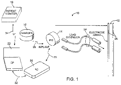
Fig. 1 is a plan view of a Deep Brain Stimulation (DBS) system constructed in
accordance with one embodiment of the invention;
Fig. 2 is a block diagram of the internal components of an implantable pulse
generator (IPG) used in the DBS system of Fig. 1;
Fig. 3 is front view of a remote control (RC) used in the DBS system of Fig.
1;
Fig. 4 is a block diagram of the internal components of the RC of Fig. 3;
Fig. 5 is a block diagram of the internal components of a clinician's
programmer
(CP) used in the DBS system of Fig. 1;
Fig. 6 is a flow diagram illustrating a method of programming the IPG of Fig.
2
using the RC of Figs. 3 and 4 or the CP of Fig. 5; and
Fig. 7 is a cross-sectional view of a patient's head showing the implantation
of
stimulation leads and an IPG of the DBS system of Fig. 1.
DETAILED DESCRIPTION OF THE EMBODIMENTS
At the outset, it is noted that the present invention may be used with an
implantable pulse generator (IPG), radio frequency (RF) transmitter, or
similar
neurostimulator, that may be used as a component of numerous different types
of
stimulation systems. The description that follows relates to a Deep Brain
Stimulation
(DBS) system. However, it is to be understood that, while the invention lends
itself well
to applications in DBS, the invention, in its broadest aspects, may not be so
limited.
Rather, the invention may be used with any type of implantable electrical
circuitry used
to stimulate tissue for the treatment of a dysfunction, such as, e.g., a motor
dysfunction.
Turning first to Fig. 1, an exemplary DBS system 10 constructed in accordance
with one embodiment of the invention generally includes one or more (in this
case, two)
11
CA 02704400 2010-04-30
WO 2009/059197 PCT/US2008/082075
07-00194-01 WO
implantable stimulation leads 12, an implantable pulse generator (IPG) 14 (or
alternatively RF receiver-stimulator), an external charger 16, a patient
monitor 18, an
external remote controller (RC) 20, and a clinician's programmer (CP) 24.
The IPG 14 is physically connected via one or more lead extensions 24 to the
stimulation leads 12, which carry a plurality of electrodes 26 arranged in an
array. In
the illustrated embodiment, the electrodes 26 are arranged in-line along the
stimulation
leads 12. In the illustrated embodiment, each stimulation lead 12 carries
eight
electrodes 26. Of course, other numbers of electrodes can be carried by each
stimulation lead 12, e.g., two, four, six, etc., and any number of stimulation
leads 12 can
be used, including a single lead. The IPG 14 comprises an outer case for
housing the
electronic and other components (described in further detail below), and a
connector
(not shown) in which the proximal end of the lead extension 24 mates with the
IPG 14,
which then at its distal end has a connector which mates with the stimulation
lead 12
mates in a manner that electrically couples the electrodes 26 to the
electronics within
the outer case. The outer case is composed of an electrically conductive,
biocompatible
material, such as titanium, and forms a hermetically sealed compartment,
wherein the
internal electronics are protected from the body tissue and fluids. In some
cases, the
outer case serves as an electrode, as will be described in further detail
below.
As will be described in further detail below, the IPG 14 includes pulse
generation
circuitry that delivers the electrical stimulation energy to the electrodes 26
in accordance
with a set of stimulation parameters. Such stimulation parameters may comprise
electrode combinations, which define the electrodes that are activated as
anodes
(positive), cathodes (negative), and turned off (zero), and electrical pulse
parameters,
12
CA 02704400 2010-04-30
WO 2009/059197 PCT/US2008/082075
07-00194-01 WO
which define the pulse amplitude (measured in milliamps or volts depending on
whether
the IPG 14 supplies constant current or constant voltage to the electrodes
26), pulse
width (measured in microseconds), and pulse rate (measured in pulses per
second).
Electrical stimulation will occur between two (or more) activated electrodes,
one of
which may be the IPG case. Simulation energy may be transmitted to the tissue
in a
monopolar manner; that is, between one of the electrodes 26 and the IPG case,
or
multipolar manner (e.g., bipolar, tripolar, etc.); that is, between two or
more of the
electrodes 26.
The external charger 16 is a portable device used to transcutaneously charge
the
IPG 14 via an inductive link 28. For purposes of brevity, the details of the
external
charger 24 will not be described herein. Details of exemplary embodiments of
external
chargers are disclosed in U.S. Patent No. 6,895,280.
The patient monitor 18 is used to measure a physiological end-function
indicative
of the changed status of the dysfunction from which the patient suffers. For
the
purposes of this specification, a physiological end-function is a
physiological function
that manifests itself outside of the brain. The physiological end-function is
preferably
measured using a non-invasive means (i.e., without having to create an opening
within
the patient) or otherwise a means that does not require penetration into the
patient's
brain. Various non-invasive means for measuring the physiological end-function
are
described in further detail below. Alternatively, the physiological end-
function may be
invasively measured. The measured physiological end-function may be, e.g., a
kinematic action, an electrical muscle impulse, or a speech pattern. The
dysfunction
may be a motor dysfunction, e.g., a gait dysfunction, posture dysfunction,
balance
13
CA 02704400 2010-04-30
WO 2009/059197 PCT/US2008/082075
07-00194-01 WO
dysfunction, motor control dysfunction (e.g., spasticity, bradykinesia,
rigidity), a speech
impediment, etc., which may be caused by any one of a variety of diseases,
including
Parkinson's Disease, essential tremor, dystonia, and epilepsy. The dysfunction
may
also be a non-motor dysfunction, e.g., psychological, hormonal, etc. The
patient monitor
18 may optionally quantify the dysfunction based on the measured physiological
end-
function; for example, by assigning a numerical value to the dysfunction
(e.g., from 1 to
10, with 1 meaning that the dysfunction is non-existent and 10 meaning that
the
dysfunction is extreme). As will be described in further detail below, the
measured
physiological end-function or quantified dysfunction information can be used
to adjust
the stimulation parameters in accordance with which the stimulation energy is
delivered
from the IPG 14.
The patient monitor 18 may be physically located in a clinical setting where
direct
physician/assistant control may be exercised under control conditions, or may
be
located with the patient at a remote setting to allow more limited and/or
gradual
adjustment of the stimulation parameters. Thus, the patient monitor 18 can be
utilized
at any time during the treatment continuum to record pre-implant performance,
post-
implant performance, and follow-up adjustment opportunities.
The RC 20 may be used to telemetrically control the IPG 14 via a bi-
directional
RF communications link 30 by transmitting stimulation parameters to the IPG 14
or
otherwise adjusting the stimulation parameters stored in the IPG 14. Such
control
allows the IPG 14 to be turned on or off and to be programmed with different
stimulation
programs after implantation. Once the IPG 14 has been programmed, and its
power
14
CA 02704400 2010-04-30
WO 2009/059197 PCT/US2008/082075
07-00194-01 WO
source has been charged or otherwise replenished, the IPG 14 may function as
programmed without the RC 20 being present.
The CP 22 provides clinician-specified stimulation parameters for programming
the IPG 14 in the operating room and in follow-up sessions. The CP 22 may
perform
this function by communicating with the RC 20 via an IR communications link 32
to
indirectly program the IPG 14 with the stimulation parameters. The CP 22 may,
at the
same time, program the RC 20 with the stimulation parameters, so that the RC
20 can
subsequently program or otherwise control the IPG 14 using the stimulation
parameters
programmed into the RC 20. Alternatively, the CP 22 may directly program the
stimulation parameters into the IPG 14 via an RF communications link (not
shown)
without the aid of the RC 20.
Significantly, the CP 22 may operate in a manual mode or an automated mod. In
a manual mode, the CP 22 can be used to program stimulation parameters into
the IPG
14 in a conventional manner. In the automated mode, the CP 22 can be used to
automatically program stimulation parameters into the IPG 14. In particular,
the CP 22
can automatically determine the stimulation parameters to be programmed into
the IPG
14 based on the physiological end-function measured by the patient monitor 18.
To this
end, the CP 22 may receive measured physiological end-function information
from the
patient monitor 18 via an IR communications link 34. Alternatively, the CP 22
may be
coupled to the patient monitor 18 via a cable (not shown). If the patient
monitor 18
quantifies the dysfunction based on the measured physiological end-functions,
the CP
22 may receive the quantified dysfunction information from the patient monitor
18 via
the IR communications link 34, and automatically determine the programmed
CA 02704400 2010-04-30
WO 2009/059197 PCT/US2008/082075
07-00194-01 WO
stimulation parameters based on the quantified dysfunction information.
Alternatively,
the CP 22, itself, may quantify the dysfunction based on the measured
physiological
end-function information received from the patient monitor 18. Notably, the CP
22 may
automatically determine the stimulation parameters to be programmed into the
IPG 14
without user intervention, or may, e.g., provide suggested stimulation
parameters, which
can be selected by the clinician to ultimately adjust the stimulation
parameters
programmed into the IPG 14. In any event, the programmed stimulation
parameters
determined by the CP 22 are intended to improve the status of the dysfunction
suffered
by the patient.
For example, the CP 22 may control the stimulation energy output by the IPG 14
by adjusting the stimulation parameters in the IPG 14. The patient monitor 18
may
measure the physiological end-function of the patient again to determine the
effect that
the adjustment of the stimulation parameters had on the dysfunction. This
process can
be repeated until optimized or otherwise effective or improved stimulation
parameters
are determined, which can then be programmed into the IPG 14. Any delay
between
the change in the stimulation parameters and the measurement of the
physiological
end-functions would be controlled and would be affected by the type of
dysfunction,
physical condition of the patient, the effects of any drugs, etc., allowing
the changes in
stimulation to take effect before another measurement of physiological end-
functions is
performed again. Changes due to disease progression, motor re-learning, or
other
changes that effect the status of the dysfunction can be triggered for re-
evaluation of the
stimulation parameters programmed into the IPG 14.
16
CA 02704400 2010-04-30
WO 2009/059197 PCT/US2008/082075
07-00194-01 WO
The RC 20 can be operated in a manual mode that allows a patient to program
stimulation parameters into the IPG 14 in a conventional manner. In
alternative
embodiments, wherein the patient monitor 18 is located within the patient in a
remote
setting, the RC 20 may operated in an automated mode in which it automatically
determines the stimulation parameters to be programmed into the IPG 14 based
on the
physiological end-function measured by the patient monitor 18 or the
dysfunction
quantified by the patient monitor 18, in which case, the RC 20 may be coupled
to the
patient monitor 18 via an IR communications link (not shown).
The CP 22, or alternatively the RC 20, may determine the improved stimulation
parameters based on the measured physiological end-function or quantified
dysfunction
in any one of a variety of manners to improve the status of the dysfunction.
In one
embodiment, the stimulation parameters are adjusted using a heuristic
approach.
For example, a value of at least one of the stimulation parameters may be
incrementally adjusted in one direction (e.g., increasing the pulse amplitude,
pulse
width, or pulse rate) if the measured physiological end-function indicates an
improvement in the status of the dysfunction, and incrementally adjusted in
another
direction (e.g., decreasing the pulse amplitude, pulse width, or pulse rate)
if the
measured physiological end-function indicates a degradation in the status of
the
dysfunction. The value of the stimulation parameters may be incrementally
adjusted in
the one direction until the measured physiological end-function indicates no
further
improvement in the status of the dysfunction or until a parameter limit is
reached.
These stimulation parameters can then be selected as the stimulation
parameters to be
programmed into the IPG 14.
17
CA 02704400 2010-04-30
WO 2009/059197 PCT/US2008/082075
07-00194-01 WO
As another example, different combinations of electrodes may be selected that
improve the status of the dysfunction. In one embodiment, the stimulation
energy may
be gradually steered up or down the leads 12. That is, the stimulation energy
may be
gradually steered in one direction if the measured physiological end-function
indicates
an improvement in the status of the dysfunction, and gradually steered in
another
direction if the measured physiological end-function indicates a degradation
in the
status of the dysfunction. The improved stimulation parameters, and in this
case, the
electrode combination, resulting from this process can then be programmed into
the
IPG 14. Details regarding the steering of stimulation energy amongst
electrodes are
further disclosed in U.S. Patent No. 6,052,624.
In another embodiment, the improved stimulation parameters may be determined
by correlating the measured physiological end-functions to a desired
performance, and
with knowledge of past performance and the operational constraints of the IPG
14,
determining the stimulation parameters to be programmed into the IPG 14. For
instance, normative data for a physiological end-function may be known in the
literature
and used as a reference for improving the performance of the patient by
adjustment of
stimulation parameters as described above. Furthermore, past patient
physiological
performance profiles may be recorded in a database for the patient and
compared to for
the adjustment methods. An example of this could be gait performance coupled
with
energy consumption in which speed of gait, stride length, cadence, and joint
excursions
coupled with the energy utilized (as measured by oxygen uptake) could be used
act as
a reference for future stimulation parameter adjustments.
18
CA 02704400 2010-04-30
WO 2009/059197 PCT/US2008/082075
07-00194-01 WO
Turning next to Fig. 2, the main internal components of the IPG 14 will now be
described. The IPG 14 includes analog output circuitry 60 capable of
individually
generating electrical stimulation pulses via capacitors C1-C16 at the
electrodes 26
(designated E1-E16) of specified amplitude under control of control logic 62
over data
bus 64. The duration of the electrical stimulation (i.e., the width of the
stimulation
pulses), is controlled by the timer logic circuitry 66. The analog output
circuitry 60 may
either comprise independently controlled current sources for providing
stimulation
pulses of a specified and known amperage to or from the electrodes 26, or
independently controlled voltage sources for providing stimulation pulses of a
specified
and known voltage at the electrodes 26 or to multiplexed current or voltage
sources that
are then connected to the electrodes 26. The operation of this analog output
circuitry,
including alternative embodiments of suitable output circuitry for performing
the same
function of generating stimulation pulses of a prescribed amplitude and width,
is
described more fully in U.S. Patent Nos. 6,516,227 and 6,993,384.
The IPG 14 further comprises monitoring circuitry 68 for monitoring the status
of
various nodes or other points 70 throughout the IPG 14, e.g., power supply
voltages,
temperature, battery voltage, and the like. The monitoring circuitry 68 is
also configured
for measuring electrical parameter data (e.g., electrode impedance and/or
electrode
field potential). The IPG 14 further comprises processing circuitry in the
form of a
microcontroller (pC) 72 that controls the control logic 62 over data bus 74,
and obtains
status data from the monitoring circuitry 68 via data bus 76. The IPG 14
additionally
controls the timer logic 56. The IPG 14 further comprises memory 78 and
oscillator and
clock circuit 80 coupled to the pC 72. The pC 72, in combination with the
memory 78
19
CA 02704400 2010-04-30
WO 2009/059197 PCT/US2008/082075
07-00194-01 WO
and oscillator and clock circuit 80, thus comprise a microprocessor system
that carries
out a program function in accordance with a suitable program stored in the
memory 78.
Alternatively, for some applications, the function provided by the
microprocessor system
may be carried out by a suitable state machine.
Thus, the iC 72 generates the necessary control and status signals, which
allow
the pC 72 to control the operation of the IPG 14 in accordance with a selected
operating
program and stimulation parameters. In controlling the operation of the IPG
14, the pC
72 is able to individually generate stimulus pulses at the electrodes 26 using
the analog
output circuitry 60, in combination with the control logic 62 and timer logic
66, thereby
allowing each electrode 26 to be paired or grouped with other electrodes 26,
including
the monopolar case electrode, to control the polarity, amplitude, rate, pulse
width and
channel through which the current stimulus pulses are provided. The pC 72
facilitates
the storage of electrical parameter data measured by the monitoring circuitry
68 within
memory 78.
The IPG 14 further comprises a receiving coil 82 for receiving programming
data
(e.g., the operating program and/or stimulation parameters) from the external
programmer (i.e., the RC 20 or CP 22) in an appropriate modulated carrier
signal, and
charging, and circuitry 84 for demodulating the carrier signal it receives
through the
receiving coil 82 to recover the programming data, which programming data is
then
stored within the memory 78, or within other memory elements (not shown)
distributed
throughout the IPG 14.
The IPG 14 further comprises back telemetry circuitry 86 and a transmission
coil
88 for sending informational data to the external programmer. The back
telemetry
CA 02704400 2010-04-30
WO 2009/059197 PCT/US2008/082075
07-00194-01 WO
features of the IPG 14 also allow its status to be checked. For example, when
the
external programmer initiates a programming session with the IPG 14, the
capacity of
the battery is telemetered, so that the external programmer can calculate the
estimated
time to recharge. Any changes made to the current stimulus parameters are
confirmed
through back telemetry, thereby assuring that such changes have been correctly
received and implemented within the implant system. Moreover, upon
interrogation by
the external programmer, all programmable settings stored within the IPG 14
may be
uploaded to the external programmer.
The IPG 14 further comprises a rechargeable power source 90 and power
circuits 92 for providing the operating power to the IPG 14. The rechargeable
power
source 90 may, e.g., comprise a lithium-ion or lithium-ion polymer battery or
other form
of rechargeable power. The rechargeable battery 90 provides an unregulated
voltage to
the power circuits 92. The power circuits 92, in turn, generate the various
voltages 94,
some of which are regulated and some of which are not, as needed by the
various
circuits located within the IPG 14. The rechargeable power source 90 is
recharged
using rectified AC power (or DC power converted from AC power through other
means,
e.g., efficient AC-to-DC converter circuits, also known as "inverter
circuits") received by
the receiving coil 82. To recharge the power source 90, an external charger
(not
shown), which generates the AC magnetic field, is placed against, or otherwise
adjacent, to the patient's skin over the implanted IPG 14. The AC magnetic
field
emitted by the external charger induces AC currents in the receiving coil 82.
The
charging and forward telemetry circuitry 84 rectifies the AC current to
produce DC
current, which is used to charge the power source 90. While the receiving coil
82 is
21
CA 02704400 2010-04-30
WO 2009/059197 PCT/US2008/082075
07-00194-01 WO
described as being used for both wirelessly receiving communications (e.g.,
programming and control data) and charging energy from the external device, it
should
be appreciated that the receiving coil 82 can be arranged as a dedicated
charging coil,
while another coil, such as coil 88, can be used for bi-directional telemetry.
As shown in Fig. 2, much of the circuitry included within the IPG 14 may be
realized on a single application specific integrated circuit (ASIC) 96. This
allows the
overall size of the IPG 14 to be quite small, and readily housed within a
suitable
hermetically-sealed case. Alternatively, most of the circuitry included within
the IPG 14
may be located on multiple digital and analog dies, as described in U.S.
Patent
Application Publication No. 2007-0038250. For example, a processor chip, such
as an
application specific integrated circuit (ASIC), can be provided to perform the
processing
functions with on-board software. An analog IC (AIC) can be provided to
perform
several tasks necessary for the functionality of the IPG 14, including
providing power
regulation, stimulus output, impedance measurement and monitoring. A digital
IC
(DigIC) may be provided to function as the primary interface between the
processor IC
and analog IC by controlling and changing the stimulus levels and sequences of
the
current output by the stimulation circuitry in the analog IC when prompted by
the
processor IC.
It should be noted that the diagram of Fig. 2 is functional only, and is not
intended to be limiting. Given the descriptions presented herein, one should
be able to
readily fashion numerous types of IPG circuits, or equivalent circuits, that
carry out the
functions indicated and described, which functions include not only producing
a stimulus
current or voltage on selected groups of electrodes, but also the ability to
measure
22
CA 02704400 2010-04-30
WO 2009/059197 PCT/US2008/082075
07-00194-01 WO
electrical parameter data at an activated or non-activated electrode. Such
measurements allow impedance to be determined (used with a first embodiment of
the
invention) or allow electric field potentials to be measured (used with a
second
embodiment of the invention), as described in more detail below.
Additional details concerning the above-described and other IPGs may be found
in U.S. Patent No. 6,516,227, U.S. Patent Publication Nos. 2003/0139781, and
2005-
0267546. It should be noted that rather than an IPG, the DBS system 10 may
alternatively utilize an implantable receiver-stimulator (not shown) connected
to the
stimulation leads 12. In this case, the power source, e.g., a battery, for
powering the
implanted receiver, as well as control circuitry to command the receiver-
stimulator, will
be contained in an external controller inductively coupled to the receiver-
stimulator via
an electromagnetic link. Data/power signals are transcutaneously coupled from
a
cable-connected transmission coil placed over the implanted receiver-
stimulator. The
implanted receiver-stimulator receives the signal and generates the
stimulation in
accordance with the control signals.
The patient monitor 18 may take the form of any one of a variety of monitoring
devices, several of which are commercially available. The patient monitor 18
may
include a peripheral device that measures the physiological end-function of
the patient,
and a processor, such as a computer, that quantifies the dysfunction of the
patient
based on the measured physiological end-function. The processor may be
separate
from the CP 22 (or RC 20), or a portion or the entirety of the processor may
be
incorporated into the CP 22 (or RC 20).
23
CA 02704400 2010-04-30
WO 2009/059197 PCT/US2008/082075
07-00194-01 WO
For example, the patient monitor 18 may be a quantitative motor assessment
system that objectively quantifies dysfunctions that involve muscle spasticity
(tremor) or
muscle limitations (e.g., bradykinesia or rigidity). Exemplary quantitative
motor
assessment systems designed specifically for patients suffering from
Parkinson's
Disease are marketed by CleveMed under the trademarks ParkinSenseTM and
KinesiaTM. The ParkinSenseTM and KinesiaTM systems are portable, wireless
devices
that can be attached to the patient using a ring sensor that is placed on a
finger of the
patient to perform physiological measurements and a wrist module that is
electrically
coupled to the wrist module via a cable and provides battery power, memory,
and real-
time transmission. The ring sensor is capable of performing three-dimensional
motion
detection (using three gyroscopes to obtain orthogonal angular rates, and
three
accelerometers to obtain orthogonal accelerations). Additional electrodes
electrically
coupled to the wrist module may be attached to the patient's skin to detect
muscle
activity (electromyograms). The resulting physiological data is wirelessly
transmitted
(using Bluetooth radio communication) from the wrist module to a computer,
which
quantifies the movement disorder based on the data. The computer has a
software
interface that provides a database to manage and review recorded data files,
and
clinical videos to guide the patient or clinician through a motor exam based
on the
Unified Parkinson's Disease Rating Scale, which results in an objective score.
As another example, the patient monitor 18 may be an isokinetic dynamometer
that objectively quantifies dysfunctions that involve neuromuscular torque and
power
and resulting limb movement. An exemplary isokinetic dynamometer specifically
designed for performing neuromuscular testing is marketed by Biodex under the
24
CA 02704400 2010-04-30
WO 2009/059197 PCT/US2008/082075
07-00194-01 WO
trademark Biodex System 3TM. The Biodex System 3TM includes a positioning
chair in
which the patient can be positioned to perform a variety of physical exercises
involving
movement of the patient's limbs, and a computer system for controlling and
implementing the physical exercises, and quantitatively measuring the
patient's
neuromuscular ability.
As still another example, the patient monitor 18 may be a balance testing
device
that objectively quantifies dysfunctions that involve balance. An exemplary
balance test
device specifically designed for performing balance testing is marketed by
Biodex under
the trademark Balance System SDTM. The Balance System SID TM includes a base
on
which a patient stands and a computer system with a visual biofeedback display
that
guides the patient through a variety of balancing tests. The base can be
manipulated
by the computer system to perform the tests in either a static (base remains
stable) or
dynamic format (base moves). The computer system displays a variety of
biofeedback
prompts for performing balancing tests, and quantifies the patient's ability
to balance
based on the performance of these balancing tests.
As still another example, the patient monitor 18 may be a motion tracking
system
that objectively quantifies dysfunctions that involve any number of aspects,
including
posture, balance, motor control, and gait. An exemplary motion tracking system
is
marketed by Vicon under the trademark Peak MotusTM. The Peak MotusTM motion
tracking system includes a number of high speed video cameras mounted around a
room, a number of reflective markers mounted to various locations on the
patients body,
and a computer for tracking the motion of the patient's limbs, including joint
flexion/extension, based on the detected images of the reflective markers as
the patient
CA 02704400 2010-04-30
WO 2009/059197 PCT/US2008/082075
07-00194-01 WO
moves about. Based on the tracked motion, the computer can quantify the
posture,
balance, motor control, and gait of the patient.
While non-invasive means for measuring physiological end-functions have been
described herein, invasive means for measuring physiological end-functions may
be
used. For example, a goniometer could be implanted within the limbs of a
patient to
measure joint flexion/extension of the limb. Use of an invasive means, such as
a
goniometer, is advantageous in that it will allow for continuous measurements
(or at
least more repeatedly) of the physiological end-functions.
Referring now to Fig. 3, one exemplary embodiment of an RC 20 will now be
described. As previously discussed, the RC 20 is capable of communicating with
the
IPG 14, patient monitor 18, or CP 22. The RC 20 comprises a casing 100, which
houses internal componentry (including a printed circuit board (PCB)), and a
lighted
display screen 102 and a button pad 104 carried by the exterior of the casing
100. In
the illustrated embodiment, the display screen 102 is a lighted flat panel
display screen,
and the button pad 104 comprises a membrane switch with metal domes positioned
over a flex circuit, and a keypad connector connected directly to a PCB. The
button pad
104 includes a series of buttons 106, 108, 110, and 112, which allow the IPG
22 to be
turned ON and OFF, provide for the adjustment or setting of stimulation
parameters
within the IPG 14, and provide for selection between screens.
In the illustrated embodiment, the button 106 serves as an ON/OFF button that
can be actuated to turn the IPG 14 ON and OFF. The button 108 serves as a
select
button that allows the RC 20 to switch between screen displays and/or
parameters. The
buttons 110 and 112 serve as up/down buttons that can actuated to increment or
26
CA 02704400 2010-04-30
WO 2009/059197 PCT/US2008/082075
07-00194-01 WO
decrement any of stimulation parameters of the pulse generated by the IPG 14,
including pulse amplitude, pulse width, and pulse rate. For example, the
selection
button 108 can be actuated to place the RC 16 in an "Pulse Amplitude
Adjustment
Mode," during which the pulse amplitude can be adjusted via the up/down
buttons 110,
112, a "Pulse Width Adjustment Mode," during which the pulse width can be
adjusted
via the up/down buttons 110, 112, and a "Pulse Rate Adjustment Mode," during
which
the pulse rate can be adjusted via the up/down buttons 110, 112.
Alternatively,
dedicated up/down buttons can be provided for each stimulation parameter.
Alternatively, rather than using up/down buttons, any other type of actuator,
such as a
dial, slider bar, or keypad, can be used to increment or decrement the
stimulation
parameters. Thus, it can be appreciated that any stimulation parameters
programmed
into the RC 20, and thus, the IPG 14, can be adjusted by the user via
operation of the
keypad 104. The RC 20 may have another button (not shown) that can be actuated
to
place the RC 20 either in a manual programming mode or an automatic
programming
mode, as previously discussed.
Referring to Fig. 4, the internal components of an exemplary RC 20 will now be
described. The RC 20 generally includes a processor 114 (e.g., a
microcontroller),
memory 116 that stores an operating program for execution by the processor
114, as
well as stimulation parameters, input/output circuitry, and in particular,
telemetry
circuitry 118 for outputting stimulation parameters to the IPG 22 and
receiving status
information from the IPG 14, and input/output circuitry 120 for receiving
stimulation
control signals from the button pad 104 and transmitting status information to
the
display screen 102 (shown in Fig. 3). As well as controlling other functions
of the RC
27
CA 02704400 2010-04-30
WO 2009/059197 PCT/US2008/082075
07-00194-01 WO
20, which will not be described herein for purposes of brevity, the processor
114
generates new stimulation parameters in response to the user operation of the
button
pad 104. These new stimulation parameters would then be transmitted to the IPG
14
via the telemetry circuitry 118, thereby adjusting the stimulation parameters
stored in
the IPG 14 and/or programming the IPG 14 with the stimulation parameters. The
telemetry circuitry 118 can also be used to receive stimulation parameters
from the CP
22 and/or physiological end-function information or quantified dysfunction
information
from the patient monitor 18. Further details of the functionality and internal
componentry of the RC 20 are disclosed in U.S. Patent No. 6,895,280.
As briefly discussed above, modifying and programming the stimulation
parameters in the programmable memory of the IPG 14 after implantation can
also be
performed by a physician or clinician using the CP 22, which can directly
communicate
with the IPG 14 or indirectly communicate with the IPG 14 via the RC 16. As
shown in
Fig. 1, the overall appearance of the CP 22 is that of a laptop personal
computer (PC),
and in fact, may be implemented using a PC that has been appropriately
configured to
perform the functions described herein. Thus, the programming methodologies
can be
performed by executing software instructions contained within the CP 22.
Alternatively,
such programming methodologies can be performed using firmware or hardware. In
any event, the CP 22 determines the improved stimulation parameters based on
the
measured physiological end-functions or quantified dysfunction information and
for
subsequently programming the IPG 14 with the optimum or effective stimulation
parameters.
28
CA 02704400 2010-04-30
WO 2009/059197 PCT/US2008/082075
07-00194-01 WO
To this end, the functional components of the CP 22 will now be described with
reference to Fig. 5. The CP 22 generally includes a processor 122 (e.g., a
central
processor unit (CPU)), memory 124 for storing software that can be executed by
the
processor 122 to allow a clinician to selectively adjust stimulation
parameters to be
programmed into the IPG 14, and when the CP 22 is in the automated mode,
automatically determining stimulation parameters to be programmed into the IPG
14
based on the measured physiological end-functions or quantified dysfunction
information received from the patient monitor 18. The CP 22 further comprises
a
standard user interface 124 (e.g., a keyboard, mouse, joystick, display, etc.)
to allow a
clinician to input information and control the process), and telemetry
circuitry 126 for
receiving the physiological end-function information or quantified dysfunction
information from the patient monitor 18, and outputting stimulation parameters
to the
IPG 14 for adjustment or programming of the stimulation parameters stored in
the IPG
14. Further details discussing CPs are disclosed in U.S. Patent No. 6,909,917.
Having described the structure and function of the DBS system 10, its
operation
will now be described with reference to Fig. 6. First, the stimulation leads
12, the
extensions 24 and the IPG 14 are implanted within the patient (step 130). In
particular,
and with reference to Fig. 7, the stimulation leads 12 are introduced through
a burr hole
164 formed in the cranium 166 of a patient 160, and introduced into the
parenchyma of
the brain 162 of a patient 160 in a conventional manner, such that the
electrodes 26 are
adjacent a target tissue region whose electrical activity is the source of the
dysfunction
(e.g., the ventrolateral thalamus, internal segment of globus pallidus,
substantia nigra
pars reticulate, subthalamic nucleus, or external segment of globus pallidus).
Thus,
29
CA 02704400 2010-04-30
WO 2009/059197 PCT/US2008/082075
07-00194-01 WO
stimulation energy can be conveyed from the electrodes 26 to the target tissue
region to
change the status of the dysfunction.
The IPG 14 may be generally implanted in a surgically-made pocket in the torso
of the patient (e.g., the chest or shoulder region). The IPG 14 may, of
course, also be
implanted in other locations of the patient's body. The lead extensions 24,
which may
be subcutaneously advanced underneath the scalp of the patient to the IPG
implantation site, facilitates locating the IPG 14 away from the exit point of
the
stimulation leads 12. In alternative embodiments, the IPG 14 may be directly
implanted
on or within the cranium 166 of the patient, as described in U.S. Patent No.
6,920,359.
In this case, the lead extensions 24 may not be needed. After implantation,
the IPG 14
is used to provide the therapeutic stimulation under control of the patient.
Next, the CP 22 is operated by the clinician to program stimulation parameters
within the IPG 14 (steps 132-140). The CP 22 may be operated in either a
manual
mode or an automated mode (step 132) to program the stimulation parameters
within
the IPG 14. If the CP 22 is operated in the manual mode, the clinician
determines the
stimulation parameters to be programmed into the IPG 14 a conventional manner
(step
134), and then programs these stimulation parameters into the IPG 14 via the
CP 22
(step 136). If the CP 22 is operated in the automated mode, the patient
monitor 18 is
operated to measure the physiological end-function indicating a change in the
status of
the dysfunction and optionally quantify the dysfunction based on the measured
physiological end-function (step 138), and the CP 22 automatically determines
the
stimulation parameters (preferably, the optimum or most effective) based on
the
measured physiological end-function or quantified dysfunction (step 140). In
one
CA 02704400 2010-04-30
WO 2009/059197 PCT/US2008/082075
07-00194-01 WO
exemplary method, the CP 22 may be operated in the manual mode to utilize the
expert
judgment of the clinician as a starting point for determining the stimulation
parameters,
and then operated in the automated mode to fine-tune the stimulation
parameters. The
CP 22 may, e.g., automatically determine the stimulation parameters by using
the
heuristic or correlation approaches discussed above. The CP 22 then programs
these
stimulation parameters into the IPG 14 without or without the aid of the
clinician (i.e., by
either automatically programming the IPG 14 with the stimulation parameters or
suggesting stimulation parameters to the clinician who can then prompt the RC
14 to
program the suggested stimulation parameters into the IPG (step 136).
Once the DBS system 10 is properly fitted to the patient, the stimulation
parameters programmed into the IPG 14 may be adjusted at a remote site outside
of
the clinical setting (steps 142-154). In particular, the RC 20 may optionally
be operated
between a manual mode and an automated mode (assuming that the patient monitor
18
is ambulatory or otherwise cost efficient to maintain within the patient's
home) in a
similar manner as the CP 22 (step 142). Notably, it may be necessary to limit
the range
of effects that could take place during the automated may, which may otherwise
require
the judgment or intervention of a clinician to oversee full automated
operation of the
process. If the RC 20 is operated in the manual mode, the patient may
determine the
stimulation parameters to be programmed into the IPG 14 in a conventional
manner
(typically, simply by using the RC 20 to adjust the stimulation parameters
already
programmed into the IPG 14) (step 144), and then may reprogram the adjusted
stimulation parameters into the IPG 14 via the RC 20 (step 146). If the RC 20
is
operated in the automated mode, the patient monitor 18 is operated to measure
the
31
CA 02704400 2010-04-30
WO 2009/059197 PCT/US2008/082075
07-00194-01 WO
physiological end-function indicating a change in the status of the
dysfunction and
optionally quantify the dysfunction based on the measured physiological end-
function
(step 148), the RC 20 automatically determines the stimulation parameters
(preferably,
the optimum or most effective) based on the measured physiological end-
function or
quantified dysfunction (step 150), and programs these stimulation parameters
into the
IPG 14 without or without patient intervention (step 152). Operation of the RC
20 in the
automated mode and can be performed continuously (by iteratively performing
steps
148-152) to compensate for changes in the dysfunction as a result of disease
progression, motor re-learning, etc. If a follow-up programming session is
necessary
(step 154), steps 132-140 can be repeated.
It should be noted that, while the DBS system 10 and method of using the same
has been described in the contact of programming an IPG or other implantable
device,
an external device, such as an external trial stimulation (ETS) (not shown)
may be
programmed in the same manner. The major difference between an ETS and the IPG
14 is that the ETS is a non-implantable device that is used on a trial basis
after the
stimulation leads 12 have been implanted and prior to implantation of the IPG
14, to test
the responsiveness of the stimulation that is to be provided. Further details
of an
exemplary ETS are described in U.S. Patent No. 6,895,280.
Although particular embodiments of the present inventions have been shown and
described, it will be understood that it is not intended to limit the present
inventions to
the preferred embodiments, and it will be obvious to those skilled in the art
that various
changes and modifications may be made without departing from the spirit and
scope of
the present inventions. Thus, the present inventions are intended to cover
alternatives,
32
CA 02704400 2010-04-30
WO 2009/059197 PCT/US2008/082075
07-00194-01 WO
modifications, and equivalents, which may be included within the spirit and
scope of the
present inventions as defined by the claims.
33