Note : Les descriptions sont présentées dans la langue officielle dans laquelle elles ont été soumises.
CA 02724978 2016-01-05
IMPROVED IMAGING OF IMMUNOMAGNETICALLY ENRICHED RARE CELLS
BACKGROUND
The present invention relates to improved apparatus and methods for performing
qualitative and quantitative analysis of microscopic biological specimens. In
particular, the
invention relates to such apparatus and methods for isolating, collecting,
immobilizing, and/or
analyzing microscopic biological specimens or substances which are susceptible
to
immunospecific or non-specific binding with magnetic-responsive particles
having a binding
agent for producing magnetically-labeled species within a fluid medium. As
used herein, terms
such as "magnetically-labeled specimen" shall refer to such biological
specimens or substances
of investigational interest which are susceptible to such magnetic labeling.
U.S. Patent No. 5,985,153 describes an apparatus and method wherein an
external
magnetic gradient is employed to attract magnetically labeled target specimens
present in a
collection chamber to one of its surfaces, and where an internal magnetic
gradient is employed
to obtain precise alignment of those specimens on that surface. The movement
of magnetically
labeled biological specimens to the collection surface is obtained by applying
a vertical
magnetic gradient to move the magnetically labeled biological specimens to the
collection
surface. The collection surface is provided with a ferromagnetic capture
structure, such as
plurality of ferromagnetic lines supported on an optically transparent
(viewing) face of a sample
chamber.
Once the magnetically labeled biological specimens are pulled sufficiently
close to the
surface by the externally applied gradient, they come under the influence of
an intense local
gradient produced by the ferromagnetic collection structure and are
immobilized at positions
laterally adjacent thereto. The local gradient preferably exceeds adhesion
forces which can hold
the biological specimens to the transparent surface after they collide with
the surface.
Alternatively, the adhesiveness of the surface must be sufficiently weak to
allow the horizontal
magnetic force to move the magnetically labeled biological specimens towards
the
1
DOCSTOR. 5387045\1
CA 02724978 2016-12-05
ferromagnetic structures. The smoothness and the hydrophobic or hydrophilic
nature of the
surface are factors that can influence the material chosen for the collection
surface or the
treatment of this surface to obtain a slippery surface.
U.S. 10/733829 and U.S. 6,790,366 describe methods and apparatus for
separating,
immobilizing, and quantifying biological substances in a fluid sample,
incorporating the
principles of the externally applieftgradient described above, and further
incorporate a high
internal gradient magnetic capture structure on the transparent collection
wall. The capture
structure encourages a uniform alignment of captured biological substances for
quantitative
analysis with automated enumeration techniques.
US 11/447562 describe small V-shaped grooves on the fluid side of the
optically
transparent (viewing) face of the chamber to align the target specimens for
automated optical
analysis. The small V-shaped grooves on the fluid side of the optically
transparent (viewing)
face of the chamber, and with the optimum dilution of magnetically-labeled
specimens provides
an alignment surface for automated optical analysis. Magnetically-labeled
specimens and
unbound magnetic particles move toward the inner surface of the chamber's
viewing face, under
the influence of the externally applied magnetic gradient. When they approach
the surface, they
come in contact with the slope of the V-shaped groove, forcing the
magnetically-labeled
specimens and unbound magnetic particles to move to the top of the groove.
US 11/344757 describe a device and method for automated collection and image
anlaysis
of detectably labeled rare target cells. These magnetically labeled rare cells
are subjected to
Time Delay Integration Imaging (TDI) in the CellTracks Platform. However when
assessing
circulating tumor cells (CTC) in the blood of cancer patients in order to
correlate with disease
activity, unbound magnetic particles that are left over from immunomagnetic
separation will
distort the image and interfere with confirmation of a captured target as a
CTC.
The present invention provides a small groove design that allows for complete
removal
of the unbound magnetic particles in the analysis of labeled rare cells using
TDI in the
CellTracks platform.
SUMMARY
In one embodiment, there is provided a method for optically analyzing rare
cells suspended in a
blood sample, which method comprises: a. mixing the blood sample obtained from
a patient
suspected of having cancer with ferrofluid particles linked to an antibody
specific for the
EpCAM antigen on the surface of epithelial cells; b. removing unbound
ferrofluid from bound
ferrofluid; and c. viewing magnetically responsive constituents in the blood
sample using Time
Delay Integration in a viewing chamber wherein said magnetically responsive
constituents are
2
CA 02724978 2016-12-05
uniformly-distributed within preformed alignment structure along the viewing
surface on the
inner surface of the optically-transparent face of said chamber.
BRIEF DESCRIPTION OF THE FIGURES
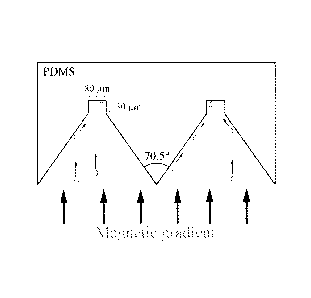
FIG 1 is a schematic diagram of the cell alignment structure.
FIG 2 shows a picture of the PDMS imprint using a scanning electron microscope
with the
smooth alignment and imaging surfaces.
FIG 3 shows the transmission spectrum of a PDMS slab at 1.3 mm and a PMMA slab
at 0.5 mm
thickness compared with a typical glass microscope slide of 1.0 mm thick.
2a
CA 02724978 2016-01-05
FIG 4 shows the effect of excess unbound magnetic particles on the intensity
of the image.
Increasing levels of EpCAM ferrofluid results in a decrease in image intensity
and an
increase in variation in assessing intensity.
FIG 5 shows the difference in total intensities when with and without 40 g/m1
ferrofluid
present. The distribution is worse with ferrofluid present.
FIG 6 shows the combined Bright field, DAPI and PE image showing aligned SKBR3
cells.
DETAILED DESCRIPTIONS
Circulating tumor cells (CTC) in blood of cancer patients are known to
correlate with
disease activity. The CellTracks platform provides a magnetic enrichment and
image analysis of
suspect target cells for enumeration and correlation with disease state.
Targets such as cells, cell
debris, and cell components are collected against a collection surface of a
vessel without
subsequent alignment adjacent to a ferromagnetic collection structure. These
cells include white
blood cells, cells of epithelial origin, endothelial cells, fungal cells, and
bacterial cells. The
collection surface is oriented perpendicular to a magnetic field gradient
produced by external
magnets. The resultant image includes magnetic nanoparticles and magnetically
labeled
biological specimens, collected in a substantially homogeneous distribution on
the optically
transparent face of the chamber while non-selected entities remain below in
the fluid medium.
The incorporation of Time Delay Integration (US 11/344757) into the CellTracks
platform in
rare cell analysis is used to rapidly detect and characterize CTC.
Immunomagnetic separation of
target rare cells (CTC) is accomplished by EpCAM positive selection.
Circulating cells
expressing the antigen for the epithelial cell adhesion molecule (EpCAM) are
selected by the
monoclonal antibody specific or EpCAM and covalently linked to magnetic
nanoparticles. The
complex is separated from the remaining blood constituents when exposed to a
magnetic field.
Subsequent analysis of the captured target is determined through a series of
fluorescent
labeling of components within the cell. The fluorescent dye DAP1 is used to
label the nuclei of
CTC and non-specifically selected leukocytes, the cytoskeleton of CTC is
labeled with
Cytokeratine-PE and the leukocytes with CD45-APC. The labeled cells are
magnetically
manipulated to align along an analysis surface. However in this system,
unbound magnetic
particles that are left over from immunomagnetic separation will cause image
distortion and
reduce the ability to confirm the presence of a CTC.
To provide for spatially patterned collection of target specimens for
qualitative and
quantitative analysis of microscopic biologic samples, the present invention
relates to making
and using pre-molded structures on the inner surface of the imaging chamber.
Generally, pre-
3
DOCSTOR. 5387045\1
CA 02724978 2016-01-05
molded grooves are long v-shaped grooves, pre-molded into the inner portion of
the viewing
surface on the imaging chamber. These structures provide an alignment of cells
as good as or
even better than previously reported Ni lines. Furthermore, they are made from
a highly
transparent material, optically suited for imaging the entire cell.
Figure 1 illustrates the principle of cell alignment using the grooves of the
present invention.
Magnetically induced cell movement within the chamber occurs in the present of
a magnetic
gradient. Here, the magnetically labeled cells will either collide with the
inclined surface of the
structure (alignment surface) and slide into the top of the groove (indicated
imaging surface), or
they will directly hit the top of the imaging surface. In either situation,
the cells will align in the
groove, allowing for subsequent imaging.
In order for sufficient movement along the inclined surface of the groove, the
surface should
be flat and cells prohibited from sticking to the walls. To achieve a smooth
precise design,
known wafer etching technologies are used. However because of expense and
optical
requirements, silicon wafers are not appropriate, rather polydimethylsiloxane
(PDMS) replica
molding provides a composition that will meet these requirements. Compositions
that will meet
this criteria are also considered in the present invention. Structures, etched
onto a silicon wafer,
are the inverse of the eventual design, and provide the PDMS mold with the
correct shape when
poured onto the silicon mold. After curing, this shape is cut into dimensions
that would allow
replacement of the glass surface of the imaging chamber.
Etching can be accomplished on any optically transparent material that can be
used in the
manufacture of the chamber. By example, silicon wafers can be used in etching
because of the
ease of precision, fine detail, and easily reproducible. Any material with
similar characteristics
and known in the art is considered in the present invention. Etching of the
structure uses two
common etching techniques. First an etch mask that is needed to etch the
grooves is created.
This mask is created using BHF (Buffered Hydrofluoric acid) etching. Once BHF
etching is
complete, thin layers of Si02 are left on the silicon wafer at places where no
groove should be
etched. Anisotropic etching is also used to etch the grooves. Here, KOH is
used as etchant.
When this process is applied to a properly orientated wafer, V-grooves are
etched, limited by the
crystal plane of the silicon wafer. Accordingly, a highly reproducible and
constant etch angle is
produced. The angle depends on the wafer orientation with one embodiment as
shown in Figure
1 at 70.5 degrees. Another technique is Deep Reactive Ion Etching (DRIE). By
using this
technique it is possible to etch structures with a high aspect ratio (ratio
between length and width
of the structure). DRIE cyclically alternates between etch and deposition
steps forming
scalloped sidewalls.
4
DOCSTOR 5387045 \ 1
CA 02724978 2016-01-05
PDMS molding is used to obtain a positive imprint on the fabricated wafer.
PDMS or
Polydimethylsiloxane (Dow Corning (Sylgard 184, Dow Corning, Midland, MI, USA)
is a
polymer containing the siloxane bond between Si (Silicon) and 0 (Oxygen). The
polymers
molecules are linked together to form longer polymers with an average number
around 50 to
100.
The final PDMS is obtained with the addition of a cross-linker. The cross-
linker connects
with the polymers to form long networks of polymers, resulting in a clear,
elastic, chemically
inert, thermally stable material. After polymerization, the PDMS forms a clear
flexible
substance which adheres to very few materials and is not hygroscopic, thus
preventing any
sticking of cells to the sides due to the fact that PDMS adheres to very few
materials.
Furthermore, it is thermally stable and transparent from approximately 300 to
900 nm. These
characteristics are all important for its use in a fluorescent imaging system
and the transmission
of visible light. After formation, the grooves are cut into the dimensions of
the viewing face of
the chamber.
In order to remove most of the excess magnetic particle with minimal cell
loss, the cell
alignment structure shown in Figure 1 was developed for the viewing surface.
Figure 1 provides
an overview of the cell alignment structure. A PDMS microstructure shown in
Figure 1 was
developed to align the cells in predetermined areas and reduce imaging time.
The design
consists of a PDMS chip of 30x2.7 mm2 containing 6 channels that are 80 um
wide and 30 mm
long. These PDMS chips are imprinted from a wafer mold. We chose PDMS because
of its
excellent transmission properties, replicating capabilities and ease of use.
This configuration
reduces the imaging time for a sample from 32 to 12 minutes.
Figure 2 shows a scanning electron micrograph of a PDMS imprint having the
structure used
in the present invention. The picture shows the smooth alignment and imaging
surfaces on the
viewing surface.
Figure 3 compares the transmission spectrum of the thickness of the PDMS used
in the
present invention with a typical glass microscope slide. With a thickness of
0.5 mm, PMMA
tracks the transmission of a glass microscope slide between 400 nm and 800 nm
wavelengths.
Excess of conjugated magnetic particles (EpCAM-ferrofluid) are used to ensure
maximum
CTC recovery. However, during imaging the excess ferrofluid forms elongated
aggregates on
the imaging surface, which influences quantitative and qualitative imaging of
the cells. Figure 4
shows the effect of excess unbound magnetic particles on imaging. The total
nuclear intensity
drops sharply for concentrations of ferrofluid higher than 2 ug/ml (circles),
while the CV rises to
40% for concentrations that are normally found in patient samples, such as 40
ug/ml (triangles).
5
DOCSTOR. 5387045\1
CA 02724978 2016-01-05
>
The effect of excess ferrofluid on imaging and the results of the removal
method are shown
in Figure 5. SKBR-3 cells imaged with 40 ug/m1 ferrofluid concentrations and
with 0 ug/ml
ferrofluid. The distribution of total intensities is significantly worse for
the sample with excess
ferrofluid.
The present invention avoids this issue and provides a method that spins the
sample inside a
conical tube at 900 rpm. This forces all the cells in the tube outwards. The
ferrofluid particles
are much smaller (-175 nm in diameter) and remain randomly distributed in the
sample. After
several minutes, 60% of the sample is automatically aspirated from the bottom
of the rotating
tube. This process is repeated 5 times and the SKBR-3 cells aligned along the
viewing surface
using the PDMS structure in the present invention. Figure 6 shows the combined
bright field,
DAPI and PE image of captured SKBR-3 cells aligned along the viewing surface.
Using the method of the present invention, 95% to 97% of excess ferrofluid can
be removed,
brining the final concentration in a patient sample down to less than 2 ug/ml.
This removes
enough excess ferrofluid to make optimal quantitative measurements and
considerable
improvements in the quality of images. Also, the recovery of CTC's ranted
between 95% and
99%. Accordingly, ferrofluid removal, using the methods of the present
invention, provides for
sufficient removal of excess ferrofluid for optimal quantitative measurements.
The quality of
the images are improved and bright field imaging is possible to obtain
additional morphological
data on individual CTC's.
While certain of the preferred embodiments of the present invention have been
described
and specifically exemplified above, it is not intended that the invention be
limited to such
embodiments. Various modification may be made thereto. The scope of the claims
may be
given the broadest interpretation consistent with the description as a whole.
6
DOCSTOR 5387045\1
