Note : Les descriptions sont présentées dans la langue officielle dans laquelle elles ont été soumises.
CA 02728309 2010-12-16
WO 2009/149502 PCT/AU2009/000731
1
DENTURES, DENTAL ARCHES AND METHODS OF MANUFACTURE
FIELD OF THE INVENTION
The present invention relates to dentures, dental arches and methods of
manufacture. In particular, although not exclusively, the present invention
relates to improved upper and lower dentures, clinical and laboratory methods,
materials and apparatus for their production.
BACKGROUND TO THE INVENTION
Replacement, artificial or false teeth are required where all teeth have
been lost or have had to be removed for one reason or another, such as
medication, accidental atrophy, disease or wear with old age. The most
common form of full edentulous artificial teeth are in the form of removable
dentures, which typically comprise a full set of upper and/or lower teeth,
usually
minus the wisdom teeth. The denture is sized and shaped to rest on the soft
tissues of the patient's jaw referred to as the oral ridge. Hundreds of
millions of
dentures are in use worldwide.
Advances in materials have enabled dentures to be more durable and
more natural looking and the development of denture designs has improved
comfort and chewing efficiency. However, the process for making dentures, for
both clinical and laboratory procedures, has changed little for decades and
because dentures are tailored to each patient, they cannot be mass produced.
The manufacturing process therefore remains both time consuming and labour
intensive. Furthermore, the patient is inconvenienced by the delays associated
with manufacturing the denture and once the denture is finished the patient
may
still experience discomfort from ill-fitting denture(s) caused by patient jaw
relation
inconsistencies, clinical and laboratory errors, including transit mishaps,
all
affecting the manufacturing process.
CA 02728309 2010-12-16
WO 2009/149502 PCT/AU2009/000731
2
The conventional process for producing a denture typically includes
multiple clinical consultations between the patient and the denture
practitioner
and each clinical consultation is typically followed by work being performed
by a
dental technician. Often the premises or laboratory of the dental technician
who
performs the clinical stages required to finish the dentures are remote from
the
denture practitioner's surgery. Therefore, impressions, casts and especially
an
articulation apparatus used to make the dentures, as will be described
hereinafter, need to be transported back and forth between the denture
practitioner's surgery and the laboratory of the dental technician. This
exacerbates the delay associated with producing the denture and incurs
transport costs and further labour costs, ultimately for the practitioner and
therefore for the patient.
Following an initial assessment of the patient by the denture practitioner,
the process of manufacturing a denture commences with primary impressions of
the patient's upper and lower mouth being taken using stock trays. The primary
impressions are sent to the technician who casts impressions in stone from
which special trays are produced for the patient. The special trays are sent
to the
denture practitioner who takes secondary impressions using the special trays.
The technician casts secondary impressions in stone and produces wax
registration rims from the stone secondary impressions. The registration of
the
patient's jaw relations are taken with the aid of wax registration rims which
are
then temporarily attached together according to the patient's tempro-
mandibular
joint (TMJ) positions and centric and vertical dimensions including occlusal
planes and the cuspid regions as points of reference. A face bow and more
intricate articulation systems are sometimes used by practitioners with a more
refined and precise attitude towards the denture construction for their
patient.
The wax registration rims are then returned once more to the technician.
The technician places the attached wax registration rims on an articulator
and follows the markings and dimensions placed on the rims for accurate
setting
CA 02728309 2010-12-16
WO 2009/149502 PCT/AU2009/000731
3
up of a dentition. A range of articulators are available having varying
degrees of
complexity, accuracy and cost. Unfortunately, the preferred articulators that
provide the best results are not always used. The artificial teeth selected
for the
patient are to be mounted one-by-one accurately and according to the
prescribed dimensions on the wax registration rims by the technician and once
waxed and sculpted meticulously and cleaned, the wax base and set up dentition
is returned to the denture practitioner for trying by the patient.
At this stage, the dentition may be a good fit and have the desired
appearance and all the appropriate physiological dimensions. However, often
the practitioner is faced with having to deal with the fit and/or the teeth
being
misaligned and/or the bite being incorrect, causing the aesthetic appearance
and/or function to be wrong. Such problems can be caused by clinical
misinterpretation of one or more of the factors necessary to determine the
perfect position of the patient's jaw relationship, caused either by improper
practitioner procedures and/or imprecise and/or inconsistent control by the
patient and their jaw relationship positions at the previous registration
appointment. Other problematic factors may be caused in the laboratory due to
wrongful preparation and/or protocol, physical impacts to the dentition and/or
distortion factors inflicted on one or more of the materials and apparatus
used,
for example, in transit, or by the temperature sensitivity of the wax
supporting the
dentition or damage of any other sort. If such problems exist, the dentition
must
be returned to the technician who must remove and reset all of the artificial
teeth,
meaning that all of the extensive laborious work done in relation to
registration,
articulation, setting of the dentition and the wax sculpturing is wasted. The
re-
articulated, reset and re-waxed dentition is then returned to the denture
practitioner for a re-try with the patient. This process is repeated until
both the
denture practitioner and the patient are happy with the result. Only then can
the
technician progress to a finishing stage in which the finished denture is
produced. It should be appreciated that each time the technician receives the
CA 02728309 2010-12-16
WO 2009/149502 PCT/AU2009/000731
4
impression, registration rims or dentition from the dentist, it must be washed
and
sterilized before work is commenced.
The finishing stage is another labour intensive and time consuming
process in which the final denture is produced from either poly-
methylmethacrylate (PMMA) or MMA acrylics or other acrylics, either by
conventional flasking, injection moulding or UV light cured methods. In
summary, a negative stone cast of the wax with the dentition is produced
within
the flask. A separator liquid or release agent is added to all plaster and/or
stone
surfaces to create a non- cohesive layer between the uncured acrylic and
surrounding plaster and/or stone cast to enable the ultimately hardened
acrylic to
be freely removed once the process is complete in the flask method. If the
flasking method is used, the soft poly-methylmethacrylate acrylic compound is
added to the flask and pressed for increased compact density. The excess is
removed and re-pressed before being heated to initiate the chemical reaction
causing the compound to harden. The heating process can take between one to
eight hours depending on the type of compound being used. Once cooled, the
denture is de-flasked, cut, festooned and polished before being sent to the
denture practitioner. The other options and methods, such as the injection
moulding process and composite UV appliance process, may also be used.
The patient tries the denture and checks are made to ensure that the fit
and the bite is correct and there are no pain spots or unwanted discrepancies,
such as premature contact, fulcrum tilting or any displeasing aesthetic
factors. If
such problems exist and can not be remedied or corrected in the clinic, the
denture must be returned to the technician to make the appropriate further
adjustments by re-articulating and undertaking either minor or major
corrections
until the fit is adequate and both the practitioner and patient are happy with
both
the fit and appearance of the denture. Based on conservative estimates and
when the process runs smoothly, the aforementioned conventional process for
producing a denture, including clinical and laboratory time, can take in the
region
CA 02728309 2010-12-16
WO 2009/149502 PCT/AU2009/000731
of at least thirteen hours in total, excluding the transport time between the
premises of the denture practitioner and the technician.
Another disadvantage of imperfect protocol being employed in the
conventional dentures process is that when the dentures are finished and ill-
fitting, sometimes excessively so, the dentures must often be remade from
start
to finish, because they can be totally irreparable at the most important
stage,
such as the finished stage. If for some unforseen reasons the attempted minor
and/or major adjustments can not be corrected in the laboratory by the
technician or by the denture practitioner whilst the patient is in the chair,
the
appliance is rendered a complete failure. The faulty denture needs to be
discarded and the aforementioned laborious process repeated to a large extent
to create a new denture. Even in the absence of specific irreparable damage,
on
average dentures need to be replaced every five years or so due to wear,
physiological atrophy of the oral ridge leading to osseo depletion or any
tissue
surface change thus creating discomfort.
The prior art base is replete with attempts to improve the efficiency of the
denture manufacturing process, the clinical process, denture construction
and/or
the quality consistency of the resulting dentures. However, none of these
attempts appreciably expedite and/or improve the clinical processes,
laboratory
construction, manufacturing process and/or address the aforementioned
problems.
In this specification, the terms "comprises", "comprising", "includes",
"including" or similar terms are intended to mean a non-exclusive inclusion,
such
that a method, system or apparatus that comprises a list of elements does not
include those elements solely, but may well include other elements not listed.
CA 02728309 2010-12-16
WO 2009/149502 PCT/AU2009/000731
6
OBJECT OF THE INVENTION
It is an object of the present invention to address, or at least ameliorate,
one or more of the aforementioned problems associated with the known
methods of producing dentures.
It is a preferred object of the present invention to reduce the time taken to
produce dentures and/or improve the quality of the dentures produced and/or
the
method of production of dentures.
SUMMARY OF THE INVENTION
In one form, although it need not be the only or indeed the broadest form,
the invention resides in a support for a dental arch, the support comprising
an
elongate member curved to substantially follow a curve of the dental arch.
Preferably, the elongate member is metallic and made of, for example,
titanium, stainless steel, high carbon steel or a metal alloy, although other
materials, such as ceramics, carbon fibre, at least one polymer or a fibre
composite may be used.
Preferably, a face of a front or anterior region of the elongate member is
substantially perpendicular to faces of rear or posterior regions of the
elongate
member.
Preferably, the elongate member comprises a transitional region between
the anterior region and each posterior region.
Suitably, the elongate member comprises a twist between the anterior
region and each posterior region.
Preferably, elongate member is twisted such that a face of the elongate
member is substantially parallel to surfaces of artificial teeth of the dental
arch.
Preferably, the face of the anterior region of the elongate member is
substantially parallel to one or more front surfaces of artificial incisor
teeth of the
dental arch.
Preferably, the faces of the posterior regions of the elongate member are
CA 02728309 2010-12-16
WO 2009/149502 PCT/AU2009/000731
7
substantially parallel to one or more biting occlusal surfaces of artificial
molar
teeth and/or artificial bicuspid teeth of the dental arch.
Suitably, the anterior region of the elongate member morphs into the
posterior regions of the elongate member approximately at the post cuspid
regions, flattening towards a more horizontal formation at the second bi-
cuspid
regions and comprises substantially horizontal, planar regions approximately
under the first and second molar posterior regions.
Suitably, each posterior region includes a textured surface for added
mechanical retention.
Suitably, the support can include one or more apertures through the
elongate member.
Suitably, the support comprises at least one joint between at least two
parts of the support.
Suitably, the at least one joint is provided substantially centrally in the
anterior region of the support.
Suitably, the at least one joint is provided in at least one of the posterior
regions of the support.
Suitably, the support comprises a joint in a left hand posterior region, a
joint in a right hand posterior region and a joint in the anterior region of
the
support.
Suitably, the artificial teeth are in the form of clip-on artificial teeth,
which
clip on to the support. The clip-on artificial teeth may be in the form of a
single
clip-on artificial tooth or may be in the form of clip-on units comprising
multiple
artificial teeth. The clip-on units may be anterior units or posterior units.
Suitably, the support comprises artificial teeth fixed to the support and one
or more spaces for attaching a clip-on artificial tooth or a clip-on unit
comprising
multiple artificial teeth.
In another form, although again not necessarily the broadest form, the
invention resides in a joint between a first part and a second part of a
support for
CA 02728309 2010-12-16
WO 2009/149502 PCT/AU2009/000731
8
a dental arch, the joint comprising:
the first part having a projection comprising a bean shaped aperture
therethrough;
the second part comprising a bean shaped recess to receive the bean-
shaped projection; and
a pin passing through the bean shaped aperture in the projection and
through an aperture in the recess about which the first part can pivot
relative to
the second part in a single plane.
Preferably, the joint comprises three contact points.
In another form, although again not necessarily the broadest form, the
invention resides in a dental arch comprising:
a metallic elongate member curved to substantially follow a curve of the
dental arch; and
a plurality of artificial teeth affixed to the elongate member.
Suitably, the artificial teeth are either permanently or adjustably affixed to
the elongate member.
Suitably, one or more of the artificial teeth are affixed to the elongate
member via a fastener passing through one of the apertures in the elongate
member for attachment to a respective back of the one or more artificial
teeth.
Suitably, the respective back of the one or more artificial teeth comprises
a recess for engaging an end of the fastener. Alternatively, the respective
back
of the one or more artificial teeth comprises a male projection for engagement
by
a female socket in an end of the fastener.
Suitably, vertical and/or lateral positions of the artificial teeth are
adjustable with respect to the apertures.
Suitably, angles of incisal inclination of the artificial teeth with respect
to
the anterior region of the elongate member are adjustable.
Suitably, the artificial teeth are in the form of clip-on artificial teeth,
which
clip on to the support.
CA 02728309 2010-12-16
WO 2009/149502 PCT/AU2009/000731
9
In another form, although again not necessarily the broadest form, the
invention resides in an arcuate member for assessing a size of a dental arch,
the
arcuate member comprising:
a pair of anterior apertures in the left and right anterior region to indicate
the positions of the cuspid teeth; and
at least one pair of posterior apertures in the left and right posterior
region
to indicate positions of the molar teeth.
Preferably, the pair of posterior apertures indicates the mesio-buckle
cusps of the first molar teeth.
Alternatively, the pair of posterior apertures indicates the positions of the
second molar teeth and more particularly the positions of the center fossa of
the
second molar teeth.
The arcuate member may comprise two pairs of posterior apertures, a
first pair of posterior apertures in the left and right posterior region to
indicate
positions the mesio-buckle cusps of the first molar teeth and a second pair of
posterior apertures in a more posterior region than the first pair of
posterior
apertures to indicate positions of the second molar teeth and more
particularly
the positions of the center fossa of the second molar teeth.
Preferably, the relative positions of the pair of anterior apertures, the
first
pair of posterior apertures and/or the second pair of posterior apertures
correspond to the size of the dental arch.
Preferably, the arcuate member comprises one or more indicia adjacent
each anterior aperture, alignment of one of the indicia with the cuspid teeth
being
indicative of a tapered arch form or a square arch form.
Preferably, a handle extends from the arcuate member to facilitate use.
The arcuate member can be used on a patient's mouth or on a model of
the patient's mouth.
In a further form, although not necessarily the broadest form, the invention
resides in a system for assessing a size of a dental arch, the system
comprising
CA 02728309 2010-12-16
WO 2009/149502 PCT/AU2009/000731
a series of arcuate members, each arcuate member comprising a pair of anterior
apertures in the left and right anterior region to indicate the positions of
the
cuspid teeth and at least one pair of posterior apertures in the left and
right
posterior region to indicate positions of the molar teeth wherein the relative
positions of the pair of anterior apertures and the pair of posterior
apertures of
each arcuate member correspond to a size of the dental arch.
Suitably, the series comprises three or more arcuate members
corresponding to a scheme comprising three or more dental arch sizes. One
particular scheme comprises five dental arch sizes.
Suitably, a connector can be inserted into one or more of the anterior
apertures and/or one or more of the posterior apertures of the arcuate member
for connecting the arcuate member to a base tray.
In a yet further form, although again not necessarily the broadest form,
the invention resides in a method of determining a size of a dental arch
including:
placing one or more of a series of arcuate members of different sizes on
the dental arch, each arcuate member comprising a pair of anterior apertures
in
the left and right anterior region to measure the positions of the cuspid
teeth and
at least one pair of posterior apertures in the left and right posterior
region to
measure positions of the molar teeth; and
determining the size of the dental arch based on the arcuate member that
best matches the positions of the cuspid teeth and the molar teeth.
In another form, although not necessarily the broadest form, the invention
resides in a base plate material for dentures comprising an acrylic composite
sheet embedded with a flexible biocompatible reinforcing mesh.
Suitably, the reinforcing mesh is biocompatible flexible fibreglass.
In a further form, although again not necessarily the broadest form, the
invention resides in a length of flexible acrylic composite material
comprising a
series of arcuate cut-outs for alignment with artificial teeth of a denture.
CA 02728309 2010-12-16
WO 2009/149502 PCT/AU2009/000731
11
Suitably, the arcuate cut-outs may be aligned with cervical regions of the
artificial teeth.
Suitably, the arcuate cut-outs may be aligned with composite or acrylic
collars surrounding cervico-neck regions.
In a yet further form, although not necessarily the broadest form, the
invention resides in a mold for a length of flexible acrylic composite
material, the
mold comprising a strip having a surface pattern for imprinting embossed
regions
on the flexible acrylic composite material.
Preferably, the strip is metallic.
In another form, although not necessarily the broadest form, the invention
resides in a flexible, cushioning insert for use in taking a mold of a dental
ridge,
the insert having a shape approximating to the shape of a dental arch and
comprising a gel in a sealed outer layer of the insert.
Suitably, the gel and/or the outer layer of the insert are transparent.
Suitably, the insert for taking a mold of a lower dental ridge has an
arcuate shape approximating to the shape of a lower dental arch.
Suitably, the insert for taking a mold of a lower dental ridge has a
substantially U-shaped cross section which follows a cross sectional shape of
the lower dental ridge.
Suitably, the insert for taking a mold of an upper dental ridge also takes a
mold of the upper palate.
Suitably, the insert for taking a mold of an upper dental ridge and upper
palate has a cross sectional shape approximately corresponding to that of the
upper dental ridge and the upper palate.
In a further form, although not necessarily the broadest form, the invention
resides in a base tray for an upper or lower denture formed from an acrylic
composite sheet embedded with a flexible biocompatible reinforcing mesh,
wherein at least one portion of the flexible biocompatible reinforcing mesh is
exposed.
CA 02728309 2010-12-16
WO 2009/149502 PCT/AU2009/000731
12
Preferably, the at least one exposed portion of the flexible biocompatible
reinforcing mesh is a vault of the base tray for an upper denture.
Suitably, the at least one exposed portion of the flexible biocompatible
reinforcing mesh is a peripheral edge of the base tray.
Suitably, a base tray for a lower denture comprises flexible labial and/or
lingual regions.
In another form, although not necessarily the broadest form, the invention
resides in an indenting member for attachment to a posterior region of a
tissue
contact side of an upper base to improve retention of an upper denture on the
upper palate.
Preferably, the indenting member extends the width of the soft palate,
between the left and right tuberosity of the upper ridges at the vibrating
line.
Suitably, the indenting member comprises two adjacent tapering regions
extending from a base of the indenting member to the compressive soft tissue
over the transverse palatine suture of the palate.
In a further form, although again not necessarily the broadest form, the
invention resides in a method of producing a denture including:
temporarily affixing a base to an oral ridge of a patient;
temporarily affixing a dental arch onto the base with one or more light-
curable dabs of composite material;
adjusting the position of the dental arch with respect to the base and the
patients' dental dimensions until the desired position is achieved; and
light-curing the dabs of composite material.
Preferably, the method includes interdigitating the occlusal surfaces of an
opposing lower dental arch to a related upper dental arch after the desired
position and appropriate dimensions of the upper dental arch with respect to
an
upper base and the patients' dental dimensions are achieved.
Alternatively, the method may include achieving the desired position and
appropriate dimensions of the lower dental arch with respect to a lower base
and
CA 02728309 2010-12-16
WO 2009/149502 PCT/AU2009/000731
13
then interdigitating the occlusal surfaces of an opposing upper dental arch to
the
related lower dental arch.
Further forms and features of the present invention will become apparent
from the following detailed description.
BRIEF DESCRIPTION OF THE DRAWINGS
In order that the invention may be readily understood and put into
practical effect, reference will now be made to embodiments of the present
invention with reference to the accompanying drawings, wherein like reference
numbers refer to identical elements. The drawings are provided by way of
example only, wherein:
FIG 1 shows a perspective view of a support for a dental arch;
FIG 2 shows a plan view of the support shown in FIG 1;
FIG 3 shows a side view of the support shown in FIG 1;
FIG 4 is a partial cross-sectional view of a dental arch;
FIG 4A is a perspective view of part of the support shown in FIG 1
comprising artificial teeth in predetermined, fixed positions;
FIG 5 is a perspective view of a support for a dental arch according to
another embodiment;
FIG 6 is a rear view of the support shown in FIG 5;
FIG 7 is a perspective view of a dental arch comprising the support shown
in FIG 5;
FIG 8 is a side view of the support shown in FIG 5 with an artificial
anterior central tooth fixed to the anterior portion of the support;
FIG 9 is a perspective view of an artificial tooth and fastener;
FIG 9A shows a perspective view and a partial, enlarged side elevation of
an artificial tooth and fastener according to an alternative embodiment;
CA 02728309 2010-12-16
WO 2009/149502 PCT/AU2009/000731
14
FIG 10 is a perspective view of an artificial tooth affixed loosely to the
support of the dental arch;
FIGS 10A-10F shows a range of different positions and angles in which
artificial teeth can be situated on the support;
FIG 10G is a perspective view of an artificial tooth being fixed in position
on the support of the dental arch with UV cured composite material or wax;
FIG 10H is a perspective view of an artificial tooth and fastener according
to alternative embodiments;
FIG 10J is a cross-sectional view of the artificial tooth and fastener of FIG
10H coupled together affixing the artificial tooth loosely to the support of
the
dental arch;
FIG 10K is an enlarged side view of parts of the artificial tooth and
fastener of FIG 10H;
FIGS 11A-11 D are side views of a dental arch showing an artificial incisor
tooth at different angles of inclination;
FIG 12 is a perspective view of an arcuate member for assessing the size
of a dental arch;
FIG 13 is a schematic drawing showing the correspondence between
relative positions of first and third pairs of apertures of arcuate members
and the
size of the dental arch;
FIG 14 shows a model of a lower dentate ridge, the positions of the left
and right cuspid regions and the positions of the left and right molar
regions;
FIG 15 is a general flow diagram showing a method of determining a size
of a dental arch;
FIG 16 is an exploded view of a base plate material for making dentures;
FIG 17 is an apron with festooning contours for finishing the labial and
buckle regions of a dental appliance;
FIG 18 is a plan view of a flexible, cushioning lower insert for use in taking
molds with the composite material of dental lower ridges;
CA 02728309 2010-12-16
WO 2009/149502 PCT/AU2009/000731
FIG 18A is a plan view of a flexible, cushioning upper insert for use in
taking molds with the composite material of upper dental ridge and upper
palate;
FIG 19 is a cross sectional view of the lower insert shown in FIG 18;
FIG 20 is a perspective view of a tray for taking molds of lower dental
ridges;
FIG 20A is a perspective view of a tray for taking molds of upper dental
ridges and the upper palate;
FIG 20B is another perspective view showing a position of a section
through the tray in FIG 20A;
FIG 21 is a cross sectional view of the base plate material of FIG 16, the
insert of FIG 18 and the tray of FIG 20 in use;
FIG 21A is a cross 'sectional view of the base plate material of FIG 16, the
insert of FIG 18A and the tray of FIG 20A in use;
FIG 22 is a first part of a general flow diagram showing a clinical
procedure and a laboratory method of the construction and manufacturing of a
denture;
FIG 23 is a second part of the general flow diagram of FIG 22;
FIG 23A is a third part of the general flow diagram of FIG 22;
FIG 23B is a fourth part of the general flow diagram of FIG 22;
FIG 24 is a chart showing a range of dental arch shapes;
FIG 25 shows examples of different teeth shapes;
FIGS 25A-25D show different occlusal classifications and bites;
FIG 25E shows further occlusal set up and contact classifications
accommodated by the dental arches of the present invention;
FIG 26A shows a dental arch comprising dabs of light curable composite
material;
FIGS 26B and 26C show a dental arch temporarily attached to an upper
base with dabs of light curable composite material; still in its malleable
consistency not yet cured.
CA 02728309 2010-12-16
WO 2009/149502 PCT/AU2009/000731
16
FIGS 27A and 27B show an upper base and a dental arch attached with
dabs;
FIG 27C shows a four unit appliance comprising upper and lower bases
with interdigitated upper and lower dental arches;
FIG 27D shows filling lingual and labial areas of a denture with composite
material;
FIG 27E shows an alternative method of festooning the denture;
FIG 27F shows a partially festooned denture;
FIG 28 is a perspective view an indenting member for an upper base;
FIG 29 shows a depression in the posterior region on a model of the
upper oral ridge and palate;
FIG 30 is a sectional view of a palate showing an indent in the soft palate
created by the indenting member 140 of FIG 28;
FIG 31 is a sectional view of the model of FIG 29 showing the depression;
FIG 32 is a plan view of a two-part support comprising a joint;
FIG 33 shows plan views of a two-part support comprising a joint and a
three-part support comprising two joints;
FIG 34 is a plan view showing various configurations of a four-part
support comprising three joints;
FIG 35 is a side view of two parts of a support showing male and female
profiles comprising the joint between the two parts;
FIG 35A is a plan view of a joint between anterior and posterior parts of a
support;
FIG 36 is a plan view of a male profile having a bean-shaped aperture;
FIG 37 is a plan view of a female profile for receiving the male profile
shown in FIG 36;
FIGS 38-40 shown the range of movement of a joint comprising the male
and female profiles shown in FIGS 36 and 37;
CA 02728309 2010-12-16
WO 2009/149502 PCT/AU2009/000731
17
FIG 41 is a plan view of a support and posterior and anterior clip-on units
comprising artificial teeth;
FIG 42 is a plan view of a support, posterior clip-on units comprising
artificial teeth and individual artificial anterior teeth;
FIG 42A shows further examples of upper and lower anterior and
posterior clip-on units comprising artificial teeth;
FIG 43 is a sectional side view of the support and, an anterior clip-on unit
comprising at least one artificial tooth;
FIG 44 is a sectional end view of the support and a posterior clip-on unit;
FIG 44A is perspective view of support and a posterior clip-on unit
according to another embodiment;
FIG 45 is a perspective view of a lower base tray and handle;
FIG 46 is a perspective view of an upper base tray with the handle
detached;
FIG 47 is a perspective view of an inverted lower base tray and handle
being filled with two-stage silicone based composite material;
FIG 48 is a rear perspective partially cut-away view of an upper base;
FIG 49 is a front perspective exploded view of the upper base of FIG 48;
FIG 50 is a rear view of the upper base of FIG 48;
FIG 51 shows a rear perspective exploded view of an upper base, a
peripheral composite rod and a completed upper base;
FIG 52 is a perspective view of an arcuate member for use with upper and
lower base trays;
FIG 53 is a perspective view of a clip on connector for use with the
arcuate member of FIG 52;
FIG 54 is a cross sectional view of the connector of FIG 53 connected to
the arcuate member of FIG 52;
FIG 55 is a perspective view of an upper base plate comprising a flexible
reinforcing mesh vault and the connectors of FIG 53; and
CA 02728309 2010-12-16
WO 2009/149502 PCT/AU2009/000731
18
FIG 56 is a perspective view of a lower base plate comprising a flexible
reinforcing mesh skirt.
Skilled addressees will appreciate that elements in the drawings are
illustrated for simplicity and clarity and have not necessarily been drawn to
scale.
For example, the relative dimensions of some of the elements in the drawings
may be distorted to help improve understanding of embodiments of the present
invention.
DETAILED DESCRIPTION OF THE INVENTION
The structure of dental arches and accessories in accordance with
embodiments of the present invention will be described followed by a
description
of methods of manufacturing dentures using the dental arches and other aspects
of the present invention.
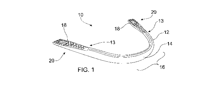
Referring to FIGS 1-3, a support 10 for a dental arch is provided in
accordance with embodiments of the present invention. The support 10
comprises a metallic elongate member 12 curved to substantially follow a curve
of a dental arch. The curve can have different oral shapes, such as tapered,
square, round (ovoid) or other shapes, depending on the general shape of the
mouth of the patient for whom the dental arch is intended. The size of the
arch
will also depend on the size of the patient's mouth, the determination of
which
will be discussed in further detail hereinafter in accordance with another
aspect
of the present invention.
The elongate member comprises a transitional region 13 between a front
or anterior region 16 and each rear or posterior region 20 such that the
anterior
region 16 gradually blends into or otherwise morphs into the posterior regions
20. According to some embodiments, the elongate member 12 is twisted such
that a face 14 of the elongate member 12 is substantially parallel to surfaces
of
artificial front or anterior teeth of the dental arch. According to the
embodiment
shown in FIGS 1-3, a face 14 of the anterior region 16 of the elongate member
CA 02728309 2010-12-16
WO 2009/149502 PCT/AU2009/000731
19
12 is substantially perpendicular to faces 18 of the posterior regions 20 of
the
elongate member. The relationship between the faces of the support 12 and the
surfaces of artificial teeth of the dental arch are described in further
detail with
reference to FIG 4 hereinafter.
According to the embodiments shown in FIGS 1-3, the elongate member
12 comprises a twist 22 between the anterior region 16 and each posterior
region 20. The anterior region 16 of the elongate member morphs into the
posterior regions 20 of the elongate member 12 approximately at the post
cuspid
regions, flattening towards a more horizontal formation at the second bi-
cuspid
regions and comprises substantially horizontal, planar regions approximately
under the first and second molar posterior regions. Each posterior region 20
includes a textured surface 24 and each textured surface 24 comprises an
aperture 26 through the elongate member 12. Composite material, such as ultra
violet light cured (UVLC) resin, or any other available acrylic or usable
material is
used to bond artificial teeth to the support 10 as will be described
hereinafter.
The textured surfaces 24 and the apertures 26 aid in mechanical retention of
the
material applied to the support 10.
According to preferred embodiments, the support 10 is formed from a
single length of high tensile metal to provide the required strength and
rigidity to
the dental arch and to the dentures made therefrom. A support made from
materials such as titanium or high tensile metal cannot be easily bent, will
not be
distorted in normal use and is biocompatible. It is envisaged that other
biocompatible metals or alloys can be used for the support 10, such as high
grade stainless steel or high carbon based metals. It is also envisaged that
other biocompatible materials of sufficient strength can be used for the
support
10, such as ceramics, one or more polymers, fibre composites or carbon fibre
materials.
With reference to FIG 4, the dental arch 28 comprises the support 10 with
a complete set of artificial teeth 29 (minus the wisdom teeth) bonded thereto
with
CA 02728309 2010-12-16
WO 2009/149502 PCT/AU2009/000731
composite material 30, or other material such as a cross-linked acrylic. For
the
sake of clarity, FIG 4 shows part of the support 10 with the adjacent teeth in
phantom and the remainder of the dental arch 28 without the support 10 being
visible. The face 14 of the front or anterior region 16 of the elongate member
12
is substantially parallel to one or more front surfaces 31 of artificial
incisor teeth
32 of the dental arch 28. The faces 18 of the rear or posterior regions 20 of
the
elongate member 12 are substantially parallel to one or more masticatory
occlusal surfaces of artificial molar teeth 34 and/or artificial bicuspid
teeth 36 of
the dental arch 28. Hence, the support 10 provides strength to the dental arch
28 in accordance with the masticatory forces that are typically encountered in
the
different regions.
With reference to FIG 4A showing a perspective view of part of the
support 10, in this embodiment, the artificial teeth 29 of the dental arch are
permanently bonded or affixed to the elongate member 12 in predetermined,
fixed positions.
FIGS 5 and 6 show a perspective view and a rear view respectively of a
support 10 for a dental arch in accordance with an alternative embodiment of
the
present invention. In this embodiment, the support 10 comprises a plurality of
apertures 26 in the elongate member 12. In the embodiment shown, apertures
26 are provided in the anterior region 16 and in the posterior regions 20 of
the
elongate member 12, but not in the transitional regions 13 achieved, for
example, by the twists 22 of the elongate member 12. The apertures 26 are
provided to loosely affix artificial teeth to the support 10 such that the
positions of
the artificial teeth are adjustable with respect to the support 10, which will
be
described in detail in relation to FIGS 9 and 10.
FIG 7 shows an upper dental arch 38 according to another embodiment in
which at least some of the artificial teeth 40 are affixed to the elongate
member
12 using the apertures 26 and fasteners (not shown). Some of the artificial
teeth
40, such as those adjacent the transitional regions 13, such as the twist 22
in the
CA 02728309 2010-12-16
WO 2009/149502 PCT/AU2009/000731
21
elongate member 12, are affixed to the elongate member 12 using acrylic 30
and/or composite bonding material and/or other material.
FIG 8 shows a single artificial anterior incisor tooth 32 affixed to the
anterior portion 16 of the elongate member 12 via one of the apertures 26,
with
remaining apertures 26 available for further artificial teeth to be attached.
It
should be appreciated that the support 12 comprising the artificial anterior
incisor
tooth 32 is for an upper denture and is inverted (pointing upwards) in FIG 8.
A
support 12 according to embodiments of the present invention for the lower
arch
has a similar structure, except that the dimensions will differ from those of
the
support for the upper arch to suit the smaller dimensions of the lower
dentition.
For example, the anterior lower dentition is smaller in size than the anterior
upper dentition.
Referring to FIGS 9 and 10, in accordance with the present invention, an
artificial tooth 40 comprises a channel 42 in the back of the tooth and a
threaded
recess 44 for receiving a threaded end 46 of a fastener 48. The fastener 48
can
be made of acrylic or metal, such as titanium, although other materials of
similar
strength can be used. The channel 42 comprises a back wall 43, an upper
surface 45 and a lower surface 47 and has a height greater than the height of
the elongate member 12 of the support 10. As shown in FIG 10, according to
some embodiments, the artificial tooth 40 is positioned on the support 10 such
that part of the elongate member 12 is received within the channel 42 and can
abut against the back wall 43, upper surface 45 and lower surface 47, either
firmly or loosely. The artificial tooth 40 is held in place on the elongate
member
12 via the fastener 48 passing through one of the apertures 26 in the elongate
member aligned with the artificial tooth 40 and screwed into the threaded
recess
44. The apertures 26 have a width and a height greater than the diameter of a
shaft 49 of the fastener 48, but less than a diameter of a head 51 of the
fastener
48, not only to allow passage of the fastener 48 through the aperture 26, but
also
to enable the artificial tooth 40 to be loosely attached in a range of
different
CA 02728309 2010-12-16
WO 2009/149502 PCT/AU2009/000731
22
positions and angles, whilst preventing the tooth 40 from being removed or
detached easily. Hence, the vertical positions, the lateral positions and the
angles of the artificial teeth are adjustable with respect to the apertures 26
providing a high degree of maneuverability in achieving the desired appearance
and anterior tooth position of the dental arch and denture produced therefrom.
In the example shown in FIG 10, an anterior lateral artificial tooth 40 is
positioned in a loose format abutting against the front 14 of the elongate
member
12.
In alternative embodiments, the recess 44 and the fastener 48 may not be
threaded. Instead, the recess 44 can be in the form of a female socket that
receives an enlarged end of the fastener 48. The enlarged end can be of a
complementary shape to the female socket. In such embodiments, the enlarged
end of the fastener 48 can be resilient such that the enlarged end is snap-
fitted
into, and engaged by, the female socket.
With reference to FIG 9A, in another embodiment, the recess is in the
form of a slightly tapered female socket 44B within the back wall 43 of the
artificial tooth 40. The end 46B of the shaft 49 of the fastener 48 is also
gently
tapered and is sized to be received within and engaged by recess 44B. End 46B
is held within the recess 44B, but can be removed by hand by pulling with
sufficient force on head 51A, which no longer requires a slot for screwing the
fastener 48 into place.
Further examples of the range of different positions and angles in which
the artificial teeth 29 can be situated with reference to the support 10 are
shown
in FIGS 10A-10F.
FIG 10A shows two artificial teeth 29A, 29B loosely attached to the
elongate member 12 of the support 10 with fasteners 48A, 48B. Tooth 29A is
loosely attached with a front surface 31A substantially parallel to the face
14 of
the elongate member 12. Tooth 29B is loosely attached with a front surface 31
B
angled with respect to the face 14 of the elongate member 12.
CA 02728309 2010-12-16
WO 2009/149502 PCT/AU2009/000731
23
FIG 10B shows a side elevation of an artificial tooth 29 loosely attached to
the elongate member 12. Shaft 49 of fastener 48 passes through aperture 26
and is secured tightly in the recess 44 of back wall 43 of channel 42. In the
embodiment shown in FIG 10B, the artificial tooth 29 is forwardly inclined
with
respect to the elongate member 12. The arrows show the various orientations in
which the artificial tooth 29 can be moved with respect to the elongate member
12.
FIG 10C illustrates how the artificial tooth 29 can be moved with respect
to the elongate member 12 to vary the angle of the front facial surface 31 of
the
tooth 29 with respect to the face 14 of the elongate member 12.
FIG 10D shows how the artificial tooth 29 can be moved forwards and
backwards with respect to the elongate member 12 to vary how much the front
surface 31 of the tooth 29 protrudes from the elongate member 12.
FIG 10E illustrates how the artificial tooth 29 can be rotated about an axis
of the fastener 48 to vary the angle of the tooth 29 with respect the vertical
V and
with respect to the elongate member 12.
FIG 10F shows part of the support 10 and six artificial anterior teeth 29
held loosely in place on the elongate member 12 by their respective fasteners
48. The artificial teeth 29 are shown in a range of positions with respect to
the
elongate member 12. Reference lines highlight the degree of rotation of each
tooth with respect the vertical and the angles of the front surfaces 31 with
respect to the elongate member 12. A range of angles of inclination and
different degrees of protrusion are also illustrated as well as an overlap 29A
between two adjacent artificial teeth 29. The blocks 55 in the foreground
illustrate schematically the position and orientation of each tooth 29. This
vast
range of orientations allows almost any position of the artificial anterior
teeth 29
desired by the patient or practitioner.
With reference to FIG 10G, once the desired position of the tooth 29 has
been determined, it can be temporarily stabilized or fixed in its preferred
position
CA 02728309 2010-12-16
WO 2009/149502 PCT/AU2009/000731
24
with wax, or fixed more permanently in position with either self cure acrylic
or UV
light cured composite material 30 to maintain the preferred aesthetics from
both
the patient's and the practitioner's perspective.
FIGS 10H, 10J and 10K show alternative embodiments of the artificial
teeth and fasteners. Artificial tooth 600 comprises many of the features of
the
earlier embodiments described herein, such as channel 42 comprising back wall
43, upper surface 45 and lower surface 47 wherein the channel 42 has a height
greater than the height of the elongate member 12 of the support 10. However,
in this embodiment, a male projection 602 extends from the back wall 43 rather
than back wall 43 comprising the recess 44 of previous embodiments. Artificial
tooth 600 is held in place on the elongate member 12 via the fastener 604
passing through one of the apertures 26 in the elongate member 12 aligned with
the artificial tooth 600. Apertures 26 have a width and a height greater than
the
diameter of the shaft 49 of the fastener 604, but less than a diameter of the
head
51 of the fastener 604. In this embodiment, an end of the fastener 604
opposite
the head 51 comprises a recess or female socket 606 for engaging male
projection 602 of artificial tooth 600. Male projection 602 can comprise an
enlarged end of a substantially complementary shape to the female socket 606
of the fastener 604. In such embodiments, the female socket 606 can be
resilient such that the enlarged end is snap-fitted into, and engaged by, the
female socket 606. A wall 608 of the female socket 606 can comprise one or
more notches or cut-outs 610 to facilitate the resilient engagement of the
male
projection 602 by the female socket 606. The length of the male projection 602
and therefore the depth of the female socket 606 can vary between
embodiments. According to some embodiments, the male projection 602
extends from the back wall 43 by at least about 1 mm and therefore the depth
of
the female socket 606 is at least 1 mm to accommodate the male projection 602.
Referring to FIGS 11A-11D, according to some embodiments, the angles
of inclination of the artificial teeth 29 with respect to the elongate member
12 are
CA 02728309 2010-12-16
WO 2009/149502 PCT/AU2009/000731
adjustable, showing vertical deflective positions, vertical angular
inclinations and
mesio-distal-overlaps, as shown in FIG 10F as 29A. FIG 11A shows a dental
arch comprising artificial teeth 29 attached to the elongate member 12 on a
far
side and no artificial teeth affixed to the near side of the elongate member
12.
An artificial incisor tooth 32 is affixed to a front region 16 of the elongate
member
12 such that the tooth 32 is inclined at an acute angle with respect to the
substantially horizontal elongate member 12. Hence, the artificial incisor
tooth
32 is angled labialy with the incisal tip prominently forward. In FIG 1113,
the
artificial incisor tooth 32 is substantially vertical and incisorly tipped
inwardly or
lingualy, compared to 11A. In FIG 11C, the artificial incisor tooth 32 is
inclined
and even more incisorly tipped inwardly or lingually than 11A or 11 B. The
range
of movement can be placed incisorly even more predominately forward than as
shown in FIG 11A if preferred, as shown in FIG 11 D.
The dental arch 28 is provided in a range of sizes to fit different mouth
sizes. The dental arch is at least provided in small, medium and large sizes
and
can also be provided in further sizes as discussed further hereinafter. The
dental arch 28 is also provided in a range of shapes to match the general
shape
of the mouth of the patient. The dental arch 28 can be, for example, generally
square, round (ovoid), tapered or other shapes depending on the degree of
curvature of the jaw. The shape of the dental arch 28 is based on the support
10
of the same shape. For example, a tapered dental arch will be based on a
support having tapered shaped teeth and so on.
In accordance with other aspects of the present invention and with
reference to FIG 12, an arcuate member 50 is provided for assessing the size
of
the dental arch. The arcuate member 50 can be formed from transparent
plastics material that is capable of being sterilized. The arcuate member 50
comprises a pair of anterior apertures 52 in the left and right anterior
region
closest to a handle 56 to indicate positions of the cuspid teeth left and
right. The
arcuate member 50 also comprises at least one pair of posterior apertures 53
in
CA 02728309 2010-12-16
WO 2009/149502 PCT/AU2009/000731
26
the left and right posterior region to indicate positions of molar teeth. In
particular, the pair of second apertures 53 indicate the positions of the
mesio-
labial buckle cusps of the first molar teeth left and right. According to some
embodiments, as shown in FIG 12, the arcuate member 50 can comprise a
second pair of posterior apertures 54 in a more posterior region to indicate
positions of the second molar teeth left and right. In particular, the second
pair
of posterior apertures 54 are posterior of the first pair of posterior
apertures and
indicate the positions of the center fossa of the second molar teeth.
According to
some embodiments, the arcuate member 50 comprises anterior apertures 52
and posterior apertures 54. The handle 56 extends from the arcuate member 50
to facilitate use and the arcuate member can be used directly on a patient's
mouth or on a model of the patient's mouth.
FIG 12 shows one of a series of arcuate members 50 used in assessing
the size of the dental arch. Each arcuate member corresponds to a particular
size of dental arch and in FIG 12 the arcuate member 50 is marked with a "4"
indicating a size 4 arcuate member. With reference to the scheme shown in FIG
13, the relative positions of the pair of anterior apertures 52 and the pair
of
posterior apertures 54 on each arcuate member correspond to the size of the
dental arch. For example, in a scheme that comprises, for example, five dental
arch sizes, there are five different arcuate members. For the smallest size,
e.g.
size 1, the pair of anterior apertures 52 and the pair of posterior apertures
54 of
the arcuate member 50 are represented by the innermost circles shown in FIG
13. For the largest size, e.g. size 5, the pair of anterior apertures 52 and
the pair
of posterior apertures 54 of the arcuate member 50 are represented by the
outermost circles shown in FIG 13. The intermediate sizes correspond to the
circles in between the smallest and largest sizes. According to some
embodiments, in FIG 13 each circle is a horizontal distance of 1 mm and a
vertical distance of 1 mm from an adjacent circle. However, other horizontal
and/or vertical spacings can be used.
CA 02728309 2010-12-16
WO 2009/149502 PCT/AU2009/000731
27
FIG 14 shows a model 58 of the lower dentate ridge and. positions 60 of
the second molar teeth, positions 61 of the mesio-buckle cusps of the first
molar
teeth and positions 62 of the cuspid teeth.
It will be appreciated that the scheme is not limited to five different dental
arch sizes. For example, the scheme can comprise three, four or more than five
arcuate members 50 corresponding to a scheme comprising three, four or more
than five dental arch sizes. In a scheme comprising three sizes, the sizes can
correspond to small, medium and large.
According to some embodiments, the arcuate member 50 comprises one
or more indicia 57A, 57B adjacent each anterior aperture 52. Alignment of one
of the indicia 57A, 57B with the cuspid teeth is indicative of a tapered arch
form
or a square arch form. For example, if the indicium 57A aligns with the cuspid
teeth, this is indicative of a square arch form. If the indicium 57B aligns
with the
cuspid teeth, this is indicative of a tapered arch form. According to some
embodiments, the indicia 57A, 57B are in the form of apertures in the arcuate
member 50.
Hence, another aspect of the present invention is a system for assessing
a size of a dental arch, the system comprising a series of arcuate members 50,
each arcuate member comprising a pair of anterior apertures 52 in the left and
right anterior region to indicate positions of cuspid teeth and at least one
pair of
posterior apertures 53, 54 in the left and right posterior region to indicate
positions of molar teeth, wherein the relative positions of the pair of
anterior
apertures 52 and the pair of posterior apertures 53, 54 of each arcuate member
50 correspond to a size of the dental arch. In particular, the pair of
posterior
apertures 53 indicate the positions of the mesio buckle cusps of the first
molar
teeth left and right. As described above, according to some embodiments, each
arcuate member 50 in the series can also or alternatively comprise a second
pair
of posterior apertures 54 in a more posterior region to indicate positions of
the
second molar teeth left and right. In particular, the second pair of posterior
CA 02728309 2010-12-16
WO 2009/149502 PCT/AU2009/000731
28
apertures 54 are posterior to the first pair of posterior apertures 53 and
indicate,
for example, the positions of the mid-center fossa of the second molar teeth.
With reference to FIG 15, utilizing the aforementioned system, another
aspect of the invention is a method 70 of determining a size of a dental arch.
The method 70 includes at 72 placing one of a series of the aforementioned
arcuate members 50 on the dental arch. At 74, the method includes determining
whether the pairs of anterior apertures 52 and the first and/or second pair of
posterior apertures 53, 54 align with the positions of the cuspid teeth and
the
molar teeth respectively. This can include aligning the indicia 57A, 57B as
described above to determine the shape of the arch. If not, the method
includes
at 76 selecting another size of arcuate member 50 and repeating steps 72 and
74. If the pairs of anterior and first and/or second posterior apertures 52,
53, 54
align with the positions of the cuspid teeth and the molar teeth, the method
includes at 78 determining whether the arcuate member 50 is the best fit. If
not,
at 76 the method includes selecting another size of arcuate member 50 and
repeating steps 72, 74 and 78. If the arcuate member 50 is the best fit, the
method includes at 80 determining the size and shape of the dental arch based
on the arcuate member that best matches the positions of the molar teeth and
the cuspid teeth. The colour of the artificial teeth to be used can also be
determined at this stage.
With reference to FIG 16, another aspect of the present invention is a
non-cured, flexible composite sheet 90 with flexible strengthening mesh for
making more resilient and stronger dentures. The exploded view in FIG 16
shows the components of an embodiment of the flexible composite sheet 90,
which comprises a composite material, such as flexible acrylic, embedded with
a
biocompatible reinforcing mesh 92, such as biocompatible fibreglass. Although
the flexible composite sheet 90 will be provided as a single unit ready to
use, the
flexible composite sheet 90 can be made by compressing the reinforcing mesh
92 between two layers 94, 96 of composite material, as shown in FIG 16. As
CA 02728309 2010-12-16
WO 2009/149502 PCT/AU2009/000731
29
described in further detail hereinafter, the composite material 94, 96 is
currently
in use in the manufacture of dentures and the reinforcing mesh 92 of the novel
flexible composite sheet 90 adds further strength to denture bases.
With reference to the embodiment shown in FIG 17, a further aspect of
the invention is a length of flexible, acrylic composite material 100 in the
form of
a dental apron. The dental apron comprises a series of arcuate cut-outs 102
for
alignment with the necks of the artificial teeth 29 of a conventional pre
finished
dental set up, or composite collars surrounding the dental arches of a pre-
finished denture. For example, the arcuate cut-outs 102 can also be aligned
with
the cervical regions of the artificial teeth 29. The dental apron has a semi-
cured
outer composition and therefore has some outer layer stiffness. The dental
apron comprises a plurality of raised or embossed regions 104 called
festooning
simulating the appearance of the gums and its underlying root structure. The
dental apron can be used for instant festooning of the labial and buckle
regions
of the denture, i.e. when blending in the artificial teeth of the dental arch
with the
artificial gum using composite material to create an authentic and natural
appearance. The dental apron is provided in different sizes to suit to the
different size arches. The dental apron improves efficiency in manufacturing
the
denture by reducing the time taken normally to festoon and characterize the
labial and buckle portions of the denture by hand during manufacturing.
The dental apron can also be formed of rubber material for a reusable
format or of wax material for the conventional wax-up format, having a series
of
arcuate cut-outs 102 for alignment with artificial teeth for use in festooning
wax
rims around the dentition arches already set in the desired position. The
dental
aprons for these formats are also provided in different sizes to suit to the
different size arches. The dental apron here, once again improves efficiency
in
manufacturing the denture by reducing the time taken normally to festoon and
characterise the denture by hand during manufacturing.
The dental apron can also be self made by pressing the flexible acrylic
CA 02728309 2010-12-16
WO 2009/149502 PCT/AU2009/000731
composite material 100 or wax material on a mold in the form of a festooning
module in accordance with another aspect of the present invention. The
festooning module is an elongate metal strip having a negative surface pattern
that imprints the embossed regions 104 on the flexible acrylic composite
material
100 or rubber or wax strip. Hence, a labial apron is created having festooned
markings or imprints ready for use.
Referring now to FIGS 18 and 19, a yet further aspect of the invention is a
flexible, cushioning insert 110 for use in taking molds of oral ridges.
According
to one embodiment, the insert 110 has an arcuate shape approximating to the
shape of an oral arch of the lower ridge and comprises a super clear gel 112
in a
sealed clear outer layer 114. As shown in FIG 19, the insert 110 of the lower
ridge has a substantially U-shaped cross section and is provided in a range of
sizes to fit a range of correspondingly shaped clear trays described
hereinafter.
For example, with reference to the aforementioned size schemes for dental
arches, the inserts 110 can be provided in three sizes, such as small, medium
and large, four sizes, five sizes, such as sizes 1-5 or another number of
sizes.
The dotted lines in FIG 19 illustrate the flexible nature of the insert 110 at
the
lingual and labial portion that fit in and around the lingual, labial, and
buckle oral
sulcuses.
FIG 20 shows a lower tray 120 for taking molds of lower oral ridges. The
tray can be sterilized and can be made of any suitable material such as clear,
high impact plastics material and is preferably highly transparent in nature.
The
tray 120 has an arcuate shape approximating to the shape of a dental arch and
comprises a handle 122 to facilitate use of the tray. The tray 120 is provided
in a
range of sizes in accordance with one of the aforementioned size schemes. FIG
20, for example, shows a size 4 tray used in a scheme comprising five sizes.
FIG 21 shows a cross sectional view of the insert 110 shown in FIG 18
and the tray 120 shown in FIG 20 being used to take an impression of an oral
ridge 124. The lower tray 120 has a substantially U-shaped cross section and
CA 02728309 2010-12-16
WO 2009/149502 PCT/AU2009/000731
31
FIG 21 shows the oral ridge 124 of a patient with a layer of composite
material
126 over the oral ridge 124. The insert 110 is placed in the tray 120 and lies
between the composite material 126 and the tray 120. The flexible, cushioning
insert 110 ensures that the composite material 126 is snuggly held against the
oral ridge 124 to achieve a close fitting and faithful impression of the oral
ridge
124 without distortion of the oral ridge and without pain or injury to the
patient.
The flexible, cushioning insert 110 prevents excessive pressure being applied
to
the tissues that would distort and displace the ridges dimensions. A hand held
UV light 128 is used to cure the composite material 126 to a solid consistency
in
the patient's mouth to retain the impression of the oral ridge 124. Once
removed
from the patient's mouth, a larger UV light 130 or UV appliance can be used to
completely cure the composite material 126 such that it sets completely.
With reference to FIG 20A and 20B, it should be appreciated that an
upper tray 123 made of the same material as the lower tray 120 is also
provided
for taking molds of the upper dental ridge and upper palate. The upper tray
123
comprises a handle 122A to facilitate use. The upper tray 123 is shaped to fit
the upper dental ridge and the upper palate and is provided in a range of
sizes in
accordance with one of the aforementioned size schemes.
With reference to FIG 18A, a flexible, cushioning insert 125 having the
same characteristics as the insert 110 described above in relation to FIG 18
and
19 is provided for use with the upper tray 123 for taking molds of the upper
oral
ridge and upper palate.
FIG 21A shows a cross sectional view of the insert 125 shown in FIG 18A
and the tray 123 shown in FIG 21A being used to take an impression of an oral
ridge and upper palate 124A. The upper tray 123 has a substantially M-shaped
cross section and FIG 21A shows a layer of composite material 126A over the
oral ridge and upper palate 124A. The insert 125 is placed in the upper tray
123
and lies between the composite material 126A and the tray 123. The flexible,
cushioning insert 125 ensures that the composite material 126A is snuggly held
CA 02728309 2010-12-16
WO 2009/149502 PCT/AU2009/000731
32
against the upper oral ridge and upper palate 124A to achieve a close fitting
and
faithful impression of the upper oral ridge and palate 124A without distortion
of
the oral ridge and without pain or injury to the patient. The flexible,
cushioning
insert 125 prevents excessive pressure being applied to the tissues that would
distort and displace the ridge dimensions. A hand held UV light 128 is used to
cure the composite material 126A to a solid consistency, in the patient's
mouth
to retain the impression of the oral ridge 124A. Once removed from the
patient's
mouth, a larger UV light 130 or UV appliance can be used to completely cure
the
composite material 126A such that it sets completely.
Methods of manufacturing a denture in accordance with embodiments of
the present invention will now be described with reference to the general flow
diagram shown in FIGS 22 and 23.
Referring to FIG 22, a method 200 of manufacturing a denture includes at
205 a clinical consultation between the patient and the dental practitioner to
review the patient's medical and dental history.
At 210, the method 200 includes determining the size of the patient's arch
using the aforementioned system comprising the series of arcuate members 50
as described above with reference to FIGS 12-15. The patient's arch size can
be
determined to be small, medium or large, if a scheme comprising three sizes is
being used, or an intermediate size, for example, size 4, if another scheme is
being used, such as a scheme with five sizes 1-5.
The method includes at 215 determining the patient's arch shape, such as
tapered, square or ovoid. A skilled denture practitioner can determine the
patient's arch shape by eye or it can be determined with reference to a chart
showing a range of arch shapes, as shown in FIG 24. FIG 24 shows different
arch shapes for one particular size. Other sizes, shapes and formats are also
available, and FIG 24 is only one example. At 215, the dental practitioner, in
consultation with the patient, can also determine the general shape of the
patient's teeth, which can be tapered, square or ovoid. Examples of such teeth
CA 02728309 2010-12-16
WO 2009/149502 PCT/AU2009/000731
33
shapes are shown in FIG 25. The teeth can also be rectangular, narrow
rectangular or asymmetrically tapered. The appropriate colour or shade of
teeth
can also be determined.
At 220, the method includes selecting the particular dental arches 28 for
the upper and lower bases from which the denture will be made. In addition to
the selected dental arches being of the correct size and shape, it will also
comprise the appropriate size, shape and colour or shade of teeth for the
patient.
According to some embodiments, this process includes ascertaining the
patient's
occlusal vertical dimension (OVD) and rest vertical dimension (RVD).
With reference to FIGS 25A-25D, the method also includes determining
the patient's occlusion, classification and bite, which are categorized in
three
classes. As shown in FIG 25A, Class I is a normal occlusion with an overbite
and over jet varying between 1-3mm. FIG 25B shows a normal occlusion where
the overbite is greater than 3mm. Class II is a post-normal occlusion wherein
the over jet is greater than the overbite, as shown in FIG 25C. Class III is a
pre-
normal occlusion with a reverse over jet whereby the incisors can meet edge-to-
edge, as shown in FIG 25D. The dentition can be a flat cusp, a semi high cusp
or a physiologically natural cusp, which is determined by the available height
or
degree of atrophy of the residual oral ridges 124 of the lower.
The dental arches are also made with a cross bite occlusion for patients
with that situation, whereby the lower mandible is larger on one side and
forces
the posterior teeth on that side to be positioned more buckley than the upper
buckle arch. The upper buckle cusps fit into the centre fossas of the
posterior
lower dentition where normally they would fit and interdigitate buckley to the
buckle posterior lower dentition.
With reference to FIG 25E, the dental arches according to embodiments
of the present invention can be shaped to accommodate the different types of
balanced occlusion in a flat plane, as shown in FIG 25E, such as monoplane,
combination, lingual contact, semi-anatomic or anatomic. Alternatively, the
CA 02728309 2010-12-16
WO 2009/149502 PCT/AU2009/000731
34
dental arches according to embodiments of the present invention can be shaped
to accommodate different types of curvature of the occlusal plane, such as the
curve of Spee, the curve of Wilson and the curve of Monson.
At 225, the method includes taking an impression of the patient's residual
oral ridge 124 and upper palate. According to some embodiments, this can be
achieved using the soft composite flexible composite sheet 90 shown in FIG 16
and the gel inserts 110, 125 and upper and lower trays 120, 123 shown in FIGS
18-21, 18A, 20A, 20B and 21A. The flexible composite sheet is trimmed for the
patient's upper and/or lower ridges as required. Usually, both an upper and a
lower are required because the patient requires a full maxillary and
mandibular
denture. According to some embodiments of the method 200, the composite
material is molded directly onto the patient's residual oral ridge 124 and
upper
palate and cured using ultraviolet light to produce instant rigid upper and
lower
bases and then trimmed with a bure to suit appropriate oral fit and
extensions,
without the conventional impression materials used for molds to be made.
At this point, optionally, an impression of the patient's old denture can be
made with conventional laboratory putty and when hardened the bases can also
be made at this stage by applying a meshed composite blank sheet over these
hardened putty bases, pressing into place and trimming the peripheries with a
sharp implement at the sulcule extensions. The bases are then hardened in a
UV curer and the peripheral regions are trimmed to suit. The bases are
described in further detail below in relation to FIGS 45-51.
With reference to the upper bases 134 and the dental arches 28 shown in
FIGS 26B and 26C, with the upper and lower bases stabilized in the patient's
mouth, for example with denture adhesive, such that the bases are temporarily
attached to the patient's oral ridges 124, if required, the method then
includes at
230 pressing small blobs or dabs 132 of light-curable composite material of an
oversized dimensional height onto the upper base 134 in a plurality of
positions,
such as in the cuspid and first molar positions. Alternatively, the composite
CA 02728309 2010-12-16
WO 2009/149502 PCT/AU2009/000731
blobs or dabs 132 of light-curable composite material can be pressed onto the
upper dental arch 28 comprising support 10 selected for the patient in similar
positions, as shown in FIG 26A. A composite bonding gel may be added to each
contact end of the dabs 132, to affirm cohesion to the base and to the arch
underbody. If a practioner prefers to locate the lower arch first, this system
can
also be successful by careful placement of the lower arch in the correct
position
first, curing it firm and then placing and fixing the upper arch to the lower
arch.
Dabs are then placed on the upper arch and base and the patient is asked to
close their mouth until the required position is achieved.
Although the upper dental arch 28 is attached to the upper base via the
dabs 132, the dabs have not yet been cured. Therefore, the method includes at
235 the dental practitioner positioning the dental arch 28 with respect to the
upper base 134 to achieve the correct centric and occlusal positions and
planes
as well as the best aesthetic position for the patient. The denture
practitioner is
able to move and manipulate the dental arch 28 as required and can also check
that there are no obstructions, especially at the retro or distal region.
The method includes at 240 either light curing the dabs 132 with a general
UV diffuser in the patient's mouth to cure all composite dabs together or
curing
each of the dabs 132 individually with a conventional hand held UV light such
that the desired position of the dental arch 28 with respect to the base 134
is
maintained. FIGS 27A and 27B show the dental arch 28 secured to the upper
base 134 with the light cured dabs 132. FIGS 27A and 27B also show a mesio-
buckle cusp marker 137, a cuspid marker 138, a centric marker 138A and a
posterior fossa line 139 that can be used as guides for alignment of the
dental
arch 28 with the upper base 134.
At 245, the method includes checking the occlusal plane and the centric
positions with a plane determining instrument, such as a fox plane or any
other
available conventional method used currently to determine the occlusal plane.
For some reason, the positions may not be. correct, for example, due to
CA 02728309 2010-12-16
WO 2009/149502 PCT/AU2009/000731
36
misplacement or other error, such as the dabs 132 not having been completely
cured and/or not completely bonded, therefore causing uncertainty regarding
the
correct positions. The method then includes at 250 simply separating the
dental
arch 28 from the upper base 134 and repeating steps 235, 240 and 245 until the
correct position of the dental arch 28 with respect to the upper base 134 has
been achieved.
If the positions are correct, the method includes at 255 removing the
upper base 134 and attached arch from the patient's mouth and interdigitating
the associated lower dental arch with the upper dental arch, i.e. the lower
arch is
positioned correctly with respect to the upper dental arch such that the
interdigitation and bite are correct. The lower arch is temporarily secured in
the
correct position to the upper arch with molten sticky wax.
At 260, the method includes placing the three unit appliance in the
patient's mouth. The three unit appliance comprises the upper base 134 with
the
attached upper dental arch 28 and the lower dental arch interdigitated in
perfect
occlusion with, and attached to, the upper dental arch.
With the lower base also in the patient's mouth secured well to the oral
ridge 124 with denture adhesive, the method includes at 265 trialling the
registration position accurately until satisfied that the perfect centric and
interrelation position of both the upper and lower jaws and therefore bases is
achieved, making sure that the mandible and TMJ position is at the most re-
truded rest position. Confirming a constant closing position is imperative for
the
patient to be assured a single, reoccurring position, without any premature
contact or obstruction to the necessary jaw positions and vertical dimensions.
The method includes at 270 placing a number of composite dabs 132
between the lower dental arch and the lower base, for example, in the cuspid
and first molar positions. The dabs can be placed on the lower dental arch or
on
the lower base. A composite bonding gel may be added to each contact end of
the dabs 132 to affirm cohesion to the lower base and to the arch underbody.
CA 02728309 2010-12-16
WO 2009/149502 PCT/AU2009/000731
37
At 273, the patient slowly closes their mouth so that the lower dental arch
of the all-in-one, three unit appliance and the lower base is in the correct
position
and maintains the correct interdigitation, registration and occlusal plane.
This
includes checking the occlusal plane, centric position and OVD.
With reference to FIG 27C, at 275, the method includes spot curing the
lower composite dabs well before removing the four unit appliance 160 from the
patient's mouth. The sequential formation of the four unit appliance 160 is
the
upper base 134 connected to the upper dental arch 28 via upper composite
dabs, the upper dental arch 28 connected to the lower dental arch 28A via
sticky
wax 161 and the lower dental arch 28A connected to the lower base 136 via
lower composite dabs. The upper base and upper arch with the attached lower
base and lower arch are then separated from each other at the point of the
interdigitating dental arches by separating the two arches secured together
with
sticky wax, after removing all remaining sticky wax at the interdigitating and
occluding parts of the arches. The unfinished appliance is now ready to trial
check the bite registration position and fit of the denture prior to finish.
It must be
noted that it doesn't matter that the sticky wax may break or dislodge whilst
the
process of removing the four unit appliance from the mouth is undertaken. The
four unit appliance 160 can be removed as a two unit, separated upper and
lower appliance because the two separate units, each unit comprising a base
and an arch, is now stabilized and affixed by the hardened and cured composite
dabs.
The method includes at 280 returning the upper and lower bases 134, 136
with their attached dental arches 28, 28A to the patient's mouth.
At 285, the method includes checking the fit, interdigitation and occlusal
contacts, centric and appearance of the dentures by asking the patient to
carefully open and close their jaw until contact of the occlusion of the two
dental
arches interdigitate, occlude and sit correctly. This includes checking the
occlusal plane, centric position, OVD and RVD.
CA 02728309 2010-12-16
WO 2009/149502 PCT/AU2009/000731
38
If there is any inconstant malocclusion or related problem, at 290, the
method includes separating the lower dental arch 28A from the lower base 136
and repeating the method from step 255. If the positions are correct and both
the denture practitioner and the patient are happy, the method includes at 295
removing the upper and lower denture from the patient's mouth and separating
the upper and lower dental arches from each other. The denture adhesive used
to secure the bases to the oral ridge and upper plate is removed and
thoroughly
cleaned from the under bases.
At 300, the method includes the dental practitioner deciding whether to
complete the denture using either a composite method in accordance with
embodiments of the present invention or using a conventional method. Method
200 continues in accordance with the composite method at 305 in FIG 23A.
Method 200 continues in accordance with the conventional method at 340 in FIG
23B.
Referring to FIG 23A, if the practitioner uses the composite method, the
method includes at 305 placing a self cure composite gel in the upper and
lower
bases 134, 136, which are then placed back in the patient's mouth asking the
patient to maintain position until the composite gel is partially cured to a
solid
consistency in its first setting/hardening stage, which usually takes about 2-
3
minutes. The gel is allowed to flow outwards whilst still soft, thus filling
the
sulculus surrounding the peripheral extensions of the bases giving a natural
roll
on the peripheries completely around the bases and filling the bases with a
more
accurate lining of material for a more accurate and final fit.
The method includes at 310 removing the upper and lower dentures from
the patient's mouth and placing them in a UV appliance to fully cure. The
dentures are now ready for the finishing stage. Hence, the clinical
consultation
between the denture practitioner and the patient is complete in only one
clinical
visit.
The dentures at this point may either be sent to a denture laboratory for
CA 02728309 2010-12-16
WO 2009/149502 PCT/AU2009/000731
39
completion or be completed in-house at the clinic and placed in a UV curing
appliance to be fully cured.
The method includes at 315 filling both dentures with composite material
in both the lingual and labial areas, as shown in FIG 27D. A length of
flexible,
acrylic composite material 100 in the form of a labial festooned apron as
shown
in FIG 17 can be used to finish the denture with a festooned and naturally
characterised appearance. The surfaces of the soft composites are smoothed
out and the edges are feathered in well and integrated for smooth transition
between hard and soft composite compounds. With reference to FIG 27E, the
festooning can also be hand made by cutting out a series of substantially
triangular shaped wedges 164 from a composite sheet and placing them under
each selected tooth neck to resemble the root structures. FIG 27F shows a
partially festooned denture with half of the upper base 134 yet to be
festooned.
The thickness created with the composite material around the peripheries can
be
varied to produce the desired effect. A brilliant lustre can be produced with
a
final finish layer placed on top, which means that the denture may not even
need
to be high shine polished mechanically resulting in further efficiencies.
At 320, the method includes adding an indenting member 140 in the form
of a post-dam indenter on the posterior region of the tissue contact side of
the
upper base to improve retention of the upper denture on the upper palate by
virtue of an improved seal. An example of the post-dam indenter and its use is
described below with reference to FIGS 28-31.
Returning to the method, at 325, the method includes returning the two
denture bases to the UV appliance for full curing. At 330, the dentures now go
through the normal process of being trimmed, polished and sterilized after
which
they are ready for their second and final visit in the clinic.
At 335, the method 200 includes fitting the denture in the patient's mouth,
checking for any tissue pain and trimming if necessary and the process is
complete.
CA 02728309 2010-12-16
WO 2009/149502 PCT/AU2009/000731
The method includes manufacturing the dentures from the upper and
lower bases 134, 136 with the respective upper and lower dental arches 28, 28A
secured in the correct positions in one of the conventional manners. The
dentures can be made using the conventional known flasking, injection molding
or composite manufacturing methods. Optionally, time can be saved when using
the finishing method for the dentures by utilizing the UV composite adapt-and-
fill
method in the lingual section of the denture and the labial section by using
the
composite dental apron described above with reference to FIG 17.
Referring to FIG 23B, if the practitioner uses a conventional method,
method 200 includes at 340 taking accurate impressions of the ridges more
accurately with a rubber base impression material, as would normally be done
in
a denture relining process. This process includes placing the lower back in
the
patient's mouth and taking an impression of the upper asking the patient to
close
to the pre-recorded vertical dimension and making sure that interdigitation
and
tooth position and centric is perfect. The process is repeated for the lower
base
impression.
Once both bases have been recorded with the accurate impression
material, the method 200 includes at 345 removing them from the mouth, rinsing
and washing in sterilizing fluid and sending to the laboratory for waxing and
processing for finish, as would be the process followed in a reline or rebuild
method and process. The method includes at 350 forming the denture at the
laboratory using a conventional method. Once the denture has been formed,
they are then trimmed, polished, sterilized at 355 and returned to the clinic.
The
method 200 includes at 360 fitting the denture in the patient's mouth,
checking
for any tissue pain and trimming if necessary and the process is complete.
According to alternative embodiments of the method 200, the method
includes following steps 205 to 225 as described above with reference to FIG
22.
Models can then be cast from the primary impressions of the patient's mouth
using conventional materials either by the denture practitioner or in a
laboratory.
CA 02728309 2010-12-16
WO 2009/149502 PCT/AU2009/000731
41
The upper and lower bases can then be molded from the cast models. If
conventional wax registration rims are not used, steps 230 to 335 of the
method
200 can then be followed as described above.
In other alternative embodiments of the method 200, if conventional wax
registration rims are used, the method includes taking all the wax dimensions
and markings and sending the attached upper and lower wax registration rims to
the laboratory for articulation and for the setting of the dental arches 28.
Hence,
the dental arches can be supplied to either the denture practitioners and/or
to the
laboratories. The dental arches are set in perfect interdigitation quickly by
placement of the upper arch in the appropriate markings of the registration
rim in
a single step and then placing the lower arch in a perfectly intergiditating
relationship with the upper arch. Wax is assimilated all the way around the
base
rims to the composite acrylic 30 of the dental arches 28. The rims are
festooned, optionally using a wax version of the apron as described above with
reference to FIG 17, and the rims are sent to the clinic for fitting with the
patient.
The method 200 can then be resumed from step 280 as shown in FIGS 23-23B.
With reference to FIG 28, according to embodiments of another aspect of
the invention, an indenting member 140 in the form of a post-dam indenter is
provided for attachment to an upper base 134. The indenting member 140 is
designed to extend the width of the soft palate between the left and right
tuberosity of the upper ridges at the vibrating line and is attached to the
posterior
region of the tissue contact side of the upper base 134 to improve retention
of
the upper denture on the upper palate. The indenting member 140 is a layer of
composite material comprising two adjacent tapering regions 142 having sides
144 and points 146 extending from a base 145 of the indenting member 140.
Each tapering region 142 has a raised profile 143 along a line extending from
a
respective point 146 to the base 145 and the raised profile decreases in
height
toward each side 144. The tapering regions 142 are designed to extend up to
the transverse palatine suture of the palate and are provided in various sizes
CA 02728309 2010-12-16
WO 2009/149502 PCT/AU2009/000731
42
according to the patient's particular mouth size and shape. The indenting
member 140 can be provided on a backing sheet 148 ready for application.
FIG 29, which shows a stone model 141 of the upper oral ridge and upper
palate, shows a depression 140A in the stone model 141 to create a
conventional post dam indenter which would consequently be created with the
flasking or injection process at the finishing stage, by filling the model 141
with
acrylic. FIG 29 shows the position and shape of the depression that will be
made in the soft palate by the indenting member 140 at the vibrating line
found
between the soft and hard palate.
Referring to FIG 30, the indenting member 140 is attached to the posterior
region of the upper base 134 and reduces in thickness from a most posterior,
rounded, thicker end 150 at the base 145 along tapering regions 142 to points
146. Hence, the indenting member 140 blends into the tissue contact side of
the
upper base 134. The rounded, thicker end 150 of the indenting member 140
creates an indent or depression 151 in the soft palate 152 of approximately 2-
3mm posterior of the hard palate 154 covered by the mucosa or soft tissue 156.
FIG 31 shows a cross section of the stone model 141 and the carved out
depression 140A in the model depicting the position, depth and contour of the
post dam on the stone model 141. The model 141 shows the location of the
depression 140A between the soft palate region 152A of the model 141, at the
vibrating line, and the hard palate region 154A of the model 141. FIG 31 also
shows a depiction of the upper oral ridge 135. The indenting member 140
improves the seal between the upper base 134 of the denture and the soft
palate
152 to help keep the upper base in place.
Yet further embodiments of the present invention relating to articulated
supports and clip-on artificial teeth will now be described with reference to
FIGS
32-44.
Referring to FIG 32, an embodiment of the support 10 comprises at least
one joint 400 between at least two parts of the support in the form of a first
CA 02728309 2010-12-16
WO 2009/149502 PCT/AU2009/000731
43
elongate member 402 and a second elongate member 404. In this embodiment,
the joint 400 is provided substantially centrally in the anterior region 16 of
the
support 10. The joint 400 allows the first and second elongate members 402,
404 to pivot relative to each other to enable the support to be adjusted to
precisely the desired shape of the patient's dental arch. FIG 32 shows three
different positions for each of the first and second elongate members 402,
404,
achieved by movement of the first and second elongate members 402, 404
indicated by the arrows. Hence, FIG 32 shows nine different configurations for
the support 10. However, it will be appreciated that there is a range of
positions
for the first and second elongate members 402, 404 and not merely the discrete
positions shown in FIG 32. Furthermore, whilst the support 10 is shown in FIG
32 comprising apertures 26 for attaching artificial teeth, apertures 26 can be
omitted and artificial teeth can be affixed to the support 10 by any of the
other
methods described herein.
Referring to FIG 33, in other embodiments of the support 10, the at least
one joint 400 is provided in at least one of the posterior regions 20 of the
support. FIG 33 shows two embodiments of the support 10 - embodiment A in
which the support 10 comprises a joint 400A in a right posterior region and
embodiment B in which the support 10 comprises joints 400A, 400B in both right
and left posterior regions. In embodiment A, support 10 comprises two parts in
the form of an elongate and arcuate member 406 and an elongate member 408.
This embodiment enables the angle of the support 10 in one of the posterior
regions to be adjusted with respect to the remainder of the support 10. In
embodiment B, support 10 comprises three parts in the form of an arcuate
member 410 in an anterior region 16 and two elongate members 412, 414 in the
posterior regions 20 joined to the arcuate member 410 at joints 400A, 400B
respectively. This embodiment enables the angles of the support in both
posterior regions 20 to be adjusted with respect to the anterior region 16 of
the
support 10.
CA 02728309 2010-12-16
WO 2009/149502 PCT/AU2009/000731
44
With reference to FIG 34, embodiments of the support 10 can comprise
four parts and three joints with the support 10 comprising a joint 400A in a
right
posterior region 20, a joint 400B in a left posterior region 20 and a joint
400C in
the anterior region 16 of the support. Hence, the support 10 comprises two
anterior parts and two posterior parts. FIG 34 shows three of the many
different
configurations of the four-part support 10 comprising three joints, but it
will be
appreciated that many other configurations are achievable with this support
other
than the discrete configurations shown in FIG 34.
The adaptability of the support 10 to patients' dental arches increases
with the number of joints 400 in the support 10. However, even with a single
joint 400, the support 10 is adaptable to a wide range of arches. The
articulated
dental supports are particularly useful for patients with asymmetric jaws, but
the
articulated supports can be used with symmetric or reasonably symmetric jaws.
The first and second elongate members 402, 404, the elongate and
arcuate member 406, the elongate member 408, the arcuate member 410 and
the two elongate members 412, 414 shown in FIGS 32-34 can be provided in a
range of sizes and shapes to provide even more adaptability. The members
402-414 can be provided in small, medium and large sizes or in one of the
sizes
1-5 in the size scheme described herein. The members 402-414 can be shaped
to suit square, tapered or ovoid shaped dental arches.
Referring to FIG 35, according to some embodiments, the joint 400
between two parts of the support 10 is achieved with one part having a male
profile 416 and the other part having a female profile 418. FIG 35 shows a
side
view of a joint 400 between arcuate member 410 in an anterior region 16 and an
elongate member 414 in the left posterior region 20. The male profile 416
comprises a projection 420 having an aperture 422 therethrough. Female profile
418 comprises a recess 424 and apertures 426, 428 in a base 430 and roof 432
respectively of the recess 424. Projection 420 is received within recess 424
and
a pin 434 is passed through aligned apertures 422, 426 and 428 and secured in
CA 02728309 2010-12-16
WO 2009/149502 PCT/AU2009/000731
place by any suitable means, such as press-pinning, which allows arcuate
member 410 and elongate member 414 to pivot with respect to each other.
FIG 35A shows another variation of the joint 400, which also comprises
one, first part of the support 10, such as a posterior part, having a
projection or
male profile 416 and the other, second part of the support 10, such as the
anterior part, having a recess or female profile 418. In this embodiment, the
male and female profiles 416, 418 are substantially circular. However, the pin
434 of the previous embodiment is omitted and the male profile 416 is held or
engaged within the female profile 418 by the closeness of the fit between the
two
profiles. An opening 435 of the female profile 418 is narrower than the width
of
the male profile 416 and the male profile 416 cannot be removed through the
opening 435. The male profile 416 is inserted into the female profile 418 from
above or below and pivotal movement between the first part and the second part
of about 7 either side of a central position is permitted.
With reference to FIGS 36-40, according to some embodiments, the
projection 420 and the aperture 422 therethrough of the male profile 416 and
the
recess 424 of the female profile 418 have specific shapes to form a joint 400
that
restricts the angle through which adjacent parts can pivot. FIG 36 shows the
projection 420 and the aperture 422 therethrough having a bean shape and FIG
37 shows the recess 424 having a bean shape to receive the bean-shaped
projection 420. Apertures 426 and 428 (not shown) of the female profile 418
are
circular. With the pin 434 passing through apertures 422, 426 and 428, the
movement shown in FIGS 38-40 of one part of the support 10 relative to the
other is achieved. The joint 400 thus comprises three-point contact and in
some
embodiments, movement of about 7 either side of a central position is
permitted.
The joints described herein are machined to a high tolerance to provide
an accurate fit between the parts of the support and to allow relative
movement
of adjacent parts of the support 10 in one plane only.
CA 02728309 2010-12-16
WO 2009/149502 PCT/AU2009/000731
46
Referring now to FIGS 41 and 42, according to some embodiments, the
artificial teeth are in the form of clip-on artificial teeth, which clip on to
the support
10. The clip-on artificial teeth can be in the form of a single clip-on
artificial tooth
or, as shown in FIG 41, can be in the form of a clip-on unit 440 comprising
multiple artificial teeth 442. The clip-on units 440 can be anterior clip-on
units
444 or posterior clip-on units 446. In the example shown in FIG 41, the
anterior
unit 444 comprises six anterior artificial teeth 448 and the two posterior
units 446
each comprise four posterior artificial teeth 450. According to preferred
embodiments, the clip-on units 440 comprise artificial teeth 442 and a region
of
artificial gum 443 made of any suitable material such as acrylic.
The clip-on artificial teeth can be used with any of the supports 10
according to embodiments of the present invention described herein. For
example, with reference to FIG 42, two posterior units 446 are used in the
less
visible posterior regions 20 with a support 10 comprising apertures 26
therein. In
the anterior region 16, individual artificial teeth 40 are affixed to the
support 10
via fasteners 48 passing through apertures 26 as described above. This allows
an individual configuration of the more visible anterior teeth to be achieved.
However, it will be appreciated that individual artificial teeth can be used
in the
posterior regions 20 if desired irrespective of whether a clip-on unit 440 or
individual artificial teeth 40 are used in the anterior region 16.
According to some embodiments, the support comprises artificial teeth
fixed to the support in the form of a dentition and one or more spaces for
attaching artificial teeth. In the spaces individual clip-on artificial teeth
or a clip-
on unit comprising multiple artificial teeth can be attached. Alternatively,
individual artificial teeth 40 can be affixed to the support 10 in the one or
more
spaces via fasteners 48 passing through apertures 26.
FIG 42A shows further examples of the clip-on units 440 for clipping on to
upper and lower supports 10 for both anterior and posterior regions. FIG 42A
shows clip-on units comprising three, six and seven artificial teeth 442, but
it will
CA 02728309 2010-12-16
WO 2009/149502 PCT/AU2009/000731
47
be appreciated that the clip-on units can comprise any number of artificial
teeth
442 from a single artificial tooth to a full set of artificial teeth. The clip-
on units
440 are provided in a range of sizes and shapes to suit the size and shape of
the
patient's dental arch. Sizes can include small, medium and large or can be
provided in accordance with a size scheme as described herein, such as sizes 1-
5. Different shapes can include tapered, ovoid and square. The artificial
teeth
442 are provided in a range of colours or shades to match a patient's
remaining
teeth or as required by the patient, as well as, a range of cusp shapes,
heights
and angles to accommodate the patients requirements.
Further details of the clip-on teeth will now be described with reference to
FIGS 43 and 44. FIG 43 shows a sectional side view of an anterior clip-on unit
444 clipped on to the support 10. The support 10 is shown with an aperture 26
therethrough, although the aperture 26 is not used to attach the clip-on unit
444
to the support 10. The clip-on unit 444 comprises a channel 452 having a back
wall 454 shaped to compliment the face 14 of the anterior region 16 of the
support 10. The channel 452 comprises a curved upper surface 456 and a
curved lower surface 458 to accommodate curved upper and lower surfaces of
the support 10 and the channel 452 is of sufficient height to accommodate the
height of the support 10. However, the height of an opening 460 of the channel
452 is smaller than the height of the support 10 such that the anterior clip
on-unit
444 has a snap-fit with the support 10. Opening 460 comprises rounded ridges
462 to facilitate a smooth snap-fit of the support 10 into the channel 452 of
the
anterior clip-on unit 444.
FIG 44 shows a sectional end view of a posterior clip-on unit 446 attached
to the support 10. The posterior clip-on unit 446 comprises channel 463 having
a
base 464 shaped to compliment the underside of the posterior region 20 of the
support 10. The channel 463 comprises curved side surfaces 466, 468 to
accommodate curved side surfaces of the support 10 and the channel 452 is of
sufficient width to accommodate the width of the support 10. However, the
width
CA 02728309 2010-12-16
WO 2009/149502 PCT/AU2009/000731
48
of an opening 470 of the channel 463 is smaller than the width of the support
10
such that the posterior clip on-unit 446 has a snap-fit with the support 10.
Opening 460 comprises rounded ridges 472 to facilitate a smooth snap-fit of
the
support 10 into the channel 463 of the posterior clip-on unit 446.
According to other embodiments, the clip-on units 440 can attach to the
support 10 utilising the apertures 26 in the support. For example, as an
alternative to, but preferably in addition to, the aforementioned channels
452,
463, the clip-on units can comprise a projection (not shown) for insertion
through
a respective aperture 26 in the support 10. The projection can comprise a
resilient end to provide a snap-fit once inserted. The resilient end can be
compressed to facilitate removal from the support if necessary.
With reference to FIG 44A, according to other embodiments of the clip-on
units 440, and in particular the posterior clip-on unit 446, a fastener 650
passing
through one or more of the apertures 26 in the support 10 can be used to
secure
the clip-on units to the support. This can be in a similar manner to the
embodiments of the artificial teeth described above with reference to FIGS 9-
10G. FIG 44A shows a fastener 650, such as a threaded screw, passing
through aperture 26 in the support 10 and into a threaded recess 652 in
posterior
clip-on unit 446. Support 10 can comprise an indentation 654 to accommodate
at least part of a head of the fastener 650. It will be appreciated that the
fastener
650 and threaded recess 652 may be used as an alternative-to, or in addition
to,
the clip-on feature of the artificial teeth and as an alternative to, or in
addition to
using biocompatible adhesives for attaching the artificial teeth to the
support 10.
In yet further embodiments of the artificial teeth according to some
embodiments of the present invention, the artificial teeth can comprise an
elongate recess or channel for receiving part of the support 10 such that the
artificial teeth can be slid onto the support 10. For example, artificial
teeth for a
posterior region in the form of a unit, similar to the clip-on units shown in
FIG
44A, can comprise an elongate channel along opposite inner walls of the unit.
CA 02728309 2010-12-16
WO 2009/149502 PCT/AU2009/000731
49
The elongate channels can be sized and shaped to receive a portion of the
support 10, such as substantially horizontal, planar regions of each posterior
region 20 of the support 10. In this example, posterior units comprising
artificial
teeth slide onto posterior regions 20 of the support 10 corresponding to the
first
and second molar posterior regions. It is also envisaged that anterior units
comprising artificial teeth could comprise an upper and lower elongate channel
for slidably receiving the anterior portion 16 of the support 10.
Further embodiments of the present invention relating to bases, methods
of production and methods of producing a denture will now be described with
reference to FIGS 45-51. At least some of the following methods are
particularly
useful in cases where a denture has been damaged, lost or it is the first time
a
patient has needed a denture.
FIG 45 shows a base tray 500 for producing a lower denture comprising a
handle 502 attached to the base tray 500. The handle 520 can be any suitable
metal or plastics material, but stainless steel is an example of a preferred
material. The handle 520 can be secured in a plurality of slots 504 in the
base
tray 500 as shown or by using one or more light-curable dabs of composite
material as described herein. Base tray 500 comprises retention grooves 505 to
aid adhesion of the support 10 and artificial teeth to the tray 500. FIG 46
shows
a base tray 506 for producing an upper denture with the handle 502 removed
from the plurality of slots 504.
The base trays 500, 506 can be produced relatively quickly and easily by
using standard dental laboratory putty and a catalyst mixed together and, for
example, placing the mixture directly on the patient's existing upper and/or
lower
denture(s) or on a pre-existing model, if available. Alternatively, the base
trays
500, 506 can be produced in a similar manner using a reinforced acrylic, such
as
PMMA, or another polymer, such as polypropylene, if conventional rubber base
impressions will be produced.
CA 02728309 2010-12-16
WO 2009/149502 PCT/AU2009/000731
As a further alternative, the base trays 500, 506 can be produced quickly
and easily by placing the mesh reinforced flexible composite base material 90,
as described herein according to embodiments of the present invention,
directly
on the patient's existing upper and/or lower denture(s). Where the denture(s)
are not available, the mesh reinforced flexible composite base material 90 can
be placed on the patient's oral ridge and gently pressed to the shape of the
oral
ridge. The shaped reinforced composite material is light cured and trimmed
around the edges to quickly obtain upper and/or lower base trays 500, 506
tailor-
made for the patient. The base trays 500, 506 can then be used to produce
accurate dentures using the supports 10, artificial teeth, light-curable dabs
of
composite material, and methods of articulation in the patient's mouth as
described herein.
With reference to FIG 47, alternatively, the base trays 500, 506 can be
filled with a two-stage silicone based composite material 508, such as Ufi Gel
available from VOCO America, Inc., or others, to take an impression of the
patient's oral ridge. Alternatively, a rubber-based impression material can be
used. The dentures can then be finished off in-house or outsourced to a dental
technician or dental laboratory.
Referring to FIG 48, another embodiment of the composite upper base
tray 506 comprises the flexible biocompatible reinforcing mesh 92 therethrough
as previously described herein. With reference to the exploded view in FIG 48
and the rear view in FIG 50, at least one portion of the flexible
biocompatible
reinforcing mesh 92 of the tray 506 is exposed. In the embodiments shown, the
tray 506 initially comprises a flexible reinforcing mesh vault 510 without the
composite material. The size and shape of the vault varies between patients
and the inventor has identified that the flexible reinforcing mesh vault 510
allows
a particularly accurate fit of the upper tray 506, and therefore the upper
denture,
to the patient. Separate upper and lower sheets 512, 514 of soft, flexible,
uncured composite material are affixed to the upper tray 506 above and below
CA 02728309 2010-12-16
WO 2009/149502 PCT/AU2009/000731
51
the flexible mesh vault 510 respectively and bonded thereto -using any
suitable
bonding process, such as a liquid bonding agent. The vault of the denture can
then be accurately moulded to the patient's unique vault regardless of any
osseo-palatal abnormalities and then light cured. An unnecessarily thick vault
is
avoided and therefore a comfortable fit is achieved and material is not
wasted.
With reference to FIG 51, a rod 516 of composite material can be affixed
to the periphery of the upper base tray 506 by any suitable means moulded to
the patient's mouth to achieve a perfect fit with the extremities of the
patient's
mouth.
According to some embodiments, the composite material can also be
initially omitted from the periphery of the upper base tray 506 as well as, or
alternatively to, the vault 510. In such embodiments, the upper base tray 506
comprises a flexible reinforcing mesh skirt 518. The rod 516 of composite
material can then be affixed to the periphery of the upper base tray 506
around
the skirt 518 to achieve an accurate fit to the extremities of the patient's
mouth.
The lower base tray 500 can also comprise a flexible reinforcing mesh
skirt in the labial and/or lingual regions of the tray 500 to achieve an
accurate fit
to the patient's mouth. Variations in the thickness of patients' oral ridges
can be
accommodated by achieving just the correct thickness and shape of composite
material around the periphery to maximize accuracy of fit and therefore
comfort
of the denture.
Further variations and embodiments of the present invention are shown in
FIGS 52-56. FIG 52 is a perspective view of an arcuate member 700 for use with
upper and lower base trays as described herein, such as lower and upper base
trays 500, 506. Arcuate member 700 can be in the form of arcuate member 50
as described above, for example, in relation to FIGS 12-14. Arcuate member
700 comprises a handle 56 extending above the plane of the arcuate member
700 and a plurality of apertures in the form of a pair of anterior apertures
52 in
the left and right anterior region closer to the handle 56 and at least one
pair of
CA 02728309 2010-12-16
WO 2009/149502 PCT/AU2009/000731
52
posterior apertures 53 in the left and right posterior region. Arcuate member
700
comprises central aperture 702 and a trimming line 704 on each arm 706
between anterior apertures 52 and posterior apertures 53 along which arms 706
can be cut to reduce the size of the arcuate member 700 to suit the size of
the
patient's mouth. Arcuate member 700 can be provided in a range of sizes, such
as five different sizes, according to a sizing scheme as described above. FIG
52
also shows a connector 708 which is removably insertable into the apertures
52,
53 for connecting the arcuate member 700 to base trays as described below.
FIG 53 shows an enlarged perspective view of the connector 708, which
can be clipped on to the arcuate member 700. Connector 708 comprises a body
710 having a protrusion 712 extending therefrom. Protrusion 712 comprises an
enlarged end 714 and a waist 716 between the end 714 and body 710 of the
connector 708. Protrusion 712 can be formed of resilient material allowing it
to
be inserted at least partially through one of the apertures 52, 53 in arcuate
member 700, as shown in the cross sectional view in FIG 54. A perimeter of one
of the apertures 52, 53 formed by one of the arms 706 engages with the waist
716 to hold the connector 708 in place in the aperture. Some embodiments of
the connector 708 comprise an adhesive 712 applied to an underside of the
body 710 for attaching the connectors 708 to upper or lower base trays, as
shown in FIG 55. The adhesive 712 can be covered by a removable cover 714
to maintain its adhesive characteristic until it is ready to be used.
Alternatively,
connectors can be attached to upper or lower base trays with conventional
biocompatible adhesive.
FIG 55 shows an upper base tray 506 comprising a pair of connectors
708A attached to posterior regions of the base tray 506 and a connector 708B
about to be attached to an anterior region. Arcuate member 700 can then be
attached to the connectors 708A, 708B via posterior apertures 53 and central
aperture 702 respectively. Alternatively, connectors 708A, 708B can first be
engaged by apertures 53, 702 of the arcuate member 700 and then adhered to
CA 02728309 2010-12-16
WO 2009/149502 PCT/AU2009/000731
53
base tray 506. The upper base tray 506 illustrated in FIG 55 also comprises
flexible reinforcing mesh vault 510 and flexible reinforcing mesh skirt 518 as
described above. FIG 56 shows a lower base tray 500 comprising a flexible
reinforcing mesh skirt 518 ready for attachment of the arcuate member 700
using connectors 708.
Hence, the supports 10, dental arches 28, dentures, arcuate members 50,
inserts 110, 125, and post dam indenter 140, systems, methods and other
apparatus disclosed herein address at least some of the aforementioned
problems of the prior art by vastly reducing the time taken to produce a
denture
for a patient and significantly assuring tooth arrangement and improving the
quality and strength of the denture. The denture can be accurately fitted
typically
in only two visits to the denture practitioner and the process is much more
accurate because the patient's actual physiological articulation is being used
directly to construct the denture, rather than using a conventional mechanical
articulating device. The dental arches 28 comprising the support 10 and the
reinforced flexible composite base plate material 90 provide additional
strength
and durability to the denture. The method of accurately fitting the dental
arches
to the upper and lower bases is vastly simplified compared with the prior art.
The accurate fitting of the dental arches to the bases is conducted purely and
solely by the denture practitioner with the patient's actual natural jaw
directly in
one session. The patient is the articulator for the dentures rather than using
a
pseudo-positioning mechanical device, such as conventional articulators in
their
basic or most intricate form, which achieve much less accurate results. Any
errors in the clinical process are thus quickly identified and easily
rectified in the
same session without any laboratory interaction or interference.
The high frequency of transporting impressions, registration rims, fully
setup try-ins and the like between the clinic and the laboratory encountered
with
the prior art systems and methods is vastly reduced, and even totally
eliminated
in some cases. This reduces and/or removes the number of interactions
CA 02728309 2010-12-16
WO 2009/149502 PCT/AU2009/000731
54
between the denture practitioner, laboratory technician and transporters,
which
also reduces the opportunities for errors to be made due to mishaps and/or
miscommunication between the denture practitioner and the laboratory
technician and/or due to damage in transit. Patients will be able to receive
their
completely finished comfortable dentures in a much shorter timeframe thus
minimizing the inconvenience of being without their dentures. Vastly reduced
errors in the process enable the denture practitioners and laboratory
technicians
to attend to the needs of more patients and increase profits. In addition, the
dentures made in accordance with the present invention are also vastly
stronger
due to the metal support 10 surrounding the base of the dentition and
strengthening mesh 92 covering the total fitting base and surrounding flanges
of
both upper and lower. Dentures according to embodiments of the present
invention also demonstrate superior performance by virtue of features such as
the indenting member 140.
There is a dramatic improvement in the aesthetics of the dentures
produced in accordance with embodiments of the present invention by virtue of
the ability to position individual artificial teeth in the desired position
and
orientation, which produces a much more natural looking denture and
accommodates the patient's bite. Festooning around the flanges of the dentures
with the apron also produces a more natural looking denture and its simplicity
of
application reduces production times whilst maintaining quality.
The articulated supports provide yet further adaptability of the supports to
the wide range of sizes and shapes of patients' dental arches to maintain
accurate and natural looking dentures even with abnormally sized and shaped
arches and bites.
The clip-on units comprising one or more artificial teeth further enhance
the systems and methods described herein by providing a simple yet robust
attachment mechanism that further speeds up the denture production process.
The clip-on units can also be used with other forms of artificial teeth
described
CA 02728309 2010-12-16
WO 2009/149502 PCT/AU2009/000731
herein, such as the individual teeth fastened to the support via apertures
therein.
The upper and lower base trays described herein are particularly useful in
cases where a denture has been damaged, lost or it is the first time a patient
has
needed a denture. The upper and lower base trays further simplify and speed
up the dentures production process and the base trays with the flexible
reinforced mesh vault and/or skirt further improve the accuracy of fit of the
denture.
The systems, methods and apparatus according to the various
embodiments and aspects of the present invention can be used by denture
professionals, laboratory technicians and associates in conjunction with
conventional systems or methods currently in use and can be introduced at any
stage of the conventional process currently used.
Throughout the specification the aim has been to describe the invention
without limiting the invention to any one embodiment or specific collection of
features. Persons skilled in the relevant art may realize variations from the
specific embodiments that will nonetheless fall within the scope of the
invention.
For example, some of the steps of the methods do not necessarily need to be
performed in the order described and can be performed in a different order.
For
example, in method 200, determining the sizes and shapes of the dental arch
and artificial teeth can be performed before or after taking an impression of
the
dental arch.