Note : Les descriptions sont présentées dans la langue officielle dans laquelle elles ont été soumises.
CA 02732735 2011-01-28
WO 2010/014421 PCT/US2009/050800
SWING PRISM ENDOSCOPE
CROSS-REFERENCES TO RELATED APPLICATIONS
[0001] This application claims the benefit of Provisional Application Serial
No. 61/084,949,
filed July 30, 2008, the contents of which are incorporated by reference.
FIELD OF THE INVENTION
[0002] The present invention relates generally to medical apparatus and
methods and more
particularly to devices and methods to facilitate endoscopic viewing within
the ear, nose, throat,
paranasal sinuses or cranium.
BACKGROUND
[0003] Functional endoscopic sinus surgery (FESS) is currently the most common
type of
surgery used to treat chronic sinusitis. In a typical FESS procedure, an
endoscope is inserted into
the nostril along with one or more surgical instruments. The surgical
instruments are then used
to cut tissue and/or bone, cauterize, suction, etc. In most FESS procedures,
the natural ostium
(e.g., opening) of at least one paranasal sinus is surgically enlarged to
improve drainage from the
sinus cavity. The endoscope provides a direct line-of-sight view whereby the
surgeon is
typically able to visualize some but not all anatomical structures within the
surgical field. Under
visualization through the endoscope, the surgeon may remove diseased or
hypertrophic tissue or
bone and may enlarge the ostia of the sinuses to restore normal drainage of
the sinuses. FESS
procedures can be effective in the treatment of sinusitis and for the removal
of tumors, polyps
and other aberrant growths from the nose.
[0004] The surgical instruments used in prior art FESS procedures have
included applicators,
chisels, curettes, elevators, forceps, gouges, hooks, knives, saws, mallets,
morselizers, needle
holders, osteotomes, ostium seekers, probes, punches, backbiters, rasps,
retractors, rongeurs,
scissors, snares, specula, suction cannulae and trocars. The majority of such
instruments are of
substantially rigid design.
[0005] In order to adequately view the operative field through the endoscope
and/or to allow
insertion and use of rigid instruments, many FESS procedures of the prior art
have included the
1
CA 02732735 2011-01-28
WO 2010/014421 PCT/US2009/050800
surgical removal or modification of normal anatomical structures. For example,
in many prior
art FESS procedures, a total uncinectomy (e.g., removal of the uncinate
process) is performed at
the beginning of the procedure to allow visualization and access of the
maxilary sinus ostium
and/or ethmoid bulla and to permit the subsequent insertion of the rigid
surgical instruments.
Indeed, in most traditional FESS procedures, if the uncinate process is
allowed to remain, such
can interfere with endoscopic visualization of the maxillary sinus ostium and
ethmoid bulla, as
well as subsequent dissection of deep structures using the available rigid
instrumentation.
[0006] More recently, new devices, systems and methods have been devised to
enable the
performance of FESS procedures and other ENT surgeries with minimal or no
removal or
modification of normal anatomical structures. Such new methods include, but
are not limited to,
uncinate-sparing procedures using Balloon SinuplastyTM tools and uncinate-
sparing
ethmoidectomy procedures using catheters, non-rigid instruments and advanced
imaging
techniques (Acclarent, Inc., Menlo Park, Calif.). Examples of these new
devices, systems and
methods are described in incorporated U.S. patent application Ser. Nos.:
10/829,917, entitled
Devices, Systems and Methods for Diagnosing and Treating Sinusitis and Other
Disorders of the
Ears, Nose and/or Throat; 10/944,270, entitled Apparatus and Methods for
Dilating and
Modifying Ostia of Paranasal Sinuses and Other Intranasal or Paranasal
Structures; 11/116,118,
entitled Methods and Devices for Performing Procedures Within the Ear, Nose,
Throat and
Paranasal Sinuses; and 11/150,847, entitled Devices, Systems and Methods
Useable for Treating
Sinusitis, each of which is hereby incorporated herein, in its entirety.
Procedures using Balloon
SinuplastyTM tools, such as those described in the above-noted applications,
for example, may be
performed using various types of guidance, including but not limited to C-arm
fluoroscopy,
transnasal endoscopy, optical image guidance and/or electromagnetic image
guidance.
[0007] In FESS and Balloon SinuplastyTM procedures, the surgeon typically
holds an
endoscope with one hand while using the other hand to manipulate surgical
instruments.
Recognizing the desirability of integrating an endoscope with an operative
device so that both
could be moved with a single hand, application Ser. No. 11/193,020, entitled
Methods and
Apparatus for Treating Disorders of the Ear, Nose and Throat (hereby
incorporated by reference)
describes a number of transnasally insertable sinus guides coupled or
integrated with
endoscopes.
2
CA 02732735 2011-01-28
WO 2010/014421 PCT/US2009/050800
[0008] Currently available endoscopes used in ear, nose and throat procedures
are generally
rigid endoscopes that view in only one direction-i.e., either straight ahead
or at a fixed angle.
At the same time, the nasal/paranasal anatomy is one of many folded and curved
structures made
of bone covered with soft tissue, thus often making it very challenging to
advance and view
anatomy with a rigid unidirectional endoscope. For example, it may be quite
challenging to
advance an endoscope into the nose and around the uncinate process to view the
ostium of the
maxillary sinus. In fact, this is at least one reason why the uncinate process
is removed in
traditional FESS procedures. Although angled endocsopes are available, to view
the anatomy as
desired a surgeon may often need to use multiple different endoscopes during a
procedure,
switching between endoscopes as different views are desired. This can be quite
awkward and
cumbersome as well as expensive.
[0009] Therefore, there is a need for new devices and methodology to
facilitate endoscopic
viewing of anatomy, guidewires, catheters and/or other devices in intracranial
procedures, such
as ear, nose and throat procedures like paranasal sinus surgery. Ideally, such
devices and
methods would involve direct viewing of anatomy and surgical tools using an
endoscope. Also
ideally, such an endoscope would be easy to manipulate and use and would be
compatible with a
variety of surgical tools and systems. At least some of these objectives will
be met by the
embodiments of the present invention.
3
CA 02732735 2011-01-28
WO 2010/014421 PCT/US2009/050800
SUMMARY
[0010] Various embodiments are directed to a variable direction of view, swing
prism
endoscope for use in ear, nose, throat and possibly other intracranial
procedures. Such an
endoscope is useful when the axis of movement is at an angle with respect to
the working or
interventional site. The scope allows the user to view anatomy, such as a
paranasal sinus ostium,
without using/exchanging multiple endoscopes during a procedure or removing
tissue as may be
required in a traditional FESS procedure. Such a scope may also allow a
physician to view
anatomy and surgical tools without using fluoroscopy or image guidance
systems, or at least with
limited use of such systems, so that a procedure might be performed in a
clinic or procedure
room setting rather than in an operating room. Eliminating the use of
fluoroscopy during a
Balloon SinuplastyTM or other ear, nose and throat procedure makes such a
procedure more
convenient for the physician, as a C-arm fluoroscope is not required in the
operating room or
procedure room. Eliminating or reducing the use of fluoroscopy may also be
advantageous for
physician and patient because they both receive less (or no) radiation dose.
[0011] One embodiment includes a method for advancing a therapeutic device
into or
through an opening or passageway into a paranasal sinus. The paranasal sinus
opening may
include a maxillary sinus ostium, at least one of a frontal sinus ostium or a
frontal sinus outflow
tract, a sphenoid sinus ostium, or a natural or man made opening of an ethmoid
sinus. The
method includes introducing a variable direction of view endoscope into a
nasal cavity with the
endoscope adjusted to a first direction of view between about 0 degrees and
about 15 degrees
relative to a longitudinal axis of the endoscope. A therapeutic device is
introduced into the nasal
cavity and the endoscope is adjusted to a second direction of view directed
toward the sinus
opening or passageway. The method also includes advancing the therapeutic
device into or
through the sinus opening and viewing at least one of the sinus opening or
passageway or the
therapeutic device using the endoscope adjusted to the second direction of
view.
[0012] In one embodiment, the therapeutic device used in this procedure
includes a balloon
dilation catheter and a balloon of the catheter is dilated to expand the
opening or passageway into
the paranasal sinus. The method may also include introducing a guide catheter
into the nasal
cavity. Introduction of the guide catheter may occur before the direction of
view of the
4
CA 02732735 2011-01-28
WO 2010/014421 PCT/US2009/050800
endoscope is adjusted. However, the direction of view of the endoscope may be
adjusted before
the guide catheter is introduced.
[0013] The therapeutic device may include a flexible device. Further, the
therapeutic device
may be advanced through a lumen of the guide catheter into or through the
paranasal sinus
opening. A guidewire may also be advanced through the lumen of the guide
catheter and into the
paranasal sinus before advancing the balloon catheter over the guidewire and
through the guide
catheter to position a balloon of the catheter in the sinus opening. In one
embodiment, the
guidewire may be a lighted guidewire having an illuminating distal end, and
the lighted
guidewire is used for transilluminating the paranasal sinus while the
illuminating distal end is
located in the sinus.
[0014] In one embodiment of the method for treating a paranasal sinus, the
therapeutic
device includes an irrigation catheter, and the paranasal sinus is irrigated
using the irrigation
catheter when at least one aperture of the irrigation catheter is located
within the sinus. The
therapeutic device may also include a drug delivery reservoir that is
implanted in at least one of
the sinus or the opening or passageway into the sinus.
[0015] Further, during the procedure, the endoscope may be adjusted to the
first direction of
view or to a third direction of view to view at least one of the therapeutic
device or anatomy of
the nasal cavity.
[0016] In another embodiment, the endoscope includes a swing prism endoscope.
In this
embodiment, adjusting the direction of view includes rotating a prism of the
endoscope.
[0017] Another embodiment includes a method for viewing anatomy in a head of a
human or
animal subject by introducing a variable degree of view endoscope into the
subject's head with
the endoscope adjusted to a first degree of view. Also, the anatomy in the
head is viewed using
the endoscope with the first degree of view, and a first portion of a handle
of the endoscope is
rotated about a longitudinal axis of the endoscope to adjust the endoscope to
a second degree of
view. The first portion of the handle rotates relative to a shaft of the
endoscope. Also, the
anatomy in the head is viewed using the endoscope with the second degree of
view. The method
may include rotating a second portion of the handle about the longitudinal
axis to rotate the shaft
of the endoscope without rotating the rest of the handle. Also, rotating the
first portion of the
handle adjusts the endoscope to the first degree of view or to a third degree
of view.
CA 02732735 2011-01-28
WO 2010/014421 PCT/US2009/050800
[0018] In one embodiment, the step of introducing the endoscope includes
passing the
endoscope into a nasal cavity. Once the endoscope has been introduced into the
nasal cavity, the
viewed anatomy may consist of the nasal cavity anatomy, an opening or
passageway into a
paranasal sinus ostium, a paranasal sinus, a Eustachian tube opening, an oral
cavity, a
nasopharynx, a throat, a larynx, and a trachea.
[0019] A physician or user may view a direction of view indicator on the
endoscope
indicating the direction of view in which the endoscope is pointing. The user
may also view at
least one medical or surgical device introduced into the subject's head with
the endoscope.
[0020] One embodiment of a variable direction of view endoscope configured to
pass into a
head of a human or animal subject is also disclosed. The endoscope includes an
elongate shaft
having a proximal end, a distal end, and an outer diameter of no more than
approximately 5 mm.
A viewing window is disposed along the shaft at or near the endoscope's distal
end, and a
pivotable prism is disposed in the shaft near the distal end to change a
direction of view of the
endoscope. The viewing window extends from the distal end of the shaft
proximally along one
side of the shaft. There may also be a handle coupled with the proximal end of
the elongate
shaft. The handle includes a first rotating dial for adjusting the viewing
angle of the endoscope
by pivoting the prism, and the first rotating dial rotates about a
longitudinal axis of the shaft. The
handle may further include a second rotating dial for rotating the shaft of
the endoscope without
rotating the rest of the handle. In certain embodiments, the first and second
dials are sealed to
allow the endoscope to be sterilized in an autoclave without damaging the
endoscope.
[0021] In one embodiment of the variable direction of view endoscope, the
first dial is
coupled to the prism via a magnet drive mechanism. Also, the endoscope may
include a self-
focusing lens disposed in the shaft, configured to automatically focus the
view acquired through
the viewing window as the prism pivots.
[0022] A field of view of the endoscope is between approximately 60 degrees
and
approximately 70 degrees or from about 5 degrees to about 100 degrees. Also, a
direction of
view of the endoscope ranges from between about 0 degrees to about 120
degrees. In use, the
endoscope is compatible with 300 Watt Xenon light sources. Also, the endoscope
may include a
handle attachment attached to the handle for facilitating holding the handle.
6
CA 02732735 2011-01-28
WO 2010/014421 PCT/US2009/050800
[0023] Further aspects, elements and advantages of the present invention will
be described in
further detail below in reference to the attached drawing figures. Although
the various
embodiments will typically be described in the context of paranasal sinus
surgical procedures, in
many embodiments the devices, systems and method described herein may be used
in other ear,
nose and throat procedures and/or in other intracranial procedures.
7
CA 02732735 2011-01-28
WO 2010/014421 PCT/US2009/050800
BRIEF DESCRIPTION OF THE DRAWINGS
[0024] FIG. 1 depicts a perspective view of a swing prism endoscope according
to one
embodiment of the present invention;
[0025] FIG. 2 depicts a side view, depicting viewing ranges of an endoscope
equipped with
a swing prism, according to one embodiment of the present invention;
[0026] FIG. 3 depicts a cross-sectional view of a distal end of a swing prism
endoscope,
according to one embodiment of the present invention;
[0027] FIG. 4 depicts a cross-sectional view of a distal end of a swing prism
endoscope,
according to one embodiment of the present invention;
[0028] FIG. 5 depicts a cross-sectional view of a distal end of a swing prism
endoscope,
according to another embodiment of the present invention;
[0029] FIG. 6 is a cross-sectional view of a distal end of a swing prism
endoscope, according
to yet another embodiment of the present invention;
[0030] FIG. 7 depicts a side view of a proximal body member or handle of a
swing prism
endoscope equipped with turning dials to control the rotation of the endoscope
shaft and rotation
of the swing prism;
[0031] FIGS. 8-10 depict three different embodiments of a handle that can be
attached to the
handle of the swing prism endoscope;
[0032] FIG. 11 depicts a cross-sectional view of the handle of a swing prism
endoscope
showing a sealed chamber and a driving mechanism using magnets to control the
rotation of the
swing prism;
[0033] FIG. 12 depicts a cross-sectional view of the handle of a swing prism
endoscope
showing a sealed chamber and a driving mechanism using bellows to control the
rotation of the
swing prism;
[0034] FIGS. 13 and 14 depict a washing system disposed over a swing prism
endoscope in a
resting state;
8
CA 02732735 2011-01-28
WO 2010/014421 PCT/US2009/050800
[0035] FIG. 15 depicts the washing system shown in FIGS. 13 and 14 in a
forward position
or activated state;
[0036] FIG. 16 depicts viewing angles of a typical endoscope having a flexible
or steerable
shaft;
[0037] FIG. 17 depicts viewing angles of a swing prism endoscope having a
flexible or
steerable shaft;
[0038] FIG. 18 depicts a reduced number of optical fibers that lapped at
various angles to
create a wider illuminating field;
[0039] FIG. 19 depicts a divergent lens positioned at a distal end of optical
fibers to create a
wider illuminating beam;
[0040] FIG. 20 depicts a partial view of a miniaturized endoscope having first
and second
prisms and a divergent lens to increase the field of view;
[0041] FIG. 21 depicts a partial view of a miniaturized endoscope having a
first prism and a
divergent lens to increase the field of view;
[0042] FIG. 22 depicts a partial view of a miniaturized endoscope having first
and second
prisms and using a divergent lens in combination with a concave lens to
increase the field of
return image capture;
[0043] FIG. 23 depicts a partial view of a miniaturized endoscope having a
first prism and
using a divergent lens in combination with two concave lenses to increase the
field of return
image capture;
[0044] FIG. 24A depicts an embodiment of an endoscope having a handle with an
open
configuration;
[0045] FIG. 24B depicts a cross-sectional view of the handle of the endoscope
shown in FIG.
24A;
[0046] FIG. 24C depicts an embodiment of an endoscope without a light post on
a handle;
[0047] FIG. 25 depicts a cross-sectional view of handle of an endoscope that
includes ferric
fluid seals;
9
CA 02732735 2011-01-28
WO 2010/014421 PCT/US2009/050800
[0048] FIG. 26A depicts a swing prism endoscope introduced into a nostril of a
human or
animal subject, according to one embodiment of the present invention;
[0049] FIG. 26B depicts the endoscope of FIG. 26A advanced farther into the
paranasal
anatomy, with the swing prism adjusted to view at an angle relative to the
longitudinal axis of the
swing prism scope;
[0050] FIG. 27A-27D depict partial sagittal sectional views through a human
head showing
various steps of a method of using a swing prism scope to view and facilitate
accessing a
paranasal sinus using a sinus guide, according to one embodiment of the
present invention;
[0051] FIG. 28 depicts a perspective view of one embodiment of a guide system;
[0052] FIG. 29 depicts a perspective view of the guide system in use on a
human subject;
[0053] FIG. 30A depicts a side view of the guide catheter of the system of
FIG. 28.
[0054] FIG. 30B depicts a cross sectional view through line 30B-30B of FIG.
30A;
[0055] FIG. 30C depicts a cross sectional view through line 30C-30C of FIG.
30A; and
[0056] FIG. 31 depicts a side view of the connector/camera/light cable
assembly of the
system of FIG. 28.
CA 02732735 2011-01-28
WO 2010/014421 PCT/US2009/050800
DETAILED DESCRIPTION
[0057] In the following description, where a range of values is provided, each
intervening
value, to the tenth of the unit of the lower limit unless the context clearly
dictates otherwise,
between the upper and lower limits of that range is also specifically
disclosed. Each smaller
range between any stated value or intervening value in a stated range and any
other stated or
intervening value in that stated range is encompassed within the invention.
The upper and lower
limits of these smaller ranges may independently be included or excluded in
the range, and each
range where either, neither or both limits are included in the smaller ranges
is also encompassed
within the invention, subject to any specifically excluded limit in the stated
range. Where the
stated range includes one or both of the limits, ranges excluding either or
both of those included
limits are also included in the invention.
[0058] Unless defined otherwise, all technical and scientific terms used
herein have the same
meaning as commonly understood by one of ordinary skill in the art to which
this disclosure
belongs. Although any methods and materials similar or equivalent to those
described herein can
be used in the practice or testing of the present disclosure, the preferred
methods and materials
are now described. All publications mentioned herein are incorporated herein
by reference to
disclose and describe the methods and/or materials in connection with which
the publications are
cited.
[0059] As used herein and in the appended claims, the singular forms "a",
"an", and "the"
include plural referents unless the context clearly dictates otherwise. Thus,
for example,
reference to "a channel" includes a plurality of such channels and reference
to "the endoscope"
includes reference to one or more endoscopes and equivalents thereof, and so
forth.
[0060] The publications discussed herein are provided solely for their
disclosure prior to the
filing date of the present application. Nothing herein is to be construed as
an admission that the
present disclosure is not entitled to antedate such publication by virtue of
prior invention.
Further, the dates of publication provided may be different from the actual
publication dates
which may need to be independently confirmed.
[0061] The following detailed description, the accompanying drawings and the
above-set-
forth Brief Description of the Drawings are intended to describe some, but not
necessarily all,
11
CA 02732735 2011-01-28
WO 2010/014421 PCT/US2009/050800
examples or embodiments of the disclosure. The contents of this detailed
description do not
limit the scope of the disclosure in any way.
[0062] FIG. 1 shows a variable degree of view endoscope 10 according to one
embodiment.
The endoscope 10 may include an elongate shaft 30 with a distal end 70 and a
proximal end 71,
the latter being attached to a proximal body member or handle 52 that can be
adapted to engage
and attach to the adjustable scope/lock extension, and a swing prism (not
shown, but described in
relation to FIGS. 3 et seq. below) for adjusting the viewing angle of the
endoscope 10. The shaft
30 may house an image fiber bundle or optic fibers 54 that extends coaxially
through its center,
with light transmitting fibers 56 disposed about the periphery. In one
embodiment, the shaft 30
may be a braided polyimide sheathing that has a maximum outer diameter of
0.0375 inches and a
length of two feet. Preferably, the image fiber bundle is made up of 10,000
thin image fibers,
and the light transmitting fibers are illumination fibers with a diameter of
between about 0.008
and 0.020 inches, with a minimum lux of about 10,000. In another embodiment,
the endoscope
may use rod lens technology instead of using image fiber bundles.
[0063] Referring now to FIG. 2, the distal end 70 of the endoscope shaft 30 is
shown with
angular measurements according to one embodiment. In describing FIG. 2, "field
of view"
means the angular width/height viewed at any one time via the endoscope,
"direction of view"
means the direction in which the center of view is pointing at any one time
(also can be called
the "degree of view" as in "variable degree of view endoscope") and "total
range of view" means
the total angular distance across which the endoscope can view when the swing
prism is moved
from one extreme direction of view to the opposite extreme direction of view.
The angles
referred to are in relation to the longitudinal axis of the endoscope shaft
30, which is the zero
angle.
[0064] In some embodiments, for example, the endoscope 10 may have a range of
directions
of view from about -5 to about 150 and more likely from about 0 to about
120 or from about
5 to about 100 . In some embodiments, the endoscope may have a field of view
from about 50
to about 100 or more likely from about 60 to about 70 . From the ranges of
the directions of
view and the fields of view, the total ranges of view may be determined. For
example, in one
embodiment the endoscope 10 may have directions of view ranging from about 5
to about 100
and may have a field of view of about 60 . In this embodiment, the total range
of view would be
12
CA 02732735 2011-01-28
WO 2010/014421 PCT/US2009/050800
from about -25 to about 130 . If the ranges of directions of view were
instead from about 0 to
about 120 and the field of view were about 60 , then the total range of view
would be from
about -30 to about 150 . In various embodiments, the endoscope 10 may have
any of a number
of different combinations and ranges of directions of view, fields of view and
total ranges of
view.
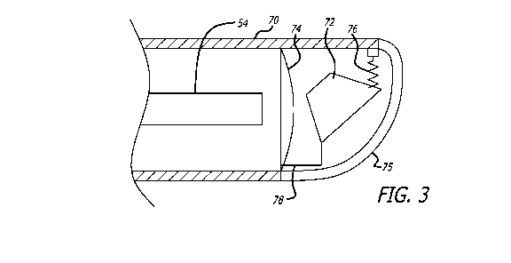
[0065] Referring now to FIGS. 3-6, various configurations distal portions 70
of the variable
degree of view endoscope 10 are shown, each having different configurations of
a swing prism
72 and/or mechanisms for mounting a swing prism 72. In a first approach, the
swing prism 72 is
mounted for rotation between a biasing spring 76 and an actuator 78. Here, the
actuator 78 can
come in the form of a wire which extends from the distal portion 70 of the
endoscope 10 to a
proximal portion which is conveniently accessible and manipulatable by an
operator. In this
regard, the actuator can be attached to a sliding member or configured to be
taken up by a
rotating dial (not shown). As so configured, images can be captured and
received through a
window 75 and transmitted through the swing prism 72 and self-focusing lens 74
to the image
fiber bundle 54. The swing prism 72 provides the desired seventy degree field
of view
throughout a viewing range of zero degrees to ninety five degrees by
manipulating the actuator
78.
[0066] In another approach shown in FIG. 4, the swing prism 72 can be mounted
in a
housing 82 placed in operative association with a rotatable shaft 84 which
extends proximally to
an operator. A distal portion of the shaft 84 is provided with threaded
structure 86 arranged to
engage teeth 88 formed on the housing 82. Rotating the shaft accomplishes
positioning the
swing prism 72 as desired. Again, these components can be arranged to provide
a one hundred
sixty five degree range of viewing.
[0067] In yet another approach shown in FIG. 5, the swing prism 72 can be
mounted in a
housing 90 placed in operative association with a flat bar 92 including teeth
94 which extends
proximally to an operator. The housing 90 can be mounted on a pin (not shown)
attached to the
distal end portion 70 of the endoscope shaft 30, where the housing and swing
prism pivot on the
pin. There are also teeth 98 on the housing that engage teeth 94 on the flat
bar. Moving the flat
bar in either the proximal or distal directions accomplishes positioning the
swing prism 72 as
13
CA 02732735 2011-01-28
WO 2010/014421 PCT/US2009/050800
desired. Again, these components can be arranged to provide one hundred sixty
five degree
range of viewing.
[0068] In an approach shown in FIG. 6, the swing prism 72 is mounted for
rotation between
a torsion spring 100 and a pull wire 102. The torsion spring can be any
spring, such as an
extension spring, leaf spring, or the like. Here, the pull wire 102 may extend
from the distal
portion 70 of the endoscope shaft 30 to a proximal portion which is
conveniently accessible and
manipulatable by an operator. In this regard, the pull wire can be attached to
a sliding member
or configured to be taken up by a rotating dial. Images can be captured and
received through the
window (not shown) and transmitted through the swing prism 72 and self-
focusing lens 74 to the
image fiber bundle 54. In this embodiment, there is always tension on swing
prism between the
torsion spring and pull wire, so there is no lag or buckling in the pull wire
during operation.
Further, use of the pull wire and torsion spring to move the swing prism
allows the diameter of
the endoscope to be smaller.
[0069] The images collected by the image fiber bundle 54 can be transmitted to
a monitor
(described below) to thereby provide the operator with visual data concerning
the particular
interventional procedure being performed. In one embodiment, the endoscope 10
be compatible
with a 300 Watt Xenon source and be configured with a universal light guide
connector, thus
making the assembly useable with conventionally available devices. In one
embodiment, the
endoscope shaft 30 may have an outer diameter of approximately 4 mm and a
working length of
about 175 mm. Moreover, the endoscope shaft 30 is preferably provided with
rounded surfaces
thus making the assembly atraumatic in use. It has also been found useful to
construct the
endoscope 10 in a manner and embodying material which permit the endoscope 10
to be
sterilized using an autoclave.
[0070] In certain approaches, it may be useful to configure the endoscope 10
with indicia
indicating the direction of view of the swing prism and/or a rotational
position of the endoscope
10. Thus, a proximal portion of the actuator 78 of FIG. 3, for example, can be
coupled with a
dial which includes markings indicative of the angle of the swing prism 72.
Similarly, a
proximal end of the shaft 84 of FIG. 4 can be attached to a dial including
indicia providing
information relative to the angle of the swing prism 72. Moreover, the
external surface of the
endoscope 10 can include marking indicators rotational positioning of the
overall assembly.
14
CA 02732735 2011-01-28
WO 2010/014421 PCT/US2009/050800
[0071] The swing prism endoscope 10 may be freely advanced within anatomy
along with a
sinus guide in order to facilitate endoscopic viewing of the desired
anatomical structures and/or
to view, guide and/or verify the positioning of the sinus guide device or a
working device that
has been inserted through the sinus guide. The ability to advance the tip of
the endoscope 10
within anatomy to view the end of the sinus guide allows the devices to be
positioned closer to
anatomy or to reach spaces in the paranasal sinuses that the devices cannot
travel due to size
constraints.
[0072] As discussed above with reference to FIGS. 3 through 6, the rotation of
the swing
prism may be controlled by a dial. As shown in FIG. 7, a proximal dial 104 is
disposed on the
handle 52 of the endoscope 10 for controlling the rotation of the swing prism.
The proximal dial
104 has a circular configuration and includes ridges 106 that provide leverage
for turning the
proximal dial or dial to a desired position. Further, the ridges provide a
tactile feel for the dial
location and grooves 108 between the ridges provide an area for the user's
fingers to rest. In one
embodiment, there are eight ridges evenly placed around the proximal dial 104,
however, there
may be fewer or more ridges placed around the dial. The height of the ridges
is approximately
0.05 inches, and can be increased or decreased depending on user preferences.
Also, the spacing
between each ridge is approximately 0.228 inches, and can be increased or
decreased depending
on the number of ridges disposed on the dial and the width of the ridges.
[0073] Still referring to FIG. 7, the handle 52 of the endoscope may include
indicia 107
adjacent the proximal dial 104 to provide information relative to the angle of
the swing prism 72.
In this embodiment, there is also a marker 108 on the proximal dial itself
indicating the relative
angle of the swing prism 72. As shown, the indicia 107 adjacent the proximal
dial indicate the
relative angle of the swing prism 72 anywhere from 0 degrees to 180 degrees.
[0074] In one embodiment, a distal dial or shaft dial 110 is disposed on the
handle 52 of the
endoscope as shown in FIG. 7, and the shaft dial 110 controls rotation of the
endoscope shaft 30.
A marker 112 is shown on the shaft dial 110 to indicate the relative position
of the endoscope
shaft 30. More particularly, the marker 112 on the shaft dial indicates the
relative position of the
window 75 (see FIG. 3) at the distal portion 70 of the endoscope 10. As shown
in FIG. 7, since
the marker 112 is on the top side of the endoscope, the window 75 is also
pointing towards the
top side of the endoscope 10, allowing the endoscope 10 to view the
surroundings in the same
CA 02732735 2011-01-28
WO 2010/014421 PCT/US2009/050800
general direction. Rotating the shaft dial 110 allows the endoscope to view
its surroundings in a
full three-hundred and sixty degrees of rotation. Having a rotating shaft dial
110 that rotates the
endoscope shaft 30 without rotating the entire handle 52 may be advantageous
because it allows
for rotation of the endoscope shaft 30 without rotating the light post 109.
[0075] FIG. 8 shows a handle attachment 114 attached to the handle 52 of the
endoscope 10.
The handle attachment 114 facilitates turning the dials 104 and 110 while the
user is holding the
endoscope 10. The handle attachment 114 is affixed to the handle 52 and/or may
be snap fit onto
the light post 109 stemming from the handle 52. A light post portion 116 of
the handle
attachment 114 snaps onto the light post 109 and shields the user from the
heat radiating from
the light post 109. When holding the handle attachment 114 and endoscope 10,
the crook
between the user's thumb and extended index finger is positioned at the curve
118 under the light
post portion 116 of the handle while the palm of the user's hand rests on the
body 120 of the
handle attachment 114. The handle attachment 114 may provide the user with
comfort and
balance while holding the endoscope and may also provide additional torque to
turn the dials 104
and 110. Holding the endoscope 10 with the handle attachment 114 allows the
user to turn the
proximal dial 104 with the thumb and index fingers, and the distal dial 110
can be accessed with
the ring or pinkie finger.
[0076] Another embodiment of a wrap-around handle attachment 122 that is snap-
fit onto the
handle 52 of the endoscope is shown in FIG. 9. The wrap-around handle
attachment 122 allows
the user to grip the endoscope tightly without impinging the rotation of the
dials 104 and 110. A
handle attachment back 124 is designed to be relatively long and rounded to
fit a variety of
positions within the palm of the user. With a light post cut out 126, the
handle attachment 122
can be moved or positioned around the handle 52 about two hundred and seventy
degrees to
facilitate a variety of holds by the user. The handle attachment 122 includes
an opening 128 that
allows the handle attachment 122 to more than half-way overlap the dials and
handle 52 of the
endoscope 10 while still allowing access to the dials 104 and 110.
[0077] Yet another embodiment of a handle attachment 130, including legs 132
that snap-fit
onto the handle 52 of the endoscope 10, is shown in FIG. 10. Handle attachment
130 includes a
back 134 that fits against the user's palm and a dial cover 136 that extends
over the proximal dial
104. A light post slot 138 can also be seen in FIG. 10 to accommodate the
light post 109. The
16
CA 02732735 2011-01-28
WO 2010/014421 PCT/US2009/050800
user is allowed to freely engage the dials 104 and 110 with his fingers when
holding the
endoscope 10 with the handle 130.
[0078] The optical fibers 54 of the endoscope 10 may be enclosed in a sealed
chamber to
allow the endoscope to be autoclaved. In one embodiment shown in FIG. 11, an
outer magnet
140 attached to a housing 142 is controlled in longitudinal motion by the
proximal dial 104
which drives a screw mechanism. A pin 144 is attached to the proximal dial 104
and extends
into the handle 52 and through a curved slot 146. The curved slot may spiral
around the housing
142. As the proximal dial 104 is turned, the pin moves along the curved slot
and moves the
housing 142 in a proximal or distal direction along the longitudinal axis of
the endoscope. As
the outer magnet moves forward and backward, it drives an inner magnet 148
that has an
opposite charge as the outer magnet. The inner magnet is disposed within an
inner shield 150
that creates the sealed chamber 151 for the optical fibers. The inner magnet
is also attached to a
push/pull mechanism 152 that rotates the swing prism at the distal end of the
endoscope. The
push/pull mechanism may be an actuator, pull wire, bar, hypotube, or the like,
that is attached to
the swing prism. As the inner magnet is driven forward or backward by the
movement of the
outer magnet, the inner magnet pushes or pulls the driver or push/pull
mechanism for the
rotatable prism.
[0079] In another embodiment shown in FIG. 12, a middle bellow joint 154 is
attached to the
housing 142 and is controlled in longitudinal motion by the proximal dial 104
which drives a
screw mechanism similar to the embodiment as shown in FIG. 11. The pin 144
attached to the
proximal dial extends into the handle 52 and through the curved slot disposed
in the housing 142.
There is also a proximal bellow joint 156 and a distal bellow joint 158 that
are fixed within the
endoscope on an inner shield 160, and there are flexible bellows 162 that are
disposed between
the bellow joints 154, 156, and 158. As the proximal dial 104 is turned, the
pin moves along the
curved slot and moves the housing 142 in a proximal or distal direction along
the longitudinal
axis of the endoscope and moves the middle bellow joint. As the middle bellow
joint moves
forwards and backwards, it drives the swing prism by moving the push/pull
mechanism 152 that
is attached to the middle bellow joint. The inner shield 160 creates the
sealed chamber 151 for
the optical fibers 54. The push/pull mechanism may be an actuator, pull wire,
bar, hypotube, or
the like, that is attached to the swing prism. In this embodiment, the bellow
joints can easily
17
CA 02732735 2011-01-28
WO 2010/014421 PCT/US2009/050800
transmit torque for rotation of a hypotube or rotatable shaft that can be
attached to the middle
bellow joint 154.
[0080] In one embodiment, the endoscope 10 is a re-usable instrument.
Conventionally,
endoscopes are processed between uses via steris, autoclave or other known
processes. The time
required to process the endoscope can be significant, resulting in delays
between cases or the
need to purchase multiple endoscopes for procedures occurring one after the
other. One
embodiment includes a disposable sterile sleeve 164 (see FIG. 1) that is used
with the endoscope
10. The sterile sleeve is low-profile and optically clear at the distal tip to
allow viewing with the
prism. The sterile sleeve spans the full length of the endoscope that is
inserted into the patient for
the procedure so that there is no direct contact between the patient and the
endoscope. Also, the
sterile sleeve may cover the proximal end of the endoscope and camera so there
is no direct
contact between the user and the endoscope. Once a procedure is complete, the
user simply
removes and discards the sterile sleeve and then inserts a new sterile sleeve
over the endoscope
for the next case. Using the sterile sleeve may eliminate the need to process
the endoscope
between cases or in the office environment.
[0081] During case procedures, endoscopes have the tendency to lose visual
clarity because
of the debris, blood, and/or mucus adhering to the distal tip of the
endoscope. Conventionally,
surgeons or users remove the endoscope from the patient frequently to clean
the distal tip of the
endoscope. Alternatively, some surgeons use scope washing systems having an
open sheath over
the endoscope shaft to deliver fluid and/or vacuum to enable in situ cleaning.
Each washing
sheath is specifically designed for the endoscope geometry, and because
endoscope distal tip
geometries vary by viewing angles, there are also multiple cleaning sheaths
that must be
correspondingly used. Therefore, when a user wants to change the scope viewing
angle during a
procedure, the washing sheath must also be changed. In one embodiment
described below, a
washing system and sheath is used with the endoscope 10. As described above,
the geometry of
the endoscope 10 does not change when the direction of the desired view
changes, and therefore,
a single fixed sheath may be used with the swing prism endoscope described
herein.
[0082] A washing system 168 is shown disposed on the endoscope 10 in FIGS. 13
through
15. The washing system includes a button 170 positioned between first and
second cones 172
and 174. The first and second cones 172, 174 are connected together by a
spring 176 (FIG. 14).
18
CA 02732735 2011-01-28
WO 2010/014421 PCT/US2009/050800
In this embodiment, the first cone 172 is fixed to the endoscope and the
second cone 174 is
connected to a wiping sheath 178. A distal end of the wiping sheath includes a
cloth 180, which
may be a hydrophilic elastomer. As shown in FIGS. 13 and 14, the washing
system 168 is in its
resting state, with the extension spring 176 in a drawn state and the first
and second cones at a
minimum distance from one another. In the resting state, the cloth 180 is
positioned proximal to
the lens 75 of the endoscope as shown in FIG. 13.
[0083] To move the washing system 168 forward to clean the lens 75 of the
endoscope, the
button 170 is pushed, such that it moves off the central axis of the endoscope
in any direction.
This movement of the button causes the second cone 174 to move forward since
the first cone
172 is fixed to the endoscope. Driving the second cone 174 forward or in the
distal direction
causes the wiping sheath 178 to also move forward and push the cloth 180 over
the lens 75 since
it is attached to the second cone. The activated state of the washing system
is shown in FIG. 15.
The cloth 180 is elastomeric, and therefore, it conforms to the shape of the
lens75 and wipes any
debris, mucus, and/or blood off of the lens. The porous hydrophilic cloth also
absorbs any fluid
which has collected on the lens. Once the button 170 is released, the spring
recoils and draws the
cloth back over the lens into a position proximal to the lens.
[0084] In one embodiment, the cloth 180 may have a supporting structure, such
as, rods,
mesh, or the like, to prevent the cloth from bunching or folding up when the
cloth is pushed
forward in the distal direction. Also, it has been contemplated that the
leading distal edge of the
cloth 180 may be silicone, rubber or some other hydrophilic material to wipe
the fluid forward
(distally) from the lens 75. The leading distal edge of the cloth may also
have multiple slits cut
into it to help wipe or push debris off of the lens.
[0085] In the embodiment described above, the endoscope 10 may have a
relatively rigid
shaft. However, it has been contemplated that the shaft of the endoscope 10
may also be flexible
to greatly enhance the viewing area of the endoscope. As shown in FIG. 16, a
typical endoscope
is shown that can visualize a fixed area A or B at any position within the
flexible range of the
typical endoscope. One embodiment of the current invention, shown in FIG. 17,
can visualize a
much larger range A' or B' by modifying the flex or position of the swing
prism within the
endoscope 10. It is contemplated that a flexible endoscope can be constructed
with fiber scope
or video chip technology. Such flexible endoscope may be useful for intra
nasal, intra sinus,
19
CA 02732735 2011-01-28
WO 2010/014421 PCT/US2009/050800
skull bass, laryngeal, orthopaedic, abdominal and other surgeries where a
variable and large
viewing range is desired.
[0086] In one embodiment, the endoscope 10 uses rod lens technology to acquire
and
transfer images along the shaft of the endoscope. In another embodiment, video
chip technology
as understood in the art requires rigidity around the distal portion of the
endoscope and the
images are transferred over a wire that enables the shaft of the endoscope to
be downsized.
Acquisition of the image via video chip technology may also allow the diameter
of the distal
portion of the endoscope to be downsized, without compromising the quality of
the image or the
size of the image that is viewed by the user. Current video chip technology
requires the distal
end of the endoscope to have a minimum diameter of about 1.2 mm to about 1.8
mm. With the
addition of illumination fibers and mechanics for the swing prism, an
endoscope using video
chip technology may be constructed with a diameter at the distal portion of
the endoscope of less
than 4 mm.
[0087] Certain embodiments are disclosed herein that increase the field of
illumination and
increase the field of image capture after miniaturizing an endoscope, such as
the swing prism
endoscope. When an endoscope is reduced in size or miniaturized, the number of
optical fibers
is decreased, thereby reducing the field of illumination utilizing such
optical fibers. Also,
miniaturizing an endoscope reduces the field of image capture due to the
smaller size of optical
components for the return image. As shown in FIG. 18, one embodiment of a
miniaturized
endoscope includes optical fibers 182 lapped at various angles from about 0
degrees to about 30
degrees. In this embodiment, the optical fibers can be arranged with such
angles increasing from
a chosen interior fiber 182a to the outer or edge fibers 182b, and thereby,
creating a wider
illuminating field A.
[0088] In another embodiment shown in FIG. 19, a diverging lens 184 can be
disposed at the
end of the optical fibers 182 to create a wider illuminating beam B. In this
embodiment, the
optical fibers are lapped at about 0 degrees; however, the diverging lens can
be combined with
lapped optical fibers similar to those shown in FIG. 18 to amplify the
divergence of the
illuminating beam. The diverging or expansion lens can be fabricated from a
block of glass with
the required curvature for beam divergence and then parted by using a saw or
high pressure
water jet to minimize edge defects. The non-functional sides of the individual
diverging lens
CA 02732735 2011-01-28
WO 2010/014421 PCT/US2009/050800
could be coated with nickel or gold to reduce optical leakage by creating
internally reflective
surfaces. It is noted that the input power to the optical fibers of a
miniaturized endoscope can be
increased to match the illumination intensity of a standard endoscope.
[0089] To maintain or improve the field of view captured by a return beam
through a prism,
a divergent lens may be used on a miniaturized endoscope. As shown in FIG. 20,
the
miniaturized endoscope includes a first prism 186 and a second prism 188
contacting the first
prism. There is also a divergent lens 184 disposed on the second prism 188
which increases the
field of view C. FIG. 21 shows a miniaturized endoscope with only the first
prism 186 being
used and the divergent lens 184 disposed near the first prism. As shown in
FIG. 21, 0 may be
optimized for the return beam relative to the axis of the endoscope.
[0090] In another embodiment, a concave or negative refractive power lens may
be mounted
to the distal prism 186 to increase the field of return image capture for the
return optics. As
shown in FIG. 22, a negative refractive power lens or concave lens 190 is used
in combination
with the positive refractive power lens or divergent lens 184 to achieve a
wider angle of image
capture while minimizing aberrations on the fiber optics to increase image
quality. In this
embodiment, the steering mechanism for the prism may be eliminated if the
range of the wide
angle image is sufficient to cover the target area without steering the prism.
In embodiments
where the steering mechanism is eliminated, this will create more space within
the miniaturized
endoscope for adding more illuminating fiber optics to better illuminate the
target area and
improve reliability.
[0091] In another embodiment shown in FIG. 23, two negative refractive power
lenses are
used with a single prism of a miniaturized endoscope including a prism
steering mechanism. As
shown in FIG. 23, a first negative refractive power lens or concave lens 190a
is disposed distally
of the prism 186 and a second negative refractive power lens or concave lens
190b is disposed
proximally of the prism 186. In this embodiment, the first and second concave
lenses can
operate in conjunction with one another or individually as required. Also, the
positive refractive
power lens or divergent lens 184 is positioned distally of the first concave
lens 190a. The
divergent lens 184 works with the first and second concave lenses 190a and
190b to reduce
optical aberrations in the lens system and to enhance the image quality.
21
CA 02732735 2011-01-28
WO 2010/014421 PCT/US2009/050800
[0092] Referring now to FIGS. 24A and 24B, one embodiment of the handle 52 of
the
endoscope may be open to allow fluid to freely move in and out of the handle
52. In this way,
the handle 52 of the endoscope may be cleaned and dried while the sealed
chamber 151 (see FIG.
11 or 24B) remains sealed. In one embodiment the proximal body 52 has an open
configuration
by drilling holes 192 into the housing of the handle 52. In another
embodiment, a mesh may be
used to create an open handle 52. Without an open configuration, there is a
possibility that fluid
may leak into the inner chamber of the handle 52 through a broken seal. Any
fluid that enters the
inner chamber of the handle 52 has the potential of rusting components and
allowing bacterial
growth. Therefore, providing the handle 52 with an open configuration prevents
this problem in
the inner chamber of the handle because any fluid entering will more easily
evaporate or be
drained through the holes 192.
[0093] The handle 52 of the endoscope shown in FIG. 24B is similar to the
embodiment
shown in FIG. 11, where the push/pull mechanism 152 is controlled by an outer
magnet 140 and
an inner magnet 148 as discussed above, and the inner magnet is disposed
within the inner shield
150 that creates the sealed chamber 151 for the optical fibers 54. Light fiber
194 is also shown
in FIG. 24B extending from the light post 193 and into the sealed chamber 151
or optical
chamber. In this embodiment, the light fiber must be free moving in order to
allow the
endoscope shaft to rotate in relation to the light post. In order to maintain
the seal on the sealed
chamber 151, a flexible sheath 196 covers the light fiber 194 and is affixed
to the sealed
chamber. This flexible sheath may be formed of silicone or steel. The flexible
sheath 196
allows the light fiber to move and the flexible sheath protects the light
fiber from damage.
[0094] In another embodiment shown in FIG. 24C, the light post as shown in
FIG. 24B has
been removed, and the light fiber 194 within the flexible sheath 196 exits the
handle 52. In this
embodiment, the light fiber would be connected to a light cable further away
from the
endoscope. Removing the light post prevents heat build up on the handle where
the user holds
the endoscope.
[0095] Yet another embodiment of an endoscope is shown in FIG. 25, where the
internal
mechanisms of the endoscope are sealed from the outside environment. FIG. 25
shows a cross-
sectional view of the handle 52 of the endoscope 10 with the internal driving
mechanisms
removed for clarity. In this embodiment, ferric fluid, which can be an oil
containing iron
22
CA 02732735 2011-01-28
WO 2010/014421 PCT/US2009/050800
particles mixed within, is injected into spaces 198 between the dials or dials
104 and 110 and the
inside portion of the handle 52. Teeth 199 are formed on the surfaces of the
dials 104 and 110 to
trap the ferric fluid as shown in FIG. 25. It has also been contemplated that
teeth can be formed
on the internal surface of the handle. Either the dials 104 and 110 or the
handle 52 can contain a
magnet disposed near or forming spaces 198, and these magnets can attract and
bond with the
magnetic ferric fluid. In another embodiment, both the dials and the handle
may contain
magnets at the spaces 198. As shown in FIG. 25, the teeth on the distal dial
110 are formed on a
proximal portion of the dial that is connected with the shaft of the endoscope
and positioned
within the internal chamber of the handle. Therefore, the spaces formed around
the internal
circumference of the handle become a fluid seal.
[0096] This bonding between the magnets within the dials 104 and 110 or the
handle 52 and
the ferric fluid allows the dials to move in relation to the handle with
little or no friction. Also,
this bonding seals the internal chamber of the handle from the outside
environment. These
fluidic seals will not wear like a typical O-ring and they are capable of
withstanding high
pressures.
[0097] Referring now to FIGS. 26A and 26B, one embodiment of a method of using
a swing
prism endoscope in the nasal and paranasal anatomy is described. For ease of
illustration, Figs.
26A and 26B show a nostril N, a nasal cavity 1009, and a non-specific
paranasal sinus 1022 with
a natural paranasal sinus ostium 1020. In various embodiments, the endoscope
10 may be used
in procedures addressing the maxillary, frontal, sphenoid and/or ethmoid
paranasal sinuses and
their related ostia. Figs. 27A-27D, for example, show a method involving
dilation of a natural
ostium of a sphenoid sinuse. However, it may be even more advantageous to use
a swing prism
endoscope of the present application in a procedure involving the maxillary
and/or frontal
paranasal sinuses, as the natural openings into these sinuses are usually
difficult to visualize
using an endoscope without removing one or more natural anatomical structures.
Therefore,
although Figs. 26A and 26B show a generic paranasal sinus, and Figs. 27A-27D
show a sphenoid
sinus, the endoscopes of the present invention may be used in any suitable
procedure involving
any paranasal sinus and/or nasal cavity. In further alternative embodiments,
endoscopes of the
present application may be used in procedures involving other portions of ear,
nose or throat
anatomy, such as but not limited to Eustachian tube procedures such as
dilation and/or stent
23
CA 02732735 2011-01-28
WO 2010/014421 PCT/US2009/050800
placement, repair of cranio-facial fractures, airway procedures such as
subglottic stenosis
dilation, tonsillectomy, adenoidectomy and/or the like.
[0098] As shown in FIG. 26A, in one embodiment a swing prism endoscope 10 may
be
inserted into a nostril N of a human or animal subject with the viewing angle
of the scope
adjusted to approximately 0 degrees (i.e., a straight ahead view), as
demonstrated by the ray lines
1024. In alternative embodiments, the endoscope 10 may not be capable of
viewing at 0 degrees
but maybe capable of between about 5 degrees and about 10 degrees as the most
"straight
ahead" angle. In either case, the physician may advance the endoscope 10
through the nasal
cavity 1009 using the straight ahead view, moving toward, for example, a
paranasal sinus ostium
1020, such as an ostium of a maxillary, frontal, sphenoid or ethmoid sinus.
FIG. 26B shows
endoscope 10 in a more advanced location. At some point during or after
advancing endoscope
10, the physician may adjust the swing prism of scope 30 to change its viewing
angle, for
example to look in the direction of ostium 1020. In one embodiment, endoscope
10 includes an
automatic focusing element, so that as the swing prism is adjusted and the
viewing angle
changed, endoscope 10 automatically refocuses. After viewing ostium 1020, the
physician may
decide to leave the viewing angle the same or make further adjustments to view
different
anatomy, an additional device inserted into the paranasal anatomy, and/or the
like. In some
embodiments, at any point during a procedure, a physician may be able to lock
the viewing angle
of endoscope 10 at a desired angle. When withdrawing the device from the human
or animal
subject's nostril, the physician may again adjust the swing prism viewing
angle back to 0 degrees
or may leave the angle as it was during any part of the procedure. Such a
method, or any of a
number of variations thereof, allows a physician to view anatomy of a nasal
cavity 1009,
paranasal sinus ostium 1020 and/or paranasal sinus 1022, as well as one or
more surgical
devices, during a procedure without having to switch out multiple different
endoscopes or to
remove tissue to look around corners.
[0099] FIGS. 27A through 27D are illustrations of partial sagittal sectional
views through a
human head showing various steps of a method for viewing and treating an
ostium of a paranasal
sinus, which in this example is a sphenoid sinus. In FIG. 27A, the swing prism
endoscope 10 is
introduced through a N nostril and through a nasal cavity 1012 to a location
close to an ostium
1014 of a sphenoid sinus 1016. The endoscope is used to view surrounding
anatomy using a
24
CA 02732735 2011-01-28
WO 2010/014421 PCT/US2009/050800
first, straight ahead viewing angle (or approximately straight ahead, such as
between about 5
degrees and about 10 degrees angled from the endoscope longitudinal axis).
[00100] In FIG. 27B, the angle of view of the endoscope 10 is altered to view
an ostium 1014
of a sinus 1016. In an alternative embodiment, one or more therapeutic or
diagnostic devices
may be advanced into the nasal cavity 1012 before the angle of view of the
endoscope 10 is
adjusted. In fact, the endoscope 10 may generally be advanced, adjusted,
removed and the like
in conjunction with any additional device(s) in any suitable order or manner
as desired.
[00101] As shown in FIG. 27C, in one embodiment, a guide catheter 212 may next
be
advanced into the nasal cavity 1012, in some cases but not necessarily
preloaded with a
guidewire 110 and/or a balloon catheter. The guidewire 110 may then be
advanced out of the
distal end of the guide catheter 212 such that it passes through the sinus
ostium 1014 and into the
sphenoid sinus 1016. A working device 1006, such as a balloon catheter, can be
introduced over
the guidewire 110, through the guide catheter, to position an expandable
member 213 such as an
inflatable balloon, into the sinus ostium 1014.
[00102] Thereafter, as shown in FIG. 27D, working device 1006 is used to
perform a
diagnostic or therapeutic procedure. In this particular example, the procedure
is dilation of the
sphenoid sinus ostium 1014, where the balloon of device 1006 is expanded to
enlarge the ostium
1014. After completion of the procedure, the sinus guide catheter 212,
guidewire 110 and
working device 1006 are withdrawn and removed. The entire procedure can be
observed using
the swing prism endoscope 10.
[00103] The features of the present disclosure may also be used to dilate or
modify any sinus
ostium or other man-made or naturally occurring anatomical opening or
passageway within the
nose, paranasal sinuses, nasopharynx or adjacent areas. In this or any of the
procedures
described in this patent application, the operator may additionally advance
other types of
catheters, and guidewire 110, guide catheter 212 or both may be steerable
(e.g. torquable,
actively deformable) or shapeable or malleable. Additionally, in various
alternative
embodiments, the endoscope 10 and one or more other devices, such as a guide
catheter 212,
may be integrated. In one embodiment, for example, a guide catheter 212 may
include an
endoscope lumen through which the endoscope 10 may pass.
CA 02732735 2011-01-28
WO 2010/014421 PCT/US2009/050800
[00104] The scope 30 may be useful to reduce or eliminate the need for
fluoroscopic
visualization during placement of a sinus guide and/or for visualization of
the procedure
performed by working device 1006. Being configured with a swing prism
providing a one
hundred sixty five degree viewing field, it can provide the capability to see
an opening into a
paranasal sinus and possibly even inside the sinus itself, and thus the
endoscope may provide
sufficient visual feedback for use in guiding guidewire 110 into the desired
sinus.
[00105] FIG. 28 shows one embodiment of a sinus guide system 210 which can be
used with
the swing prism endoscope 10 of the present disclosure. Sinus guide 212 maybe
straight,
malleable, or it may incorporate one or more preformed curves or bends as
further described
above, as well as in U.S. Patent Publication Nos. 2006/004323; 2006/0063973;
and
2006/0095066, for example, each of which are incorporated herein, in their
entireties, by
reference thereto. In embodiments where sinus guide 212 is curved or bent, the
deflection angle
of the curve or bend may be in the range of up to about 135 degrees. This
sinus guide system
210 comprises a sinus guide 212 and a camera/transmission/endoscope assembly
214. This
embodiment of the sinus guide 212 is shown in more detail in FIGS. 30A-30C. As
shown, this
sinus guide 212 comprises a sinus guide body 226 and an endoscope channel 228
in generally
side-by-side arrangement. As previously described, the swing prism endoscope
10 may be
inserted separately from the sinus guide system 210. In certain applications,
however, the
endoscope 10 also can be inserted through the endoscope channel 228.
Accordingly, the system
210 can also lack an endoscope channel 228. In either approach, the swing
prism endoscope can
be connected to a camera/transmission assembly and to a console 234 including
a monitor 236
and video recorder 240.
[00106] The sinus guide body 226 can embody a tube 244 having a lumen 245
(e.g., see FIG.
30B), such as a polymer tube made of biocompatible polymeric material.
Optionally, a liner 246
(FIG. 30B) may be disposed within the lumen 245 of the tube 244. Such liner
may be formed of
lubricious or smooth material such as polytetrafluoroethylene (PTFE). Also,
optionally, a
proximal portion of the tube 244 may be surrounded by an outer tube member 242
formed of
material such as stainless steel hypotube. In the embodiment shown, a distal
portion of the tube
244 extends out of and beyond the distal end of outer tube 242. This
protruding distal portion of
the tube 244 may be straight or curved. Also, it may be pre-formed at the time
of manufacture or
malleable to a desired shape at the time of use. When intended for use in
accessing the ostium of
26
CA 02732735 2011-01-28
WO 2010/014421 PCT/US2009/050800
a paranasal sinus, the distal portion of tube 244 may be curved to form an
angle A from about 0
degrees to about 120 degrees. For example, a series of sinus guides 212 having
angles A of 0,
30, 70, 90 and 110 degrees may be provided thereby allowing the physician to
select the sinus
guide angle A that is most appropriate for the particular paranasal sinus
ostium to be accessed.
[00107] Additionally, in some embodiments, a rotation grip 260 may be
positioned about a
proximal portion of the sinus guide 210, as seen in FIGS. 28, 30A and 30B.
This rotation grip
260 may have a smooth or textured round outer surface (e.g., it may be a
cylindrical tube) that
may be grasped between the fingers of the operator's hand and easily rotated,
thereby facilitating
rotation (e.g., rolling) of the sinus guide 212 as it is being used. Such
rotation of the sinus guide
212 may be desirable for a number of reasons including but not limited to
positioning of the
distal end of the sinus guide 212 at a desired location.
[00108] In the event it is desirable to configure the sinus guide system with
an endoscope
channel, it is contemplated that the channel 228 may comprise any structure
(e.g., tube, track,
groove, rail, etc.) capable of guiding the advancement of a flexible
endoscope. In the particular
examples shown in these figures, the endoscope channel 228 comprises a tube
(e.g., a polymer
tube) having a lumen 229 extending therethrough. In the embodiment seen in
FIGS. 28-30C, the
endoscope channel 228 is attached to and extends along substantially the
entire length of the
sinus guide body 226. In another embodiment, the endoscope channel 228 can be
inside the
sinus guide body 226. In other embodiments, the endoscope channel 228 may be
interrupted,
non-continuous or may extend over less than the entire length of the sinus
guide body 226. An
outer skin 240 may be heat shrunk or otherwise disposed around the sinus guide
body 226 and
endoscope channel 228 to hold the endoscope channel 228 at a desired position
on the outer
surface of the sinus guide body 226. Alternatively, the endoscope channel 228
may be attached
to the sinus guide body 226 at one or more locations by any other suitable
attachment substance,
apparatus or technique, including but not limited to adhesive, soldering,
welding, heat fusion,
coextrusion, banding, clipping, etc. The particular circumferential location
of the endoscope
channel 228 can be important in some applications, particularly when the sinus
guide body 226
includes a curve formed in its distal portion 244. In this regard, for some
applications, the
endoscope channel 228 may be affixed at a particular circumferential location
on the sinus guide
body 226 to allow an endoscope 10 inserted through the endoscope channel 228
to provide a
view from a desired or optimal vantage point, without obstruction from
adjacent anatomical
27
CA 02732735 2011-01-28
WO 2010/014421 PCT/US2009/050800
structures. It is also to be recognized that a second endoscope (not shown)
distinct from the
above described swing prism endoscope and which incorporates a swing prism or
otherwise
defines flexible structure can be inserted through the endoscope channel.
[00109] Again referring to FIGS. 28-30C, a proximal Y connector 241 may be
attached to the
proximal end of the sinus guide 212. A first arm 243b of this Y connector
comprises a female
Luer fitting that is connected to the lumen 245 of the sinus guide body 226.
The other arm 243a
is a female Luer fitting that is connected to the lumen 229 of the endoscope
channel 226.
[00110] A camera/cable/endoscope assembly 214 is attachable to arm 243a. In
the particular
embodiment shown in FIGS. 28 and 31, the camera/cable/endoscope assembly 214
comprises an
adjustable scope/lock extension 216, a camera 220 and a monitor cable 224. The
scope body 30
can be advanced through the scope/lock extension 216 and through the lumen 229
of the
endoscope channel 228. As shown in FIG. 29, the light cable 250 and monitor
cable 224 may be
connected to console 234 that houses a monitor 236, light source 238 and video
recorder 240.
Alternatively, the endoscope 10 can be directly connected to a console 234
separate from the
sinus guide system 212.
[00111] The invention has been described hereabove with reference to certain
examples or
embodiments of the invention, but various additions, deletions, alterations
and modifications
may be made to these examples and embodiments and or equivalents may be
substituted without
departing from the intended spirit and scope of the invention. For example,
any element or
attribute of one embodiment or example may be incorporated into or used with
another
embodiment or example, unless to do so would render the embodiment or example
unsuitable for
its intended use. In addition, many modifications may be made to adapt a
particular situation,
material, composition of matter, process, process step or steps, to the
objective, spirit and scope
of the present invention. All such modifications are intended to be within the
scope of the claims
appended hereto.
28