Note : Les descriptions sont présentées dans la langue officielle dans laquelle elles ont été soumises.
CA 02736625 2011-03-09
WO 2010/031036 PCT/US2009/056935
PELVIC IMPLANT SYSTEM AND METHOD
RELATED APPLICATION
This application claims priority to and the benefit of U.S. Provisional
Application No. 61/097,106, filed September 15, 2008, which is incorporated
herein
by reference in its entirety.
FIELD OF THE INVENTION
The invention relates to systems and methods for treating pelvic conditions
and, more particularly, to an implant device to secure to and/or support
pelvic tissue.
BACKGROUND OF THE INVENTION
Pelvic health for men and women is a medical area of increasing importance,
at least in part due to an aging population. Examples of common pelvic
ailments
include incontinence (fecal and urinary) and pelvic tissue prolapse (e.g.,
female
vaginal prolapse). Urinary incontinence can further be classified as including
different types, such as stress urinary incontinence (SUI), urge urinary
incontinence,
mixed urinary incontinence, among others. Other pelvic floor disorders include
cystocele, rectocele, enterocele, and prolapse such as anal, uterine and
vaginal vault
prolapse. A cystocele is a hernia of the bladder, usually into the vagina and
introitus.
Pelvic disorders such as these can result from weakness or damage to normal
pelvic
support systems.
Urinary incontinence can be characterized by the loss or diminution in the
ability to maintain the urethral sphincter closed as the bladder fills with
urine. Male
or female SUI occurs when the patient is physically stressed.
1
CA 02736625 2011-03-09
WO 2010/031036 PCT/US2009/056935
The female's natural support system for the urethra is a hammock-like
supportive layer composed of endopelvic fascia, the anterior vaginal wall, and
the
arcus tendineus. Weakening and elongation of the pubourethral ligaments and
the
arcus tendineus fascia pelvis, and weakening of the endopelvic fascia and
pubourethral prolapse of the anterior vaginal wall, may have a role in the
loss of
pelvic support for the urethra and a low non-anatomic position that leads to
urinary
incontinence.
In general, urinary continence is considered to be a function of urethral
support and coaptation. For coaptation to successfully prevent or cure
incontinence,
the urethra must be supported and stabilized in its normal anatomic position.
A
number of surgical procedures and implantable medical devices have been
developed over the years to provide urethral support and restore coaptation.
One alternative surgical procedure is a pubovaginal sling procedure. A
pubovaginal sling procedure is a surgical method involving the placement of a
sling
to stabilize or support the bladder neck or urethra. There are a variety of
different
sling procedures. Although complications associated with sling procedures are
infrequent, they do occur. Complications include urethral obstruction,
prolonged
urinary retention, bladder perforations, damage to surrounding tissue, and
sling
erosion.
Elongated fixating slings have also been introduced for implantation in the
body, to treat pelvic conditions such as prolapse and incontinence conditions.
Various systems and methods sold by American Medical Systems, Inc. under the
product names BioArc and SPARC provide a single use sling implantation tools
sold in a kit with an elongated urethral sling.
2
CA 02736625 2011-03-09
WO 2010/031036 PCT/US2009/056935
Another known implant system includes the use of a sling device having a
self-fixating tip at a distal end of an extension portion, and is sold under
the product
name MiniArc by American Medical Systems, Inc. The self-fixating tip can be
placed at and secured within internal tissue of the pelvic region to support
the
implant end extension and pelvic tissue that is supported by the implant. As
an
example, a self-fixating tip can be placed at tissue of the obturator foramen
(this
phrase referring to tissue that lies within or spans the obturator foramen,
for example
the obturator internus muscle, the obturator membrane, or the obturator
externus
muscle). Embodiments of these self-fixating tips can be designed to provide
desired
function and performance in positioning and tissue attachment. For example, a
self-
fixating tip can be designed to provide desirably low input force, desirably
high
pullout force, and reduced trauma caused by passage of the self-fixating tip
or an
associated insertion tool.
SUMMARY OF THE INVENTION
The present disclosure describes pelvic implant systems, devices and
methods for treating pelvic conditions such as incontinence (e.g., fecal
incontinence,
stress urinary incontinence, urge incontinence, mixed incontinence, etc.),
vaginal
prolapse (e.g., enterocele, cystocele, rectocele, vault prolapse, etc.), and
other like
conditions or dysfunctions. Embodiments of various implant devices including
one
or more tips, or self fixating tips, at a generally distal end of one or more
extension
portions, and one or more intermediate anchors generally attached, integrated
or
otherwise provided with the extension portion. The extension portion can be
constructed of a mesh or woven polymer or like compatible material.
3
CA 02736625 2011-03-09
WO 2010/031036 PCT/US2009/056935
The one or more anchors along portions of the extension portion can be
referred to as intermediate anchors provided in one embodiment proximate the
ends
of the mesh extension portions, or otherwise intermediate the end device tips,
to
provide or increase fixation until tissue in-growth into the implant occurs.
In
another embodiment, the one or more intermediate anchors can be disposed in
one
or more positions along a length of the mesh extension portions.
In one embodiment, the tips can be self-fixating tips securable within
internal
tissue of the pelvic region to further assist in supporting the implant
device. The one
or more intermediate anchors and tips can be configured of various sizes and
shapes.
The tips of the implant can be designed to engage a distal end of an insertion
tool to
allow the insertion tool to place the tip and intermediate anchors at a
desired tissue
location via pushing.
Embodiments of the intermediate anchors and tips can be designed to
provide desired fixation while simultaneously reducing trauma caused by
passage of
the implant and the corresponding insertion tool through and into the pelvic
region.
These functional properties can result from selecting desired overall
dimensions
(length or width) for the anchors and tips, angles of the anchor structure,
linear or
curvature designs, and other size, shape and extension configurations.
In one embodiment, the invention provides a method of treating urinary
incontinence in male and female patients (e.g., SUI) in a minimally invasive
manner
including injecting a local anesthetic; creating only one medial (e.g.,
transvaginal)
incision under the mid-urethra; inserting a urinary incontinence sling implant
through the one transvaginal incision, anchoring the urinary incontinence
sling, and
closing the incision.
4
CA 02736625 2011-03-09
WO 2010/031036 PCT/US2009/056935
Another aspect of the invention includes a combination (e.g., kit, system,
etc.) of an implant device, as described herein, including one or more
fixating tips
and one or more intermediate anchors. The kit also includes one or more
insertion
tools or systems useful for inserting, positioning and deploying the implant
device.
In another aspect, the invention relates to a method of treating a pelvic
condition. The method includes providing an implant device according to the
current description; providing an insertion tool that includes a handle and a
needle
extending from the handle, the needle including a proximal end attached to the
handle and a distal end, the distal end including a needle distal end that
removably
or selectively engages the device tip; engaging the needle distal end with the
tip;
inserting the needle distal end and tip through an incision in a patient; and
inserting
the tip and corresponding one or more intermediate anchors into tissue in the
pelvic
region such that an extension portion of the implant device supports the
targeted
pelvic tissue.
BRIEF DESCRIPTION OF THE DRAWINGS
Other features and advantages of the present invention will be seen as the
following description of particular embodiments progresses in conjunction with
the
drawings.
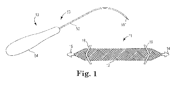
Fig. 1 is a view of a pelvic implant system, having an insertion tool and
implant device, in accordance with embodiments of the present invention.
Fig, 2 is a view of an implant device having end tips, an extension portion
and intermediate anchors, in accordance with embodiments of the present
invention.
5
CA 02736625 2011-03-09
WO 2010/031036 PCT/US2009/056935
Fig. 3 is schematic cross-section view of an implant device having end tips,
an extension portion and intermediate anchors, in accordance with embodiments
of
the present invention.
Fig. 4 is a partial cross-section view of an implant device, showing the
extension portion and an intermediate anchor, in accordance with embodiments
of
the present invention.
Fig. 5 is a partial view of an implant device having an end tip portion
coupled or in communication with an extension portion, in accordance with
embodiments of the present invention.
DETAILED DESCRIPTION OF THE INVENTION
The following description is meant to be illustrative and not limiting. Other
embodiments of this invention will be apparent to those of ordinary skill in
the art in
view of this description, claims and corresponding figures.
The present invention is directed to surgical instruments, assemblies, and
implantable articles for treating pelvic floor disorders such as fecal or
urinary
incontinence, including stress urinary incontinence (SUI), prolapse, etc.
According
to various embodiments, a surgical sling or implant device can be used to
treat a
pelvic condition, including the specific examples of implanting a support
implant to
treat a condition such as vaginal vault prolapse or incontinence (male or
female).
The sling implant or system may include portions or sections that are
synthetic (e.g., polymer) or constructed of biological material (e.g.,
porcine,
cadaveric, etc.). Extension portions may be constructed of a synthetic mesh
such as
a polypropylene, or other like materials. Examples of implant devices and
tools that
6
CA 02736625 2011-03-09
WO 2010/031036 PCT/US2009/056935
may be useful according to and with the present description include those sold
commercially by American Medical Systems, Inc., of Minnetonka MN, under the
trade names Apogee and Perigee for use in treating pelvic prolapse
(including
vaginal vault prolapse, cystocele, enterocele, etc.), and Sparc , Bioare ,
Monarc
and MiniAre 12 , for treating urinary incontinence. U.S. Patent Publication
Nos.
6,911,003, 6,612,977, 6,652,450, 2009/0192347, 2008/0119863, 2008/0045782, and
2004/0039453, and International PCT Publication No. 2008/057261, disclose
various implant devices, structures, procedures, systems and methods or
techniques
capable of use with the present invention and are, therefore, incorporated
fully
herein by reference.
Embodiments of the present invention, as shown in Figs. 1-5, include a sling
implant system 10 that can be installed to help maintain continence by
supporting
the urethra during times of increased abdominal pressure. The present
invention
also includes methods of implanting the sling. The sling system 10 can be
implanted
through a single incision in the vaginal wall for females (transvaginally), or
perineal
floor for males, and attached to (e.g., anchored) the obturator internus
muscle on
either side of the urethra. Only requiring one incision in the vaginal wall
(for
females) or perineum (for males) eliminates additional incisions such as
external
incisions used in some methods of implanting urethral slings. The sling system
10
and its methods of implantation can be, therefore, a reduced or "minimally"
invasive
treatment option for patients suffering from urinary incontinence.
In various embodiments, the sling system 10 may be anchored at other
locations besides the obturator internus muscle, such as, for example, the
obturator
membrane, the obturator externus muscle, etc.
7
CA 02736625 2011-03-09
WO 2010/031036 PCT/US2009/056935
Referring to Figs. 2-5, the implant system 10 can include an implant device
11 having an elongate extension portion 12, end or tip portions 14, 16, and
one or
more intermediate anchors 18. The extension portion 12 can include a first
generally longitudinal side 12a and a second generally longitudinal side 12b.
The
longitudinal sides 12a, 12b can extend a distance between the end or tip
portions 14,
16.
The extension portion 12 can be constructed of a polymer (e.g.,
polypropylene) mesh material, or other materials known for use with
incontinence
slings or pelvic tissue support devices. The extension portion 12 may be
woven,
knitted, sprayed, solid, or punched from a blank. In one aspect of the
invention,
extension portion 12 may include one or more woven, knitted, or inter-linked
filaments or fibers that form multiple fiber junctions. In addition, the size
of the
resultant openings or pores of a mesh embodiment of the extension portion 12
may
be sufficient to allow tissue in-growth and fixation with surrounding tissue.
Additionally, the extension portion 12 may be surface coated or impregnated
with
epithelialization-promoting agents, drugs or other materials to enhance tissue
impregnation.
Further, the extension portion 12 can include an intermediate band portion
22. The band portion 22 can be a plasma-treated print area of the sling
extension
portion 12 or a separately coupled or integrated band or indicia. The portion
22 can
facilitate tracking of the device during the surgical implant procedure.
The extension portion 12 generally extends between and is integrated or
otherwise coupled with the two end tips 14, 16. In alternative embodiments, a
single
tip, or no tip at all, could be implemented with the present invention. In
those
8
CA 02736625 2011-03-09
WO 2010/031036 PCT/US2009/056935
embodiments having one or more tips 14, 16, the tips can include an end
portion 24,
a first anchoring tine 26, a second anchoring tine 28, a body portion 30, and
a
coupling portion 32, as shown in Figs. 3 and 5. Further, the anchoring tines
26, 28
can be angled, rounded, linear, or take on a myriad of other shapes and
configurations. The coupling portion 32 is adapted for fixation with the
extension
portion 12. Fixation can be achieved by molding, mateable engagement,
clipping,
bonding, or other like techniques. The overall dimensions of the implant
device 11
may be 6 to 15 cm in length. Other proportional and dimensional embodiments
may
be employed without deviating from the spirit and scope of the present
invention.
In certain embodiments, the one or more intermediate anchors 18 can include
a plurality (e.g., two or more) of intermediate anchors 18a... 18n. The
anchors 18
can be constructed of polypropylene, polyglycolic acid (PGA), polylactide
(PLA),
copolymers of PGA and PLA, silicone, or any other material known by those
skilled
in the art, biodegradable or non-biodegradable. As such, the anchors 18 can be
generally rigid, hingeable, flexible or otherwise deformable to facilitate
placement
and fixation within the pelvic region.
As particularly illustrated in Figures 2-4, the implant device 11 can include
two intermediate anchors 18a, 18b. The intermediate anchors 18a, 18b can be
generally arcuate in shape, with end regions 36, 38 and 40, 42, respectively,
extending out from the extension portion 12. For example, Fig. 4 depicts a
length A
of the end regions extending out, e.g., generally transverse, from the
generally
longitudinal sides 12a, 12b of the extension portion 12. Further, the one or
more
intermediate anchors 18 can be configured or positioned such that they extend
out
(e.g., generally transverse) from other surfaces of the extension portion 12,
such as
9
CA 02736625 2011-03-09
WO 2010/031036 PCT/US2009/056935
the top, bottom, and like planes or surfaces of the extension portion 12. Such
anchor
configurations can be included in lieu of or in addition to anchors 18
extending out
from the longitudinal sides 12a, 12b. In various embodiments, the intermediate
anchors 18a, 18b can be generally V-shaped (Fig. 1), U-shaped, straight,
undulating,
etc. The end regions can be straight, angled, rounded, jagged or take on other
shapes
and configurations to facilitate attachment, fixation and/or retention to
tissue. In
addition, other arcuate, linear or angled shapes can be implemented for the
overall
design and shape of the anchors 18. Further, the anchors 18 can be placed at,
attached to, or proximate the tip portions 14, 16, or they can be placed in
any desired
location along a length of the extension portion 12. Various denution
surfaces,
protrusions, fibers, textures and the like can be included along portions of
the
intermediate anchors 18 (e.g., the end regions 36-42) to promote tissue
disruption
and/or fixation.
The sling extension portion, tips and anchors can exhibit desirable
"adjustability" or "positionability" features, without the need for a length-
adjusting
mechanism. Each tip or respective intermediate anchor of the implant device
can be
placed within a pelvic tissue such as tissue of the obturator foramen, with
properties
of the tips and anchors (e.g., dimensions, pullback force, number of lateral
extensions) and the implant (dimensions such as length between the tips and
corresponding anchors) being sufficient to allow placement at tissue on one or
both
sides of the pelvic region, while the sling extension portion of the implant
supports
the urethra, bladder neck, vaginal tissue, etc. Desired positioning of the
implant, the
proximity to the supported tissue (e.g., urethra), or the amount of supportive
force
CA 02736625 2011-03-09
WO 2010/031036 PCT/US2009/056935
placed on the supported tissue, can be achieved by selectively placing the
tips and
intermediate anchors.
A fixed length of implant material can be of a single piece of material
(integral), or may be of multiple pieces secured together. Pieces of the
implant 11
can be sewn or otherwise secured together, or pieces of synthetic material may
be
sewn or otherwise secured to a biologic material. For instance, as shown in
Fig. 2,
the intermediate anchors 18 can be attached to (e.g., bonded to and/or sewn or
interwoven with the strands or filaments of) the extension portion 12. Various
other
bonding or attachment methods and techniques can be employed as well.
The implant system 10 can further include an insertion or deployment tool
50. Various types of insertion tools are known, and these types of tools and
modifications thereof can be used according to this description to install an
implant.
Examples of useful tools include those described and depicted in previously-
incorporated PCT Patent Publication No. 2008/057261. In one embodiment, the
tool
50 generally includes a thin elongate needle or shaft portion 52 that attaches
to a
handle 54. A distal end 56 of the needle 52 can be adapted to engage one of
the tips
14, 16. The tip allows the needle to push the sling implant 11 through a
tissue
passage and insert the extension portion 12 and corresponding anchors 18
within,
confronting or along tissue of the pelvic region. Other embodiments can
utilize an
insertion tool 50 including a catheter delivery system, wherein at least
portions of
the implant 11 are adapted for positioning and deployment from a shaft of the
catheter.
Exemplary insertion tools for use according to the invention can be similar to
or can include features of tools described in the above-referenced patent
11
CA 02736625 2011-03-09
WO 2010/031036 PCT/US2009/056935
publications. For example, the insertion tool 50 may be used to place the
implant 11
and anchors 18 at tissue within the pelvic region through a tissue path that
does not
extend to an external incision, e.g., transvaginally. The insertion tool can
be
designed, shaped, and sized to include an elongate inserter or needle that may
be
straight or that may be curved in two or three dimensions, that can be
inserted
through a vaginal incision (for female anatomy) or through a perineal incision
(for
male anatomy), and to extend from that incision to a pelvic tissue location
for
placement of the extension portion 12, end or tip portions 14, 16, and
intermediate
anchors 18a, 18b. As such, the extension portion 12 can be positioned to
cradle,
press against or otherwise support the tissue.
According to certain methods of the invention, the tip portions 14, 16 and/or
the anchors 18 may be placed into pelvic tissue that is a fibrous tissue such
as
muscle, ligament, or tendon, with specific examples including the arcus
tendineus,
the obturator internus muscle, the levator ani, and the sacrospinous ligament.
The
end regions 36-42, or portions thereof, of the intermediate anchors 18 can be
designed for implantation within the fibrous tissue at an orientation that
places the
lateral extending end regions 36-42 in a direction that is non-parallel to the
fibers of
the fibrous tissue. As such, the end tips 14, 16 can provide an initial
placement
fixation to the tissue, with the intermediate anchors 18 further securing or
retaining
(e.g., secondary fixation) the implant 11 within or against the tissue.
One example of a method according to the invention is a method of treating
urinary incontinence by surgical implantation of a urethral sling implant I1
along a
tissue path that extends from a region of the urethra to the obturator
foramen. These
methods can advantageously involve only a single incision (a vaginal incision
in a
12
CA 02736625 2011-03-09
WO 2010/031036 PCT/US2009/056935
female or a perineal incision in a male) and can exclude the need for any
additional
incision. The elongate urethral sling 11 is attached at tissue of the opposing
obturator foramen by the respective tip portions 14, 16, with the extension
portion
12 positioned to pass below the urethra to support the urethra. The
intermediate
anchors 18a, 18b can be fixated or anchored to the tissue at or proximate the
tip
portions 14, 16 (e.g., the obturator foramen).
All patents and publications referenced herein are hereby incorporated by
reference in their entireties.
It will be understood that certain of the described structures, functions and
operations of the above-described preferred embodiments are not necessary to
practice the present invention and are included in the description simply for
completeness of an exemplary embodiment or embodiments. It will also be
understood that there may be other known structures, functions and operations
ancillary to the typical surgical procedures that are not disclosed, but that
can be
implemented to practice the present invention. It is, therefore, to be
understood that
within the scope of the appended claims, the invention may be practiced other
than
as specifically described without actually departing from the spirit and scope
of the
present invention.
13