Note : Les descriptions sont présentées dans la langue officielle dans laquelle elles ont été soumises.
CA 02737114 2016-01-20
FOOT, ANKLE, AND LOWER EXTREMITY COMPRESSION AND FIXATION SYSTEM
AND RELATED USES
BACKGROUND
[0001] There are
about 26 bones in the human foot (about 28 if you include the sesamoid
bones at the base of the big toe). These are: 1) the talus, which connects to
the tibia and fibula at
the ankle; 2) the calcaneus, which forms the heel; 3) the navicular, cuboid,
and three cuneiforms
(medial, intermediate, and lateral), which form the middle of the foot; 4) the
five metatarsals,
which radiate out to the toes; and 5) the 14 phalanges (2-3-3-3-3), which form
the toes.
[0002] The joint
named "ankle" is made up of the tibia, the fibula, and the talus. Below the
talus is another joint called the "below the talus joint" or, in another
language, the subtalar joint.
That joint between the talus and calcaneus is intricately inseparable from the
action of the
mid foot and rearfoot joints.
100031 Charcot
neuroarthropathy (CN) is defined by a bone and/or joint deformity in limbs
that have lost sensory innervation. The incidence is very high in diabetic
patients with peripheral
neuropathy. Currently, the pathogenesis of CN is largely unknown. This is
often reflected in
that diabetic patients with CN present challenging surgical candidates
secondary to diabetes-
related complications.
[00041 CN is
a progressive disorder believed to result from a disturbance in pain and
sensation as a result of peripheral neuropathy. Originally described as a
complication of
syphilitic neuropathy, the Charcot foot and ankle in the last decade has been
most commonly
associated and treated in patients with uncontrolled diabetes mellitus and
dense peripheral
neuropathy. The true etiology and nature of this debilitating condition is
still unknown, and the
- 1 -
CA 02737114 2016-01-20
treatment is variable and patient-dependent. Historically, foot and ankle
deformities as a result
of CN were treated with immobilization, total contact casting and, later, with
accommodative
footwear or bracing. Unfortunately, the evidence supporting non-operative
treatment for the
Charcot foot is equivocal. The increased risk of amputation in the non-
operative treatment of
CN should alert the treating physician to use caution and close monitoring in
the presence of a
severe deformity, fracture/dislocation, instability, and/or ulceration.
Currently, the literature has
shown variable protocols, techniques, and outcomes for surgical reconstruction
of these complex
and debilitating deformities, raising great concerns on the treatment options
for the Charcot foot
and ankle.
[0005] The
presence of ulceration, severe osseous deformity, poor bone quality,
neuropathy,
immune deficiency, obesity and multiple co-morbidities commonly seen in this
patient
population limit the use of traditional internal fixation to achieve a
successful outcome. The risk
of surgical infection is increased in diabetics due to their impaired immune
system. In the
presence of ulceration with an underlying bony prominence, this risk of
infection and future
amputation is high, and the ability to utilize internal fixation alone is
limited. Accordingly, CN
involving severe deformity, instability, ulceration, and/or infection of the
foot and/or ankle poses
difficulty in achieving limb salvage. When the surgeon is faced with this
clinical scenario, limb
salvage is often plausible through a rationale approach that, in various
treatments, incorporates
arthrodesis of affected joints to correct the deformity, plastic soft tissue
reconstruction for wound
closure, and application of an external fixation device.
[0006]
Generally, arthrodesis has been used to artificially induce joint ossification
between
two bones via surgery. A bone graft can be created between the two bones using
a bone from
elsewhere in the person's body (autograft) or using donor bone (allograft)
from a bone bank.
[0007] Bone
autograft is generally preferred by surgeons because, as well as eliminating
the
risks associated with allografts, bone autograft contains native bone-forming
cells (osteoblasts),
so the graft actually forms new bone itself (osteoinductive); it acts as a
matrix or scaffold to new
bone growing from the bones being bridged (osteoconductive). The main drawback
of bone
autograft is the limited supply available for harvest.
[0008] Bone
allograft has the advantage of being available in far larger quantities than
autograft; however, the treatment process the bone goes through following
harvest, which
- 2 -
CA 02737114 2016-01-20
usually involves deep-freezing and may also involve demineralization,
irradiation and/or freeze-
drying, kills living bone or bone marrow cells. This significantly reduces the
immunogenicity
(risk of graft rejection) such that no anti-rejection drugs are needed and,
combined with
appropriate donor screening practices, these processing and preservation
practices can
significantly reduce the risk of disease transmission. In spite of all of this
processing, cancellous
allograft bone retains its osteoconductive properties. Furthermore, certain
processing practices
have been shown to also retain the acid-stable osteo inductive proteins in
cortical bone grafts so
that many bone allografts can be considered both osteoconductive and
osteoinductive.
[0009]
Likewise, a variety of synthetic bone substitutes are commercially available.
These
are usually hydroxyapatite-based granules formed into a coralline or
trabecular structure to
mimic the structure of cancellous bone. Such materials can include, for
example, bone cement,
demineralized bone matrix, hydroxyapatite and calcium phosphate materials.
[0010] Many
of the above procedures are costly, require extensive healing and are not
always successful; accordingly, the art field is in search of novel and
improved processes and
apparatuses for treating such conditions.
[0011] In
view of the foregoing, it is an object of the present invention to provide an
apparatus for compressing and stabilizing a patient's foot, ankle, and/or
lower extremity,
particularly those conditions in which internal fixation alone is
insufficient. Hence, various
embodiments of the present invention include external fixation in combination
with internal
fixation to create compression across a larger linear gap. Such combination
systems and
methods may be advantageous for repairing diseased bone in which large defects
are present
such as, for example, CN.
SUMMARY
[0012] In
general, various embodiments of the present invention relate to therapeutic
orthopedic devices for compressing and stabilizing a patient's foot, ankle,
and/or lower
extremity. In general, the various embodiments of the present invention
include methods that
can be used to treat a variety of maladies from diseases to fractures.
[0013J In
various embodiments, a system of the present invention comprises an apparatus
or
device for compressing and stabilizing at least one bone in a patient's foot,
ankle, and/or lower
- 3 -
CA 02737114 2016-01-20
extremity, the system comprising an external fixation device, an internal
fixation device, and a
lower extremity stabilizer. The external fixation device is connected to the
lower extremity
stabilizer, and the external fixation device is adjustably connected to the
internal fixation device.
The internal fixation device is capable of being attached to at least one bone
in a patient's foot,
ankle, and/or lower extremity.
[0014]
Further embodiments of the present invention also include methods for treating
a
malady of the foot, ankle and/or lower extremity, the malady requiring
reduction of at least one
bone of a patient's foot, ankle, or lower extremity. The methods comprise
steps of attaching an
internal fixation device to at least one bone of a patient's foot, ankle, or
lower extremity;
adjustably connecting an external fixation device to the internal fixation
device with an
adjustable member to provide compression of at least one bone and stabilizing
the lower
extremity of the patient with an external stabilization member, the external
stabilization member
being fixedly connected to the external fixation device, wherein the malady is
treated. In other
embodiments, methods for treating Charcot neuroarthropathy using the systems
disclosed herein
are described.
According to one aspect of the invention there is provided a system for
compressing
and stabilizing at least one bone in a patient's foot, ankle, and/or lower
extremity in arthrodesis,
said system comprising:
an external fixation device, wherein the external fixation device is not
attached to the
at least one bone in the patient's foot, ankle and/or lower extremity;
an internal fixation device, wherein the internal fixation device attaches to
the at least
one bone in the patient's foot, ankle and/or lower extremity; and
a lower extremity stabilizer, wherein the lower extremity stabilizer comprises
at least
one rod and at least one stabilization member, and furthcr wherein the at
least one rod and the at
least one stabilization member do not attach to the at least one bone in the
patient's foot, ankle
and/or lower extremity; wherein said external fixation device is connected to
said lower extremity
stabilizer and said external fixation device is adjustably connected to said
internal fixation device,
and wherein the system compresses and stabilizes the at least one bone in the
patient's foot, ankle
and/or lower extremity and promotes arthrodesis.
According to a further aspect of the invention there is provided a method for
treating
a malady of the foot, ankle and/or lower extremity, said malady requiring
reduction of at least one
bone of a patient's foot, ankle, or lower extremity, said method comprising
the steps of:
- 4 -
CA 02737114 2016-01-20
a) attaching an internal fixation device to said at least one bone of a
patient's foot,
ankle, or lower extremity;
b) adjustably connecting an external fixation device to said internal fixation
device
with an adjustable member to provide a compression of said at least one bone,
the external fixation
device comprising a component adapted to contact the ground; and
c) stabilizing a lower extremity of said patient with an external
stabilization member,
said external stabilization member being connected to said external fixation
device, wherein said
malady is treated.
According to another aspect of the invention there is provided a method for
treating
Charcot neuroarthropathy in a patient, said method comprising a step of
attaching a system for
compressing and stabilizing at least one bone in a patient's foot, ankle,
and/or lower extremity,
said system comprising:
an external fixation device, wherein the external fixation device is not
attached to the
at least one bone in the patient's foot, ankle and/or lower extremity, and
wherein the external
fixation device comprises a component adapted to contact the ground;
an internal fixation device, wherein the internal fixation device attaches to
the at least
one bone in the patient's foot, ankle and/or lower extremity; and
a lower extremity stabilizer, wherein the lower extremity stabilizer comprises
at least
one rod and at least one stabilization member, and further wherein the at
least one rod and the at
least one stabilization member do not attach to the at least one bone in the
patient's foot, ankle
and/or lower extremity; wherein said external fixation device is connected to
said lower extremity
stabilizer and said external fixation device is adjustably connected to said
internal fixation device,
and wherein the system compresses and stabilizes the at least one bone in the
patient's foot, ankle
and/or lower extremity and promotes arthrodesis.
[0015] The foregoing has outlined rather broadly the features of the
present disclosure in
order that the detailed description that follows may be better understood.
Additional features and
advantages of the disclosure will be described hereinafter, which form the
subject of the claims.
BRIEF DESCRIPTION OF THE DRAWINGS
[0016] The foregoing summary, as well as the following detailed description
of the
invention, will be better understood when read in conjunction with the
appended drawings. It
- 4a -
CA 02737114 2016-01-20
should be understood, however, that the invention is not limited to the
precise descriptions and
instrumentalities shown herein, with emphasis instead being placed upon
clearly illustrating the
principles of the present invention. For a more complete understanding of the
present invention,
and the advantages thereof, reference is now made to the following
descriptions taken in
conjunction with the accompanying drawings, in which:
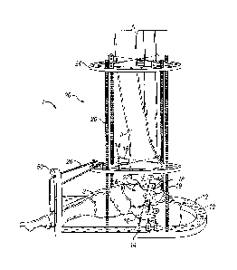
[00171 Figure
1 is an illustration of an embodiment of a device for compressing and
stabilizing a patient's foot, ankle, and/or lower extremity;
- 4b -
CA 02737114 2016-01-20
[0018] Figure 2 is
an illustration of an alternate embodiment of a device for compressing and
stabilizing a patient's foot, ankle, and/or lower extremity;
100191 Figure 3 is an illustration of an alternate perspective of the
embodiment of Figure 2;
100201 Figure 4 is
an illustration of an alternate embodiment of a device for compressing and
stabilizing a patient's foot, ankle, and/or lower extremity;
[0021] Figure 5 is
an illustration of an alternate embodiment of a device for compressing and
stabilizing a patient's foot, ankle, and/or lower extremity;
100221 Figure 6 is
an illustration of an alternate embodiment of a device for compressing and
stabilizing a patient's foot, ankle, and/or lower extremity;
[00231 Figure 7 is
an illustration of an alternate embodiment of a device for compressing and
stabilizing a patient's foot, ankle, and/or lower extremity; and
[0024] Figure 8 is
an illustration of an alternate embodiment of a device for compressing and
stabilizing a patient's foot, ankle, and/or lower extremity.
DETAILED DESCRIPTION
[00251 In the
following description, certain details are set forth such as specific
quantities,
sizes, etc. so as to provide a thorough understanding of the present
embodiments disclosed
herein. However, it will be obvious to those of ordinary skill in the art that
the present disclosure
may be practiced without such specific details. In many cases, details
concerning such
considerations and the like have been omitted inasmuch as such details are not
necessary to
obtain a complete understanding of the present disclosure and are within the
skills of persons of
ordinary skill in the relevant art.
100261
Referring to the drawings in general, it will be understood that the
illustrations are for
the purpose of describing a particular embodiment of the disclosure and are
not intended to be
limiting thereto. Drawings are not necessarily to scale.
100271 The
following definitions and explanations are meant and intended to be
controlling
in any future construction unless clearly and unambiguously modified in the
following
description or when application of the meaning renders any construction
meaningless or
essentially meaningless.
- 5 -
CA 02737114 2016-01-20
[0028] As
used herein, the term "internal fixation device" includes devices such as, for
example, at least one of wire(s), pins, nails, intramedullary nails, screws,
bolts, plates, staple(s),
brackets, fasteners, bars, and/or the like. Such internal fixation devices are
used for stabilizing
and/or compressing at least one bone in a patient's foot, ankle, and/or lower
extremity. Such
internal fixation devices may be constructed from a plurality of segments
comprising any
combination of the above components to form the complete device.
100291 As
used herein, the term "external fixation device" includes, for example,
monolateral, hybrid, circular, uni-plane, and bi-plane members, combinations
thereof and/or the
like for providing external fixation of a patient's foot, ankle and/or lower
extremity. Such
external fixation devices are used for stabilizing and/or compressing at least
one bone in a
patient's foot, ankle, and/or lower extremity.
[0030] As
used herein, the term "lower extremity stabilizer or lower extremity
stabilization
apparatus/device" includes devices for providing stability to a patient's
lower extremity,
including the foot and ankle. Such lower extremity stabilizers may be
connected to the above
referenced internal fixation devices and external fixation devices.
[0031] As
used herein, the term "radiolucent" means and refers to almost entirely
transparent
to radiation and/or almost entirely transparent in x-ray photographs and/or
almost entirely
transparent under fluoroscopy and/or other imaging modalities.
[0032] As
used herein, the term "adjustably connected" refers to a connection that can
be
tightened or loosened.
[0033] As used herein, the term "reduce" means and refers to repair.
[0034] As
used herein, the term "lower extremity" means and refers to the section of a
patient's kg below the knee.
[0035]
Various embodiments of the present invention generally relate to devices,
systems
and processes that can simultaneously reduce a malady of the foot, ankle,
and/or lower extremity
- 6 -
CA 02737114 2016-01-20
and provide compression and stabilization through the use of a combined
internal fixation device
and an external fixation device. Embodiments of the present combined system
are capable of
providing adequate compression in the medial, central and/or lateral columns
and/or sides of the
foot, ankle, and/or lower extremity, as well as the necessary stabilization
for healing and/or
therapy.
[0036] In
some embodiments, the compression is provided by the internal fixation device
and the external fixation device provides stabilization. In some other
embodiments, the external
fixation device provides compression and the internal fixation device provides
stabilization. In
still other embodiments, the compression and stabilization is provided by a
combined system of
the internal fixation device and the external fixation device. In still other
embodiments, the
internal fixation device or external fixation device alone provides the
compression. In some
embodiments, the internal fixation device is used alone, following an initial
use of the combined
system of the internal fixation device and external fixation device.
[0037] In an
embodiment, the present combined system includes an internal fixation device
adapted to have connectors such as, for example, transfixation wires, pins,
screws or
springs/spring clips fixed to it from a plurality of mounting blocks, tabs or
holes on the external
fixation device. Optionally, wires, pins, nails, staples, screws, bolts,
plates, brackets, fasteners,
bars, springs/spring clips, turnbuckles, twist locks, snap features and/or the
like may extend into
and/or through the bone segments in a patient's foot, ankle, and/or lower
extremity. In some
embodiments, the wires, pins, nails, staples, screws, bolts, plates, brackets,
fasteners, bars,
springs/spring clips, turnbuckles, twist locks, snap features and/or the like
may be attached to the
bone in such a way that they are attached substantially to the surface of the
bone. Such surface
attachment advantageously lowers the risk of infection in the patient.
[0038] The
present combined system further includes a lower extremity stabilization
assembly/device for compressing and stabilizing the foot, ankle and/or lower
extremity of a
patient. In some embodiments, the external fixation device and the lower
extremity stabilizer are
adjustably connected together. In some embodiments, the adjustable connection
comprises a
pivoting joint such as, for example, a ball joint. In some embodiments, the
external fixation
device and the lower extremity stabilizer are rigidly connected together. In
other embodiments,
the external fixation device and the lower extremity stabilizer are fixedly
connected but capable
- 7 -
CA 02737114 2016-01-20
of disengagement, such that at least one of the external fixation device
and/or the lower
extremity stabilizer can move relative to the other of the external fixation
device or the lower
extremity stabilizer. In some embodiments, a connection between the external
fixation device
and the lower extremity stabilizer can be adjusted to achieve compression. In
some
embodiments, the internal fixation device is adjustably connected to the lower
extremity
stabilizer. In other embodiments, the internal fixation device is rigidly
connected to the lower
extremity stabilizer. In embodiments wherein a rigid or fixed connection is
established, the
connection between components may be pre-tensioned while making the
connection. For
example, wires or other connectors may be stretched taut when making
connections. Alternately,
such connections may be tightened after connection such as with, for example,
a ratchet, gears or
like device.
[0039] Various embodiments of the present invention include a lower
extremity stabilizer,
which is any device capable of providing stabilization to a lower extremity.
Suitable examples
include, but are not limited to, a circumferential circular stabilizing
member, a monolateral
member, a hybrid member, a uniplane member, a biplane member, a combination of
one or more
of the aforementioned, other external fixation device configuration, and/or
the like.
[0040] A combination of the internal fixation device to the external
fixation device of the
foot, ankle and/or the lower extremity stabilizer is a system of the present
invention. In some
embodiments, at least a portion of such systems are radiolucent. In other
various embodiments,
at least a portion of such systems have radio-opaque markings. Such properties
are advantages
for monitoring therapeutic progress by various imaging techniques known to
those of ordinary
skill in the art.
[0041] In various embodiments of the present invention, at least one of the
external fixation
device, the internal fixation device and the lower extremity stabilizer may
comprise a material
that changes dimension in response to an external stimulus. Furthermore, an
adjustable member
comprising the system may also comprise such materials. For example, such
material may
expand or contract upon exposure to temperature, standing over time, and/or
interacting with
ultraviolet or microwave radiation. Illustrative materials for forming these
components of
system include certain polymers and Nitinol (Ni-Ti alloy), for example.
- 8 -
CA 02737114 2016-01-20
[0042] In any
of the various embodiments having adjustable connections, a change in
compression of the combination system may be achieved by adjusting a connector
interconnecting any one of the internal fixation device, external fixation
device and the lower
extremity stabilizer. Such connectors may include, for example, wires,
threaded wires, pins,
threaded pins, half pins, nails, staples, screws, bolts, plates, brackets,
fasteners, bars,
springs/spring clips, turnbuckles, twist locks, snap features and/or the like.
Adjustment of these
connectors may be performed using means known to those of ordinary skill in
the relevant art.
For example, in various embodiments of the invention compression may be
achieved using a
handheld tool (e.g., a ratchet or jig) to tighten screws or pins affixed to
the combination system.
Furthermore, such tightening with a handheld tool may take place while the
screws or pins are
attached to the internal fixation device alone (e.g., during surgery before
attachment of an
external fixation device). In some embodiments, an adjustable member forming
the adjustable
connection comprises a pivoting joint. The pivoting joint adjustable
connection may be
connected to at least one of the external fixation device, the internal
fixation device and the
lower extremity stabilizer. In various embodiments, springs or spring clips
are used to apply a
preloading to the adjustable member.
[0043]
Advantageous features of various embodiments of the present invention include,
but
are not limited to, the ability to provide maximum compression and
stabilization by a
combination of internal and external mechanisms of action. Further, various
embodiments allow
for an optionally fixed configuration or a configuration which allows relative
movement in
various joints of the foot, ankle and/or lower extremity. In various
embodiments of the
invention, after adequate healing of bones has been achieved, the external
fixation system is
removed such that only an internal fixation device remains in a patient's
foot, ankle, and/or lower
extremity for prolonged compression and stabilization and, in various
embodiments,
maintenance of the arthrodesis site(s). In embodiments wherein the external
fixation system is
removed, the remaining internal fixation system provides both compression and
stabilization.
When the external fixation system is removed, the internal fixation system may
be connected to
the lower extremity stabilizer, or the internal fixation system may be
unconnected. In such
embodiments, the lower extremity stabilizer is an optional component.
[0044] In
various embodiments, the present invention comprises an internal fixation
system
and an external fixation system that provides maximum compression and
stabilization
- 9 -
CA 02737114 2016-01-20
arthrodesis to all maladies of the foot, ankle, and/or lower extremity. In
various embodiments,
external threaded pins, wires, or other suitable connector means are capable
of being
incorporated with an internal fixation device to provide compression along a
single or multiple
arthrodesis site(s) across the foot, ankle, and/or lower extremity. In some
embodiments, an
interlocking screw is inserted within the internal fixation device to provide
further stabilization.
Interlocking holes of the internal fixation device, in various embodiments,
also allow the
insertion of compression screws initially, followed by further stabilization
through attachment of
externally threaded pins or other connector means that are attached to the
external fixation
device's foot, ankle and/or lower extremity stabilizer. In an alternate
embodiment, the threaded
pins or like connector means for providing compression are first used for
compression via an
external fixation device, and then internal screws or like connector means for
providing
stabilization are then applied to the internal fixation device. The threaded
pins, wires or like
connector means may then be secured to the external fixation device that is
capable of being
incorporated to the patient's foot, ankle and/or lower extremity stabilizer.
[00451 For purposes of the present disclosure, a malady of the foot, ankle,
and/or lower
extremity can be described as any injury, disease, or malformity, including,
but not limited to
Charcot neuroarthropathy, fractures, revisional foot, ankle, and/or lower
extremity surgery
including but not limited to malunions, nonunions, delayed unions, fibrous
unions, avascular
necrosis, resected osteomyelitis, incorporated autogenous and/or allogenic
bone grafts for
arthrodesis procedures, pseudoarthrosis and bones with decreased mineral
density and cortical
stiffness, congenital or acquired foot/ankle/lower extremity bone deformities
(pediatric or adult),
and/or the like for any reconstructive and/or elective surgery, and/or the
like.
[00461 A variety of technologies are capable of use for arthrodesis for the
internal fixation
device and the external fixation device of the present invention. In various
embodiments of the
present invention, athrodesis is capable of being accomplished through the use
of various
connector(s)/connecting members, including, but not limited to, plates(s),
intramedullary nail(s),
staple(s), pin(s), bolt(s), screw(s), wire(s), fastener(s), bar(s), rail(s),
spring(s)/spring clip(s),
turnbuckle(s), twist lock(s), snap feature(s) and/or the like. Arthrodesis,
according to various
embodiments of the present invention, is capable of being accomplished through
external
connection of at least one bone in the foot, ankle, and/or lower extremity;
or, reaming of at least
one bone of the foot, ankle, and/or lower extremity; or, other
internal/external connection of at
- 10 -
CA 02737114 2016-01-20
least one bone of the foot, ankle, and/or lower extremity; and/or, any other
means of connection
common in the art for athrodesis. In various embodiments, the connecting
members are
adjustable connecting members.
100471 In an
embodiment, arthrodesis is accomplished through a nail adapted for insertion
into the medullary canal of at least one bone of the foot, ankle, and/or lower
extremity which is
capable of being reamed or left unreamed. Reaming, in various embodiments, is
achieved by
drilling out the medullary canal of the bone with a malady, where the nail, or
other structure, is
inserted to stabilize and position the bone for healing. The size can vary
according to at least the
patient and the procedures, with an appropriate size being within the
understanding of one of
ordinary skill in the art.
[0048] In
various embodiments, intramedullary nail systems comprise intramedullary nails
having fastener holes at both the proximal and distal ends for the insertion
of fasteners, or
locking screws. Fasteners include all attaching means by which an implant is
capable of being
attached to bone. Such fasteners are capable of being inserted through a
fastener hole or slot,
also described as transfixation holes and screw holes.
[0049] In
various embodiments, the use of locking screws is optional depending upon, for
example, the severity of the bone damage and/or other factors. An
intramedullary nail implanted
with at least one fastener or locking screw both proximally and distally
completely locks the nail
in place (also known as static locking). Static locking neutralizes rotational
stresses while
preventing shortening of the limb. An intramedullary nail implanted with only
one locking
screw, either proximally or distally, partially locks the nail (also known as
dynamic locking).
Dynamic locking neutralizes rotational stresses on one side of the fracture
site while permitting
axial loading.
100501 In
various further embodiments, several nails exist with fasteners or locking
screws
that are much longer in shape, described as lag screws, jigs, or nail heads,
which further prevent
the rotation of separated bones. Other nails have spacers to absorb stress
associated with
repetitive, natural impact. All such devices are focused on securing bone
fragments during the
process of fracture healing. Other methods for attaching the nail to the bone
include the use of
longitudinal pins, or the use of cement injected through a cannula in a nail
to secure one end to
the nail.
- 11 -
CA 02737114 2016-01-20
[0051]
Various types of implants and intramedullary nails exist in the prior art,
each
composed of different materials and having different shapes with various
degrees of
functionality. In various embodiments nails are formed from either a solid
metal rod or a more
flexible sheet metal. In alternate embodiments, nails have been proposed which
form a rod
material containing a central longitudinal bore disposed throughout
(cannulated). Many nails
have a number of longitudinal grooves cut along the rod (known as fluting),
which allow for
more rapid revascularization within the bone. Accordingly, nails made of
various materials and
those having a differing array of shapes already exist and are capable of
being easily
manufactured. In some embodiments of the present invention, screws with hollow
bores may be
used in place of intramedullary nails.
[0052]
Further materials capable of use with various embodiments of the present
invention
include, but are not limited to nails fabricated using bioactive,
biocompatible, and bioabsorbable
material (e.g., bone cement, demineralized bone matrix, hydroxyapatite, and
calcium phosphate).
Such nails and like connectors are made from bioabsorbable polymers,
copolymers, or polymer
alloys that are self-reinforced containing ceramic particles or some type of
reinforcement fibers.
These implants and nails, as well as others, can also be made to be porous.
The prior art also
discloses methods to create human replica bones, gown in animals utilizing
human gene
technology, for ultimate use in humans, and such material is capable of being
a viable
supplement for standard intramedullary nails of today. Such techniques should
also be
considered as an appropriate part of the present invention. Likewise, in
various embodiments, a
system, or a portion of a system, is radiolucent or have radio-opaque
markings.
[0053] Now
referring to the Figures, Figure 1 is an illustration of an embodiment of the
present invention attached to a skeletal representation of a patient's foot,
ankle and lower
extremity. This particular embodiment of a system 1 is supporting the
patient's anIde 6, foot 3,
and lower extremity 8 along the medial aspect of the ankle. In general, an
embodiment of a
system of the present invention comprises an external fixation device 10 and a
lower extremity
stabilizer 20. In various embodiments, external fixation device 10 comprises
at least one
mounting block or mounting hole 12 for connection of at least one keeper or
tab 14 and
connector 16 which can be used for assisting with arthrodesis and/or
compression. Threaded
pins, half-pins, nuts, wires, bolts, screws, springs/spring clips,
turnbuckles, twist locks, snap
- 12 -
CA 02737114 2016-01-20
features and/or the like are also capable of use with various embodiments of
the present
invention in place of or in addition to tab 14 and connector 16 for support
and/or compression.
100541
External fixation device 10 is capable of being any shape that allows for
support of
the foot, ankle, and/or lower extremity. In system 1 shown in Figure 1, the
shape is that of a
horseshoe wherein the horseshoe shape lies flat along the ground. Circular,
rectangular,
spherical and other shapes also lie within the scope of various embodiments of
the
present invention. Half-ring additions 50 may also be included in the external
fixation device.
An external fixation device of an embodiment of the present invention includes
conventional
constructions such as those shown in the Figures and herein described but also
rail-type devices
and/or external fixation device configuration(s) and other orthotic
apparatuses used in foot-
related procedures.
[00551 Lower
extremity stabilizer 20 comprises at least one rod 26 and at least one
stabilization member 24. In various embodiments, holes or mounting blocks are
located along
stabilization member 24 such that connector(s) 16 can be connected to tab(s)
14 to assist in
arthrodesis and/or compression. Half-ring addition 50 is attached to at least
one of stabilization
member 24 using, for example, rods. Attachment of half-ring addition 50 may
also be
accomplished by other means such as, for example, pins and wires.
[00561 Still
referring to Figure I, an internal fixation device 18 is connected to bones of
foot
3, about ankle 6, and lower extremity 8 and comprises a series of sections 19
which are
attached/connected to the bones through at least one screw 22. At least one
connector 16 is
attached from tab 14 to a section 19 of internal fixation device 18. In
various embodiments,
there are multiple sections 19 within internal fixation device 18. In general,
internal fixation
device 18 is capable of being designed for the particular bones undergoing
arthrodesis, as shown
in subsequent Figures hereinbelow. Various shapes of internal fixation device
18 are
contemplated in the present invention including, for example, J-shape, L-
shape, straight, curved
and the like. Hence, the straight internal fixation device 18 in Figure 1
should not be considered
limiting. Further, in various embodiments, multiple internal fixation devices
18 may be affixed
to the bones undergoing arthrodesis.
[00571 Now
referring to Figure 2, a system 100, a foot 103, an ankle 106, and a portion
of a
lower extremity 108 are illustrated. In this alternate embodiment, an internal
fixation device 118
- 13 -
CA 02737114 2016-01-20
extends along at least one bone of the lateral aspect of foot 103. Also shown
in Figure 2 are
stabilization member 124, rod 126, external fixation device 110, mounting hole
112, sections
119, half-ring addition 150, screw 122 and tab(s) 114, wherein like terms are
described for
Figure 1.
[0058] Now
referring to Figure 3A, an alternative embodiment of system 1 is illustrated
wherein an internal fixation device 18 is illustrated and extends along the
medial aspect of at
least one bone of foot 3. Figures 3B and 3C show expanded views of internal
fixation device 18
showing attachment of screws 22. Figure 3C shows screw holes 60 and multi-
purpose hole 70,
having threads 71 on one side for accommodating threaded pins or screws and
non-threaded side
72 for attachment of non-threaded connectors. In some embodiments of the
present invention,
screws may be used in multi-purpose hole 70 and threaded pins may be used in
screw holes 60.
In other embodiments of the present invention, screws are used in screw holes
60, and threaded
pins are used in multi-purpose hole 70. Furthermore, the diameter of screw
holes 60 and multi-
purpose hole 70 may vary to accommodate different sizes of threaded pins
and/or screws. Multi-
purpose hole 70 may also hold both a combination of pins, screws, wires and
threaded pins.
Hole 61 may house a screw or a pin. In some embodiments, internal fixation
device 18 may be
attached to foot 3 in a reversed fashion by, for example, passing connector 16
through the foot
and ankle bones and attaching to external fixation device 10 on the opposite
side of the foot 3.
[00591 Now,
referring to Figure 4, an alternate embodiment of Figure 1 is illustrated with
the
use of an alternate external fixation device 400. Athrodesis is conducted
along the medial ankle
fusion and subtallar joint in the embodiment of Figure 4. External fixation
device 400 further
includes clamp connectors 420 operable for being connected by connectors 430.
An optional
lower extremity stabilizer is not shown. Internal fixation device 410 is
oriented as described in
Figure 1.
[0060j Now,
referring to Figure 5, an alternate embodiment of Figure 3 is illustrated with
the use of an alternate external fixation device 500. Athrodesis is conducted
along the medial
aspect of the foot in the embodiment of Figure 5. External fixation device 500
further includes
clamp connectors 520 operable for being connected by connectors 530. An
optional lower
extremity stabilizer is not shown. Internal fixation device 510 is oriented as
described in Figure
2.
- 14 -
CA 02737114 2016-01-20
[0061] Now,
referring to Figure 6, an alternate embodiment of Figure 2 is illustrated with
the
use of an alternate external fixation device 600. Athrodesis is conducted
along the lateral aspect
of the foot in the embodiment of Figure 5. External fixation device 600
further includes clamp
connectors 620 operable for being connected by connectors 630. An optional
lower extremity
stabilizer is not shown. Internal fixation device 610 is oriented as described
in Figure 2.
[0062] Now,
referring to Figure 7, an alternate perspective of the embodiment of Figures
2,
3, 5 and 6 is illustrated with the use of an alternate external fixation
device 700 and optional
lower extremity stabilizer 710. In the embodiment shown in Figure 7,
athrodesis is conducted
along the medial and lateral aspect of the foot. Two internal fixation devices
720 are oriented as
described in Figure 2. External fixation device 700 further includes clamp
connectors 730
operable for being connected by connectors 740. Lower extremity stabilizer 710
is shown
connected to external fixation device 700 through a threaded rod in Figure 7.
However, one of
ordinary skill in the art will recognize that such connection may be made
through various means
including, pins and wires, for example.
[0063] Now,
referring to Figure 8, an alternate perspective of the embodiment of Figures 1
and 4 is illustrated with the use of an alternate external fixation device 800
and optional lower
extremity stabilizer 810. In the embodiment shown in Figure 8, athrodesis is
conducted along
the lateral aspect of the ankle and subtallar joint. Internal fixation device
820 is oriented as
described in Figure 1. External fixation device 800 further includes clamp
connectors 830
operable for being connected by connectors 840. Lower extremity stabilizer 810
is shown
connected to external fixation device 800 through a threaded rod in Figure 8.
However, one of
ordinary skill in the art will recognize that such connection may be made
through various means
including, pins and wires, for example.
[0064]
Various embodiments of the present invention also relate to methods of using
systems
of the present invention for treating one or more maladies of the foot, ankle,
and/or lower
extremity. For example, in an embodiment, Charcot neuroarthropathy may be
treated using the
systems described herein.
j0065]
It will be understood that certain of the above-described structures,
functions, and operations of
the above-described embodiments are not necessary to practice the present
invention and are
- 15 -
CA 02737114 2016-01-20
included in the description simply for completeness of an exemplary embodiment
or
embodiments. It is therefore to be understood that the invention is capable of
being practiced other
than as specifically described without actually departing from the scope of
the present invention
as defined by the appended claims.
- 16 -