Note : Les descriptions sont présentées dans la langue officielle dans laquelle elles ont été soumises.
CA 02740260 2011-04-11
WO 2010/058014
PCT/EP2009/065668
1
REAL-TIME PERFUSION IMAGING AND QUANTIFICATION
Technical field
The solution according to an embodiment of the present invention relates to
the field of medical equipments. More specifically, this solution relates to
the field of
diagnostic systems.
Background of the invention
Medical tests are commonly used as tools for the diagnosis of a number of
pathologies ¨ e.g., following the report of corresponding symptoms. For this
purpose,
different techniques are available in the art.
For example, the gold standard technique for cancer diagnosis (e.g., in
prostate, liver, and breast) is biopsy, where samples of relevant tissues
(commonly
referred to as cores) are removed from a patient for examination. However,
biopsy is
a very invasive and expensive procedure. Moreover, biopsy is relatively
inaccurate in
specific applications (for example, its success rate is only approximately 70%
in
prostate cancer diagnosis, even with new strategies based on a higher number
of
cores).
Contrast-enhanced ultrasound analysis is another diagnostic technique that
finds increasing applications in the same field. Generally, this diagnostic
technique is
based on the administration of an ultrasound contrast agent (UCA) to the
patient - for
example, a suspension of phospholipid-stabilized gas-filled microvesicles (or
microbubbles); these contrast agent microbubbles act as efficient ultrasound
reflectors, and can be easily detected by applying ultrasound waves and
measuring
the echo signals that are returned in response thereto. Since the contrast
agent flows
at the same velocity as the red-blood cells in the patient, its detection and
tracking
provides information about blood perfusion in a body-part under analysis (from
which information about its condition can be derived).
CA 02740260 2016-07-05
2
Particularly, in an imaging approach, image sequences representing an
evolution of the contrast agent in the body-part during the perfusion process
are
generated (where the values of each pixel in the images represent an intensity
of the
recorded echo signal over time for a corresponding location of the body-part).
Therefore, examination of such image sequences (for example, displayed on a
monitor) provides only a qualitative indication of the blood perfusion in the
body-
part.
Conversely, in a quantitative approach, the echo signals recorded during the
whole perfusion process are fitted by mathematical model functions (for
example, as
disclosed in WO-A-2004/110279. The instances of the model functions so
obtained
can then be used to calculate different perfusion parameters (such as a wash-
in rate, a
wash-out rate, and the like). Any perfusion parameter may be calculated from a
global echo signal that is obtained in a predefined Region Of Interest (ROI)
comprising more than one pixel (with the perfusion parameter that is then
presented
as a single value). Alternatively, any perfusion parameter may be calculated
from the
echo signal of each pixel individually; a parametric image is then generated
by
graphically representing the value of the perfusion parameter for each
corresponding
pixel (preferably in a color-coded representation). The perfusion parameters
provide
a quantitative assessment of the blood perfusion in the body-part (with the
parametric
images representing a spatial map of the perfusion parameters throughout the
body-
part).
With reference in particular to prostate cancer diagnosis, studies using
targeted biopsy under contrast-enhanced ultrasound guidance have shown an
increase
of its success rate (with the possibility of reducing the number of required
cores).
Moreover, contrast-enhanced ultrasound analysis could also replace biopsy as
the
first choice in the prostate cancer diagnosis (with a dramatic reduction of
side-
effects, costs, and patient morbidity).
For this purpose, the use of contrast-enhanced ultrasound analyses as a tool
for the diagnosis of prostate cancer requires detection and characterization
of
corresponding lesions in the body-part. More specifically, the lesions are
detected
according to differences in perfusion kinetics compared to normal parenchymal
tissue (i.e., earlier and faster wash-in and wash-out of the contrast agent).
The lesions
CA 02740260 2016-07-05
3
can then be characterized (in order to differentiate benign lesions from
malignant
lesions) according to differences in their vascular properties (i.e., density
and/or
structure of corresponding microvascular networks).
Parametric analyses may be used to detect the lesions. Indeed, the
examination of parametric images based on corresponding perfusion parameters
(such as the wash-in rate and the wash-out rate) allows detecting the lesions
by
localizing regions in the body-part with high values of these wash-in and wash-
out
rates. However, reliable parametric analyses generally require images that are
spatially sub-sampled ¨ i.e. pixel values of groups of neighboring pixels are
low-pass
filtered and then sub-sampled (according to a sub-sampling factor) to produce
cell
values for corresponding cells, on which the fitting operation is then
performed. In
this way, it is possible to increase a signal-to-noise ratio (SNR) ¨ normally
very low
in the original echo signals ¨ and to reduce a computation time ¨ normally
very high
because of the complexity of the fitting operation and the large number of
pixels.
However, spatial sub-sampling generates parametric images with degraded
resolution
(which is not optimal for the characterization of the lesions). Moreover, echo
signals
must be recorded over an extended duration (encompassing the wash-in phase and
a
substantial part of the wash-out phase) in order to guarantee an acceptable
robustness
of the fitting operation (and then to provide reliable perfusion parameter
estimates).
Therefore, the echo signals are usually processed off-line (with a post-
processing
time that can easily exceed 3-8 minutes); proceeding in this way prevents any
real-
time examination of the body-part.
Imaging analyses may instead be used to characterize the lesions. Indeed, the
examination of the images representing blood perfusion in the body-part (at
full
resolution) is useful to determine its vascular properties. However,
identification of
tiny blood vessels (such as capillaries) is challenging because the local
contrast agent
concentration can be very low (with blood vessels that may even contain only a
single
contrast agent microbubble as they are being imaged).
In order to solve this problem, a solution known in the art involves the
application of a Maximum Intensity Projection (MIP) algorithm to the images
(for
example, as disclosed in US-B-6,676,606). Particularly, for each pixel the
maximum
intensity projection algorithm holds the corresponding values in the different
images
CA 02740260 2016-07-05
4
to their maximum over time. In this way, trajectories of contrast agent
particles are
projected spatially, so as to emphasize the corresponding blood vessel
morphology.
However, in this way the images become diffuse as soon as the contrast agent
starts
perfusing the parenchymal tissue surrounding the lesions; therefore, the
representation
of the vascular properties of the lesions gets blurred and looses conspicuity
(thereby
considerably reducing the effectiveness of the imaging analyses for lesion
characterization).
A Minimum Intensity Projection (mIP) algorithm is also known in the art; in
this case, for each pixel the minimum intensity projection algorithm holds the
corresponding values in the different images to their minimum over time. The
minimum intensity projection algorithm may be used before contrast agent
arrival in
the images to suppress background clutter and improve the visualization of the
contrast agent (for example, as suggested in US-B-6,436,049); however, this
algorithm is completely ineffective with respect of the above-mentioned
problems.
It should be noted that the imaging analyses based on the maximum intensity
projection algorithm might also be used to perform a qualitative detection of
the
lesions ¨ e.g., in locations of the body-part that exhibit an early
enhancement of the
contrast agent during the wash-in phase. However, the maximum values of the
echo
signals for the lesions and the parenchymal tissue may be similar, so that
their
representations after application of the maximum intensity projection
algorithm
become similar at times after reaching their corresponding peaks; therefore,
this
approach is useful to emphasize differences in the perfusion kinetics only
during the
short period of the wash-in phase. In any case, any information about the wash-
out
phase is completely lost (since the pixel values remain constant after
reaching their
peaks).
Summary
In its general terms, the solution according to an embodiment of the present
CA 02740260 2011-04-11
WO 2010/058014
PCT/EP2009/065668
invention is based on the idea of using signal monitoring techniques.
Particularly, an aspect of the present invention proposes a diagnostic system
(for example, an ultrasound scanner or a computer associated therewith). The
system
includes means for providing a plurality of input signals; the input signals
represent a
5 body-part
that is perfused with a contrast agent over time. Particularly, each input
signal is indicative of a response to an interrogating stimulus of a
corresponding
location of the body-part that possibly includes the contrast agent (for
example, an
echo signal from ultrasound pulses). The system also includes means for
generating a
plurality of filtered signals from selected input signals of selected
locations (for
example, in a region of interest); each filtered signal at each instant over
time is
generated from a corresponding selected input signal according to a portion of
the
selected input signal including said instant (for example, by applying a
maximum
intensity projection algorithm). In the solution according to an embodiment of
the
invention, means is provided for monitoring each filtered signal to detect a
peak in
the response to the interrogating stimulus of the corresponding selected
location; the
peak is detected when a stability condition is fulfilled by a corresponding
portion of
the filtered signal (for example, when the filtered signal remains constant
for a
predefined period).
In an embodiment of the invention, the means for monitoring each filtered
signal includes means for verifying the stability condition over time at a set
of
monitoring instants (for example, at each acquisition instant of the
corresponding
input signal); the verification is stopped after the stability condition has
been
fulfilled. Means is then provided for detecting the peak according to the
monitoring
instant at which the stability condition is fulfilled.
In an embodiment of the invention, the system includes means for verifying
(at each monitoring instant) whether the filtered signal remained constant in
a
stability time-window preceding the monitoring instant; the system then
includes
means for detecting the peak at an instant preceding the monitoring instant at
which
the stability condition is fulfilled by the stability time-window.
In an embodiment of the invention, the system further includes means for
CA 02740260 2011-04-11
WO 2010/058014
PCT/EP2009/065668
6
calculating one or more perfusion parameters - indicative of the perfusion of
each
selected location - according to the corresponding peak (for example, a wash-
in rate).
In an embodiment of the invention, the system includes means for generating
a linearized input signal from each selected input signal; the linearized
input signal at
each instant is substantially proportional to a concentration of the contrast
agent in
the corresponding selected location at said instant. The system also includes
means
for calculating each perfusion parameter according to the corresponding
linearized
input signal, at one or more instants that are determined by the means for
monitoring.
In an embodiment of the invention, the system includes means for providing a
sequence of input images. Each input image includes a digital representation
of the
body-part at a corresponding instant; particularly, each input image includes
a
plurality of input values each one indicative of the response to the
interrogating
stimulus of a corresponding location at the corresponding instant. The system
also
includes means for generating a sequence of filtered images from the input
images.
For each selected location, each filtered image includes a filtered value that
is
generated according to the input values corresponding to the selected location
in a set
of selected input images; the set of selected input images consists of a
corresponding
input image and one or more preceding input images. In this case, the system
includes means for monitoring the filtered values of each selected location.
In an embodiment of the invention, the system includes means for setting
each filtered value of each filtered image to a value, which is representative
of a
maximum response to the interrogating stimulus of the corresponding selected
location in the selected input images until the corresponding peak has been
detected
(for example, it is obtained by applying a maximum intensity projection
algorithm);
optionally, the filtered value may be also representative of a minimum
response to
the interrogating stimulus of the corresponding selected location in the
selected input
images after the corresponding peak has been detected (for example, it is now
obtained by applying a minimum intensity projection algorithm).
In an embodiment of the invention, the system includes means for setting the
filtered value to a value that is representative of the maximum response to
the
CA 02740260 2011-04-11
WO 2010/058014
PCT/EP2009/065668
7
interrogating stimulus between the filtered value of the selected location in
a
preceding filtered image and a comparison value until the corresponding peak
has
been detected; the comparison value is based on a set of input values of the
selected
location in a set of comparison input images (including the corresponding
input
image). Optionally, the filtered value may also be set to a value, which is
representative of the minimum response to the interrogating stimulus between
the
filtered value of the selected location in the preceding filtered image and
the
comparison value after the corresponding peak has been detected.
In an embodiment of the invention, the comparison value consists of the input
value of the selected location in the corresponding input image. In an
alternative
embodiment of the invention, the comparison input images consist of the
corresponding input image and one or more preceding input images; in this
case, the
system includes means for calculating the comparison value by applying a
smoothing
function to the input values of the selected location in the comparison input
images
(for example, a median function).
In an embodiment of the invention, the system further includes means for
generating one or more sequences of dynamic parametric images; for each
selected
location, each dynamic parametric image includes a null value before a
corresponding perfusion parameter is calculated, and a value that is
indicative of the
corresponding perfusion parameter after its calculation (for example, in a
color-
coded representation).
In an embodiment of the invention, the system includes means for
maintaining the null value for each selected location of each dynamic
parametric
image even after the calculation of the corresponding perfusion parameter,
when this
perfusion parameter does not reach a threshold value.
In an embodiment of the invention, the system further includes means for
generating a sequence of overlaid images for each sequence of dynamic
parametric
images; the overlaid images are generated by overlaying each dynamic
parametric
image on a corresponding filtered image.
In an embodiment of the invention, the system includes means for detecting
CA 02740260 2011-04-11
WO 2010/058014
PCT/EP2009/065668
8
an arrival instant (which is indicative of an instant at which the filtered
signal reaches
a significant value), and means for detecting a peak instant (which is
indicative of an
instant of detection of the peak); the system then includes means for
determining a
peak value, which is indicative of a response to the interrogating stimulus of
the
corresponding selected location at the peak instant. Optionally, the system
may also
include means for detecting a reduction instant, which is indicative of an
instant at
which the filtered signal reaches a reduction value - with the reduction value
being a
predefined fraction of the peak value (for example, a half-peak value).
In an embodiment of the invention, the system includes means for calculating
a wash-in rate (according to a ratio between the peak value and a difference
between
the peak instant and the arrival instant), means for calculating a wash-out
rate
(according to a ratio between the reduction value and a difference between the
reduction instant and the peak instant), means for calculating a product
between the
wash-in rate and the wash-out rate, or any other mathematical combination
thereof.
In an embodiment of the invention, the system further includes means for
applying a destruction pulse to the body-part (so as to cause a substantial
destruction
of the contrast agent); the system then includes means for repeating one or
more
times an actuation of the means for performing the above-mentioned operations.
Another aspect of the present invention proposes a corresponding data
processing method. Particularly, the data processing method includes the step
of
providing a plurality of input signals; the input signals represent a body-
part that is
perfused with a contrast agent over time. Each input signal is indicative of a
response
to an interrogating stimulus of a corresponding location of the body-part that
possibly includes the contrast agent. The method also includes the step of
generating
a plurality of filtered signals from selected input signals of selected
locations; each
filtered signal at each instant over time is generated from a corresponding
selected
input signal according to a portion of the selected input signal including
said instant.
In the solution according to an embodiment of the invention, each filtered
signal is
monitored to detect a peak in the response to the interrogating stimulus of
the
corresponding selected location; the peak is detected when a stability
condition is
CA 02740260 2011-04-11
WO 2010/058014
PCT/EP2009/065668
9
fulfilled by a corresponding portion of the filtered signal.
The same additional features described above with reference to the diagnostic
system apply mutatis mutandi to the data processing method (either alone or in
combination with each other).
A further aspect of the present invention proposes a corresponding computer
program. Particularly, the computer program includes code means for causing a
data
processing system to perform the steps of the above-mentioned data processing
method when the computer program is executed on the system.
A still further aspect of the present invention proposes a corresponding
computer program product. Particularly, the computer program product includes
a
computer-usable medium embodying a computer program, the computer program
when executed on a data processing system causing the system to perform the
same
data processing method.
Brief description of the drawings
The solution according to one ore more embodiments of the invention, as well
as further features and the advantages thereof, will be best understood with
reference
to the following detailed description, given purely by way of a non-
restrictive
indication, to be read in conjunction with the accompanying drawings, in
which:
FIG.1 is a pictorial representation of a medical imaging system in which the
solution according to an embodiment of the invention is applicable,
FIG.2A-2B illustrate an exemplary application of the solution according to an
embodiment of the invention,
FIG.3A-3B illustrate an exemplary application of the solution according to a
further embodiment of the invention,
FIG.4A-4B illustrate an exemplary application of the solution according to a
still further embodiment of the invention,
FIG.5A-5A' and FIG.5B-5B' show an exemplary scenario of application of
the solution according to an embodiment of the invention,
FIG.6A-6B illustrate an exemplary application of a maximum intensity
CA 02740260 2011-04-11
WO 2010/058014
PCT/EP2009/065668
projection algorithm and of a minimum intensity projection algorithm known in
the
art, respectively,
FIG.7A-7D show an example of in-vivo application of the solution according
to an embodiment of the invention compared with techniques known in the art,
5 FIG.8A-8C
show another example of in-vivo application of the solution
according to an embodiment of the invention compared with techniques known in
the
art, and
FIG.9A-9B show a diagram representing the roles of the main components
that may be used to implement the solution according to an embodiment of the
10 invention.
Detailed description
With reference in particular to FIG.1, a medical imaging system consisting of
an ultrasound scanner 100 is illustrated; the scanner 100 may be used to
analyze a
body-part 102 of a patient 103 in the solution according to an embodiment of
the
invention. The ultrasound scanner 100 includes a central unit 105 and a hand-
held
transmit-receive imaging probe 110 (for example, of the array type). The
imaging
probe 110 transmits ultrasound waves consisting of a sequence of pulses (for
example, having a center frequency between 1 and 50 MHz), and receives radio-
frequency (RF) echo signals resulting from the reflection of the ultrasound
pulses by
the body-part 102; for this purpose, the imaging probe 110 is provided with a
transmit/receive multiplexer, which allows using the imaging probe 110 in the
above-described pulse-echo mode.
The central unit 105 houses a motherboard 115, on which the electronic
circuits controlling operation of the ultrasound scanner 100 (for example, a
microprocessor, a working memory and a hard-disk drive) are mounted. Moreover,
one or more daughter boards (denoted as a whole with 120) are plugged into the
motherboard 115; the daughter boards 120 provide the electronic circuits for
driving
the imaging probe 110 and for processing the received echo signals. The
ultrasound
scanner 100 can also be equipped with a drive 125 for reading removable disks
130
CA 02740260 2016-07-05
11
(such as CD-ROMs or DVD-ROMs). A monitor 135 displays images relating to an
analysis process that is in progress. Operation of the ultrasound scanner 100
is
controlled by means of a keyboard 140, which is connected to the central unit
105 in
a conventional manner; preferably, the keyboard 140 is provided with a
trackball 145
that is used to manipulate the position of a pointer (not shown in the figure)
on a
screen of the monitor 135.
During the analysis of the body-part 102, a contrast agent (acting as an
efficient ultrasound reflector) is administered to the patient 103. For
example, the
contrast agent consists of a suspension of gas bubbles in a liquid carrier;
typically,
the gas bubbles have diameters on the order of 0.1-5 um, so as to allow them
to pass
through the capillaries of the patient. The gas bubbles are generally
stabilized by
entraining or encapsulating the gas or a precursor thereof into a variety of
systems,
including emulsifiers, oils, thickeners, sugars, proteins or polymers;
stabilized gas
bubbles are generally referred to as gas-filled microvesicles. The
microvesicles
include gas bubbles dispersed in an aqueous medium and bound at the gas/liquid
interface by a very thin envelope involving a surfactant, i.e., an amphiphilic
material
(also known as microbubbles). Alternatively, the microvesicles include gas
bubbles
that are surrounded by a solid material envelope formed of lipids or of
natural or
synthetic polymers (also known as microballoons or microcapsules). Another
kind of
contrast agent includes a suspension of porous microparticles of polymers or
other
solids, which carry gas bubbles entrapped within the pores of the
microparticles.
Examples of suitable aqueous suspensions of microvesicles, in particular
microbubbles and microballoons, and of the preparation thereof are described
in EP-
A-0458745, WO-A-91/15244, EP-A-0554213, WO-A-94/09829 and WO-A-
95/16467. An example of a commercial contrast agent comprising gas-filled
microvesicles is SonoVue by Bracco International By.
Preferably, the contrast agent is administered to the patient 103
intravenously
as a bolus - i.e., a single dose provided by hand with a syringe over a short
period of
time (of the order of 2-20 seconds). The contrast agent circulates within a
vascular
system of the patient 103, so as to perfuse the body-part 102. At the same
time, the
imaging probe 110 is placed in contact with the skin of the patient 103 in the
area of
the body-part 102. The body-part 102 is then insonated by applying a series of
CA 02740260 2016-07-05
12
ultrasound pulses with low acoustic energy (such as with a mechanical index
MI=0.01-0.1), so as to involve a negligible destruction of the contrast agent
(such as
less than 5%, and preferably less than 1% of its local concentration between
successive ultrasound pulses). The echo signals that are recorded in response
to the
ultrasound pulses over time (for each location of the body-part 102 in a
selected
scanning plan) provide a representation of a corresponding region (i.e., a
slice) of the
body-part 102 possibly including the contrast agent - during the analysis
process.
The echo signals are then converted into a sequence of digital images (or
frames) in standard Brightness mode (B-mode), which then represent the body-
part
102 at corresponding successive acquisition instants (for example, with a
frame rate
FR=10-30 images per second). Each image is defined by a matrix (for example,
with
M=512 rows and N=512 columns) of values for respective visualizing elements -
i.e.,
basic picture elements (pixels), each one corresponding to a location of the
body-part
102. Typically, each pixel value consists of a gray-scale level (for example,
coded on 8
bits) defining the brightness of the pixel; the pixel value increases from 0
(black) to
255 (white) as a function of the intensity of the corresponding echo signal
(representing the acoustical response at the corresponding location of the
body-part).
The echo signals and then the corresponding images generally result from the
superimposition of different contributions generated by the contrast agent and
the
surrounding tissue. Preferably, the ultrasound scanner 100 operates in a
contrast-
specific imaging mode so as to substantially remove, or at least reduce, the
dominant
(linear) contribution of tissue in the echo signals, with respect to the (non-
linear)
contribution of the contrast agent; examples of contrast-specific imaging
modes
include harmonic imaging (HI), pulse inversion (P1), power modulation (PM) and
contrast pulse sequencing (CPS) techniques, as described, for example, in
"Rafter et
al., Imaging technologies and techniques, Cardiology Clinics 22 (2004), pp.
181-
197".
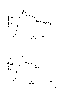
In FIG.2A, a time-intensity curve 205 (solid-line) is shown as an exemplary
response to the ultrasound waves of a generic location of the body-part ¨
representing the power of the corresponding echo signal (in terms of arbitrary
units,
or a.u.) as a function of time (in seconds). The curve 205 has an initial
portion
CA 02740260 2011-04-11
WO 2010/058014
PCT/EP2009/065668
13
wherein the echo signal increases from zero (before contrast agent arrival)
towards a
peak, as a result of a wash-in phase of the contrast agent perfusing the body-
part after
its administration; once the echo signal has reached its absolute maximum
value at
this peak, it starts decreasing towards zero as a result of a wash-out phase
of the
contrast agent that is filtered out of the patient (for example, by the lungs
and/or by
the liver).
The echo signal is at first filtered, so as to generate a filtered signal that
is
represented in the figure with a curve 210 (dashed-line). As described in
detail in the
following, at each instant the filtered signal is generated from the echo
signal
according to a corresponding portion of the echo signal including the same
instant.
For example, at the beginning the filtered signal is generated by applying the
maximum intensity projection algorithm, wherein the echo signal is held at its
maximum value over time. In this way, the corresponding portion of the curve
210
(denoted with 210a) accurately follows the curve 205 when the echo signal
increases
monotonously; however, if the echo signal momentarily decreases (for example,
due
to noise or natural fluctuation in local contrast agent concentration), its
last
maximum value is preserved until a higher value of the echo signal is
detected.
The filtered signal can be generated at any instant simultaneously with the
recording of the echo signal (or at most with a very short delay). For
example, the
maximum intensity projection algorithm only requires the knowledge of the echo
signal up to the corresponding instant (so that the filtered signal can be
generated in
real-time according to information that is already available).
In the solution according to an embodiment of the invention, the filtered
signal is subsequently monitored in order to detect its peak (at a peak
instant tp, when
the filtered signal reaches a peak value If); particularly, the peak is
detected when a
corresponding portion of the filtered echo signal fulfills a stability
condition. For
example, this happens when the filtered signal remains constant for a
predefined
period. Therefore, the peak is now detected as soon as it occurs (with a short
delay
required for fulfilling the stability condition).
The filtering of the echo signal is relatively simple and does not require
extensive computational resources, so that it can be performed at the pixel
level
CA 02740260 2011-04-11
WO 2010/058014
PCT/EP2009/065668
14
(without any spatial sub-sampling of the images). Nevertheless, the filtered
signal is
significantly smoothed (by removing any strong variations of the corresponding
echo
signal before filtering), thereby allowing the detection of the peak in a
robust way.
As a result, the proposed processing can be performed at full image resolution
with
an acceptable degree of reliability.
At the same time, the proposed solution allows obtaining the desired results
substantially in real-time; this means that the analysis process is being
carried out
while the body-part is being imaged - i.e., with a short delay from the
detection of the
peaks due to the required computations, but without the need of waiting for
the
imaging process to be completed.
In addition or in alternative, after the detection of the peak the filtering
operation is switched to the minimum intensity projection algorithm, wherein
the echo
signal is held at its minimum value over time. In this way, the corresponding
portion
of the curve 210 (denoted with 210b) now accurately follows the curve 205 when
the
echo signal decreases monotonously (during the contrast agent wash-out phase);
however, if the echo signal momentarily increases (due to noise or natural
fluctuations in the contras agent concentration), its last minimum value is
preserved
until a lower value of the echo signal is detected.
In this way, information about the wash-out phase is preserved as well. At the
same time, further examinations of the body-part may be performed again on
full-
resolution images and substantially in real-time.
The information relating to the detection of the peak can be used for
different
purposes. Particularly, as shown in FIG.2B, in an embodiment of the invention,
this
information is used to calculate one or more perfusion parameters (indicative
of the
blood perfusion in the corresponding location of the body-part). For example,
it is
possible to calculate a wash-in rate Wi (as represented in FIG.2B by a
corresponding
dashed-dotted straight line) using the following formula:
Wi= ,
Ati
wherein ziti=tp-ta measures a duration of the wash-in phase (from a contrast
agent
arrival instant ta to the peak instant tp). The arrival instant ta is defined
as the instant
CA 02740260 2011-04-11
WO 2010/058014 PCT/EP2009/065668
at which the filtered signal reaches a significant value /a exceeding a
predefined
threshold value. It should be noted that this wash-in rate Wi is very
reliable, since it
is completely independent of the instant of the contrast agent administration.
In addition or in alternative, the filtered signal is also monitored to detect
a
5 half-peak
instant tr, wherein the filtered signal falls to under a half-peak value Ip/2.
It
is then possible to calculate a wash-out rate Wo (as represented in FIG.2B by
a
corresponding dashed-dotted straight line) using the following formula:
/ /2
Wo = v ,
Ato
wherein Ato=tr-tp measures a duration of the wash-out phase (from the peak
instant tp
10 to the
half-peak instant tr). In this case as well, the wash-out rate Wo is very
reliable,
since it is again independent of the instant of the contrast agent
administration.
In an embodiment of the invention, the above-described method is used to
generate a sequence of filtered images from the (original) images representing
the
body-part. Particularly, for each pixel the corresponding pixel values in the
different
15 filtered
images are obtained by applying the maximum intensity projection algorithm
before their peak instant tp and the minimum intensity projection algorithm
afterward.
More formally, before the peak instant tp, each pixel value of the filtered
images is set to the maximum between the pixel value for the same pixel in the
corresponding original image and the running maximum of the pixel values for
the
same pixel in the preceding original images as resulting from the earlier
iterations of
the process, that is:
OP(x, y,k)= MAX [IP(x, y,k),OP(x, y,k -1)1,
wherein OP(x,y,k) is the pixel value of the pixel identified by the spatial
coordinates
x,y (row number and column number, respectively) in the filtered image with
number k
(taken at an instant t being the inverse of the frame rate of the original
images
multiplied by the image number k - i.e., t=k/FR). IP (x,y,k) and OP(x,y,k-1)
are the
pixel values of the same pixel (x,y) in the corresponding original image with
the same
number k (taken at the instant t) and in the preceding filtered image with the
number k-
1 (taken at the instant (k-1)/FR), respectively, and MAX[1 is a function
returning the
CA 02740260 2011-04-11
WO 2010/058014
PCT/EP2009/065668
16
maximum value among its arguments. After the peak instant tp, instead, each
pixel
value of the filtered images is set to the minimum between the pixel value for
the same
pixel in the corresponding original image and the running minimum of the pixel
values
for the same pixel in the preceding original images (from the peak forward) as
resulting from the earlier iterations of the process, that is:
OP(x,y,k)=MIN[IP(x,y,k),OP(x,y,k-1)],
wherein MIND is a function returning the minimum value among its arguments.
In general, each pixel value of the filtered images can then be calculated by
applying the following filtering function:
MAX[IP(x,y,k),OP(x,y,k -1)] if k k +L
OP(x,y,k)= ,
MIN[IP(x,y,k),OP(x,y,k -1)] if k > kp+ L
wherein L (with L>0) is a stability length, which represents a number of
filtered
images that are used to detect the peak, and icy (with kp>L) is a peak number
that
expresses the peak instant tp in terms of image number (with tp=kp/FR).
Particularly,
the peak number ki, is set to the image number k that satisfies the stability
condition
defined by:
kp=k for OP(x,y,k)¨ OP(x,y,k + L ¨1)= 0 .
In other words, the peak is detected as soon as the pixel values in the
filtered images
remain at the same value for a number of filtered images defined by the
stability
length L (i.e., in a stability time-window given by the product of the
stability length L
2 0 by the inverse of the frame rate of the original images). The value of
the stability
length L (and then the value of the stability time-window) is tuned according
to the
opposed requirements of high accuracy and fast response of the analysis
process.
Particularly, higher values of the stability length L allow avoiding false
detections of
the peak ¨ when the corresponding echo signal momentarily increases (for
example,
for durations corresponding to 1 or 2 original images only), with the
resulting filtered
signal that exhibits flat portions until the echo signal starts increasing
again;
however, increasing the stability length L delays the instant at which the
peak is
detected. For example, typical values of the stability length L are 3-12
(corresponding to a stability time-window of 0.3-1.2 s for a frame rate of 10
original
CA 02740260 2011-04-11
WO 2010/058014
PCT/EP2009/065668
17
images per second).
The above-described filtering algorithm (although providing reliable
perfusion parameters in most practical situations) might show some limitations
in
critical conditions (for example, when the echo signal exhibits a very low
SNR), as
illustrated for instance in FIG.3A. Particularly, when the echo signal -
represented
with a curve 205' - momentarily increases (spurious positive spike) during the
wash-
in phase or momentarily decreases (spurious negative spike) during the wash-
out
phase (for example, due to a motion artifact), the filtered signal ¨
represented with a
curve 210' - does not accurately follow the actual trend of the echo signal
any longer.
Indeed, for each positive spike the filtering algorithm holds the filtered
signal at the
spike value until the echo signal exceeds the held value; likewise, for each
negative
spike the filtering algorithm holds the filtered signal at the spike value
until the echo
signal drops to under the held value. The problem is particularly acute if a
positive
spike with a value higher than the peak value Ip occurs before the peak
instant tp. In
this case, a wrong peak would be detected at a peak instant tp'< tp wherein
the
filtered signal reaches a peak value Ip '>/v.
As shown in FIG.3B, the error propagates to the calculation of the desired
perfusion parameters (i.e., higher wash-in rate Wi ', earlier half-peak
instant tr'
wherein the filtered signal reaches a higher half-peak value Ip 72, and wrong
wash-
2 0 out rate
Wo ' in the example at issue). Similar considerations apply if a spurious
negative spike occurs in the echo signal before the occurrence of the half-
peak
instant, during the wash-out phase, said negative spike having a value lower
than the
half-peak value.
However, the above-mentioned problem can be solved by smoothing the echo
signal before filtering it according to the method mentioned before. With
reference in
particular to the wash-in phase, the running maximum of the pixel values is
now
compared with a smoothed value, which is based on a smoothing set of pixel
values
for the same pixel in the corresponding original image and in one or more
preceding
original images:
OP(x, y,k)= MAX [SP(x, y,k),OP(x, y,k -1)1
wherein SP (x,y,k) is the smoothed value for the pixel (x,y) in the original
image with
CA 02740260 2011-04-11
WO 2010/058014
PCT/EP2009/065668
18
the number k. The smoothed value is in turn defined by applying a smoothing
function on the smoothing set of pixel values:
SP(x,y,k)= SMT[IP(x,y,k)...IP(x,y,k¨m+1)],
wherein SMT[] is a smoothing function adapted to remove, or at least reduce,
short
(positive or negative) spikes in the smoothing set of pixel values, and m
(with m>2)
is a smoothing length representing a number of the pixel values in the
smoothing set
(and then the number of the corresponding original images) ¨ corresponding to
a
smoothing time-window given by the product of the smoothing length m by the
inverse of the frame rate of the original images. A typical example of
smoothing
function well suited to this purpose is the median function (wherein the
smoothed
value represents the middle value in the set of smoothing pixel values
arranged in
ascending order). The value of the smoothing length m is tuned according to
the
opposed requirements of high accuracy and fast response of the analysis
process.
Particularly, higher values of the smoothing length m allow removing spikes
with a
longer duration in the corresponding echo signal (lasting up to half the
smoothing
time-window); however, increasing the smoothing length m delays the instant at
which the (smoothed) images are available for filtering. For example, typical
values
of the smoothing length m are 2-6 (corresponding to a smoothing time-window of
0.2-0.6s for the same frame rate of 10 original images per second).
The same smoothing algorithm can also be applied to the wash-out phase. In
this case, the running minimum of the pixel values is likewise compared with
the
smoothed value (based on the same smoothing set of pixel values):
OP(x,y,k)=MIN[SP(x,y,k),OP(x,y,k -1)].
Therefore, the whole filtering function now becomes:
MAX[SP(x,y,k),OP(x,y,k -1)] if k kp+ L
OP(x,y,k)= .
MIN[SP(x,y,k),OP(x,y,k -1)] if k> kp+L
The above-described solution also allows analyzing multiple regions of the
body-part with a single bolus injection of the contrast agent. Indeed, the
analysis
process completes once the desired perfusion parameters have been calculated
(i.e.,
after the peak instant for the wash-in rate or after the half-peak instant for
the wash-
CA 02740260 2011-04-11
WO 2010/058014
PCT/EP2009/065668
19
out rate). However, a substantial amount of contrast agent may still be
circulating in
the patient; for example, in a typical application the peak is detected after
30-40 s,
while the wash-out phase ends only after 60-90 s.
Therefore, as shown in FIG.4A, it is possible to destroy the remaining
circulating contrast agent by applying one or more ultrasound pulses with high
acoustic energy (flash) to the body-part as soon as the information required
to
calculate the desired perfusion parameters has been obtained (for example, at
a flash
instant tf> tp+L after the wash-in rate Wi has been calculated); the acoustic
energy
must be sufficient (such as with a mechanical index of 1-2) to cause the
destruction
1 0 of a
substantial amount of the remaining circulating contrast agent (for example,
at
least 50% of its local concentration before the application of the flash). The
circulating contrast agent then replenishes the body-part. Therefore, if the
imaging
probe is moved to another scanning plane, the echo signals that are recorded
after the
flash instant tf represent a re-perfusion of another relevant region of the
body-part.
Particularly, a new echo signal of a generic location of the body-part now
under
analysis is represented with a curve 205f, which has a pattern similar to the
one of the
curve 205 for the (original) echo signal (i.e., increasing from zero towards a
lower
peak during a new wash-in phase, and then decreasing towards zero during a new
wash-out phase). The same operations described above (i.e., filtering the new
echo
signal to obtain a new filtered signal represented with a curve 210f,
monitoring the
new filtered signal to detect its peak, and calculating one or more perfusion
parameters based thereon) can then be repeated for this location of the body-
part.
For example, as shown in FIG.4B, it is possible to detect a new arrival
instant
taf (immediately after the destruction of the contrast agent), and a new peak
instant tpf
/ f
with a new peak value Ipf, in order to calculate a new wash-in rate Wif =
(with
At
tif= tpf- taf); in addition or as an alternative, it is also possible to
detect a new half-
peak instant trf when the filtered signal falls to under a new half-peak value
Ipf/2, in
I /2
order to calculate a new wash-out rate Wo ¨ Pf (with
Atof=trf-tpf). Of course, the
f Atof
same process may be reiterated one or more times (as long as a sufficient
amount of
CA 02740260 2011-04-11
WO 2010/058014
PCT/EP2009/065668
contrast agent remains circulating in the patient).
The above-described solution is particularly advantageous for prostate cancer
diagnosis. Indeed, as shown in FIG.5A-FIG.5A', the echo signals relating to
healthy
(parenchymal) tissue and prostate cancer have a significant different pattern.
5
Particularly, FIG.5A shows the echo signal (represented with a curve 205h) and
the
corresponding filtered echo signal (represented with a curve 210h) of a
location
relating to healthy tissue; FIG.5A' instead shows the echo signal (represented
with a
curve 205) and the corresponding filtered echo signal (represented with a
curve
210) of a location affected by cancer. As can been seen, the echo signal of
cancerous
10 tissue
(FIG.5A') exhibits an earlier and faster wash-in and wash-out, as compared to
the echo signal of healthy tissue (FIG.5A).
Therefore, as shown in FIG.5B, the application of the proposed solution to
healthy tissue allows detecting its peak at a peak instant tph with a peak
value 'ph; it is
also possible to detect its arrival instant tah and half-peak instant tch, so
as to calculate
Iph 2,A_ A õ
15 a wash-in
rate Wih = ---= 53 (vun tih ¨ I ah ) and a wash-out rate
Add,
I hi 2
Woh = =12 (with Atah=tch-tph). Likewise, as shown in FIG.5B', the
application
Atoh
of the proposed solution to cancerous tissue allows detecting its peak at a
peak
instant tpc with a peak value /pc; it is also possible to detect its arrival
instant tac and
half-peak instant tõ, so as to calculate a wash-in rate Wic = =118
(with zitic=tpc-
Atte
/ /2
20 _________________________ tac) and a wash-out rate Wo = Pc =
72 (with Atocrc- t tc) = As can be seen,
__ p
c Atoc
cancerous tissue can be easily differentiated from healthy tissue, since it
provides a
higher wash-in rate and a higher wash-out rate (i.e., Wic=118 and Woc= 72
against
Wih=53 and Woh=/2, respectively).
As a further improvement, it is possible to combine the wash-in rate and the
wash-out rate into a new perfusion parameter given by their product. This
product for
healthy tissue and for cancerous tissue is then Wh=Wih Wh0=53=12=639 and Wc=
Wic= Whc=118=72=8,469, respectively. The devised new perfusion parameter
further
CA 02740260 2011-04-11
WO 2010/058014
PCT/EP2009/065668
21
facilitates the differentiation of cancerous tissue from healthy tissue, since
their
differences are enhanced in the wash-in/wash-out rate products (Wc=8,469
against
Wh= 639).
More generally, the proposed solution facilitates the task of a physician, by
providing intermediate results that may help him/her in performing the desired
diagnosis (even though the diagnosis for curative purposes stricto sensu is
always
made by the physician himself/herself).
It should be noted that the above-described results could not be obtained by
applying the maximum intensity projection algorithm or the minimum intensity
projection algorithm alone.
Particularly, FIG.6A illustrates the application of the maximum intensity
projection algorithm alone to the same echo signal shown in FIG.2A (again
represented with the curve 205). In this case, the operation generates a
filtered signal
that is now represented with a curve 610a (dashed-line). As above, the curve
610a
accurately follows the curve 205 until it reaches the peak of the echo signal
(by
filtering any strong variations in the echo signal); however, after the peak
instant, the
filtered signal now maintains its maximum value, so that the curve 610a
remains
constant. Therefore, any information about the wash-out phase is completely
lost.
FIG.6B instead illustrates the application of the minimum intensity projection
algorithm alone to the same echo signal shown in FIG.2A (again represented
with the
curve 205). In this case, the operation generates a filtered signal that is
now
represented with a curve 610b (dashed-line). As can be seen, the filtered
signal
always maintains its baseline value corresponding to the value before arrival
of the
contrast agent; as a result, the curve 610b is a simple horizontal line at
this baseline
value. Therefore, any information about the perfusion process (in both its
wash-in
phase and wash-out phase) is completely lost.
An example of in-vivo application of the solution according to an embodiment
of the invention compared with techniques known in the art is shown in FIG.7A-
FIG.7D. For this purpose, a human prostate was analyzed by means of a
commercial
ultrasound scanner after administering a bolus of the above-mentioned SonoVue
contrast agent.
Particularly, FIG.7A shows a series of original images representing the
CA 02740260 2011-04-11
WO 2010/058014
PCT/EP2009/065668
22
prostate at different instants during the analysis process. The first image
(A.1) relates
to an early wash-in phase, the second image (A.2) relates to a late wash-in
phase, the
third image (A.3) relates to an early wash-out phase, and the fourth image
(A.4)
relates to a late wash-out phase. As shown in the images A.1 and A.2, during
the
wash-in phase, the contrast agent appears and enhances a highly vascularized
central
zone of the prostate, showing the typical symmetric enhancement of the
contrast
agent in this part thereof Another region showing early contrast enhancement
can be
seen in the left peripheral zone of the prostate indicated by an arrow (i.e.,
on the
lower-right side in the images). A region showing a similar pattern of
contrast
enhancement is instead absent in the contra-lateral part of the prostate -
i.e., in the
right peripheral zone of the prostate (on the lower-left side in the images).
Such an
asymmetric enhancement pattern, particularly in the peripheral zone of the
prostate,
is a typical indication of a suspicious region and may be related to a cancer.
Moving
to the image A.3, this typical early enhancement pattern in the suspicious
region
rapidly disappears when the wash-out in the suspicious region has started and
the
contrast agent begins perfusing the surrounding parenchymal tissue (the
suspicious
region becomes iso-echoic compared to the surrounding parenchymal tissue).
Once
the overall wash-out phase in the prostate has started (as shown in the image
A.4),
the typical enhancement pattern related to the suspicious region has
disappeared and
information about its location is completely lost.
FIG.7B instead shows a series of maximum-hold images obtained by
applying the maximum projection algorithm alone on the original images, such
images (B.1, B.2, B.3 and B.4) being taken at the same instants as above. As
shown
in the images B.1 and B.2, the early enhancement of the contrast agent in the
suspicious region remains visible during the whole wash-in phase (since the
maximum value of each pixel is preserved over time even after the peak
instant);
therefore, the suspicious region is better defined and delineated as compared
to the
corresponding original images. Moreover, the images B.1 and B.2 also show the
finest details of the microvascular network of the prostate (since the
trajectories of
the contrast agent are projected); this facilitates the examination of the
suspicious
region for its characterization. However, during the wash-out phase the images
B.3
and B4 become diffuse; this is due to the enhancement of the surrounding
CA 02740260 2011-04-11
WO 2010/058014
PCT/EP2009/065668
23
parenchymal tissue, which reaches and maintains possibly similar maximum
values,
thus reducing the conspicuity of the suspicious region.
With reference now to FIG.7C, a series of dynamic parametric images are
shown, obtained by applying the solution according to an embodiment of the
invention, such images (C.1, C.2, C.3 and C.4) being taken at the same
instants as
above. Particularly, each pixel has a value that represents the wash-in rate
that is
calculated for the corresponding location as described above (with the
brightness of
the pixel in proportion to the local wash-in rate); the pixel remains at a
null value
(represented in black) until the wash-in rate is calculated - i.e., until the
detection of
the corresponding peak.
Therefore, the images C.1 and C.2 are completely black, since during the
wash-in phase no wash-in rate is yet available. However, as shown in the
images C.3
and C.4, the suspicious region is now clearly identified against the
parenchymal
tissue (since they have very different wash-in rates); moreover, this
difference is
maintained even during the (late) wash-out phase (as shown in the image C.4).
Therefore, this enhances the conspicuity of the suspicious region throughout
the
whole analysis process. At the same time, the high resolution of the images
C.3-C.4
also reveals the finest details of the microvascular network of the prostate.
This
means that the images C.3-C.4 may be used both to detect and to characterize
any
lesion in the prostate in real-time.
FIG.7D instead shows a series of overlaid images obtained by overlaying the
dynamic parametric images on the corresponding maximum-hold images (denoted
with D.1, D.2, D.3 and D.4 in the figure for the same instants). Particularly,
each
pixel has a value as defined in the corresponding maximum-old image; this
value is
replaced with the representation of the corresponding wash-in rate as soon as
it is
calculated (and preferably if it exceeds a predefined threshold value).
Therefore, the images D.1 and D.2 clearly show the early enhancement of the
contrast agent and the finest details of the typical microvascular network for
the
suspicious region during the wash-in phase. During the wash-out phase, the
images
D.3 and D.4 then provide parametric information of the wash-in rate in the
suspicious region at high resolution (thereby maintaining the finest details
of its
microvascular network). As a result, any lesion of the prostate can be
detected in
CA 02740260 2011-04-11
WO 2010/058014
PCT/EP2009/065668
24
real-time during the wash-in phase, with the corresponding wash-in rates
appearing
during the wash-out phase, which can subsequently be used for its
characterization.
Moreover, the final image that is obtained at the end of the analysis process
provides
an overview or summary of both the perfusion kinetics and the vascular
properties of
the prostate for improved detection and characterization of any lesion.
Another example of in-vivo application of the solution according to an
embodiment of the invention compared with techniques known in the art is shown
in
FIG.8A-FIG.8C (again for prostate cancer diagnosis). Particularly, FIG.8A
shows a
series of original images of the prostate, FIG.8B shows a series of
corresponding
maximum-hold images, and FIG.8C show a series of corresponding dynamic
parametric images. The different images of FIG.8A, FIG.8B and FIG.8C are taken
at
the same instants of the analysis process; more specifically, the first set of
images
(A.1', B.1' and C.1', respectively) relate to an early wash-in phase, the
second set of
images (A.2', AB.2' and C.2', respectively) relate to a late wash-in phase,
and the
third set of images (A.3', B.3' and C.3', respectively) relate to a late wash-
out phase.
With reference in particular to FIG.8A (original images), in this case as well
a
suspicious region with an early enhancement of the contrast agent can be seen
during
the wash-in phase (images A.1' and A.2') in the right peripheral zone of the
prostate
(as indicated by an arrow on the lower-left side in the images). However, the
early
enhancement in the suspicious region rapidly disappears during the wash-out
phase
(image A.3'), so that any information about its location is completely lost.
Moving to FIG.8B (maximum-hold images), the suspicious region is better
defined and delineated during the wash-in phase (images B.1' and B.2');
particularly,
the suspicious region now becomes apparent already during the early wash-in
phase
(image B.1'), whereas in the corresponding original image (A.1' in FIG.8A) it
is
hardly visible. However, during the wash-out phase the image B.3' becomes
diffuse,
and the suspicious region is less conspicuous.
With reference now to FIG.8C (dynamic parametric images), the images C.1'
and C.2' are again completely black during the wash-in phase. Conversely, as
shown
in the image C.3', the suspicious region is now clearly identified against the
parenchymal tissue during the wash-out phase, and it maintains its conspicuity
even
at a late stage thereof; at the same time, the high resolution of the image
C.3' also
CA 02740260 2011-04-11
WO 2010/058014
PCT/EP2009/065668
reveals the finest details of the microvascular network of the suspicious
region
(which can be used for its improved detection and characterization).
A collaboration diagram representing the main software and/or hardware
components that may be used to implement the solution according to an
embodiment
5 of the
invention is illustrated in Figures 9A-9B. These components are denoted as a
whole with the reference 900; particularly, the information (programs and
data) is
typically stored on the hard-disk and loaded (at least partially) into the
working memory
of a data processing system (for example, the ultrasound scanner or a distinct
personal
computer) when the programs are running, together with an operating system and
other
1 0
application programs (not shown in the figure). The programs are initially
installed onto
the hard disk, for example, from DVD-ROM. More specifically, the figure
describes the
static structure of the system (by means of the corresponding components) and
its
dynamic behavior (by means of a series of exchanged messages, each one
representing
a corresponding action, denoted with sequence numbers preceded by the symbol
"A").
15
Particularly, an input module 903 includes a driver that controls the imaging
probe. For example, the imaging probe driver is provided with a transmit beam
former
and pulsers for generating the ultrasound pulses to be applied to the body-
part under
analysis; the imaging probe then receives the (analog RF) echo signal that is
reflected
by each location of the body-part in a selected scan plane. The RF analog echo
signal
2 0 is
supplied to a receive processor, which pre-amplifies the analog RF echo signal
and
applies a preliminary time-gain compensation (TGC); the analog RF echo signal
is
then converted into digital values by an Analog-to-Digital Converter (ADC),
and
combined into a focused beam signal through a receive beam former. The digital
signal
so obtained is preferably processed through further digital algorithms and
other linear
25 or non-linear signal conditioners (for example, a post-beam-forming TGC).
Particularly, the receive processor applies a contrast-specific algorithm to
suppress the
contribution of the tissue (such as based on the above-mentioned HI, PI, PM or
CPS
techniques). The digital signal is then demodulated, log-compressed (in order
to obtain
images with well-balanced contrast), and scan-converted into a video format.
This
process generates a sequence of (video) images, which are stored into a
corresponding
repository 906 ¨ hereinafter, the different memory structures and their
contents will be
denoted with the same references for the sake of simplicity.
CA 02740260 2011-04-11
WO 2010/058014
PCT/EP2009/065668
26
At the beginning of the analysis process, an operator of the ultrasound
scanner
actuates the imaging probe and moves it around the body-part to be analyzed
(before
administering any contrast agent). The corresponding video images 906 are
provided
in succession to a display module 907 as soon as they are acquired, so as to
obtain their
display in real-time (action "Al Initialize"). The operator chooses a scan
plane
representing the region of the body-part to be analyzed (preferably including
a
suspicious region) and keeps the imaging probe in a fixed position. A selector
909 is
then used by the operator to select a region of interest in the corresponding
video
image 906 (for example, by drawing a line around it with the help of the
trackball).
The operation generates a delimitation mask 912 (action "A2 Select"). The
delimitation mask 912 consists of a matrix of binary values with the same size
as the
video images 906; all binary values for the pixels inside the region of
interest are
assigned the logical value 1, whereas the binary values for the pixels outside
the
region of interest are assigned the logical value 0.
The contrast agent is then administered to the patient, and the ultrasound
scanner acquires a series of further video images 906 representing the
perfusion
process in the selected scan plane of the body-part (action "A3 Analyze"). A
delimiter
915 multiples each current video image 906 by the delimitation mask 912 pixel-
by-
pixel. This operation generates a corresponding delimited image; the delimited
image
includes the pixel values of the corresponding video image 906 for the pixels
inside
the region of interest (as defined by the delimitation mask 912), while the
other pixel
values are reset to 0. The delimited image is inserted into a repository 918
(action
"A4.1 Delimit"). The repository 918 consists of a shift register with a depth
equal to
the smoothing length m; therefore, for each new delimited image that is
inserted into
the repository 918, it already includes a number of preceding delimited images
equal
to m-1 (after an initial transient period).
The repository of the delimited images 918 is accessed by a smoother 921,
which applies the smoothing algorithm to each current delimited image (just
inserted
into the repository 918); this operation generates a corresponding smoothed
image
924, which is continually overridden for each new delimited image 918 (action
"A4.2 Smooth"). Particularly, the smoothed image 924 is obtained by applying
the
smoothing function for each pixel of the delimited image 918 having a value
CA 02740260 2011-04-11
WO 2010/058014
PCT/EP2009/065668
27
different from 0 in the delimitation mask 912 (i.e., inside the region of
interest).
A filter 927 applies the filtering algorithm to each new version of the
smoothed image 924, so as to generate a corresponding filtered image that is
added
in succession to a repository 930 (action "A4.3 Filter"). For this purpose,
the filter
927 also accesses a peak mask 933 consisting of a matrix of binary values with
the
same size as the video images 906; for each pixel, the peak mask 933 includes
a flag
that has the logical value 0 before the detection of the corresponding peak,
and it is
assigned the logical value 1 afterward (with the flags of all the pixels that
are reset to
the logical value 0 at the beginning of the analysis process). The filter 927
may apply
either the maximum intensity projection algorithm or the minimum intensity
projection algorithm according to the content of the peak mask 933.
Particularly, for
each pixel of the smoothed image 924 having a value different from 0 in the
delimitation mask 912 (i.e., inside the region of interest), the filter 927
calculates the
maximum (when the corresponding flag in the peak mask 933 has the logical
value
0) or the minimum (when the corresponding flag in the peak mask 933 has the
logical value 1) between the pixel value in the smoothed image 924 and the
pixel
value in the preceding filtered image 930.
The repository of filtered images 930 is accessed by a detector 936 (for
detecting the instants of interest). Particularly, the detector 936 verifies
each current
filtered image 930 to detect the arrival of the contrast agent. The detector
936
updates an arrival map 937 accordingly (action "A4.4 Start"). The arrival map
937
consists of a matrix of values with the same size as the video images 906; for
each
pixel, the value in the arrival map 937 represents the corresponding arrival
instant
(with the values of all the pixels that are reset to 0 at the beginning of the
analysis
process). For this purpose, for each pixel of the filtered image 930 having a
value
different from 0 in the delimitation mask 912 (i.e., inside the region of
interest) and
whose value in the arrival map 937 is equal to 0 (i.e., the arrival instant
has not been
detected yet), the detector 936 verifies whether the pixel value in the
filtered image
930 exceeds a predefined threshold (indicative of the presence of a
significant
amount of contrast agent in the corresponding location ¨ for example, 1-5% of
a
maximum allowable value); if so, the value in the arrival map 937 is set to
the image
number of the filtered image 930 in the corresponding sequence. In this way,
after
CA 02740260 2011-04-11
WO 2010/058014
PCT/EP2009/065668
28
the detection of the arrival of the contrast agent in the corresponding
location, each
value of the arrival map 937 will include an arrival number expressing the
arrival
instant in terms of image number (with the arrival instant equal to the
arrival number
multiplied by the inverse of the frame rate of the video images 906).
When the repository 930 includes a number of filtered images equal to the
stability length L, the detector 936 also starts monitoring the filtered
images to detect
the peaks of the echo signals. This operation generates a peak detection map
939,
which is continually overridden for each new filtered image (action "A4.5
Detect").
The peak detection map 939 consists of a matrix of values with the same size
as the
video images 906; for each pixel, the value in the peak detection map 939
represents
the corresponding peak instant in response to its detection or it is 0
otherwise. For
this purpose, for each pixel of the filtered image 930 having a value
different from 0
in the delimitation mask 912 (i.e., inside the region of interest), whose
value in the
arrival map 937 is different from 0 (i.e., the arrival instant has already
been
detected), and whose flag in the peak mask 933 has the logical value 0 (i.e.,
the peak
has not been detected yet), the detector 936 verifies whether the stability
condition is
satisfied in the filtered images 930. If so, the value in the peak detection
map 939 is
set to the image number of the filtered image 930 in the corresponding
sequence
minus the stability length L-1. In this way, at the detection of the peak of
the echo
signal in the corresponding location, each value of the peak detection map 939
will
include the peak number (expressing the peak instant in terms of image
number).
The detector 936 then updates the content of the peak mask 933 accordingly
(action "A4.6 Update"). Particularly, for each pixel having the corresponding
value
in the peak detection map 939 different from 0 (i.e., the peak has just been
detected),
the detector 936 assigns the logical value 1 to the corresponding flag in the
peak
mask 933. As a result, the peak mask 933 will accumulate the detection of the
peaks
in the different filtered images 930 by the detector 936 (so as to prevent
their loss
due to the override of the peak detection map 939 when new filtered images 930
are
processed); therefore, as soon as the peak of each pixel is detected (and the
corresponding flag in the peak mask 933 is set to the logical value 1), for
this pixel
the filter 927 switches to the minimum intensity projection algorithm and the
pixel is
discarded by the detector 936 when processing new filtered images 930.
CA 02740260 2016-07-05
29
At the same time, for each current video image 906 a linearizer 945 generates
a corresponding linearized image that is added in succession to a repository
948
(action "A4.1' Linearize"). Each pixel value of the linearized image 948 is
obtained
from the corresponding pixel value of the video image 906 by making it
directly
proportional to the local concentration of the contrast agent; for example,
this result
can be achieved by applying an inverse log-compression and then squaring the
value
so obtained (for example, as described in WO-A-2004/110279).
A processor 951 accesses the arrival map 937, each new version of the peak
detection map 939, and the repository of linearized images 948 (for
calculating the
wash-in rates). For this purpose, for each pixel having the corresponding
value in the
peak detection map 939 different from 0 (i.e., the peak has just been
detected), the
processor 951 retrieves the corresponding pixel value in the linearized image
948
with the number equal to the value in the peak detection map 939 (i.e., the
peak
number); this pixel value then represents the peak value for said pixel
(linearized so
as to be directly proportional to the concentration of the contrast agent);
the
information is used to calculate the corresponding wash-in rate - as the ratio
between
the peak value (from the linearized images 948) and the wash-in duration. The
wash-
in duration is obtained as the difference between the value in the peak
detection map
939 (i.e., the peak number) and the value in the arrival map 937 (i.e., the
arrival
number) multiplied by the inverse of the frame rate of the video images 906.
This
operation generates a wash-in image 954, which is continually overridden for
each
new filtered image (action "A4.7 Calculate"). For each pixel, the wash-in
image 954
includes the corresponding wash-in rate that has been calculated in response
to the
detection of its peak, or the value 0 otherwise. The processor 951 optionally
represents each wash-in rate with a corresponding discrete value (for example,
consisting of 64 or 128 levels that are uniformly distributed between the
lowest value
and the highest value of all the pixels, by possibly applying a gain factor).
In this case,
the processor 951 also accesses a color lookup table (not shown in the
figure), which
associates all the possible levels with the representation of corresponding
colors (that
are preferably brighter as the levels increase); for example, each color is
defined by an
index for accessing a location within a palette containing its actual
specification. The
CA 02740260 2011-04-11
WO 2010/058014
PCT/EP2009/065668
wash-in rate is then replaced with the corresponding color representation
before its
addition to the wash-in image 954.
A thresholder 957 accesses each new version of the wash-in image 954 (for
keeping significant information only). Particularly, the thresholder 957
generates a
5
corresponding thresholded image 960 (action "A4.8 Threshold"). The thresholded
image 960 is obtained from the wash-in image 954 by resetting (to the value 0)
each
pixel value that is lower than a predefined threshold (for example, ranging
from 0 to
5% of a maximum allowable pixel value in the wash-in images 954). In this way,
it is
possible to disregard non-significant wash-in rates (for example, due to a
motion
10
artifact). The threshold value may be tuned to optimize the quality of the
resulting
images; however, it should be noted that the application of this thresholding
operation may be avoided by simply setting the threshold value to 0 (so as to
obtain a
threshold image 960 that is exactly the same as the wash-in image 954).
For each new version of the thresholded image 960, a generator 963 creates a
15
corresponding dynamic parametric image that is added in succession to a
repository
966 (action "A4.9 Generate"). The dynamic parametric image 966 accumulates the
results obtained from the previous versions of the thresholded image 960 (so
as to
prevent any loss of information due to its override when new wash-in images
954 are
generated); particularly, each pixel of the dynamic parametric image 966
remains at
20 the value
0 until the corresponding wash-in rate is calculated, and then it keeps this
value afterwards.
An overlayer 969 overlays each current dynamic parametric image 966 on the
corresponding filtered image 930 (taken at the same instant); this operation
generates
an overlaid image that is added in succession to a repository 972 (action
25 "A4.10
Overlay"). Particularly, an overlay mask is generated from the dynamic
parametric image 966; the overlay mask consists of a matrix of binary values
with the
same size as the dynamic parametric image 966; each binary value of the
overlay
mask is assigned the logical value 0 if the corresponding pixel value in the
dynamic
parametric image 966 is strictly higher than 0 (and thus it is strictly higher
than the
30 threshold
value as well following the thresholding operation being performed on the
corresponding wash-in image 954), or it is assigned the logical value 1
otherwise. At
this point, the overlayer 969 generates a masked filtered image by multiplying
the
CA 02740260 2011-04-11
WO 2010/058014
PCT/EP2009/065668
31
filtered image 930 by the overlay mask pixel-by-pixel (so as to keep the pixel
values
of the filtered image 930 that are not included in the dynamic parametric
image 966,
while the other pixel values are reset to 0). The overlayer 969 then generates
the
overlaid image 972 by adding the masked filtered image and the dynamic
parametric
image 966 pixel-by-pixel. In this way, the pixel value in the filtered images
930 are
replaced by the corresponding wash-in rates (for values above the threshold)
as soon
as they are calculated.
At the same time, an inverter 975 generates an inverted delimitation mask
from the delimitation mask 912 (by exchanging its logical values 0 and 1). At
this
point, the inverter 975 generates a masked video image 978 by multiplying the
current
video image 906 by the inverted delimitation mask pixel-by-pixel (so at to
keep
information of the current video image 906 outside the region of interest
only).
A combiner 981 overlays each current overlaid image 972 on the
corresponding masked video image 978 (taken at the same instant); this
operation
generates a corresponding combined image, which is added in succession to a
repository 984 (action "A5 Combine"). For this purpose, the overlaid image 972
(having pixel values different from 0 only inside the region of interest) is
added to the
masked video image 978 (having pixel values different from 0 only outside the
region
of interest) pixel-by-pixel. As a result, the combined image 984 will include
the
2 0 information of the video image 906 outside the region of interest;
inside the region of
interest, instead, the combined image 984 will include the information of the
filtered
image 930, said information being replaced by the wash-in rates once they are
calculated. The combined images 984 are provided in succession to the display
module
907 as soon as they are generated, so as to obtain their display in real-time
(action
"A6 Display").
A similar structure (not shown in the figure for the sake of simplicity) may
also be used to monitor the filtered images for detecting the instant when the
echo
signals reach their respective half-peak values. Particularly, the detector
936
generates a half-peak detection map, which is continually overridden for each
new
filtered image 930. The half-peak detection map consists of a matrix of values
with
the same size as the video images 906; for each pixel, the value in the half-
peak
detection map represents the corresponding half-peak instant in response to
its
CA 02740260 2011-04-11
WO 2010/058014
PCT/EP2009/065668
32
detection or it is 0 otherwise. As above, this requires a half-peak mask
(consisting of
a matrix of binary values with the same size as the video images 906); for
each pixel,
the half-peak mask includes a flag that has the logical value 0 before the
detection of
the corresponding half-peak, and it is assigned the logical value 1 afterward
(with the
flags of all the pixels that are reset to the logical value 0 at the beginning
of the
analysis process). Particularly, for each pixel of the filtered image 930
having a value
different from 0 in the delimitation mask 912 (i.e., inside the region of
interest),
whose flag in the peak mask 933 has the logical value 1 (i.e., the peak has
already
been detected) and whose flag in the half-peak mask has the logical value 0
(i.e., the
half-peak has not been detected yet), the detector 936 verifies whether the
pixel value
in the filtered image 930 is lower than the corresponding half-peak value
(stored by
the detector 936 in a corresponding map when the peak is detected). If so, the
value
in the half-peak detection map is set to the image number of the filtered
image 930 in
the corresponding sequence. In this way, at the reaching of the half-peak
value of the
echo signal in the corresponding location, each value of the half-peak
detection map
will include a half-peak number expressing the half-peak instant in terms of
image
number (with the half-peak instant equal to the half-peak number multiplied by
the
inverse of the frame rate of the video images 906).
The detector 936 then updates the content of the half-peak mask accordingly.
Particularly, for each pixel having the corresponding value in the half-peak
detection
map different from 0 (i.e., the half-peak has just been detected), the
detector 936
assigns the logical value 1 to the corresponding flag in the half-peak mask.
As a
result, the half-peak mask will accumulate the detection of the half-peaks in
the
different filtered images 930 by the detector 936 (so as to prevent their loss
due to
the override of the half-peak detection map when new filtered images 930 are
processed); therefore, as soon as the half-peak of each pixel is detected (and
the
corresponding flag in the half-peak mask is set to the logical value 1), the
pixel is
discarded by the detector 936 when processing new filtered images 930.
The processor 951 accesses each new version of the half-peak detection map
and the repository of linearized images 948 (for calculating the wash-out
rates). For
this purpose, for each pixel having the corresponding value in the half-peak
detection
map different from 0 (i.e., the half-peak has just been detected), the
processor 951
CA 02740260 2011-04-11
WO 2010/058014
PCT/EP2009/065668
33
retrieves the corresponding pixel value in the linearized image 948 with the
number
equal to the value in the half-peak detection map 939 (i.e., the half-peak
number);
this pixel value then represents the (linearized) half-peak value for said
pixel; the
information is used to calculate the corresponding wash-out rate - as the
ratio
between the half-peak value (from the linearized images 948) and the wash-out
duration. The wash-out duration is obtained as the difference between the peak
number (stored by the detector 936 in a corresponding map when the peak is
detected) and the value in the half-peak detection map (i.e., the half-peak
number)
multiplied by the inverse of the frame rate of the video images 906. This
operation
generates a wash-out image, which is continually overridden for each new
filtered
image. For each pixel, the wash-out image includes the corresponding wash-out
rate
that has been calculated in response to the detection of its half-peak, or the
value 0
otherwise. The wash-out images are then processed in a way similar to the wash-
in
images as described above.
Modifications
Naturally, in order to satisfy local and specific requirements, a person
skilled
in the art may apply to the solution described above many logical and/or
physical
modifications and alterations. More specifically, although this solution has
been
described with a certain degree of particularity with reference to preferred
embodiment(s) thereof, it should be understood that various omissions,
substitutions
and changes in the form and details as well as other embodiments are possible.
Particularly, the same solution may even be practiced without the specific
details
(such as the numerical examples) set forth in the preceding description to
provide a
more thorough understanding thereof; conversely, well-known features may have
been omitted or simplified in order not to obscure the description with
unnecessary
particulars. Moreover, it is expressly intended that specific elements and/or
method
steps described in connection with any embodiment of the disclosed solution
may be
incorporated in any other embodiment as a matter of general design choice.
Particularly, similar considerations apply if the ultrasound scanner has a
CA 02740260 2011-04-11
WO 2010/058014
PCT/EP2009/065668
34
different structure or includes other units (for example, with an imaging
probe of the
linear-, convex-, phased-, or matrix- array type). Alternatively, the proposed
solution
is applied in a medical imaging system that consists of an ultrasound scanner
and a
distinct computer (or any equivalent data processing system); in this case,
the
recorded information is transferred from the ultrasound scanner to the
computer for
its processing (for example, through a digital, analogue or network
connection). In
any case, the application to any other medical imaging system - for example,
based
on Magnetic Resonance Imaging (MRI) or X-ray Computed Tomography (CT) - is
within the scope of the proposed solution. Moreover, even though in the
preceding
description reference has been made to prostate cancer diagnosis, this is not
to be
intended in a limitative manner - with the same solution that may likewise
find
application in the diagnosis of other types of cancer (for example, in liver
and
breast), or more generally to arbitrary medical tests.
The proposed solution lends itself to be put into practice with equivalent
contrast agents; for example, the contrast agent may be specific for enhancing
Magnetic Resonance imaging or X-ray Computed Tomography imaging.
Alternatively, the contrast agent may be injected in an intra-arterial,
intralymphatic,
subcutaneous, intramuscular, intradermal, intraperitoneal, interstitial,
intrathecal or
intratumoral way, as a continuous infusion (with or without the application of
destructive flashes), orally (for example, for imaging the gastro-intestinal
tract), via a
nebulizer into the airways, and the like. Similar considerations apply if the
video
images are acquired in any other way (for example, by applying a motion
compensation algorithm). Moreover, nothing prevents applying the proposed
solution to 3-D video images, to the whole video images (without selecting any
region of interest), and the like.
Alternatively, the peak may be detected with different monitoring operations,
or more generally with any operation aimed at recording, collecting, verifying
and/or
comparing the progress of the filtered signal over time (including variations
or
steadiness thereof); for example, this operation may be implemented with
continuous
or discontinuous verifications of the stability condition, said verification
being
performed either at regular or variable time intervals (i.e., at monitoring
instants that
are not necessarily the same as the acquisition instants of the video images).
In any
CA 02740260 2011-04-11
WO 2010/058014
PCT/EP2009/065668
case, although the proposed technique has been specifically designed for use
in real-
time, the examination of the obtained results off-line is within the scope of
the
solution according to an embodiment of the invention (for example, after
transferring
the recorded information from the ultrasound scanner to the computer through a
5 removable disk or a memory key).
Moreover, the stability length may be set to different values, or it may be
determined dynamically (for example, according to an estimated flow rate of
the
contrast agent). In any case, the use of alternative stability conditions is
not excluded;
for example, when the filtering operation is based on a mean function, it is
possible
10 to detect
the peak when the variations of the filtered signal remain within a
predefined range in the stability window.
As already pointed out, the information relating to the detection of the peaks
may be used for a number of purposes. For example, in a different embodiment
of
the proposed solution only the filtered images are generated (without the
calculation
15 of any
perfusion parameter); the filtered images are then displayed (possibly
overlaid
on the video images) so as to provide an enhanced visual perception of the
perfusion
process. Particularly, the application of the maximum intensity projection
algorithm
before the peak instants facilitates the detection and characterization of any
suspicious region (since their early enhancement of the contrast agent is
better
20 defined
and delineated at full-resolution). At the same time, the application of the
minimum intensity projection algorithm after the peak instants provides useful
information relating to the wash-out phase; this allows preserving the
conspicuous
representation of the suspicious region during the whole analysis process.
The video images may be linearized in a different way; for example, the
25
linearized images might be already available for other purposes (such as when
parametric analyses are implemented); in this case, it is possible to exploit
the
available information without any additional linearization operation. Anyway,
the
application of the filtering algorithm directly on the linearized images or
the use of
the video images alone is not excluded.
30 The
application of the filtering function to the video images is not to be
interpreted in a limitative manner; indeed, nothing prevents filtering the raw
echo
CA 02740260 2011-04-11
WO 2010/058014
PCT/EP2009/065668
36
signals directly in a similar way (even in the analog domain).
The implementation described above assumes a direct relation between the
intensity of the echo signal and each corresponding pixel value (i.e., a
larger intensity
of the echo signal results in a brighter pixel); conversely, in a system based
on
negative images (wherein the pixel values decrease with the intensity of the
echo
signals) all the equations given above would need to be modified to reflect
the
reverse logic. Of course, when only the wash-in rates have to be calculated,
it is
possible to apply the maximum intensity projection algorithm alone (up to the
detection of the peaks).
1 0
Alternatively, similar filtering functions may be used (even of the same type
throughout the whole analysis process); for example, it is possible to
calculate each
pixel value in the filtered images as the average of the corresponding pixel
values in
a set of video images; these video images include the one taken at the current
instant,
in addition to one or more other video images taken at preceding and/or
following
1 5 instants.
The smoothing length may be set to different values, or it may be determined
dynamically (for example, according to an estimated quality of the video
images). In
any case, the use of equivalent smoothing functions is feasible (for example,
the
mean function). However, the application of the smoothing algorithm to the
video
2 0 images (before the application of the filtering algorithm) is not
strictly necessary, and
it may be omitted in specific scenarios (for example, when the quality of the
video
images is relatively high).
The calculated perfusion parameters may be displayed in a different way; for
example, nothing prevents generating a single (static) parametric image for
each
25 perfusion parameter as soon as it has been calculated for all the pixels
of interest.
The threshold value may be set to different values, or it may be determined
dynamically (for example, according to an estimated maximum value of the wash-
in
rates). In any case, this feature is not strictly necessary, and it may be
omitted in a
simplified implementation of the proposed solution (wherein all the wash-in
rates are
30 included in the dynamic parametric images irrespectively of their values
¨ so as to
leave the assessment of the corresponding significance to the operator).
The choice of how to display the obtained results may also be left to the
CA 02740260 2011-04-11
WO 2010/058014
PCT/EP2009/065668
37
preference of the operator. For example, it is possible to display the
overlaid images
alone (i.e., without combining them with the masked video images), the dynamic
parametric images alone (i.e., without combining them with the filtered
images), the
dynamic parametric images combined with the masked video images (in order to
display the perfusion parameters against a black background in the region of
interest
for improved contrast), and the like. Alternatively, the filtered images may
be
combined with (original) non contrast-specific images - such as fundamental B-
mode
images being obtained from the echo signals directly.
Naturally, the monitoring of the filtered signals may be limited to the
detection of the instants required to calculate the desired perfusion
parameters (for
example, only the arrival instant in addition to the peak instant for the wash-
in rate).
Moreover, the calculation of the wash-out rate may be based on the detection
of
another instant when the filtered signal reaches a different percentage of the
peak
value (for example, 40-60% of its value).
In different applications of the proposed solution, it is possible to
calculate
only some of the proposed perfusion parameters ¨ down to a single one of them
(i.e.,
only the wash-in rate, the wash-out rate or the product of the wash-in and
wash-out
rates). More generally, nothing prevents calculating any other additional
and/or
alternative perfusion parameters (for example, a blood volume, a mean
velocity, a
2 0 maximum
intensity, a time-to-peak, a wash-in time, a time-of-arrival, a square-root
of the peak value divided by the square of the wash-in duration, and the
like).
The application of the proposed solution to multiple regions of the body-part
with a single bolus injection of the contrast agent may be implemented in
different
ways (for example, by applying the flash only after the detection of the half-
peaks).
In any case, this feature is merely optional (with a single examination that
is
normally performed during the whole analysis process).
The proposed solution lends itself to be put into practice with an equivalent
method (by using similar steps, removing some steps being non-essential, or
adding
further optional steps); moreover, the steps may be performed in a different
order,
concurrently or in an interleaved way (at least in part).
This solution may be implemented as a plug-in for a pre-existing control
program of the ultrasound scanner, directly in the same control program, or as
a
CA 02740260 2011-04-11
WO 2010/058014
PCT/EP2009/065668
38
stand-alone application (even running on a distinct computer or provided as a
network service). Similar considerations apply if the program (which may be
used to
implement each embodiment of the invention) is structured in a different way,
or if
additional modules or functions are provided; likewise, the memory structures
may
be of other types, or may be replaced with equivalent entities (not
necessarily
consisting of physical storage media). In any case, the program may take any
form
suitable to be used by any data processing system or in connection therewith
(for
example, within a virtual machine); particularly, the program may be in the
form of
external or resident software, firmware, or microcode (either in object code
or in
source code ¨ for example, to be compiled or interpreted). Moreover, it is
possible to
provide the program on any computer-usable medium; the medium can be any
element suitable to contain, store, communicate, propagate, or transfer the
program.
For example, the medium may be of the electronic, magnetic, optical,
electromagnetic, infrared, or semiconductor type; examples of such medium are
fixed disks (where the program can be pre-loaded), removable disks, tapes,
cards,
wires, fibers, wireless connections, networks, broadcast waves, and the like.
In any
case, the solution according to an embodiment of the present invention lends
itself to
be implemented even with a hardware structure (for example, integrated in a
chip of
semiconductor material), or with a combination of software and hardware.
The above-described solution, as well as any modification thereof, can
advantageously be used in a conventional diagnostic method. The diagnostic
method
typically includes administering the contrast agent to the body-part, and
acquiring
input signals from the body-part for the execution of the proposed operations.
As
mentioned above, the administration of the contrast agent is typically
performed
intravenously, preferably as a bolus injection. Moreover, the acquisition of
the input
signals may be performed by insonating the body part (by means of an
ultrasound
scanner generating a pulsed ultrasound wave with a predetermined center
transmit
frequency), receiving the echo signals originating from the body part, and
processing
them in a way to generate images representative of the same body part. The
diagnostic method may also include the further step of applying at least one
destruction pulse to the body-part to cause a substantial destruction of the
contrast
CA 02740260 2011-04-11
WO 2010/058014
PCT/EP2009/065668
39
agent therein. In general, each step of applying the destruction pulse is
followed by a
further respective series of steps of insonating the body-part, receiving the
corresponding echo signals and processing them as mentioned above.