Note : Les descriptions sont présentées dans la langue officielle dans laquelle elles ont été soumises.
CA 02745462 2013-11-29
TISSUE ROLL SCAFFOLDS
10 BACKGROUND
1. Field of the Invention
[0002] The present application relates generally to tissue engineering and in
particular
to apparatuses and systems suitable for use as scaffolds in the treatment of
wounds.
2. Description of Related Art
10003] Clinical studies and practice have shown that providing a reduced
pressure in
proximity to a tissue site augments and accelerates the growth of new tissue
at the tissue site.
The applications of this phenomenon are numerous, but application of reduced
pressure has
been particularly successful in treating wounds. This treatment (frequently
referred to in the
medical community as "negative pressure wound therapy," "reduced pressure
therapy," or
"vacuum therapy") provides a number of benefits, including faster healing and
increased
formation of granulation tissue. Typically, reduced pressure has been applied
to tissue through
a porous pad or other manifolding device. The porous pad contains pores that
are capable of
distributing reduced pressure to the tissue and channeling fluids that are
drawn from the tissue.
The porous pad often is incorporated into a dressing having other components
that facilitate
treatment. A scaffold can also be placed into a defect to support tissue
growth into the defect.
The scaffold is usually bioabsorbable, leaving new tissue in its place.
[0004] Scaffolds for reduced pressure treatment are described in, e.g.,
W008/091521,
W007/092397, W007/196590, W007/106594. The adequacy of current scaffolds can
be
evaluated in light of knowledge of wound healing. Injury to body tissues
results in a wound
healing response with sequential stages of healing that include hemostasis
(seconds to hours),
inflammation (hours to days), repair (days to weeks), and remodeling (weeks to
months). A
high level of homology exists across most tissue types with regards to the
early phases of the
wound healing process. However, the stages of healing for various tissues
begin to diverge as
1
CA 02745462 2011-06-01
WO 2010/078349 PCT/US2009/069718
time passes, with involvement of different types of growth factors, cytokines,
and cells. The
later stages of the wound healing response are dependent upon the previous
stages, with
increasing complexity in the temporal patterning of and interrelationships
between each
component of the response.
[0005] Strategies to facilitate normal repair, regeneration, and restoration
of function
for damaged tissues have focused on methods to support and augment particular
steps within
this healing response, especially the latter aspects of it. To this end,
growth factors, cytokines,
extracellular matrix (ECM) analogs, exogenous cells and various scaffolding
technologies
have been applied alone or in combination with one another. Although some
level of success
has been achieved using this approach, several key challenges remain. One main
challenge is
that the timing and coordinated influence of each cytokine and growth factor
within the wound
healing response complicate the ability to add individual exogenous factors at
the proper time
and in the correct coordination pattern. The introduction of exogenous cells
also faces
additional complications due to their potential immunogenicity as well as
difficulties in
maintaining cell viability.
[0006] Synthetic and biologic scaffolds have been utilized to provide three-
dimensional frameworks for augmenting endogenous cell attachment, migration,
and
colonization. To date nearly all scaffolds have been designed with the idea
that they can be
made to work with the biology. Traditional scaffolding technologies, however,
rely on the
passive influx of endogenous proteins, cytokines, growth factors, and cells
into the interstitium
of the porous scaffold. As such, the colonization of endogenous cells into the
scaffold is
limited by the distance away from vascular elements, which provide nutrient
support within a
diffusion limit of the scaffold, regardless of tissue type. In addition, the
scaffolds can also
elicit an immunogenic or foreign body response that leads to an elongated
repair process and
formation of a fibrous capsule around the implant. Taken together, these
complications can all
lead to less than functional tissue regeneration at the implantation or injury
site.
[0007] It would therefore be advantageous to provide additional systems for
the repair
and remodeling of specialized tissues. The present invention provides such
systems.
2
CA 02745462 2011-06-01
WO 2010/078349 PCT/US2009/069718
SUMMARY
[0008] The systems, apparatuses, and methods of the illustrative embodiments
described herein provide active guidance of tissue repair and regeneration
through an
implanted scaffold and manifold. In one embodiment, an apparatus for treating
a wound
having a cavity is disclosed. The apparatus comprises a scaffold including a
scaffold lamina
and a tissue lamina wherein the scaffold lamina has edges and forms a laminate
in fluid
communication with the tissue lamina. The laminate is rolled into a generally
cylindrical
shape having two end surfaces. The rolled scaffold is positioned within the
cavity of the
wound and provides reduced pressure to the wound. The apparatus further
comprises a
manifold having a port for coupling to a source of reduced pressure that is
positioned in fluid
communication with the scaffold to provide reduced pressure to the scaffold
lamina and the
wound. The apparatus may also comprise a drape formed of substantially
impermeable
material to cover the scaffold and the manifold within the wound to
substantially maintain the
reduced pressure within the wound when provided by the manifold. Reduced
pressure may
likewise be maintained within the wound by closure of the soft tissues and
skin over the
wound or application site.
[0009] In another embodiment, a method for treating a wound having a cavity is
also
disclosed and comprises positioning a scaffold lamina adjacent a tissue lamina
to form a
laminate in fluid communication with the tissue lamina, rolling the laminate
into a generally
cylindrical shape having two end surfaces, and positioning the scaffold within
the cavity of the
wound to provide reduced pressure to the wound. The method further comprises
positioning a
manifold in fluid communication with the scaffold to provide reduced pressure
to the scaffold
lamina and the wound, and then covering the scaffold and the manifold within
the wound with
a substantially impermeable material to maintain the reduced pressure within
the wound when
provided by the manifold. Reduced pressure may likewise be maintained within
the wound by
closure of the soft tissues and skin over the wound or application site.
[0010] Other objects, features, and advantages of the illustrative embodiments
will
become apparent with reference to the drawings and detailed description that
follow.
3
CA 02745462 2011-06-01
WO 2010/078349 PCT/US2009/069718
BRIEF DESCRIPTION OF THE DRAWINGS
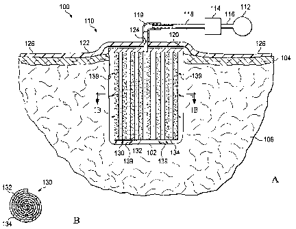
[0011] FIG. lA is a schematic cross-section of a first illustrative embodiment
of a
system for treating a surface wound on a patient including a composite
scaffold and a side-
mounted manifold;
[0012] FIG. 1B is a cross-section of the composite scaffold taken on the line
1B-1B in
FIG. 1A;
[0013] FIG. 2 is a schematic cross-section of a second illustrative embodiment
of a
system for treating a subcutaneous wound on a patient including a composite
scaffold and a
side-mounted manifold;
[0014] FIG. 3 is a schematic, perspective view, of the composite scaffold and
the side-
mounted manifold of FIGS. 1 and 2;
[0015] FIG. 4 is a schematic cross-section of a third illustrative embodiment
of a
system for treating a surface wound on a patient including a composite
scaffold and an end-
mounted manifold;
[0016] FIG. 5 is a schematic cross-section of a fourth illustrative embodiment
of a
system for treating a subcutaneous would on a patient including a composite
scaffold and an
end-mounted manifold;
[0017] FIG. 6 is a schematic, perspective view, of the composite scaffold and
the end-
mounted manifold of FIGS. 4 and 5; and
[0018] FIG. 7 is a schematic view of a fluid control system for the system
shown in
FIGS. lA and 4.
4
CA 02745462 2013-11-29
DETAILED DESCRIPTION
[0019] In the following detailed description of the illustrative embodiments,
reference
is made to the accompanying drawings that form a part hereof. These
embodiments are
described in sufficient detail to enable those skilled in the art to practice
the invention, and it is
understood that other embodiments may be utilized and that logical structural,
mechanical,
electrical, and chemical changes may be made without departing from the spirit
or scope of the
invention. To avoid detail not necessary to enable those skilled in the art to
practice the
embodiments described herein, the description may omit certain information
known to those
skilled in the art.
[0020] The term "reduced pressure" as used herein generally refers to a
pressure less
than the ambient pressure at a tissue site that is being subjected to
treatment. In most cases,
this reduced pressure will be less than the atmospheric pressure at which the
patient is located.
Alternatively, the reduced pressure may be less than a hydrostatic pressure
associated with
tissue at the tissue site. Although the terms "vacuum" and "negative pressure"
may be used to
describe the pressure applied to the tissue site, the actual pressure applied
to the tissue site may
be significantly more than the pressure normally associated with a complete
vacuum.
Reduced pressure may initially generate fluid flow in the area of the tissue
site. As the
hydrostatic pressure around the tissue site approaches the desired reduced
pressure, the flow
may subside, and the reduced pressure is then maintained. Unless otherwise
indicated, values
of pressure stated herein are gauge pressures. Similarly, references to
increases in reduced
pressure typically refer to a decrease in absolute pressure, while decreases
in reduced pressure
typically refer to an increase in absolute pressure.
[0021] Referring to FIGS. lA and 1B, a first illustrative embodiment of a
reduced-
pressure system 100 for applying reduced pressure at a tissue site in the body
of a patient to
repair a defect. As used herein the term "defect" refers to a tissue site in
need of tissue repair
or bulking. For example, the defect may be a wound such as a laceration, an
incision, a burn
or an ulcer. A defect may also be an induced defect such as an incision or
puncture made by a
surgeon in otherwise healthy tissue for the purposes of bulking the tissue
(e.g., such as in
cosmetic surgery). Examples of tissue sites that may be bulked by implantation
of an
apparatus according to the invention include, but are not limited to, the
breasts, buttocks, neck
and face (e.g., the lips, chin or cheeks). For example, FIG. lA shows a
surface wound 102
having an opening in the epidermis 104 extending into the dermis 106 and
forming a cavity.
5
CA 02745462 2011-06-01
WO 2010/078349 PCT/US2009/069718
The surface wound may extend to different depths including into the
subcutaneous tissue (not
shown) below the dermis 106. Referring to FIG. 2, a reduced-pressure system
200 is shown
with another example of a wound. The wound in FIG. 2 is a subcutaneous wound
202 having
an incisional opening in the epidermis 104 extending through the dermis 106
into a cavity
within the subcutaneous tissue 208. The reduced-pressure system 200 is
otherwise
substantially similar to the reduced-pressure system of FIG. 1 and, as such,
utilizes the same
reference numerals used in FIG. 1 for the same components.
[0022] Referring back to FIG. 1 with reference to FIG. 2 as well, the reduced-
pressure
system 100 comprises a dressing assembly 110 positioned over the surface wound
102 and a
reduced-pressure source 112 for providing a reduced pressure to the dressing
assembly 110.
The system 100 further comprises a canister 114 having a filter (not shown)
contained within
the canister 114 that is coupled in fluid communication with the reduced-
pressure source 112
via a conduit 116. The canister 114 is also in fluid communication with the
reduced pressure
dressing 110 via a second conduit 118 and a conduit connector 119. The
canister 114 may be
a fluid reservoir, or collection member, to filter or hold exudates and other
fluids removed
from the surface wound 102. In one embodiment, the canister 114 and the
reduced-pressure
source 112 are integrated into a single housing structure.
[0023] As used herein, the term "coupled" includes direct coupling or indirect
coupling via separate object. The term "coupled" also encompasses two or more
components
that are continuous with one another by virtue of each of the components being
formed from
the same piece of material. Also, the term "coupled" may include chemical,
mechanical,
thermal, or electrical coupling. Fluid coupling means that fluid is in
communication with the
designated parts or locations.
[0024] The dressing assembly 110 further comprises a distribution manifold 120
adapted to be positioned at the opening of the surface wound 102, and a drape
122 adapted to
cover the distribution manifold 120 to maintain reduced pressure beneath the
drape 122 within
the cavity of surface wound 102. The drape 122 includes an aperture 124
through which the
conduit connector 119 extends to provide fluid communication between the
second conduit
118 and the distribution manifold 120. The drape 122 also includes a periphery
portion 126
that extends beyond the perimeter of the opening of the surface wound 102 that
includes an
adhesive or bonding agent (not shown) to secure the drape 122 to the healthy
tissue adjacent
the opening of the surface wound 102. In one embodiment, the adhesive disposed
on the
drape 122 may be used to provide a seal between the epidermis 104 and the
drape 122 to
6
CA 02745462 2011-06-01
WO 2010/078349
PCT/US2009/069718
maintain reduced pressure within the surface wound 102. In another embodiment,
a seal layer
(not shown) such as, for example, a hydrogel or other material, may be
disposed between the
drape 122 and the epidermis 104 to augment or substitute for the sealing
properties of the
adhesive.
[0025] The drape 122 may be any material that provides a pneumatic or fluid
seal.
The drape 122 may, for example, be an impermeable or semi-permeable,
elastomeric material.
"Elastomeric" means having the properties of an elastomer, and generally
refers to a polymeric
material that has rubber-like properties. More specifically, most elastomers
have elongation
rates greater than 100% and a significant amount of resilience. The resilience
of a material
refers to the material's ability to recover from an elastic deformation.
Examples of elastomers
may include, but are not limited to, natural rubbers, polyisoprene, styrene
butadiene rubber,
chloroprene rubber, polybutadiene, nitrile rubber, butyl rubber, ethylene
propylene rubber,
ethylene propylene diene monomer, chlorosulfonated polyethylene, polysulfide
rubber,
polyurethane, EVA film, co-polyester, and silicones. Specific examples of
drape materials
include a silicone drape, 3M Tegaderm drape, V.A.C. TM Drape TM, acrylic
drape such as one
available from Avery Dennison, or an incise drape.
[0026] The dressing assembly 110 further comprises a composite scaffold 130
positioned within the cavity of surface wound 102 in fluid communication with
the manifold
120 for applying reduced pressure to the cavity of the surface wound 102 and
to provide a
structure for promoting the growth of tissue within the cavity of the surface
wound 102. The
composite scaffold 130 may be partially or fully in contact with the cavity
walls of the surface
wound 102 being treated. When the composite scaffold 130 is in contact with
the walls of the
surface wound 102, the composite scaffold 130 may partially or fully fill the
void of the
surface wound 102. The composite scaffold 130 may be any size, shape, or
thickness
depending on a variety of factors, such as the type of treatment being
implemented or the
nature and size of the cavity of the surface wound 102.
[0027] In one illustrative embodiment, the distribution manifold 120 is a foam
material
that distributes reduced pressure to the composite scaffold 130 and the cavity
of the surface
wound 102 when the distribution manifold 120 is in contact with or near the
composite
scaffold 130. The foam material may be either hydrophobic or hydrophilic. In
one non-
limiting example, the distribution manifold 120 is an open-cell, reticulated
polyurethane foam
such as GranuFoam dressing available from Kinetic Concepts, Inc. of San
Antonio, Texas.
In the example in which the distribution manifold 120 is made from a
hydrophilic material, the
7
CA 02745462 2011-06-01
WO 2010/078349 PCT/US2009/069718
distribution manifold 120 also functions to wick fluid away from the composite
scaffold 130
and the cavity of the surface wound 102, while continuing to provide reduced
pressure to the
composite scaffold 130 as a manifold. The wicking properties of the
distribution manifold 120
draw fluid away from the cavity of the surface wound 102 by capillary flow or
other wicking
mechanisms. An example of a hydrophilic foam is a polyvinyl alcohol, open-cell
foam such
as V.A.C. WhiteFoame dressing available from Kinetic Concepts, Inc. of San
Antonio, Texas.
Other hydrophilic foams may include those made from polyether. Other foams
that may
exhibit hydrophilic characteristics include hydrophobic foams that have been
treated or coated
to provide hydrophilicity.
[0028] Referring to FIG. 2, the reduced-pressure system 200 further comprises
a flange
portion 219 of the conduit connector 119 positioned between the drape 122 and
the epidermis
104 and a third conduit 218 supported by the flange portion 219 and extending
therefrom into
the cavity of the subcutaneous wound 202. The reduced-pressure system 200
further
comprises a distribution manifold 220 fluidly coupled to the conduit connector
119 via the
third conduit 218. The distribution manifold 220 is substantially similar to
the distribution
manifold 120 (FIG. 1), but is constructed from bioresorbable materials that do
not have to be
removed from a patient's body following use of the dressing assembly 210.
Suitable
bioresorbable materials may include, without limitation, a polymeric blend of
polylactic acid
(PLA) and polyglycolic acid (PGA). The polymeric blend may also include,
without
limitation, polycarbonates, polyfumarates, and capralactones. The distribution
manifold 220
may further serve as a scaffold for new cell-growth, or a scaffold material
may be used in
conjunction with the distribution manifold 220 to promote cell-growth. A
scaffold is a
substance or structure used to enhance or promote the growth of cells or
formation of tissue,
such as a three-dimensional porous structure that provides a template for cell
growth.
Illustrative examples of scaffold materials include calcium phosphate,
collagen, PLA/PGA,
coral hydroxy apatites, carbonates, or processed allograft materials.
[0029] Referring to FIG. 3, the composite scaffold 130 comprises a strip of
tissue such
as, for example, adipose tissue sandwiched together with a strip of scaffold
material, i. e., a
tissue lamina 134 and a scaffold lamina 132, respectively, forming a laminate
136. The
laminate 136 may then be rolled into a generally cylindrical shape as shown
with one end
portion rolled inside the composite scaffold 130, i.e., the internal end
portion 138, and the
other end portion rolled outside the composite scaffold 130, i.e., the
external end portion 137.
The surfaces of the scaffold lamina 132 are in fluid communication with the
surfaces of the
8
CA 02745462 2011-06-01
WO 2010/078349 PCT/US2009/069718
tissue lamina 134. In one embodiment, the scaffold lamina 132 is relatively
thin and best
suited for maintaining the viability of the tissue lamina 134 before and after
being transferred
to the cavities of the wounds 102, 202. In another embodiment, the scaffold
lamina 132 is
relatively thicker so that it not only maintains the viability of the tissue
lamina 134, but also
expands the tissue lamina 134 as the tissue lamina 134 grows into the scaffold
lamina 132
increasing in volume and bulk while in the cavities of the wounds 102, 202. It
should be
understood that the composite scaffold 130 or laminate 136 may be treated in
vitro with fluids
or the application reduced pressure prior to being transferred to the patient
and/or in vivo with
native fluids from the cavities of the wounds 102, 202 or with other fluids as
described below
in conjunction with the system shown in FIG. 7.
[0030] When the composite scaffold 130 is positioned in the cavity of the
surface
wound 102 as described above, the manifold 120 is in fluid communication with
the edges of
the scaffold lamina 132 as described above and shown by arrows 139 in FIGS. IA
and 3. The
scaffold lamina 132 is preferably bioabsorbable and as such will be absorbed
as the tissue in
the cavity of the surface wound 102 and the tissue lamina 134 grows in vivo to
fill the cavity.
As indicated above, the composite scaffold 130 may be rolled into any size and
shape to fill or
partially the fill the cavity of the surface wound 102 and the subcutaneous
wound 202.
[0031] The tissue lamina 134 according to the invention may be any type of
tissue
desired for implantation, such as adipose tissue. In certain embodiments, the
tissue of the
tissue lamina 134 is the same type of tissue that surrounds a defect (e.g.,
wound) site. The
tissue lamina 134 may be allograft, autograft, xenograft tissue or may be a
tissue generated in
vitro from a population of pluripotent cells. In certain aspects, the tissue
lamina 134
comprises a substantially intact slice of tissue that is shaped to fit a
scaffold lamina 132. In
certain other aspects, the tissue lamina 134 may be composed of raw
lipoaspirate or cells
separated from the lipoaspirate.
[0032] The fluid communication between the manifold lamina 132 and the tissue
lamina 134 composed of adipose tissue allows the cells in the adipose tissue
to remain viable
while the introduced tissue undergoes neovascularization (or revascularization
in the case of
graft tissue). In particular, fluid flow through the tissue removes metabolic
waste products
from the tissue and draws nutrients such as oxygen from the surrounding tissue
into the
introduced tissue. Thus, fluid flow not only maintains the viability of cells
in the tissue lamina
134, but also promotes proliferation of the cells and bulking a tissue defect
such as the surface
wound 102.
9
CA 02745462 2011-06-01
WO 2010/078349 PCT/US2009/069718
[0033] Referring to FIG. 4, a third illustrative embodiment of a reduced-
pressure
system 300 for applying reduced pressure at a tissue site in the body of a
patient to repair the
surface wound 102 is shown and comprises the same components as the reduced-
pressure
system 100 in FIG. 1 as indicated by the reference numbers. Referring to FIG.
5, a fourth
reduced-pressure system 400 for applying reduced pressure to the subcutaneous
wound 202 is
shown and comprises the same components as the reduced-pressure system 200 in
FIG. 2 as
indicated by the reference numbers. The reduced-pressure systems 300 and 400
are
substantially the same as the systems 100 and 200, respectively, other than
the manifolds and
composite scaffolds. The reduced-pressure systems 300, 400 comprise a dressing
assembly
310 and 410, respectively, each one of which includes a distribution manifold
320 and a
composite scaffold 330.
[0034] The composite scaffold 330 also comprises a scaffold lamina 332 and a
tissue
lamina 334 that form a laminate 336 which also may be rolled into a generally
cylindrical
shape as shown in FIG. 6. The rolled laminate 336 also has an external end
portion 337 and an
internal end portion 338 within the composite scaffold 330. In this
embodiment, however, the
distribution manifold 320 is fluidly coupled to the scaffold lamina 332 at the
external end
portion 337 of the composite scaffold 330 rather than at the edges of the
scaffold lamina 332.
The scaffold lamina 332 has sufficient porosity to fluidly communicate the
reduced pressure to
substantially the full length of the tissue lamina 334. The scaffold lamina
332 may have a
porosity that increases toward the inside of the composite scaffold 330 to
create a reduced-
pressure gradient within the composite scaffold 330. Otherwise, the scaffold
lamina 332 is
substantially similar to the scaffold lamina 132. When the composite scaffold
330 is
positioned in either type of wound 102, 202, the manifold 320 is fluidly
coupled to the third
conduit 218 for distributing reduced pressure to the composite scaffold 330 as
described
above. In this embodiment, however, the composite scaffold 330 is oriented
within the
cavities of the wounds 102, 202 such that the longitudinal axis of the
composite scaffold 330 is
aligned generally parallel with the epidermis 104 rather than perpendicular.
The structure and
orientation of this distribution manifold 320 and composite scaffold 330 may
be better suited
for different types of wounds such as, for example, a surface or subcutaneous
wound with an
incisional cut through the epidermis 104 and the dermis 106.
[0035] Referring to FIG. 7, the reduced pressure therapy systems 100, 200,
300, and
400 (collectively, the "systems") may further comprise a pressure sensor 140
operably
connected to the second conduit 118 to measure the reduced pressure being
applied to the
CA 02745462 2011-06-01
WO 2010/078349 PCT/US2009/069718
manifolds 120, 220, 320, and 420 (collectively, the "manifolds"). The systems
further include
a control unit 145 electrically connected to the pressure sensor 140 and the
reduced pressure
source 112. The pressure sensor 140 measures the reduced pressure within the
cavity of the
wounds 102, 202 (collectively, the "wounds") and also may indicate whether the
second
conduit 118 is occluded with blood or other bodily fluids. The pressure sensor
140 also
provides feedback to control unit 145 which regulates the reduced pressure
therapy being
applied by the reduced pressure source 112 through the second conduit 118 to
the manifolds.
The reduced pressure therapy systems may also comprise a fluid supply 150
fluidly coupled to
the second conduit 118 via a fourth conduit 152 and operatively connected to
the control unit
145. The fluid supply 150 may be used to deliver growth and/or healing agents
to the
scaffolds 130 and 330 (collectively, the "scaffolds") for the wounds
including, without
limitation, an antibacterial agent, an antiviral agent, a cell-growth
promotion agent, an
irrigation fluid, or other chemically active agents. The systems further
comprise a first valve
154 positioned in the fourth conduit 152 to control the flow of fluid
therethrough, and a second
valve 156 positioned in the second conduit 118 between the reduced pressure
supply 112 and
the juncture between the second conduit 118 and the fourth conduit 152 to
control the flow of
reduced pressure. The control unit 145 is operatively connected to the first
and second valves
154, 156 to control the delivery of reduced pressure and/or fluid from the
fluid supply 150,
respectively, to the manifolds as required by the particular therapy being
administered to the
patient. The fluid supply 150 may deliver the liquids as indicated above, but
may also deliver
air to the manifolds to promote healing and facilitate drainage at the site of
the wounds.
[0036] In the embodiment illustrated in FIG. 7, the reduced-pressure source
112 is an
electrically-driven vacuum pump. In another implementation, the reduced-
pressure source 112
may instead be a manually-actuated or manually-charged pump that does not
require electrical
power. The reduced-pressure source 112 instead may be any other type of
reduced pressure
pump, or alternatively a wall suction port such as those available in
hospitals and other
medical facilities. The reduced-pressure source 112 may be housed within or
used in
conjunction with a reduced pressure treatment unit (not shown), which may also
contain
sensors, processing units, alarm indicators, memory, databases, software,
display unites, and
user interfaces that further facilitate the application of reduced pressure
treatment to the
wounds. In one example, a sensor or switch (not shown) may be disposed at or
near the
reduced-pressure source 112 to determine a source pressure generated by the
reduced-pressure
11
CA 02745462 2011-06-01
WO 2010/078349 PCT/US2009/069718
source 112. The sensor may communicate with the control unit 145 that monitors
and controls
the reduced pressure that is delivered by the reduced-pressure source 112.
[0037] As used herein, the term "manifold" refers to a substance or structure
that is
provided to assist in directing reduced pressure to, delivering fluids to, or
removing fluids
from a tissue site. A manifold can include a plurality of flow channels or
pathways that are
interconnected to improve distribution of fluids provided to and removed from
the area of
tissue around the manifold. Examples of manifolds may include, without
limitation, devices
that have structural elements arranged to form flow channels, cellular foams
such as open-cell
foam, porous tissue collections, and liquids, gels and foams that include or
cure to include
flow channels. A detailed description of manifolds and their use according to
the invention is
provided below.
[0038] The term "scaffold" as used herein refers to a substance or structure
applied to
or in a wound or defect that provides a structural matrix for the growth of
cells and/or the
formation of tissue. A scaffold is often a three dimensional porous structure.
The scaffold can
be infused with, coated with, or comprised of cells, growth factors,
extracellular matrix
components, nutrients, integrins, or other substances to promote cell growth.
A scaffold can
take on characteristics of a manifold by directing flow through the matrix. A
manifold can
transmit flow to the scaffold and tissue; in the context of reduced pressure
treatment, the
manifold can be in fluid communication with the scaffold. A detailed
description of scaffolds
and their use according to the invention is provided below.
[0039] As such, the invention disclosed here discloses methods and apparatuses
for
controlling cellular-level based patterns of fluid flow that would allow for
control of patterned
protein organization at a microscopic, nanoscopic, or mesoscopic scale
amenable to provide a
structured manifold and, optionally, a scaffold material for cellular
migration, differentiation,
and like behavior necessary for functional regeneration of tissues. In
comparison to the
passive nature of the current state of the art with regards to tissue repair
and regeneration, the
methods, scaffolds, manifolds, flow sources and systems disclosed herein
provide an active
mechanism by which to promote the endogenous deposition of proteins and
organization of
the provisional matrix with biochemical and physical cues to direct cellular
colonization of a
scaffold or tissue space. The present invention thus enhances current
technology by exploiting
the active force of directed fluid flow, providing a framework upon which to
design manifolds
and scaffolds based upon the need of the biology under the influence of fluid
flow. Flow
vectors and pathways are utilized to enhance protein deposition and cellular
colonization. The
12
CA 02745462 2011-06-01
WO 2010/078349 PCT/US2009/069718
systems provided herein are designed to promote establishment of a provisional
matrix
network with a seamless transition from the healthy tissue edges through a
scaffold or tissue
site to promote a finictional tissue continuum.
[0040] Thus, the apparatuses, methods and systems disclosed herein provide a
means
for active guidance of tissue regeneration through an implanted scaffold or
within a tissue site
to promote functional recovery. This active guidance occurs through mechanisms
of
controlled fluid flow, which can be used to initiate or augment the early
stages of the body's
own natural healing process; a manifold can provide the active guidance
necessary to create a
controlled fluid flow. Specifically, the controlled flow vectors that the
manifolds provide can
be used to facilitate the directed influx of cells and proteins into a
scaffold. Creation of
specific flow pathways within a tissue site or scaffold can lead to patterned
deposition of
proteins, such as collagen and fibrin within the manifold, scaffold or tissue
space.
Biochemical cues from cytokines, growth factors, and cells bound within the
provisional
matrix can work in conjunction with the natural physical cues of the
provisional matrix and
extracellular matrix to guide the subsequent migration of endogenous cells
during the repair
stages of healing. These cues act as a form of track that emanates from the
healthy tissues and
passes through the scaffolding or tissue space to facilitate a continuous
guidance pathway for
organized tissue regeneration.
[0041] To that end, this disclosure provides unique manifolding technologies
designed
for specific biological needs based upon principles of fluid flow. In certain
aspects, the
invention concerns a new approach to wound healing, flow (or gradient)
activated tissue
engineering. In rudimentary form, this approach involves a source or generator
of flow that
forms a gradient for controlled movement of either endogenous or exogenous
fluids into, out
of, or through a tissue space for the organized deposition of proteins and/or
spatial
concentration of cytokines and growth factors, with subsequent formation of a
directionally
oriented provisional matrix. The tissue space being defined here includes, but
is not limited
to, the region surrounding a site of tissue deficit or damage, including a
wound or incision.
[0042] Fluid flow into, through, or out of the tissue space can be refined and
directed
through the inclusion of additional elements to the system including manifolds
and/or
scaffolds. The coordinated elements of the system are designed to create flow
parameters,
pathways, and patterns sufficiently detailed in scale as to be able to
influence and direct the
controlled adsorption of proteins, the organization of matrix, and organized
colonization of
specific cell types. Individual elements of the system are as follows.
13
CA 02745462 2011-06-01
WO 2010/078349 PCT/US2009/069718
[0043] Source or Generator of Flow. Flow is induced into, through, or out of
the tissue
space by methods or apparatuses that introduce changes in mechanical,
chemical, and/or
electrical potentials. These generators of flow provide either a gradient or a
change in
potential from the site or reservoir of endogenous or exogenous fluids to the
placement
position of the flow generator or its extension element (L e., manifold or
scaffold). In one
embodiment, the source of flow comprises a source of reduced pressure. Systems
and
apparatuses according to the invention may also comprise valves or arrays of
valves that
control the application and amount of negative pressure applied to the
manifold. In certain
aspects, scaffolds and/or manifolds described herein comprise a pressure
sensor. Thus, in
some embodiments, the amount of negative pressure applied by a source is
regulated based on
the amount of negative pressure that is sensed in the manifold or scaffold or
at the site of
tissue damage.
[0044] Manifold. The flow generators are the driving force for stimulating the
flow of
fluids. Manifolds are apparatuses for refining the pattern of flow between the
source or
generator of flow and the tissue space. The macroscale level of flow is
refined by specialized
manifolds utilized for directed localization to a single point or to a
plurality of selectively
positioned points for creating initiation sites for microscale flow pathways
within the
manifold/scaffold and, ultimately, the tissue space. The manifold may also
serve as a conduit
for the removal of fluids from and as an apparatus for the delivery of
exogenous fluids to the
tissue space.
[0045] A manifold generally refers to a physical substance or structure that
serves to
assist in applying and translating a mechanical, chemical, electrical or
similar alterations into
changes in the flow of a fluid, herein defined as the movement of liquids,
gases, and other
deformable substances such as proteins, cells, and other like moieties. As
such, this physical
device includes a single point or plurality of points for the egress or
evacuation of pressure,
fluids, and like substances capable of translating the movement of fluids in a
scaffold, as
defined above. This can include, but is not limited to, the introduction of
exogenous factors
such as cells and/or therapeutic moieties into the scaffold through the lumen
or plurality of
lumens present in the manifold. In addition, as used herein, a manifold
includes a single point
or plurality of points for the ingress or introduction of fluid from the
scaffold back towards the
point source of flow.
[0046] Flow distributed by the manifold can direct the movement of endogenous
proteins, growth factors, cytokines, and cells from their resident locations
within the host to
14
CA 02745462 2011-06-01
WO 2010/078349 PCT/US2009/069718
the tissue space or scaffold in an organized manner. The establishment of flow
along these
pathways leads to the deposition of proteins and provisional matrix that
creates an interfacial
endogenous network connecting the host to the scaffold. Extensions of this
matrix can be
established within the scaffold through selective positioning of the manifold
flow initiation
sites with flow promoting scaffolding designs. The organized protein
deposition and
provisional matrix provide a biochemical and physical framework that
stimulates the
attachment and migration of cells along directed pathways throughout the
scaffold and the
tissue space. The resulting endogenous network of proteins, growth factors,
and cells provides
a foundation upon which subsequent phases of the body's own tissue repair and
regeneration
mechanisms can build.
10047] When in place, the manifold works in conjunction with a flow generating
source and a scaffold, if present. Flow generating sources include, but are
not limited to
generators of negative pressure; generators of positive pressure; and
generators of osmotic
flow. The flow gradient established in the manifold may be further refined
through the
scaffold, to deliver a flow gradient to the scaffold to optimize flow through
the scaffold as
needed for the particular defect. Many of the embodiments disclosed herein are
manifolds
capable of translating changes in pressure and the like into controlled
movement of fluids,
optionally through a physical scaffold, for the purposes of directed tissue
regeneration. These
embodiments are generally specified for a particular application in the
regeneration of specific
tissues, but are not limited to a particular tissue therein.
[0048] In order to realize the goal of inducing flow for the purpose of tissue
regeneration, alterations in the aforementioned mechanical, chemical, or
electrical impetus
must be translated from the singular gradient source toward a physical
substrate or scaffold to
elicit cellular-level changes in protein adsorption, matrix organization, cell
migration, and
other tissue regeneration-related behaviors. These alterations are
multivariate in nature and
can include mechanical changes that elicit a physical change in pressure
applied to the scaffold
as applied to the site of the wound or desired site of tissue regeneration,
chemical changes that
elicit a gradient in protein and/or ion concentrations, which result in the
creation of osmotic
gradients capable of inducing flow, or electrical changes that create a
gradient of current/ion
exchange allowing for propagation of electrical signals from the point source.
It is to be
understood, however, that the applicants are not bound by any particular
mechanism through
which gradients and fluid flow induce advantageous results in tissue repair or
growth. In order
CA 02745462 2011-06-01
WO 2010/078349 PCT/US2009/069718
to advantageously transmit these gradients to the tissue, a physical device is
needed to direct
the path of flow from its source to the scaffold or tissue site and vice
versa.
[0049] In some embodiments, the manifold comprises a physical structure in
close
apposition to or within the contents of a scaffold and serves to propagate an
alteration in a
physical parameter, whether it be mechanical, chemical, electrical, or
something similar in
nature, for the means of directing these changes from its point source to the
scaffolding
material. The placement of this manifold with respect to its location with
regard to that of the
scaffold may be of crucial importance for facilitating controlled and directed
regeneration of
specific tissue types. For example, the manifold may be situated such that
implanted tissue,
such as a tissue lamina, is between the manifold and blood source at a tissue
site so that fluid
from the blood source or interstitial fluids can flow to or through the
implanted tissue
[0050] Manifolds may be composed of a bioabsorbable or bioinert material.
Examples
include non-bioabsorbable materials such as medical grade silicone polymers,
metals,
polyvinylchloride (PVC), and polyurethane (e.g., GranuFoam ). Bioabsorbable
polymers
such as collagen, polylactic acid (PLA), polyglycolic acid (PGA), polylactide-
co-glycolide
(PLGA), a polysaccharide (e.g., alginates), a hydrogel, or a polyethylene
glycol, or
combinations thereof, can also be used. In certain aspects, a manifold is
composed of a
mechanically stiff materials such as a calcium phosphate, hydroxyapatite, DBM,
carbonates or
bioglass. Such mechanically stiff materials may have particular use in filling
hard tissue
defects. Some manifolds are also a mix of non-bioresorbable and bioresorbable
materials. In
general material used for a scaffold may also be used to compose a manifold
and such
materials are further detailed below. In certain aspects, manifold materials
are structured to
include a high void fraction for improved bioabsorption properties. In some
embodiments, the
manifold may embody characteristics of the scaffold.
[0051] Scaffold. Biologic and synthetic scaffolds are used in the field of
tissue
engineering to support protein adhesion and cellular ingrowth for tissue
repair and
regeneration. The current state of the art in scaffold technology relies upon
the inherent
characteristics of the surrounding tissue space for the adsorption of proteins
and migration of
cells. A scaffold for use according to the invention is coupled to a manifold,
provides physical
guidance to direct the pathway of fluid flow in the tissue site, creating
avenues for the
movement and migration of adhesive proteins and cells, respectively, which are
integral to the
establishment of a provisional matrix in predetermined patterns of
organization within the
tissue space. The methods and apparatuses described for fluid flow-induced and
gradient-
16
CA 02745462 2011-06-01
WO 2010/078349
PCT/US2009/069718
induced generation of tissues have direct implications into the design of the
scaffolds. Within
this context, scaffolds serve to refine the pathways of fluid flow within the
tissue space to
cellular level patterns from the fluid source to the point(s) of flow
initiation within the
manifold. A scaffold may embody characteristics of a manifold or be combined
in
conjunction with a manifold for refinement of the flow pathways within the
tissue site. In
certain aspects, a scaffold is a reticulated structure comprising high void
fraction for improved
bioabsorption properties.
[0052] Nonlimiting examples of suitable scaffold materials include
extracellular
matrix proteins such as fibrin, collagen or fibronectin, and synthetic or
naturally occurring
polymers, including bioabsorbable or non-bioabsorbable polymers, such as
polylactic acid
(PLA), polyglycolic acid (PGA), polylactide-co-glycolide (PLGA),
polyvinylpyrrolidone,
polycaprolactone, polycarbonates, polyfumarates, caprolactones, polyamides,
polysaccharides
(including alginates (e.g., calcium alginate) and chitosan), hyaluronic acid,
polyhydroxybutyrate, polyhydroxyvalerate, polydioxanone, polyethylene glycols,
poloxamers,
polyphosphazenes, polyanhydrides, polyamino acids, polyortho esters,
polyacetals,
polycyanoacrylates, polyurethanes, polyacrylates, ethylene-vinyl acetate
polymers and other
acyl substituted cellulose acetates and derivatives thereof, polystyrenes,
polyvinyl chloride,
polyvinyl fluoride, poly(vinylimidazole), chlorosulphonated polyolefins,
polyethylene oxide,
polyvinyl alcohol, Teflon , and nylon. The scaffold can also comprise ceramics
such as
hydroxyapatite, coralline apatite, calcium phosphate, calcium sulfate, calcium
carbonate or
other carbonates, bioglass, allografts, autografts, xenografts, decellularized
tissues, or
composites of any of the above. In particular embodiments, the scaffold
comprises collagen,
polylactic acid (PLA), polyglycolic acid (PGA), polylactide-co-glycolide
(PLGA), a
polyurethane, a polysaccharide, an hydroxyapatite, or a polytherylene glycol.
Additionally,
the scaffold can comprise combinations of any two, three or more materials,
either in separate
areas of the scaffold, or combined noncovalently, or covalently (e.g.,
copolymers such as a
polyethylene oxide-polypropylene glycol block copolymers, or terpolymers), or
combinations
thereof. Suitable matrix materials are discussed in, for example, Ma and
Elisseeff, 2005, and
Saltzman, 2004.
[0053] Bioactive agents
[0054] In certain aspects, the apparatuses and methods according to the
invention
concern bioactive agents. Bioactive agents may, in some cases, be incorporated
directly onto a
manifold or scaffold material (i.e., to generate a bioactive manifold and/or
scaffold). For
17
CA 02745462 2011-06-01
WO 2010/078349 PCT/US2009/069718
example, agents that facilitate tissue growth such as collagen or fibrin may
be directly
incorporated onto or into a manifold or scaffold material. Likewise, in
applications where
aberrant immune response need be avoided (e.g., tissue grafts) immune
regulator agents such
as rapamycin may be incorporated into manifold or scaffold structures.
[0055] In further aspects soluble bioactive agents may be introduced at a site
of tissue
damage by virtue of the flow through the tissue site. For example, a manifold
may be in fluid
communication with a fluid source and a bioactive agent may be introduced into
the fluid
source and thereby into the manifold and tissue lamina.
[0056] Nonlimiting examples of useful bioactive growth factors for various
applications are growth hormone (GH), a bone morphogenetic protein (BMP),
transforming
growth factor-a (TGF-a), a TGF-13, a fibroblast growth factor (FGF),
granulocyte-colony
stimulating factor (G-CSF), granulocyte/macrophage-colony stimulating factor
(GM-CSF),
epidermal growth factor (EGF), platelet derived growth factor (PDGF), insulin-
like growth
factor (IGF), vascular endothelial growth factor (VEGF), hepatocyte growth
factor/scatter
factor (HGF/SF), an interleukin, tumor necrosis factor-a (TNF-a) or nerve
growth factor
(NGF). In certain applications, the bioactive molecule may be a molecule that
directs
vascularization such as VEGF.
[0057] Tissue repair and regeneration. The apparatuses and systems disclosed
herein
can be used for tissue repair and engineering in various contexts including
the following.
[0058] Repair and regeneration of lost tissue. A generator of flow may be
combined
with manifolds and/or scaffolds to direct the regeneration of lost tissue at a
site of injury or
compromised function. Tissues lost from traumatic injury, surgery, burns, or
other causes
(e.g., infection or autoimmune disease) can be led to regenerate using the
methods, scaffolds,
manifolds, flow sources and systems of the invention.
[0059] Retard the progression of a tissue disease state. A generator of flow
may be
combined with manifolds and/or scaffolds to retard disease progression of an
affected tissue
such as occurs, e.g., in autoimmune disease and wasting infections such as a
Staph infection.
[0060] Maintenance of tissue viability. A generator of flow may be combined
with
manifolds and/or scaffolds to maintain the viability of explanted tissues,
such as adipose
tissues, either for in vitro study, ex vivo scaffold or implant preparation,
or in vivo transplant.
A generator of flow combined with a manifold may be used to provide nutrient
fluid flow to
the tissue and to control waste removal from the tissue.
18
CA 02745462 2011-06-01
WO 2010/078349 PCT/US2009/069718
[0061] Expansion of tissue. A generator of flow may be combined with manifolds
and/or scaffolds to promote the expansion of existing tissues. The methods,
scaffolds,
manifolds, flow sources and systems of the invention can be used to direct the
growth of
tissues where additional tissue quantity is needed or desired. Tissue
expansion may be
accomplished either in vivo or ex vivo, for example in a tissue culture
environment that
provides required nutrients to the tissue wherein the nutrients are infused by
the application of
reduced pressure.
[0062] Acceleration of tissue formation or promoting new tissue formation. A
generator of flow may be combined with manifolds and/or scaffolds to
accelerate the rate of
tissue formation within a natural healing response. The methods, scaffolds,
manifolds, flow
sources and systems of the invention may be used to accelerate tissue growth
by augmenting
formation of provisional matrices, facilitating its stable positioning, and
aiding in recruitment
of cells to the tissue space. Likewise, the apparatuses and methods disclosed
herein may be
used to promote new tissue formation at a selected tissue site. Such new
tissue formation may
be used to bulk (i.e., add volume and mass) a tissue site. Such methods may be
used to rebuild
tissue features that were lost to an injury, malformed during development or
to improve the
external appearance of a feature.
[0063] Stimulating the differentiation of stem cells along specific pathways.
A
generator of flow may be combined with manifolds and/or scaffolds to stimulate
the
differentiation of stem cells or other pluripotent cells into specific
lineages. Application of
flow using the methods, scaffolds, manifolds, flow sources and systems of the
invention may
be used to direct pluripotent cells into specific cell lineages needed to
foster growth in the
tissue space. For example, adipose (e.g., brown or white adipocyte) progenitor
cells may be
provided as part of a tissue lamina and grown on a matrix either in vitro or
at a tissue site in
vivo.
[0064] Introducing proteins, matrix, cells, or pharmaceuticals into the in
vivo
environment. A generator of flow may be combined with manifolds and/or
scaffolds to
introduce exogenous growth factors, proteins, cells, or pharmaceutical agents
into the tissue
space to augment tissue repair, regeneration, and/or maintenance.
[0065] Creating matrices in vitro for implantation in vivo. A generator of
flow may be
combined with manifolds and/or scaffolds to facilitate formation of matrices
in vitro that may
subsequently be used for in vivo transplantation.
19
CA 02745462 2011-06-01
WO 2010/078349 PCT/US2009/069718
[0066] Promoting integration of transplanted tissue. A generator of flow may
be
combined with manifolds and/or scaffolds to promote integration of
transplanted tissue into
the host environment. This can be applied to autograft, allograft, or
xenograft transplants.
Transplanted tissues may be whole sections of tissue excised from surrounding
tissue, or
substantially disrupted tissues such as lipoaspirate. In such applications
manifold material
may include immune suppressing agents to reduce the chance of tissue
rejection.
[0067] Directing extracellular matrix (ECM) deposition and orientation in
vitro. A
flow generator may be combined with manifolds and/or scaffolds to guide the
directed
deposition and orientation of ECM expressed by cells and tissues. The directed
orientation of
ECM has an impact in organizing and directing the attachment and colonization
of subsequent
cell layers and tissues.
[0068] References
U.S. Patent No. 4,787,906
U.S. Patent No. 6,103,255
U.S. Patent No. 6,135,116
U.S. Patent No. 6,365,146
U.S. Patent No. 6,695,823
U.S. Patent No. 6,696,575
U.S. Patent No. 6,767,334
U.S. Patent No. 6,814,079
U.S. Patent No. 6,856,821
U.S. Patent No. 6,936,037
U.S. Patent No. 6,951,553
U.S. Patent No. 6,994,702
U.S. Patent No. 7,004,915
U.S. Patent No. 7,070,584
U.S. Patent No. 7,077,832
U.S. Patent No. 7,108,683
U.S. Patent No. 7,160,553
U.S. Patent No. 7,186,244
U.S. Patent No. 7,214,202
U.S. Patent No. 7,279,612
U.S. Patent No. 7,316,672
CA 02745462 2011-06-01
WO 2010/078349
PCT/US2009/069718
U.S. Patent No. 7,346,945
U.S. Patent No. 7,351,250
U.S. Patent No. 7,384,786
U.S. Patent Publn. 2003/0225347
U.S. Patent Publn. 2005/0260189
U.S. Patent Publn. 2007/0123895
U.S. Patent Publn. 2008/0033324
U.S. Patent Publn. 2008/0208358
U.S. Provisional Patent Appin. 61/142,053
U.S. Provisional Patent Appin. 61/142,065
Anderson et al., Tissue Eng., 13:2525-38, 2007.
Brody et al., J Biomed Mater. Res. B: AppL Biomater, 83:16-43, 2007.
Gemmiti et al., Tissue Eng., 12:469-79, 2006.
Lago et al., IEEE Trans. Biomed. Eng., 54:1129-37, 2007.
Ma et al., Scaffolding in Tissue Engineering, 2005.
Manwaring et al., Biomaterials, 22:3155-3168, 2001.
Manwaring et aL, Biomaterials, 25:3631-3638, 2004.
Mercier et al., Biomaterials, 26:1945-1952, 2005.
Mikos et aL, J Biomed. Mater. Ref, 27:183-189, 2004.
Norman et al., Ann Biomed Eng., 34:89-101, 2006.
PCT Appin. WO 00/38755A2
PCT Appin. WO 00/61206A1
PCT Appin. WO 03/018098A2
PCT Appin. WO 03/092620A2
PCT Appin. WO 04/060148A2
PCT Appin. WO 04/105576A2
PCT Appin. WO 05/009488A2
PCT Appin. WO 05/033273A2
PCT Appin. WO 06/004951
PCT Appin. WO 06/127853
PCT Appin. WO 07/067685A2
PCT Appin. WO 07/092397A2
PCT Appin. WO 07/106589A2
21
CA 02745462 2013-11-29
PCT Appin. WO 07/106590A2
PCT Appin. WO 07/106591A2
PCT Appin. WO 07/106592A2
PCT Appin. WO 07/106594A2
PCT Appin. WO 07/133555A2
PCT Appin. WO 07/133556A2
PCT Appin. WO 07/143060A2
PCT Appin. WO 07/196590
PCT Appin. WO 08/013896A2
PCT Appin. WO 08/036162A2
PCT Appin. WO 08/036359A2
PCT Appin. WO 08/036361A2
PCT Appin. WO 08/042481A2
PCT Appin. WO 08/091521A2
Pfister et al., Neurosurgery, 60:137-41, 2007.
Saltzman, Tissue Engineering: Engineering Principles for the Design of
Replacement
Organs and Tissues, 2004.
Sachlos et al., Cells and Mat., 5:29-40, 2003.
Segvich et al., 1 Biomed. Mater. Res. B: Appl. Biomater., 84B:340-349, 2008.
Shimko et al., J Biomed Mater. Res. B: Appl. Biomater., 73:315-24, 2005.
Takahashi et al., Cell, 126:663-76, 2006.
Tan et al., Bone, 41:745-751, 2007.
Tan et a , Biochem. Biophys. Res. Comm., 369:1150-1154, 2008.
Walsh et al., Tissue Eng., 11:1085-1094, 2005.
Wen et al., Handbook of Nanostructured Biomaterials and Their Applications in
Nanobiotechnology, 1-23, 2005.
[0069]
The discussion of the references herein is intended merely to summarize the
assertions made
by the authors and no admission is made that any reference constitutes prior
art. Applicants
reserve the right to challenge the accuracy and pertinence of the cited
references.
[0070] In view of the above, it will be seen that the advantages of the
invention are
achieved and other advantages attained. As various changes could be made in
the above
methods and compositions without departing from the scope of the invention, it
is intended
22
CA 02745462 2011-06-01
WO 2010/078349
PCT/US2009/069718
that all matter contained in the above description and shown in the
accompanying drawings
shall be interpreted as illustrative and not in a limiting sense.
23