Note : Les descriptions sont présentées dans la langue officielle dans laquelle elles ont été soumises.
CA 02746243 2011-06-08
EQUIPMENT FOR INFRARED VISION OF ANATOMICAL STRUCTURES AND
SIGNAL PROCESSING METHODS THEREOF
FIELD OF THE INVENTION
The present invention relates to the field of photonics, image acquisition,
image processing, vision enhancement and information extraction applied to
life
sciences, mainly focused on the medical and biomedical fields and specially,
but not
exclusively, on the fields of endoscopy, fetoscopy and laparoscopy.
More specifically, the invention relates to a medical device or equipment
comprising means for acquiring multimodal or multispectral images of a living
subject, including illumination means and a digital image processing platform
associated to said means, with embedded algorithms to extract and/or enhance
specific image information with the objective of assisting physicians in their
decisions, for example, when diagnosing, monitoring and/or performing a given
therapy or surgical operation.
BACKGROUND OF THE INVENTION
In the medical field a number of = imaging systems have been devised to
display and enhance the visualization of an area under examination, either
with
diagnostic purposes or to guide therapeutic surgical procedures. It is
apparent that
any relevant information of the area under inspection plays an important role
in the
clinical assessment of the subject's condition which is of major importance
when
making clinical decisions that may ultimately affect the well being and
quality of life
of the patient. This is of critical importance for instance, in endoscopic
surgery,
where an accurate vision is essential for the results of the operation. In
general, the
visualization of critical structures (i.e. blood vessels and nerves) involves
three
typical situations: (i) said structures cannot be distinguished due to poor
visualization conditions, (ii) the structures are hidden beneath a layer of
other tissue
and/or (iii) the structure of interest is not distinguishable from the
surrounding
structures. Particularly relevant to this field is the task of enhancing
and/or
generating image contrast and improving the definition of the visualization of
blood
CA 02746243 2011-06-08
-2-
vessels and/or different functional aspects about these vessels, which are
poorly or
non visible by endoscopy with the presently available technology without using
an
exogenous contrast agent.
Despite endoscopy is an advanced surgical technique which greatly
minimizes surgical procedure risks, some problems still persist, like the risk
of
accidentally cutting a mayor blood vessel. Even though the use of minimally
invasive
endoscopy offers clear advantages to the patient, it imposes certain
disadvantages
to the physicians, like a constrained vision of the surgical field or a poor
contrast
and/or definition. The need to accurately identify blood vessels under such
conditions may represent a serious challenge to any surgeon, rendering surgery
extremely dependent on the surgeon's experience, resulting in prolonged
operations
due to bleeding episodes, and occasionally resulting in major hemorrhagic
complications.
A number of inventions have been devised to improve medical imaging
relating to the visualization of blood vessels. Some of the previous
inventions refer
to specific solutions for problems in the above-discussed field. For instance
there
are several patents describing particular lighting systems, like US patents
7,041,054
or 6,730,019. These patents describe illumination and image acquisition
systems
with particular preferred embodiments. It should be noted that the present
invention
does not rely on a particular illumination method. Although illumination is
needed in
the system, the system is independent of the method employed to achieve that
illumination. Moreover, the system is able to cope with and adapt to different
illumination schemes to achieve its purpose, that is, to enhance and/or
generate
image contrast and improve definition for the visualization of anatomical
structures.
Other inventions relate to a particular method for capturing the images or
disclose
embodiments of specific image capturing devices, like US 2008/0208006. Again
as
with the illumination, the present invention is independent of the method of
capturing
the image; it can be used according to different image capturing approaches.
Patent US 5,255,087 by Olympus describes a video system comprising a well
detailed lighting system used in combination with a standard endoscope, a
control
system and an image capturing and processing unit. The goal of said system is
to
CA 02746243 2011-06-08
-3-
improve the images of an endoscopic system. In order to achieve this goal
three
techniques are described by the inventor: auto fluorescence imaging (AFI),
narrow
band imaging (NBI) and Infrared Imaging (IRI). AFI is based on the principle
of the
auto fluorescence of certain tissues; NBI is based on a well know technique of
using
contrast agents and an illumination of a particular wavelength to which the
contrast
agent is sensitive; and IRI is a specific combination of the two previous
techniques
which uses an exogenous contrast agent like indocyanine green (ICG) to detect
submucosal blood vessels, but it is used only for diagnostic purposes. That
limitation
to a purely diagnostic use is because the injected dye rapidly dissolves into
the
bloodstream, and the five minutes that it lasts would not allow using it in
therapy or
surgery, which require much longer duration. However, another disadvantage
with
respect to the present invention is that it requires a contrast agent to
enhance the
visualization of blood vessels, both superficial (that is apparent to the
naked eye)
and submucosal (running under the mucosa and therefore normally non visible to
simple inspection), whereas the present invention makes use of an algorithm to
perform such feature, making it less invasive and hence more appropriate in
the
surgery field. In addition to the fact that IRI is not designed as an
assistance vision
system for surgery, the said technique does not provide other additional
features
like: image segmentation, image mapping of the surgical field, or functional
assessment of blood vessels (by obtaining relevant information such as, for
example, the amount of oxygen carried by the blood or the coagulation state of
the
vessels). Those features are supplied by the present invention, and are
differential
and provide useful information when used for surgical endoscopic procedures,
including laparoscopy or fetoscopy. In those surgical techniques the
availability of a
complete vascular map of the surgical field or the capability to distinguish
the
coagulation status of a vessel, might represent extremely valuable information
to
assist the surgeon during the operation.
Patent US 2005/0182321 discloses a similar invention as the one previously
commented concerning IRI, based on a medical imaging enhancing system using
visible and infrared images in combination with a dye agent. A mayor
disadvantage
of this system compared with the present invention is that it requires of a
contrasting
agent or dye to be injected to the blood stream of the patient, consequently
rendering this system non-usable in any surgical procedure for the reasons
above
CA 02746243 2011-06-08
-4-
mentioned, namely the rapid dilution of the contrast agent into the
bloodstream with
the consequent inefficiency to assist physicians in carrying out therapy or
surgery.
Additionally, said patent only contemplates the use of a visible and a single
near
infrared (NIR) channel, constraining the image capturing process to a total of
four
spectral bands without mentioning the possible use of more NIR channels or
other
imaging modes that could improve the detection of vessels and the extraction
of
vessel functional information. A further weakness is that it only focuses on
the ability
of detecting blood vessels and does not provide specific embedded methods as,
for
example, image segmenting, image mapping and assessing vessel functionality.
Patent US 2008/0097225 explicitly mentions specific optical techniques, namely
optical coherence tomography (OCT) and spectrally-encoded endoscopy (SSE),
with the aim to reduce the size of the endoscope and increase its resolution.
A
significant disadvantage of said techniques is their technical complexity,
since they
necessary comprise a scanning unit and a complex optical assembly. Although
the
said patent mentions that the wavelength can be chosen to assess the amount of
oxygen carried by the blood, it does not take into account the use of this
information
as an integrated tool for assisting the surgeon or the physician by means of
enhancing the images displayed. A further disadvantage, due to the small field
of
view of such small instrument, is that the physician's angle of vision is
substantially
restricted, thus limiting considerably the feasibility of such system for
surgical
applications. These problems are overcome with the present invention.
A similar case is patent EP 1,839,561, which discloses an endoscopic apparatus
that is a combination of a standard visible endoscopy in conjunction with an
OCT
arrangement, the said apparatus being a particular solution to apply to a
known
optical technology in a particular way. A disadvantage of said apparatus is
that it can
only obtain information to generate an enhanced image for a narrow portion of
the
area under study; furthermore it does not compose a substantially enhanced
image.
The said invention does not seem to have optimum use for blood vessel enhanced
imaging.
Patent US 6,353,753 describes a device for the acquisition of images from deep
anatomical structures. The main disadvantage of said device is that it does
not
CA 02746243 2011-06-08
-5-
include specific image analysis processing intended for segmenting and
displaying
the information in conjunction . with the visual image; it also lacks image
reconstruction functions.
Regarding the state of the art related to image enhancement through the
capture of
visible and infrared light, some advances have been achieved as, for example,
in
patent US 2008/0079807, where a multispectral imaging system is described with
special emphasis in charge couple devices (CCDs) and optoelectronics. In this
case
that patent focuses particularly on the image collection device technology.
Major
drawbacks of said patent in comparison with the present invention include the
lack
of tracking (defined as a signal processing procedure for tracking and
localizing of
blood vessels between consecutive scenes from the images generated), lack of
image mapping, lack of functional analysis such as the analysis of tissue
(blood
vessel) oxygenation or the lack of use of more than one infrared spectral
band.
The state of the art in image processing and segmentation describes various
algorithms to enhance medical images and to segment certain tissues. However,
the
state of the art also comprises real-time platforms of different natures such
as
graphic processing units (GPUs), field-programmable gate arrays (FPGAs) or
systems based on central processing units (CPUs). Although said algorithms and
platforms describe general approaches for medical image processing that are
intended to be used in the same applications of the present invention, none of
them
refers to an intergraded real-time tool that performs the function of the
current
invention. Precisely the systems disclosed in the state of the art do not
combine the
image processing techniques with optical illumination and image
capturing/processing techniques, without the need of using contrast agents.
Additionally, those other state of the art algorithms and platforms are not
intended
for the service of physicians with the aim of assisting surgical procedures in
real
time.
To sum up, prior art disclosures focus on solving specific technical problems
in the field of image illumination and image capture, but do not explicitly
include the
function of image processing and enhancing, anatomical structures image
CA 02746243 2011-06-08
-6-
segmentation and large-area composition using multispectral and multimodal
input
signals.
There remains, thus, a need for enhancing imaging systems capable of
generating a better image of anatomical structures and/or blood vessels in
terms of
quality, contrast and information provided to the physician, without the use
of
contrast agents, with the ability to clearly highlight the presence of even
tiny or
submucosal vessels, hidden to the naked eye. Besides, the methods disclosed in
the state of the art do not allow to map large vascular areas of interest, and
to obtain
functional information about vessels which can assist in decision during an
operation. One of the objects of the present invention is to provide a new
form of
imaging system for endoscopic surgery that overcomes the limitations of the
presently available technology.
Throughout the text, the term multimodal designates the use of more than
one image acquisition method in different bands with the application of
different
optical techniques such as, for instance, the acquisition of red, green and
blue
(RGB) and NIR images, in combination with the application of polarizing
filters,
optical filters, digital filters, digital image processing algorithms,
polarization imaging,
multiphoton imaging, laser speckle imaging, dynamic speckle imaging, optical
coherence tomography, . two photon fluorescence, harmonic generation,
optoacustics, coherent anti-Stokes Raman spectroscopy (CARS) and/or other
optical elements and techniques that can generate contrast in the image
generation.
The term multispectral refers to the use and detection of more than one
spectral
band such as, for instance, RGB detection in combination with NIR detection.
SUMMARY OF THE INVENTION
The scope of the present invention lays on the industrial sector dedicated to
the manufacture of medical devices in general and in particular to robotized
equipment and devices with audio visual and computerized tools. The invention
is
intended to assist or as guidance of physicians during medical procedures and
surgical operations.
CA 02746243 2011-06-08
-7-
More specifically, the present invention relates to an equipment for infrared
enhanced vision of anatomical structures, applicable to assist physicians
during
endoscopic, fetoscopic or laparoscopic procedures and/or treatments and the
methods to improve said vision. The equipment and methods disclosed by the
present invention constitute a novelty in this field which provides remarkable
improvements and innovative features that surpass the systems currently known
for
the same purpose, being adequately reflected in the characterizing features
that
distinguish the said invention from the state of the art in the claims
accompanying
this technical description.
One aim of the present invention is to solve the technical difficulties that
currently exist in the surgery for complications of monochorionic twins
pregnacies, in
order to locate and identify blood vessels coagulated by the use of a laser
source for
therapeutic purposes, achieving improved safety and repeatability in such
surgical
operations.
Other possible applications of the invention include any type of endoscopy
surgery, such as gastrointestinal tract endoscopy, respiratory tract
endoscopy,
arthroscopy, gynecologic endoscopy, colposcopy, urologic endoscopy, otoscopy,
plastic surgery endoscopy or a wide range of other medical procedures, such as
skin or open surgical procedures, among others.
Other possible applications of the invention include the use of the disclosed
invention in conjunction with robotized surgery. It is of relevant interest
for the
automated, semi-automated or remote equipment to perform surgery as the
presented methods are particularly suitable to be used automated, semi-
automated
or remote equipment to perform surgery as they provide objective and
quantitative
data and reproducible behavior.
One object of the invention is an equipment designed to assist the guidance
of surgical operations by means of the representation of the surgical site and
its
surroundings, being composed of two basic units that work together:
- a multimodal or multispectral image acquisition unit, comprising an image
CA 02746243 2011-06-08
-8-
capturing device, preferably an endoscopic image acquisition device comprising
an
endoscope, a fetoscope or a laparoscope and additional optical systems, to
acquire
multimodal or multispectral images from the interior of the human body,
transferring
said images to an enhanced imaging unit.
- an image processing unit, wherein said unit comprises an image-
processing device with a navigation interface responsible for processing and
displaying the enhanced images of the human body, preferably the vascular map
of
the human body, and the location of the endoscope to the surgeon. For this
purpose, the specific hardware and software constituting this unit and
implemented
in GPUs, FPGAs, CPU-based systems or any other hardware performing real-time
processing through local, distributed or parallel computing, comprises at
least five
signal processing methods consisting essentially of:
1. Normalization: Signal processing method to normalize the amount of light
that illuminates the tissue, by real-time comparing of the intensities in each
of the
image points of the visible light (red, green and blue) and infrared light
with the
intensities obtained by the application of a spatial low-pass filter
implementing
image-blurring functions on the images. By this method the amount of incident
infrared light is estimated in a reproducible manner.
2. Segmentation: Signal processing method to segment the images of the
anatomical structures or tissues, preferably vascular structures such as blood
vessels, based on the real-time multimodal analysis of infrared and visible
light.
3. Tracking: Signal processing method for real-time tracking and co-localizing
of the anatomical structures or tissues, preferably the vascular structures
such as
blood vessels between two consecutive images from images generated by previous
methods (normalization and segmentation).
4. Mapping: Signal processing method to generate the real-time map of the
anatomical structures or tissues, preferably the vascular structures from
individual
images and the tracking coordinates obtained by normalization and
segmentation.
CA 02746243 2011-06-08
-9-
5. Fusion: Signal processing method to fuse in real-time the visible image
(produced by a standard endoscope) with information obtained after the mapping
step.
The ability to navigate or to view vascular characteristics is greatly
improved
by means of the present invention since the surgeon, in addition to the
standard
obtained visualization, has at least the above-referred five options or new
ways of
visualization.
A further object of the invention is an equipment for infrared-enhanced
imaging of anatomical structures and tissues, preferably vascular structures,
to
assist in endoscopic, fetoscopic and laparoscopic surgery, where the
multimodal
image acquisition unit comprises an endoscope, a fetoscope or a laparoscope
with
at least one channel from where the video images from inside the human body
are
acquired, to which an infrared light source and a white light source (or
comprising at
least light in blue, green and red wavelengths) are coupled. That source of
light is
coupled to the video channel of the endoscope by using different optical
elements
such as beam splitters, hot mirrors, cold mirrors, dichroic mirrors,
polarizers,
diffusers, diffractive optical elements, analyzers, holographic optical
elements,
phase plates, acusto-optic materials, dazzlers, shapers, partial mirrors,
dichroic
prism systems, tunable optical filters, multibifurcated light guides,
polarization beam
splitters or any other optical devices able to modify their transmission or
reflection
conditions depending on the wavelength, polarization or other optical property
in
order to split or combine the optical path for either or both detection and
illumination,
also including the encapsulation in optical fiber when the optical path is a
fiber optic
path.
A further object of the invention is an equipment wherein the same channel
in the endoscope, fetoscope or laparoscope may be employed for the detection
by
using elements such as hot mirrors or optical fibers with embedded built-in
mirrors
(encapsulated mirrors); and where the use of additional optical elements such
as
filters and lenses is also envisaged, in order to form images in one or more
video
cameras, like for example a charge couple device, a complementary metal oxide
semiconductor (CMOS) or an electron-multiplying charge couple device (EM-CCD)
CA 02746243 2011-06-08
-10-
camera, etc., digitizing said images for later processing by the image
processing
unit.
Another object of the invention is an equipment where, in case that the
signals detected are very weak or they possess a low quality, image
intensifiers are
provided to the video cameras.
A further object of the invention is an equipment wherein, alternatively and
with the aim to simplify the multimodal images, the light sources are coupled
to the
video systems by using different channels of the endoscope. The use of more
than
one channel allows the definition of different light paths, thus simplifying
the
employment of optical elements in each channel.
A further object of the invention is an equipment wherein, alternatively, at
least one channel in the endoscope, fetoscope or laparoscope is used only for
the
illumination in combination with optical elements; and at least one other
channel is
used only for the detection, wherein the equipment optionally further
comprises
additional optical elements such as filters and lenses.
A further object of the invention is an equipment wherein a CCD, CMOS or
EM-CCD camera is installed at the probe of the endoscope and coupled to an
electric connection for the detection of different bands or wavelengths
sequentially
emitted by light sources, wherein at least one filter in the camera can
optionally be a
color filter array (CFA) or a color filter mosaic (CFM) for the separation of
one or
more infrared spectral bands.
A further object of the invention is an equipment wherein the image
acquisition unit comprises as image capturing device an optical objective
adapted to
skin and open surgical procedures.
A further object of the invention is a procedure of signal processing of
images of anatomical structures and tissues, preferably vascular structures
such as
blood vessels, comprising at least five signal processing methods.
CA 02746243 2011-06-08
-11-
A further object of the invention is an image processing unit comprising at
least five signal processing methods.
A further object of the invention is the use of an equipment, a procedure or
an image processing unit in endoscopy, fetoscopy or laparoscopy.
A further object of the invention is the use of an equipment, a procedure or
an image processing unit in treatments of monochorionic twins pregnacies.
A further object of the invention is the use of an equipment, a procedure or
an image processing unit applied to endoscopy surgery procedures, such as
gastrointestinal tract endoscopy, respiratory tract endoscopy, arthroscopy,
gynecologic endoscopy, colposcopy, urologic endoscopy, otoscopy, or plastic
surgery endoscopy, among others.
A further object of the invention is the use of an equipment, a procedure or
an image processing unit for infrared-enhanced imaging of anatomical
structures
applied to skin and open surgical procedures by the replacement of the
endoscope,
laparoscope or fetoscope by an optical objective adapted to its application in
said
procedures.
A further object of the invention is the use of an equipment, a procedure or
an image processing unit in order to report functional information on the
anatomical
structures such as the amount of oxygen level in tissues or vessels to
distinguish
between arteries and veins, or to assess the collagen structure of the
tissues.
It is important to highlight that the system has the advantage that it does
not
need contrast agents to carry out the task of representing the vascular map,
being
that feature an essential property to perform foetal surgery (avoiding the use
of
substances potentially dangerous for the fetus when administered in a
considerable
amount or during a long period of time) and reducing, in general, the
invasiveness of
the rest of the surgical procedures.
Additionally, the equipment of the invention includes a device that generates
CA 02746243 2011-06-08
-12-
a global map of the patient vascular surgical sites; in particular, in
operations of
complications in monochorionic twins pregnacies it facilitates viewing the
vasculature of the placenta thus achieving a better surgeon's orientation.
The equipment of the invention is also able to report functional information
on the anatomical structures giving an enhanced view with rich and relevant
data of
the field that is being imaged with not only spatial or temporal dependent
information
but also with information on the functional performance of the anatomical
structure
such amount of oxygen level in tissues or vessels and to distinguish between
arteries and veins amongst others.
DESCRIPTION OF THE FIGURES
To complete the current description and in order to better understand the
features of the invention here described, a set of drawings with illustrative,
but not
restrictive, purpose is presented:
Figure 1 - Block diagram with the schematic representation of a preferred
embodiment of the multimodal image acquisition unit integrated on the
equipment of
the invention, to appreciate their key elements and the interrelationship
between
them.
Figure 2 - Block diagram of an alternative embodiment of the multimodal
image acquisition unit, in this case including two video channels for the
endoscope.
Figure 3 - Block diagram of an alternative embodiment of the multimodal
image acquisition unit, in this case including a video channel and an
illumination
channel.
Figure 4 - Block diagram of an alternative embodiment of the multimodal
image acquisition unit wherein a CCD, CMOS or EM-CCD camera is installed at
the
probe of the endoscope and coupled to an electric connection for the detection
of
different bands or wavelengths sequentially emitted by the light sources.
CA 02746243 2011-06-08
-13-
Figure 5 - Diagram of the image processing unit built-in the equipment of the
invention, to appreciate the main elements comprised therein, and the
arrangement
and relationship between them.
Figure 6 - (a) Local imaging obtained by the equipment described by the
present invention coupled to a standard endoscope or festoscope; (b) surface
vessel NIR detection; (c) digital superposition of (a) and (b); (d) detection
and
reconstruction of the vascular map after manual scanning by the surgeon during
the
operation; (e) digital superposition and mosaicing of the vascular map.
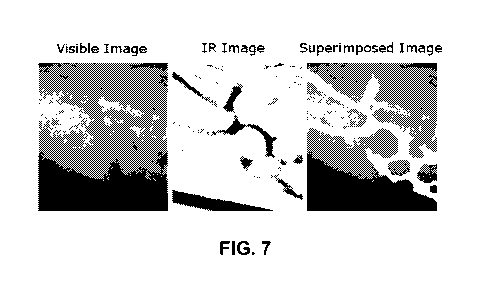
Figure 7 - Images obtained by the application of the techniques described by
the present invention to the detection of vessels over the forearm's surface,
by
fusing visible modes with NIR images.
DETAILED DESCRIPTION OF THE INVENTION
In view of the aforementioned figures and according to the numbering
adopted in them, different embodiments of the invention are described
hereunder.
Thus, as shown in said figures, the equipment comprises a multimodal image
acquisition unit (1) and an image processing unit (2).
The multimodal image acquisition unit (1), whose preferred implementation
as shown in Figure 1 includes an image capturing device, preferably an
endoscopic
image acquisition device comprising an endoscope, a fetoscope or a laparoscope
and additional optical systems, comprising said systems at least one channel
from
which the video images from the inside of the patient are acquired, and at
least one
light source to illuminate the observed tissues.
In a preferred embodiment of the invention, the video channel or channels
that are available on the endoscope are coupled to an infrared light source
(4) and a
white light source (5) or a light source that contain at least three
wavelengths within
the blue, green and red.
CA 02746243 2011-06-08
-14-
The infrared light source (4) is, preferably:
- A source belonging to the NIR (ranging from 750nm to 1600nm).
- A source ranging from 800nm to 900 nm.
- A source ranging from 1050 to 1150nm.
- A monochromatic source centered at a wavelength between 800 and 900nm.
- A monochromatic source centered at a wavelength between 1050 and 1150nm.
- A laser Nd: YAG source (centered at 1064nm).
- A source based on titanium-sapphire laser (Ti: Sap), focusing on 700nm to
1100nm.
- Ytterbio based laser source (Yb: KYW, Yb: KGW, etc.)
- Ytterbio laser source based on Chromium, Cr: Forsterite 1230 to 1270nm.
- An infrared source based on parametric conversion methods (Optical
Parametric Oscillators, Optical Parametric Amplifiers, Nonlinear Crystals,
etc.).
- Lights or LEDs with emission spectrum wavelengths in the NIR between 750 -
1600nm.
- Lights or LEDs with emission spectrum wavelengths in the NIR between 800 -
900nm.
- Lights or LEDs with emission spectrum wavelengths in the NIR between 1050 -
1150nm.
- Lights or LEDs with infrared emission spectrum in combination of optical
filters.
- Light sources with coupled optical filters to restrict the radiation within
the
infrared spectrum, optionally motor controlled.
Additionally, the infrared light source (4) for its application to operations
of
complications in monochorionic twins pregnacies is, preferably:
- A monochromatic source centered between 815 - 835nm, preferably centered at
821 nm. The latter value corresponds to a wavelength of optimal transmittance
in
the amniotic fluid.
- A monochromatic source centered at 1050 - 1090nm, preferably centered at
1070nm. The latter value corresponds to a wavelength of optimal transmittance
in
the amniotic fluid.
The light can be coupled to the video channel of the endoscope using
different optical elements (6) such as beam splitters, hot mirrors (intended
as
infrared-reflecting mirrors), cold mirrors (intended as visible light-
reflecting mirrors),
CA 02746243 2011-06-08
-15-
dichroic mirrors, polarizers, diffusers, diffractive optical elements,
analyzers,
holographic optical elements, phase plates, acusto-optic materials, dazzlers,
shapers, partial mirrors, dichroic prism systems, tunable optical filters,
multibifurcated light guides, polarization beam splitters or any other optical
devices
able to modify their transmission or reflection conditions depending on the
wavelength, polarization or other optical property in order to split or
combine the
optical path for either or both detection and illumination, also including the
encapsulation in optical fiber when the optical path is a fiber optic path.
The same channel can also be used for detection by the employment of
filters (8) and lenses (9) to form the images on a video camera (CCD, CMOS, EM-
CCD, etc.), in order to digitize them to be further processed by the image
processing
unit (2).
Additionally, an image intensifier can be added to the video cameras (10),
(11) if the detected signals are very weak or they show a low quality.
In order to simplify the multimodal image acquisition unit (1), light sources
(4), (5) can be coupled to the video systems (10), (11) by using two channels
of the
endoscope (3), as shown in Figure 2.
Also a separate channel can be used only for illumination, employing
different optical elements (6), as shown in Figure 3.
In another embodiment of the invention, a CCD, CMOS or EM-CCD camera
(10), installed at the probe of the endoscope and coupled to an electric
connection
(22), is employed for the sequential detection of different bands or
wavelengths
sequentially emitted by the light sources (4), (5), as shown in Figure 4.
Optionally, at
least one filter (8) in the camera (10) can be a color filter array (CFA) or a
color filter
mosaic (CFM) for the separation of one or more infrared spectral bands.
The image processing unit (2) forming part of the equipment of the present
invention is a device responsible for processing and displaying the enhanced
images to the surgeon in real time after having been acquired by the
multimodal
CA 02746243 2011-06-08
-16-
image acquisition unit (1). Said device comprises at least each of the methods
listed
below, as shown in the diagram of Figure 5, by the implementation of the
appropriate hardware and software in CPUs, FPGAs, CPU-based systems or any
other hardware performing real-time processing through local, distributed or
parallel
computing. In Figure 5, for better understanding, the infrared image has been
referenced with (12), the visible image with (13), the reflected image in red,
green
and blue, with (14a), (14b) and (14c) respectively, the different methods with
(15),
(16), (17), (18) and (19), enhanced local display with (20) and the enhanced
overall
display with (21). The essential tasks that said hardware and software
execute, i.e.
the procedures of signal processing to improve the imaging of the equipment
that
makes this unit are:
Method 1. Normalization (15): signal processing procedure to normalize the
amount of light that illuminates the tissue (7), by real-time comparing the
intensities
in each of the points in the image of the intensity of visible light (red,
green and blue)
and infrared light and the use of low-pass filter on the images, estimating
the
amount of incident infrared light in a reproducible manner.
- Inputs:
Reflected red image RR(x, y) (14a), wherein (x, y) refers to the two-
dimensional
pixel coordinates in the image obtained.
Reflected green image Rc (x, y) (14b).
Reflected blue image RB(x, y) (14c).
Reflected infrared image RN,R(x, y) (12).
- Outputs:
Estimated illumination image IN,R(x, A .
Method 2. Segmentation (16): Signal processing procedure to real-time
segment the blood vessel images based on spectral analysis of infrared and
visible
light.
- Inputs:
CA 02746243 2011-06-08
-17-
Estimated illumination image IN,R(x, y) .
Reflected infrared image R. (x, y) .
- Outputs:
Blood vessel probability P,, (vessellx, y) , m=1,2, ... M. M corresponds to
the different
image-acquisition modes different from the RGB mode.
Blood vessel segmented image V(x, y) .
- Essential Steps:
1. Using the ratio of infrared light reflected and estimated incident light a
probability
to each point can be assigned forming a new image that contains the
probability of
being "blood vessel" for each point on the screen by a sigmoid curve, for
example:
P, (vessel) x, y) = 1
I + exp - a R. (x, Y)
1NIR(x,Y)
where a is a constant manually or automatically chosen, RN,R(x, y) is the
infrared
reflected image and 1NIR(x, y) is the estimated image using method 1.
2. By low-pass filtering the probabilities, a new probability image is
generated, which
averages the probabilities within a neighborhood, PP (vessel) x, y) .
3. The essential steps 1 and 2 can be repeated for each of the wavelengths or
optical imaging modes that are available for the multimodal imaging unit (1),
thus
generating a range of images of probability Pn,(vessellx, y) for m =1,2,...M .
4. Using a threshold over P(vessellx, y) and the application of morphologic
operations, the image is segmented between "blood vessel" with a value of 1
for
V(x, y) and "not blood vessel" with a value of 0 for V(x, y) .
5. The incorporation of image acquisition modes in the multimodal imaging unit
(1)
improves the accuracy of the segmentation and/or obtains a , greater number of
CA 02746243 2011-06-08
-18-
segmented classes, such as arteries and veins using additional wavelengths, or
collagen structure, by using polarizers. The latter application is
particularly relevant
for dermatology.
Method 3. Tracking (17): Signal processing procedure for real-time tracking
and co-localizing blood vessels between two consecutive scenes from images
generated by Methods 1 and 2.
- Inputs:
Blood vessel probability image P(vessel) x, y) , m=1,2,... M.
Blood vessel segmented image V(x, y) .
Previous blood vessels probability images P' (vessellx, y) , m=1,2.... M.
Previous blood vessel segmented image V'(x, y) or vascular map image T(x, y).
- Outputs:
Displacement vector between two images d(x,y), used for measuring displacement
distances.
Cross correlation coefficient between images Cv.
- Essential steps:
= Option A:
1. A predictive model favors the blood vessels natural direction and smoothes
the blood vessels edges of the previous V'(x,y) and the current V(x,y) images,
resulting in Vp' (x, y) and Vp(x, y) , respectively.
2. The maximum of the normalized crossed correlation between Vp'(x,y) and
Vp(x, y) is detected.
3. The distance of the maximum to the origin of coordinates gives the
displacement distance d(x,y).
4. Cross correlation coefficient is calculated, as the maximum of the
normalized
CA 02746243 2011-06-08
-19-
cross correlation.
= Option B:
1. A predictive model which favors the blood vessels natural direction and
smoothes the blood vessels edges of the previous V'(x,y) and the current
V(x,y)
images, resulting in Vp'(x,y) and Vp(x, y) , respectively.
2. The area which delimitates the full width half maximum of the cross
correlation between Vp'(x, y) and Vp(x, y) is detected.
3. The distance of the centroid or center of mass of the said area, weighted
or
not, respect to the origin gives the displacement distance d(x,y). Centroid
and center
of mass calculations are intended as usual image-processing operations for
calculating the center of an area.
4. The quotient of the cross correlation is the weighted average of the
normalized cross correlation.
= Option C:
1. The most probable displacement is found, d(x,y), maximizing likelihood, by
comparing the previous and current probability images P,,,'(vessel x, y) and
P(vessel) x, y), respectively.
2. The overlapping area of the previous V'(x,y) and current V(x,y) is
calculated and normalized with respect to the total area of the field of view
of the
image, this gives Cv.
Method 4. Mapping (18): Signal processing procedure to generate the map
of the anatomical structures or tissues, preferably the vascular structures in
real-
time, based on images and tracking coordinates obtained from methods 1 and 2.
- Inputs:
CA 02746243 2011-06-08
-20-
Position vector p(x,y).
Displacement vector between the two images d(x,y).
Cross correlation coefficient between images Cv.
Reflected red image RR (x, y) (14a).
Reflected green image RG(x, y) (14b).
Reflected blue image RR(x,y) (14c).
- Outputs:
Vascular map image T(x, y).
Global image G(x, y, c) (Note: c refers to colors red, green, blue).
Previous blood vessels probability images P,,,' (vessel) x, y) , m=1,2, ... M.
Previous blood vessel segmented image V'(x, y) .
- Essential steps:
These techniques are known as Stitching or Mosaicing and are used in computer
vision. A possible implementation is:
1. A threshold > 0.5 is applied over the cross correlation coefficient, Cv.
2a. If Cv < 0.5, the automatic system assumes that the current image contains
errors and does not use it for the vascular map stitching.
3a. Search the current image V(x,y) in the global vascular map T(x,y) through
the Tracking algorithm (Method 3). New parameters d(x,y) and Cv are obtained.
4a. If Cv > 0.5 proceed to step 2b, else skip the rest of the steps and wait
until next
image acquisition.
2b. If Cv > 0.5, the current image V(x,y) is placed on the global image T(x,
y) in a
way that the previous position p(x,y) and its displacement d(x,y) is taken
into
account.
CA 02746243 2011-06-08
-21-
3b. The current image which belongs to the visible in the red reflected image
RR (x, y) (14a), green reflected image RG (x, y)14b and blue reflected image
RB(x, y) (14c) in the global image G(x,y,c) in a way that the previous
position p(x,y)
and its displacement d(x,y) is taken in to account, where c, for instance,
refers to the
color in a standard video image c=R, G or B.
4b. Prepare the system for a new iteration. Transfer the current image V(x,y)
to
the previous image V'(x, y), i.e., V'(x, y) = V(x, y).
5b. Transfer the current probabilities to the previous ones.
Pm' (vessel) x, y) = Pm (vessel) x, y) .
6b. Update the position by d(x,y) and p(x,y).
Method 5. Fusion (19): Signal processing procedure to merge in real-time the
image of the visible (produced by a standard endoscope) with information from
method 3.
- Inputs:
Vascular map image T(x, y) .
Global image G(x, y, c) .
Reflected red image RR(x, y) (14a).
Reflected green image RG(x, y) (14b).
Reflected blue image RB(x, y) (14c).
Blood vessels segmented image V(x, y).
- Outputs:
Color image of local enhanced vision VEL(x,y,c).
Color image of global enhanced vision VEG(x,y,c).
- Essential steps:
1. Image VEL(x,y,c) is obtained by the weighted adding of the segmented blood
CA 02746243 2011-06-08
-22-
vessel image V(x,y) overlapped onto one or many Visible images: reflected red
image RR(x, y) (14a), reflected green image RG(x, y) (14b) and reflected blue
image RB(x, y) (14c).
2. Image VEG(x,y,c) is obtained by adding the segmented vascular map image
T(x,y) overlapped onto one of the channels or colors c of the global image
G(x, y, c) .
3. Achieving a digital image that can be sent to one or several monitors,
projectors
or generic device able to represent a digital or analog image.
4. A user interface is created to choose the viewing modality to display in
each of
the monitors (or equivalent): VEL(x,y,c), VEG(x,y,c), V(x, y) , T(x,y) or G(x,
y,c) .
In order to clarify the effect of the described methods, different vision
modes
available to the equipment described by the present invention are depicted in
Figure
6, showing (a) the vision mode offered by a standard endoscope, (b) the
segmentation (16) of blood vessels through NIR analysis, (c) fusion (19) of
visible
and NIR images, (d) mapping (18) reconstruction and (e) mosaic reconstruction
by
tracking (17) of consecutive images.
To sum up, the signal processing procedure to improve infrared vision of
anatomical structures with the equipment of the invention is performed in the
image
processing unit (2) with the specific hardware and software implemented in
GPUs,
FPGAs, CPU-based systems or any other hardware performing real-time processing
through local, distributed or parallel computing, comprising said procedure at
least
the following methods:
Method 1. Normalization (15): Signal processing procedure to normalize the
amount
of light that illuminates the tissue (7), by real-time comparing of the
intensities in
each of the points in the image of the intensity of visible light (red, green
and blue)
and infrared light; and use of low pass filter on the images. The amount of
incident
infrared light is estimated in a reproducible manner.
CA 02746243 2011-06-08
-23-
Method 2. Segmentation (16): Signal processing procedure to segment the
anatomical structures or tissues, preferably the vascular structures, based on
real-
time spectral analysis of infrared and visible images.
Method 3. Tracking (17): Signal processing procedure for real-time tracking
and co-
localization of the anatomical structures or tissues, preferably the vascular
structures, between two consecutive images generated by Methods 1 and 2.
Method 4. Mapping (18): Signal processing procedure to generate the real-time
map
of the anatomical structures or tissues, preferably the vascular structures
from the
images and tracking coordinates obtained from methods 1 and 2.
Method 5. Fusion (19): Signal processing procedure to fuse the visible image
(produced by a standard endoscope) with information from method 3.
The equipment can further integrate more image modes by using additional
sources of light (both visible and infrared) and/or additional optical systems
to
acquire different imaging modes in the multimodal imaging unit (1).
The present invention offers, additionally, relevant applications to any type
of
endoscopy surgery, such as gastrointestinal tract endoscopy, respiratory tract
endoscopy, arthroscopy, gynecologic endoscopy, colposcopy, urologic endoscopy,
otoscopy, or plastic surgery endoscopy, among others. The invention further
provides applications to other medical procedures, such as skin or open
surgical
procedures, by the replacement of the endoscope, laparoscope or fetoscope by
an
optical objective (intended as a lens, a mirror or other optical instrument
that gathers
the light coming.from the object being observed) adapted to its employment in
said
medical procedures. As an example, Figure 7 shows the images obtained by the
use of the techniques of vascular detection here described applied to the
surface of
the forearm, where visible modes are fused to the NIR image.
The disclosed invention also offers the possibility to perform functional
analysis of the anatomical structures. Other modalities of the present
invention offer
CA 02746243 2011-06-08
-24-
the classification of different anatomical structures, such as collagen by the
use of
polarization imaging and/or second harmonic. It can also be used to
distinguish
between variations in the same anatomical structures to detect anomalies that
lead
to diagnose clinic conditions. All this automated and quantitative data
acquisition is
not only adaptable to the guide surgery but also to the robotized remote or
automated surgery.
Having sufficiently described the nature of the present invention, as well as
how to implement it, it is not considered necessary to extend the explanation
for any
expert in the field to understand its scope and the advantages that derive
from it, but
highlighting that, within its fundamental nature, it can be put into practice
in other
embodiments that differ in the details from that indicated though the
examples, and
which remain covered by the claimed protection providing that the fundamental
nature is not altered, changed or modified.
CA 02746243 2011-06-08
-25-
DESCRIPTION OF THE NUMERICAL REFERENCES USED
Reference Description
(1) Multimodal or multispectral image acquisition unit
(2) Image processing unit
(3) Endoscope, fetoscope or laparoscope
(4) Infrared light source
(5) White light source
(6) Optical elements
(7) Anatomical structure, tissue or vascular structure
(8) Filter
(9) Lens
(10) Video camera
(11) Video camera
(12) Infrared image
(13) Visible image
(14a) Red image
(14b) Green image
(14c) Blue image
(15) Normalization method
(16) Segmentation method
(17) Tracking method
(18) Mapping method
(19) Fusion method
(20) Enhanced local display
(21) Enhanced overall display
(22) Electric connection