Note : Les descriptions sont présentées dans la langue officielle dans laquelle elles ont été soumises.
CA 02746851 2011-06-13
WO 2010/026557 PCT/IB2009/053897
Coated medical device and method of coating a medical device to
reduce, fibrosis and capsule formation.
FIELD OF THE INVENTION
The present invention relates to a coating technology modulating cell
adhesion on the surface of medical devices. The coating prevents
capsule formation and decreases fibrosis induced by foreign bodies such
as medical devices.
BACKGROUND OF THE INVENTION
Medical devices in contact with living cells and tissues induce a foreign
body reaction due to their physical and chemical properties. This
process results in a capsule formation around the implant that often
contracts and results in malfunctioning of the device and clinically
relevant complications such as pain and dysmorphism of the patient that
ultimately require additional surgical operations.
The reaction of cells to a foreign material such as for example a silicone
breast and aesthetic implant, a gastric band, a dental implant, an
orthopedic implant, hearth heart valve, etc., which cannot be
phagocytated, enzymatically digested or otherwise eliminated, is the
formation of fibrotic capsule around it.
1
CA 02746851 2011-06-13
WO 2010/026557 PCT/IB2009/053897
It has been reported by Prantl L., and al. (Clinical and morphological
conditions in capsular contracture formed around silicone breast
implants, Plast Reconstr Surg 2007, 120:275-284) that in up to 20-30%
of implanted prosthesis develop capsule formation. The capsule
formation is excessive and leads to clinically relevant, painful
contractures around the device. Patients with excessive capsular
formation need to receive a second operation to remove the capsule
around the device which considerable increases in the morbidity and
costs. This process significantly shortens the lifespan of the device.
This capsule is the result of a chronic inflammatory process around the
material that resolves only after the formation of the capsule itself.
Myofibroblasts play a main role in capsule formation and contraction in
healing tissues. The capsule itself is deposited and contracted by
myofibroblasts. In the wound healing process, these highly active cells
proliferate and growth to occupy tissue defects and replaces them with
a scar. Around the implant, myofibroblast deposit collagen fibers around
the foreign material and eventually contract it. This reaction produces
capsular contraction.
The problem of capsule formation has also been discussed in patent
literature. In US 4,772,285 a collagen coated soft tissue prosthesis is
described for reducing capsule formation. While this patent proposes a
strategy to decrease capsule formation, no mechanism is provided to
effect myofibroblast formation. Further the clinical evidence is lacking.
2
CA 02746851 2011-06-13
WO 2010/026557 PCT/IB2009/053897
US 4,955,907; 4,731,081; 5,571,183; 5,207,709; 5,354,338;
4,428,082 and 4,298,998 also propose solutions to avoid or diminish
capsule formation.
All these documents have several common denominators which have
the potential of making them unsuitable for resolving this problem in
human beings.
For example US 4,298,998 disclose causing a capsule to form at a
predetermined, controlled distance from the surface of the implant, thus
resulting in the same capsule but at a different location. The end result
clinically appears to be a hard capsule for the patient and not resolving
the problem.
Similarly, the implant described in US 5,207,709 includes a plurality of
fine projections extending from the outer surface arrayed in a basket
weave-like, herringbone-like, or other suitable pattern to create a
sinuous path for collagen formation around the implanted device. It
appears that this implant actually creates or invites collagen formation
again in another location around the implant and again therefore is not
resolving the problem.
Still other patents relate to the implant being surrounded by a medical
grade elastomer or as described in US 4,944,749 a viscous gel coating
with the membranes constructed of a suitable material such as medical
grade silicone rubber, which does not react with human tissue. The
outer membrane contains an amount of viscous gel, for example a
silicone rubber gel of medical grade silicone.
3
CA 02746851 2011-06-13
WO 2010/026557 PCT/IB2009/053897
It appears in the end that this patent still has a silicone tissue interface
that has accounted for problems.
US 4,610,690 is directed to an implant with a lubricious layer of an
acrylamide polymer radiation bonded to at least one wall surface of a
silicone shell or bag. Potential long-term effects in human beings of an
acrylamide polymer interface are not discussed.
All these aforementioned patents continue to have unnatural chemicals
as interface with human tissue, which is exactly what patients do not
want in their body and what usually causes problems.
In US 4,995,882 an organic fill solution is proposed to solve the
problem. The implant is proposed the use a triglyceride fill substance
such as peanut oil or sunflower seed oil as a filling substance.
Although some implants of this kind were implanted in Europe, they
were never authorized for implantation in the United States and were
subsequently taken off the market worldwide because of various
problems.
Although many strategies have been developed to reduce capsule
formation, from modifications of the surface of the implants to chemical
coatings, only mixed results have been reported and, with the increase
in the demand for implantable devices, the clinical problem is on the
rise.
4
CA 02746851 2011-06-13
WO 2010/026557 PCT/IB2009/053897
SUMMARY OF THE INVENTION
It is therefore an object of the invention to provide a coating of a
medical device and a method to produce it with which the fibrosis and
capsule contraction can be further diminished or even avoided.
This can be achieved by a medical device wherein a surface of the
medical device is coated with cell-adhesive proteins deposited in a
matrix of islets with specific size, area and distribution, object of this
invention.
This coated surface, which is supposed to come in contact with living
cells and tissues to modulate cell adhesion is composed from two
specific regions:
1. Area (islets) where cell (tissue) can specifically attach (coated part),
and
2. Other region where cell (tissue) cannot specifically attach (non coated
part).
This invention relates in general to a method and device for guiding
cellular adhesion on medical devices. The invention relates to a method
comprising protein islets to coat devices (implantable and external) in
contact with any cell and tissue of the body.
Specifically, the invention contemplates the use of such technique in
combination with devices implanted or externally applied to the body.
5
CA 02746851 2011-06-13
WO 2010/026557 PCT/IB2009/053897
This proposal outlines a novel strategy based on islands of proteins to
decrease the formation of fibrosis reaction and contraction around
medical devices.
According to a preferred embodiment of the invention the medical
device comprises silicone as a material of the medical device.
Other preferred embodiments include the use of plastic, metal and/or
collagen as a material of the medical device.
According to one embodiment of the invention the medical device
according to the invention comprises islets of proteins with uniform
geometric shapes.
Another possibility would be the use of different geometric shapes.
Generally, to reduce the myofibroblast differentiation and fibrosis, the
area of the single islets is less than 12 pmt.
The topography of islets that best reduces fibrosis is composed of single
islets, wherein the islets have preferably a length that is <6 pm, a width
that is <2 pm and distance between them that is <6 pm
In a preferred embodiment of the invention the medical device further
comprises an additional substrate to facilitate the transfer of the islets of
proteins.
6
CA 02746851 2011-06-13
WO 2010/026557 PCT/IB2009/053897
The substrate which is preferably applied to the medical device before
coating same with proteins could be silicone, plastic, bio resistant
materials, etc..
Further the invention describes a method of coating a medical device,
wherein the medical device is coated with cell-adhesive proteins in form
of single islets.
As already mentioned it could be in some embodiments of advantage if
the medical device is coated by a substrate before being coated by the
proteins.
According to one embodiment of the invention the proteins are applied
to the medical device by a stencil or mask (a template with holes of the
size and distribution of the islets).
For applying the proteins according to another embodiment the stencil
or mask could be brought in contact with the surface of the medical
device and the proteins are transferred to the surface through holes of
the stencil.
The fixing of the proteins to the medical device could be done by every
possibility known from the art. As an example, it should be mentioned
covalent binding, electro deposition or precipitation of proteins on the
medical device and/or the substrate.
The medical device as described can be used for example for breast
implants, tissue expanders, inflatable bumps, implants, transplants,
7
CA 02746851 2011-06-13
WO 2010/026557 PCT/IB2009/053897
prosthesis, insulin pumps, drug delivery systems, cardiovascular
devices, skin substitutes, wound dressings and/or tubes.
DETAILED DESCRIPTION OF THE INVENTION
These and other objects, features, aspects and advantages of the
present invention will become more apparent from the following detailed
description of the present invention when taken in conjunction with the
accompanying drawings.
Figure 1 shows a medical device before and after being coated
according to one embodiment of the invention; and
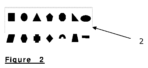
Figure 2 shows examples of islet shapes according to a
preferred embodiment of the invention.
The invention describes a new method of micro-deposition of cell-
adhesive proteins and molecules onto the surface of medical devices,
such as, but not limited to highly deformable and elastic substrates
(such as, but not limited to silicone), Titanium, Plastic, Polyurethane and
generally all materials used for medical devices and implants.
The micro-deposited islets of proteins guide cell adhesion on medical
devices with the objective to reduce the fibrotic reaction of the cells and
tissues in contact with the device.
8
CA 02746851 2011-06-13
WO 2010/026557 PCT/IB2009/053897
As shown in Figure 1 according to an embodiment of the invention a
microperforated stencil or mask is used to transfer proteins to coat a
medical device. 1. Deposition of the proteins and molecules is in the
form of specific islets. 2. Protein islets include any possible geometrical
shape and spatial organization and individual area. Protein islets (2) are
deposited to modulate cell and tissue adhesion to medical devices (1).
The distance between the islets may vary from 1 to 50 pm. The area of
the single islets being less than 12 pmt.
The distribution of protein islets according to a preferred embodiment of
the invention to best reduce fibrosis includes single islets, wherein the
islets have a length that is <6 pm, a width that is <2 pm and distance
between them that is <6 pm.
In another embodiment, deposited islets could have any geometrical
shape. Examples to which the invention is not limited are illustrated in
Figure 2.
The mask or a stencil to transfer the islets is micro fabricated using
technology as for example photolithography, dry and wet etching, laser
cutting.
As type of mask or stencil should be mentioned soft stencils made from
silicone and flexible polymers and hard stencils from silicon, hard
polymer and metal.
9
CA 02746851 2011-06-13
WO 2010/026557 PCT/IB2009/053897
To deposit proteins onto medical device as for example an implant
surface, the stencil is first brought in conformal contact with the surface
and then the proteins are transferred on the surface through the micro-
holes of the stencil by covalent binding using 3-
aminopropyltriethoxysilane (APTES) and glutaraldehyde or
formaldehyde. After the crosslinking of proteins, the stencil is removed
and the pattern remains on the implant surface.
Another possible method is electro deposition or precipitation of proteins
on implant or medical device surface through the micro-holes of stencil
or mask. These possibilities are just examples and should not limit the
invention to this approach of deposition.
Our results in vitro and in vivo illustrate the importance of the size of
the islets in reducing fibrosis and contraction around implants proving
the relevance of this invention. We found that the main mechanism
responsible for limiting the differentiation of human dermal fibroblasts in
myofibroblasts (the main cell responsible for fibrosis and contraction) is
the area of adhesion.
In our experiments, the islets represent areas of adhesion (focal
adhesions) for the cells and dimensions of the islets of a length that is
<6 pm, a width that is <2 pm and distance between them that is <6 pm
impaired the formation of myofibroblasts (Figure 3).
CA 02746851 2011-06-13
WO 2010/026557 PCT/IB2009/053897
The specific islet size and distribution with a length <6 pm, a width <2
pm and distance between them <6 pm does not allow fibroblasts to
exert forces on the surface where they attach and become
myofibroblasts (the main cell responsible for fibrosis and contraction).
In vitro, we showed that this specific size and distribution of proteins
reduced 10-fold the differentiation of human dermal fibroblasts to
myofibroblasts compared to devices coated with other size and
distribution of proteins (Figure 4). The deposition of islets of higher size
allowed the differentiation of fibroblasts in myofibroblasts. These later
cells were seen in high percentage (up to 20%) when the surface was
coated with larger islets than the specific ones provided by this
invention (Figure 5).
In vivo, silicone pads (1x1 cm) covalently coated with a stencil or mask,
as described in the methods, on both side with protein islets with a
length <6 pm, a width <2 pm and distance between them <6 pm, were
inserted in subcutaneous pockets, in female Wistar rats (250-350 g). On
the scapular region of the dorsum of these animals, 4 coated silicone
pads were placed. Each animal received four implants: two implants
coated with islets with a length <6 pm, a width <2 pm and distance
between them <6 pm; and two non coated silicone implants, alternating
the location on successive animals. Results at 6 months show a 3-fold
decrease in capsule formation around implants coated with islets of
proteins with a length <6 pm, a width <2 pm and distance between
them <6 pm (Figure 6).
In vitro and in vivo results show that the transformation of fibroblasts
into myofibroblasts is crucial in the development of fibrosis and capsule
11
CA 02746851 2011-06-13
WO 2010/026557 PCT/IB2009/053897
contraction around medical implants and underline the importance of
this invention specifically limiting this event.
The optimized protein islets deposition with e.g. a length <6 pm, a
width <2 pm and distance between them <6 pm can be applied to any
medical device such as, but not limited to:
= Silicone breast implants
= Tissue expanders and inflatable pumps
= Bone, and cartilage and orthopaedic implants
= Tendon, nerve, and ligament transplants, substitutes and implants
= Implantable pumps, such as insuline pumps and drug delivery
systems
= Cardiovascular devices, such as pace makers, vascular prosthesis,
heart valves, vascular stents
= Skin substitutes and cell scaffolds
= Wound dressings, including dressings and wound interfaces
connected to a vacuum (negative pressure dressings)
= Acoustic waves (such as shockwave therapy)
= Ear, throat, nose and eye implants
= Brain, central and peripheral nervous system implants and
prosthesis
= Tubes, connecting tubes, drainage tube systems
12
