Note : Les descriptions sont présentées dans la langue officielle dans laquelle elles ont été soumises.
I1
HEART SUPPORT DEVICE
The invention relates to a heart support device for pulsatile delivery of
blood.
Heart support devices, particularly mechanical systems for support of the
blood
circulation (VAD: Ventricular Assist Device) are implanted into the body of a
patient suf-
fering from cardiac insufficiency. Such devices will take over a part of the
pumping work,
thus stabilizing the blood circulation, e.g. until a donor organ has become
available. Re-
cent search has revealed that, during use of a heart support device, the
cardiac function can
improve to an extent favorable enough to allow for explantation of the system
without
subsequent heart transplantation.
Artificial heart pumps can be adapted to the most diverse requirements and, in
con-
trast to donor hearts, are readily available without a waiting period.
However, such heart
support devices have to meet high demands with regard to the chosen technology
and the
tolerability of the implants. For instance, the blood may happen to be damaged
by the
pumping work. The power supply to the electrically operated systems, e.g. via
cables pass-
ing through the abdominal wall, harbors a considerable risk of infection to
the patient.
Further, low degrees of efficiency will cause high energy consumption and will
heat the
surrounding tissue. Often, the assisted blood circulation has to be maintained
through
months or years. The systems are subjected to strong mechanical stresses.
Since it is not
possible to exchange blood pumps quickly, such pumps must have a very high
failure
safety.
DE 40 201 20 describes a heart support system which is operated hydraulically.
A
displacement pump is provided for pumping a hydraulic liquid alternately into
a first and a
second hydraulic chamber, the hydraulic liquid being separated by a flexible
membrane
from the blood - contained in the hydraulic chamber - that is to be conveyed.
By filling the
first hydraulic chamber with hydraulic liquid, the blood within the chamber
will be dis-
placed and, via a valve, be conveyed into the circulation. The hydraulic pump
is arranged
between the two hydraulic chambers and partially within the hydraulic chambers
them-
selves.
A disadvantage of the above described device resides in that the two hydraulic
chambers, having two blood chambers arranged laterally adjacent thereto, as
well as the
hydraulic pump located between the hydraulic chambers, together have a large
construc-
t
I1
tion height and thus can be implanted into a patient's body only with
considerably diffi-
culty.
It is an object of the invention to provide a heart support device having a
low con-
struction height.
According to the invention, the above object is achieved by the features
defined in
claim 1.
A heart support device for pulsatile delivery of blood comprises a first and a
sec-
ond ventricle as well as a pump. Each of said two ventricles comprises a fluid
chamber
and a blood-conveying chamber, each fluid chamber being able, with the aid of
said pump,
1o to be filled with a fluid or to be evacuated in a manner causing an
expansion or contraction
of the fluid chamber.
Preferably, no blood is included in the fluid chamber.
According to the invention, expansion of a fluid chamber of a ventricle will
result
in compression of the blood-conveying chamber of the same ventricle. In this
manner, it is
possible to convey blood into the blood circulation, while both the left and
the right ven-
tricle can be supported. A volume compensating reservoir compensating for the
conveyed
volume is not required.
According to the invention, said pump is arranged outside said first and
second
fluid chambers and/or outside said first and second ventricles.
Thereby, it is rendered possible to place or implant the pump and the two
ventricles
at different sites in or on a patient's body. Preferably, for this purpose,
the pump can be
connected to the fluid chambers via a fluid conduit of corresponding length.
Thus, the de-
vice of the invention has a reduced construction height and can be implanted
also into the
body of smaller patients in a less problematic way.
Preferably, the first and the second ventricles are arranged adjacent to each
other
and, e.g., comprise a common partition wall. Particularly, the two ventricles
can be located
adjacent to each other while separated from each other exclusively by a
partition wall.
By the design of the heart support device as provided by the invention, it is
possi-
ble to arrange the pump at a distance from the two ventricles and/or the fluid
chambers.
Said distance can e.g. be larger than 10 cm and with particular preference
larger than 15
cm. This offers the possibility to arrange the drive pump in an electronics
housing, ar-
ranged separate from the heart pump, which also accommodates rechargeable
batteries for
2
I1
operation of the pump as well as electronics for control. The resultant
advantage lies in the
minimizing of the volumes of the implanted components. This will enhance a
full im-
plantability of the components. Particularly, the pump can be arranged
laterally next to the
first and the second ventricle. Also an arrangement above or below the two
ventricles is
possible. Further, for instance, the two ventricles can be arranged at a
lateral offset from
each other on the left and right sides while preferably being directly
adjacent to each other,
i.e. abutting each other. In this case, the pump can be connected to the two
ventricles via a
fluid conduit and can be arranged, e.g., above the two ventricles, i.e.
cranially relative to
the ventricles. Thus, the pump is preferably not arranged between the first
and the second
lo ventricle and/or not between the first and the second fluid chamber.
The fluid conveyed by the pump preferably is a hydraulic fluid, wherein the
fluid
in the sense of the invention is not the blood of a patient. Instead, the
fluid can be, e.g., a
liquid causing the blood to be conveyed by corresponding contraction of the
blood-
conveying chambers.
According to a particularly preferred embodiment, said partition wall by which
the
two ventricles are separated from each other, is arranged stationary relative
to the heart
support device. This means that, when the fluid chamber is being expanded, the
partition
wall is not displaceable, so that an expansion of the fluid chamber will cause
a compres-
sion of the blood-conveying chamber and will thus effect a delivery of blood
into the
2o blood stream.
Said partition wall is preferably made of the same material as the ventricles
them-
selves. For instance, the two pump chambers and the partition wall can be
produced in a
sole manufacturing process and, at a later time, be easily integrated into an
enclosing
housing. The common wall, i.e. the partition wall, preferably has a thickness
slightly lar-
ger than that of the ventricle walls so that its stiffness is increased. In
spite of its larger
thickness, the partition wall is preferably flexible. First, this flexibility
will have the effect
of a certain yieldingness during the pumping operation, thus allowing for the
blood to be
discharged in a gentle manner. Thereby, the pump can be used with little wear,
and the
useful life of the system can be increased. Second, thereby, a flow-optimized
conveyance
can be achieved in the pumping chambers. In addition to these benefits, the
system can be
given a still more compact design so that the blood conduits to and from the
heart can be
3
I1
kept at minimum length. Thus, one can choose an implantation site close to the
heart. In
the presently described embodiment, said membrane is preferably elastic.
The fluid chambers can each be separated from the blood-conveying chambers by
a
flexible elastic membrane. This membrane is preferably of a two-layered design
wherein,
between the two layers of the membrane, a liquid-filled gap can be provided
for reducing
the friction between the two layers. The gap can be filled e.g. with silicone
oil.
An independent invention relates again to a heart support device for pulsatile
de-
livery of blood, comprising a first and a second ventricle and a pump. In
correspondence
to the invention as described above, the two ventricles comprise a respective
fluid cham-
1 o ber and a respective blood-conveying chamber, wherein, with the aid of
said pump, each
fluid chamber can be filled with a fluid or be evacuated, thus causing an
expansion or con-
traction of the fluid chamber. An expansion of the fluid chamber of a
ventricle will result
in a compression of the blood-conveying chamber of the same ventricle. An
essential fea-
ture of the second invention resides in the provision of a respective,
preferably stiff pres-
sure plate arranged between a fluid chamber and the corresponding blood-
conveying
chamber, said pressure plate being displaceable in the direction toward the
respective
blood-conveying chamber.
In this arrangement, the fluid chamber can be formed as a bellows, squib or
bal-
loon. Via said pressure plate, an expansion of the fluid chamber will entail a
compression
of the respective blood-conveying chamber.
The pressure plate can have a surface larger in size than the base surface of
the
fluid chamber so that, in the fully expanded state of the fluid chamber, the
volume of the
fluid will be smaller than the volume of the conveyed blood. Thus, the use of
a preferably
stiff pressure plate makes it possible to deliver a larger blood volume with
the aid of a
smaller quantity of fluid. There will occur a translation of the pressure and
of the volumes.
The heart support device according to the second invention can comprise all
features of
the first invention. Particularly, a stationary and thickened partition wall
can be provided
between the two ventricles.
According to a preferred embodiment, the fluid chamber comprises stiff lateral
walls which extend vertically to the pressure plate so as to prevent the
lateral walls from
yielding due to the applied pressure. The connection between said two lateral
walls, i.e.
4
I1
the side of the fluid chamber facing in the direction of the pressure plate,
can be flexible
and stretchable.
For instance, the steps of said squib, when seen in sectional view, can form a
la-
mellar structure which is able to expand under the influence of the fluid
pressure and thus
will press onto the pressure plate in the manner of a telescope.
Said squib can further be realized as a balloon which is ball-shaped and has a
smaller volume than the blood chamber. The amount of the diameter of the
balloon is only
as large as the width of the blood chamber when viewed in lateral cross
section.
The heart support devices according to the first invention and the second
invention
can further comprise the features described hereunder. There can be provided
stiff housing
walls, with the ventricles arranged therebetween. Further, the first and
second blood-
conveying chambers can be flexible and, particularly, stretchable.
According to a particularly preferred embodiment, the ventricles are of a
stiff na-
ture, with the fluid chambers being separated by an elastic membrane from the
respective
blood-conveying chamber of a stiff ventricle. Thus, the volume of the first
and second
blood-conveying chambers can be enlarged particularly by an underpressure in
the respec-
tive fluid chamber and the resultant elastic deformation of the elastic
membrane. With
particular preference, the volume of the first and second blood-conveying
chambers can be
reduced, subsequent to its enlargement caused by said underpressure, by an
inherent ten-
sion of the elastic membrane, particularly without using an overpressure in
the respective
fluid chamber.
Preferably, the shape of the stiff ventricle is selected to achieve an
optimization of
the flow. When using heart support devices, it is imperative to keep the
ventricles free of
regions with little or no movement of the blood because such occurrences would
have a
thrombogenic effect. It is thus important to achieve an optimized flow in the
blood-
conveying chamber and in the conduits. This can be realized in that the stiff
ventricle is
given a flow-optimizing shape, i.e. a shape which will enhance the blood flow.
In this re-
gard, it is of essence that the membrane separating the fluid chamber from the
blood-
conveying chamber is elastic so that, in the filled state of the blood-
conveying chamber,
the membrane will be in full abutment on the inner wall of the stiff ventricle
and thus will
have the flow-optimizing shape of the stiff ventricle. During discharge of the
blood, the
surface of the membrane will change due to the membrane's elasticity so that
the mem-
5
I1
brane, once it has reached its relaxed state, will not present any kinking or
warping which
could have an adverse effect on the blood flow.
The above described embodiment can also be operated by hydraulic actuation
wherein the blood-conveying chamber can be enlarged by an underpressure and
the blood
can be discharged again by an overpressure generated by a hydraulic liquid.
For discharge
of the blood, there can be additionally used the inherent tension of the
elastic membrane.
Preferably, by use of said pump, the fluid chamber of the first ventricle and
the
fluid chamber of the second ventricle can be alternately filled with fluid and
evacuated of
fluid.
1.0 For replenishing fluid that has been diffused, a replenishment port can be
provided
which is accessible externally of the patient's body, e.g. via a syringe.
In each of the above described heart support devices, use can be made of
electric
pumps as known from the state of the art. Advantageously, the electric pumps
should have
a low construction height and a high running performance.
Preferred embodiments of the invention will be explained hereunder with
reference
to the Figures.
In the Figures, the following is illustrated:
Fig. 1 is a schematic view of a first embodiment of the heart support device
according to
the first invention,
Fig. 2 is a schematic view of the first ventricle of the embodiment according
to Fig.
1,
Figs. 3 a and 3b are schematic views of an embodiment of the heart support
device
according to the second invention,
Figs. 4a and 4b are schematic views of a further embodiment of the heart
support
device according to the first invention, and
Fig. 5 is a schematic view of a squib.
As shown in Fig. 1, a heart support device for pulsatile delivery of blood 12
com-
prises a first 14 and a second 16 ventricle. Each ventricle comprises a fluid
chamber
14a,16a and a blood-conveying chamber 14b,1 6b. Said fluid chambers are filled
with hy-
3 o draulic liquid. Fluid chamber 14a is connected to a pump 18 via a fluid
conduit 38a. Fluid
chamber 16a is connected to pump 18 via a fluid conduit 38b. By means of pump
18, hy-
draulic liquid will be pumped alternately into said first and second fluid
chambers I4a,16a,
6
I1
causing an alternate expansion of the two fluid chambers 14a,16a. Expansion of
fluid
chamber 14a of ventricle 14 will result in compression of blood-conveying
chamber I4b
of the same ventricle 14. The same applies to the second ventricle 16.
Pump 18 is arranged outside the first and second ventricles 14,16 as well as
outside
the first and second fluid chambers 14a,16a. As can be seen in Fig. 1, pump 18
is ar-
ranged, e.g., on the right-hand side of the two ventricles 14,16. Thus, the
pump is located
at a lateral offset from the ventricles 14, 16. However, the ventricles 14,16,
instead of be-
ing arranged above each other as depicted in Fig. 1, can also be disposed
laterally of each
other in the body of a patient so that pump 18 can be located e.g. above or
below the ven-
1o tricles 14,16.
According to Fig. 1, the two ventricles 14,16 are arranged adjacent to each
other
and are separated from each other by a stationary partition wall 22. The heart
support de-
vice is delimited by a first and a second housing wall 32,34.
The fluid chamber 14a,16a and the blood-conveying chamber 14b,16b are sepa-
rated from each other by a respective double-layered membrane 13.
Fig. 2 is a lateral view of the first ventricle 14 of Fig. 1. The blood-
conveying
chamber 14b comprises a blood inlet conduit 15a and a blood outlet conduit
15b, which
conduits will be connected to blood vessels of the patient during the
implantation process
of the heart support device. Said conduits 15a,15b can include valves for
control of the
2o blood flow.
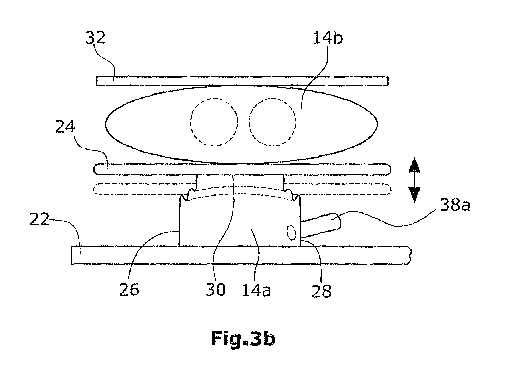
According to a second invention shown in Figs. 3a and 3b, a stiff pressure
plate 24,
adapted to be displaced in the direction of the respective blood-conveying
chamber
14b,16b, is arranged between each fluid chamber 14a, 16a and the respective
blood-
conveying chamber 14b,I6b. In Figs. 3a and 3b, only the first ventricle 14 is
illustrated,
the second ventricle 16 being arranged below the partition wall 22 of the
housing in mirror
image to the first ventricle 14.
In the illustrated embodiment, fluid chamber 14a is formed as a squib.
According
to Fig. 5, said squib, comprising a base surface of any desired shape, can be
designed in
the manner of an accordion or a bellows. The folds are made of a material with
tensile
strength so as to allow for stretching only in the direction of the
longitudinal axis of the
squib. Stretching in the other two directions, however, is prevented. In the
non-expanded
state, the squib is flat while, on the other hand, its base surface is not
significantly
7
I1
enlarged. Via said hydraulic line 38a, which is arranged laterally of squib
14a near the
base surface of the latter, pressurized air or a hydraulic fluid can be
supplied. When the
squib is expanded, a force F can be exerted on the target object. Squib 14a is
connected to
pump 18 via hydraulic line 38a, wherein a delivery of hydraulic liquid into
squib 14a will
cause the squib to expand. Thereby, the stiff pressure plate 24 will be
pressed into the di-
rection of blood-conveying chamber 14, with resultant compression of blood-
conveying
chamber 14b. Since squib 14a has a base surface which is smaller than the
surface of pres-
sure plate 24, the volume of the hydraulic liquid 20 required for expansion of
squib 14a is
smaller than the volume of the conveyed blood 12. Pressure plate 24 can also
be integrated
into the distal end of squib 14a. On the left side of Fig. 5, a non-expanded
squib 14a is
shown, while an expanded squib 14a is shown on the right side.
It is preferred that the squib 14a is formed as non-segmented component, i.e.
in one
piece, thus preventing the occurrence of leakage problems. Further, in
comparison to an
actuator consisting of several segments, friction losses at the segment
contact regions
which may cause a reduction of the mechanical efficiency of the pump, can be
reduced.
Preferably, the squib 14a does not need bearings, seals, tractive connections
or the like for
establishing a safe and reliable operation. When using a squib in the
embodiments of the
invention, the pressure plate 24 may be omitted so that the side or wall of
squib 14a facing
toward the blood-conveying chamber 14b will press directly onto the blood-
conveying
chamber 14b. Thus, squib 14a can be formed as a part of blood-conveying
chamber 14b.
Further, the squib can be designed in such a manner that its sectional surface
corre-
sponds to the projection surface of the blood-conveying chamber or the
membrane.
Thereby, the force will be equally distributed onto the blood-conveying
chamber, and the
device can be realized with a low construction height. Also in case of this
design, it is not
necessary to provide a separate pressure plate. Preferably, the common wall
between the
squib and the blood-conveying chamber or the membrane has a suitable shape or
surface
for enhancing the blood flow.
The side walls 26,28 of squib 14a extend vertically to pressure plate 24 and
are of a
stiff nature, thus allowing an expansion of squib 14a to occur only in the
direction of
3o blood-conveying chamber 14b. The side 30 of fluid chamber 14a facing in the
direction of
pressure plate 24 and facing also in the direction of the blood-conveying
chamber 14b, is
flexible and stretchable.
8
I1
In Fig. 3b, a blood-conveying chamber 14b is shown in its compressed state.
A further embodiment of the heart support device of the invention is shown in
Figs. 4a and 4b. Illustrated in these Figures is only one ventricle 14, which
is of a stiff na-
ture. A elastic membrane 36, preferably bonded to the inner wall of ventricle
14, separates
the fluid chamber 14a from the blood-conveying chamber I4b of the stiff
ventricle 14. The
volume of blood-conveying chamber 14b can be enlarged by an underpressure in
fluid
chamber 14a in that the elastic membrane will be elastically deformed (see
Fig. 4). For
generating an underpressure in fluid chamber 14a, the fluid will be pumped out
from the
stiff ventricle 14 via fluid conduit 38a, with the effect that the pressure in
blood-conveying
chamber 14b will be higher than the pressure in fluid chamber 14a. In this
state, according
to Fig. 4b, blood is supplied via blood inlet conduit 15a into blood-conveying
chamber
14b. Subsequent to the enlargement of the volume of blood-conveying chamber
14b
caused by the underpressure, the volume will be reduced again. This takes
place due to the
inherent tension of the elastic membrane 36, so that the blood will be pressed
out of blood-
conveying chamber 14b via blood outlet conduit 15b.
In order to avoid dead zones in blood-conveying chamber 14b, it is important,
apart from a flow-enhancing shape of the stiff ventricle 14, to find a
suitable site where the
elastic membrane 36 is connected to the inner wall of the ventricle 14. The
surrounding
connecting line on which the membrane 36 is connected to the inner wall of
ventricle 14,
is arranged in such a manner within ventricle 14 that no dead zones will
develop within
blood-conveying chamber 14b. Thus, for instance, the elastic membrane 36 can
be con-
nected to the inner wall at the narrowest site of ventricle 14. Preferably,
the connection site
is selected in such a manner that, in the filled state of blood-conveying
chamber 14b, the
elastic membrane 36 extends tangentially to the outer wall of blood inlet
conduit 15a. The
same applies to blood outlet conduit 15b. Since the stiff ventricle 14 is
formed with a con-
striction below the inlet and outlet lines I5a,15b, it is particularly
preferred that the mem-
brane 36 is connected to ventricle 14 at the apex of said constriction, i.e.
at the narrowest
site of ventricle 14, thus preventing a development of dead zones in the blood-
conveying
chamber above or below this connecting line. Preferably, the ventricle 14 is
designed for
flow-enhancement by having no undercuts and particularly by having a round
uniform
shape.
9