Note : Les descriptions sont présentées dans la langue officielle dans laquelle elles ont été soumises.
CA 02765972 2011-12-19
WO 2010/146160 1 PCT/EP2010/058647
A SURFACE PLASMON RESONANCE SENSING METHOD AND SENSING
SYSTEM
Technical field
[0001] The present invention generally relates to a surface plasmon resonance
(SPR) sensing method and to a SPR sensing system. The invention more
particularly relates to a SPR sensing method and to a SPR sensing system
suitable for use i.a. in chemical, biochemical, biological, biomedical,
pharmaceutical and physical testing.
Background Art
[0002] There are many known sensors using the excitation of surface plasmons,
termed Surface Plasmon Resonance (SPR) Sensors, for detecting refractive index
changes in a sample adjacent to a sensor surface. Such SPR sensors are used
e.g. for quantifying concentrations of substances in chemical, biochemical,
biological, biomedical or pharmaceutical research, in clinical or food
diagnosis or
in environmental measurements (e.g. detection of gas or wastewater), etc. Many
SPR sensors can perform fast, parallel and massive inspections, which make
these sensors also convenient for quantifying molecular interactions, in
particular
for studying the affinity and the real-time reaction kinetics between two or
more
interacting molecules.
[0003] SPR sensors rely on the well-known SPR phenomenon, which involves
one or more surface-bond electromagnetic waves that propagate at an interface
between a metallic material (typically gold or silver) and a dielectric
material. Each
surface-bond electromagnetic wave, which is due to a collective oscillation of
free
electrons at the metal-dielectric interface, propagates with its highest
intensity
parallel to this interface and decays exponentially away from this interface.
[0004] Conventionally, a SPR sensor comprises a sensor surface supporting
surface plasmons, where SPR can be optically excited. It is well known that
light
can excite the resonance of surface plasmons at a metal-dielectric interface
if an
interface-parallel component of the incident light and a surface-bond
electromagnetic wave of the SPR both have matching frequencies and matching
wavelengths. In the resonance condition, the incident light is absorbed by the
CA 02765972 2011-12-19
WO 2010/146160 2 PCT/EP2010/058647
metal-dielectric interface so as to couple with the surface-bond
electromagnetic
wave. It is then possible to observe this absorption by detecting for example
a
reduction in the intensity of the light that is transmitted or reflected by
the metal-
dielectric interface. The coupling condition between light and surface plasmon
waves being very sensitive to refractive index changes of the dielectric
medium
close to the metal-dielectric interface, SPR sensors take advantage of this
sensitivity in the resonance coupling condition for detecting changes in the
refractive index of a dielectric medium by measuring the decrease in intensity
of
light reflected from the metal-dielectric interface, while the latter is
illuminated with
an SPR exciting light beam.
[0005] SPR finds particular application in biosensor systems capable of
detecting
interactions between biomolecules or biochemical molecules, for example
interactions between antigens and antibodies, enzymes and ground substances,
endocrines and receptors, nucleic acids and nucleic acids, etc. In particular,
many
SPR biosensor systems have receptors or ligands attached on their sensor
surface so as to detect changes in the light-SPR coupling condition caused by
refractive index changes at the sensor surface when biochemical molecules or
biomolecules interact with (bind to) these receptors or ligands. Such
biosensor
systems are suitable for measuring for example concentrations of biomolecules
or
biochemical molecules in solutions, etc.
[0006] Currently, there is a variety of laboratory equipments that are based
on
such SPR sensing. US patent application No. 2009/021,727, e.g., describes a
SPR sensing method and device for detecting refractive index changes of a
dielectric medium, in particular for detecting biomolecules. According to the
sensing method described in this document, a transversal magnetic polarized
light
is directed towards a magnetized metallic layer so as to excite SPR on this
metallic layer, wherein the light is at least partly reflected by the metallic
layer
towards a detector. The detector then detects a feature of the reflected light
and
produces a signal that is thereafter analyzed for determining an absolute
value of
a refractive index, a magnitude and/or an indication of occurrence of a change
in
refractive index of a dielectric medium adjacent to the metallic layer.
[0007] Another SPR biosensor system for detecting biochemical molecules is
known from US 2008/316,490. This system comprises a sensor featuring a
CA 02765972 2011-12-19
3
WO 2010/146160 PCT/EP2010/058647
metallic detection film arranged on a glass substrate, where the metallic
detection
film is covered by a metallic grating structure. The metallic material used
for this
sensor comprises gold, silver or copper. Micelles are deposited on the sensor
surface formed by the detection film and the grating structure so as to enable
reaction with biomolecules. A liquid sample containing biomolecules is then
disposed on this surface, whereby analyte biomolecules will react with these
micelles and thereby induce a change in the refractive index at the sensor
surface.
This change in refractive index is detected by illuminating the sensor surface
so as
to excite SPR thereon and by detecting an intensity change of the reflected
light.
[0008] EP 1729110 discloses an optical biosensor having noble metal
nanoparticles. Light is irradiated from a light source to the noble metal
nanoparticles through an optical fiber and reflected light is introduced to
one or
more optical detecting units through another optical fiber. The optical
detecting unit
separately measure the intensity of the input light in a main band including a
maximum absorption wavelength, as well as in a couple of auxiliary bands that
have respectively have longer and shorter wavelength. The auxiliary bands are
in
spectral proximity of the main band and are used to evaluate the amount of the
shift of the resonant wavelength of the noble metal nanoparticles due to the
change of the refractive index.
[0009] The above-mentioned SPR sensing systems are however subject to
multiple detection and/or measurement errors of the properties that are of
interest,
such as refractive indexes or refractive index changes, etc. Such errors can
may
be caused by external effects such as e.g. temperature variations of a sensor
surface and/or of an examined medium, changes or fluctuations in a measured
light intensity and/or in a measured polarization of a light beam along its
optical
path, instabilities of light sources, noise signals interfering with a sensed
signal,
mechanical shocks on the sensor, etc. It is interesting to note that such
extraneous
effects can cause undesired changes in the intensity or polarization of a
light beam
to be detected or measured, which are frequently referred to as artefacts.
Moreover, the above-mentioned SPR based sensing systems are not adapted to
detect if an artefact has occurred, nor are they adapted to correct such
artefacts.
CA 02765972 2016-03-31
4
H8312484CA
Technical problem
[0010] Hence, there is a need for a SPR sensing method or SPR sensing system
that is able to detect and/or take into account the occurrence of such
artefacts for
an improved reliability.
[0011] This is achieved by a SPR sensing method according to the present
invention.
General Description of the Invention
[0012] In order to be able to detect the occurrence artefacts during SPR
sensing,
the present invention proposes, in addition to monitoring the surface plasmon
resonance condition related to the sample under investigation, to also monitor
the
reflected or transmitted intensity of a reference light that does not excite
SPR.
[0013] Indeed, monitoring reflected or transmitted light intensity under non-
SPR
conditions can advantageously be used to check fluctuations or drifts that are
not
affected by the phenomenon of interest, i.e. the shift in SPR due to
variations in
refractive index at the sensor surface. The present inventors have in fact
observed
that fluctuations of such transmitted or reflected reference light is caused
by
extraneous phenomenons, e.g. temperature variations or instabilities of the
light
source.
[0014] According to the present method, a resonance condition is monitored by
illuminating the sensor surface with at least one test light beam so as to
excite
SPR, and the reflected or transmitted test light is sensed and preferably
measured. It will be understood that, in order to excite SPR at the sensor
surface,
the at least one test light beam has one or more frequencies that match to one
or
more frequencies of surface-bond electromagnetic waves at the sensor surface.
Simultaneously or alternatively, the sensor surface is illuminated by at least
one
reference light beam under conditions selected so as not to excite surface
plasmon resonance at said sensor surface, and the reflected or transmitted
intensity is measured. According to an important aspect of the invention, the
sensed or measured reflected or transmitted intensity of the reference light
beam
is taken into account in the determination of a light property, e.g. the
intensity, of
the at least one test light beam as transmitted or as reflected by the sensor
surface.
CA 02765972 2011-12-19
WO 2010/146160 PCT/EP2010/058647
[0015] Taking into account the measured intensity of the reflected/transmitted
reference light while performing SPR permits determining the occurrence of an
artefact and even more interestingly a systematic correction of the measured
test
values. Accordingly, sensed reference light beam can be as a basis for
filtering or
correcting the sensed/measured test light beam.
[0016] Preferably, the reference light beam covers a spectral band the
spectral
limits of which are at a spectral position far at least the double of the Full
Width at
Half Maximum of the Surface Plasmon Resonance from the (closest) Surface
Plasmon Resonance peak (considering the centre of the peak). Such reference
light beam (respectively the corresponding monitored band) preferably has a
narrow spectral width, e.g. in the order of 100 nm or less.
[0017] The present invention can be implemented based on any kind of SPR
sensing technology, e.g. relying on the conventional Kretschmann approach or
on
the more recent use of periodic metallic nanogratings as surface sensing
layer, or
other suitable surface sensing layer configuration supporting localized and/or
delocalized SPR, inasmuch the selected technology allows monitoring the
reflectivity/transmittivity of a non-SPR motivating reference light beam at
the
sensor surface.
[0018] It may be noted that since resonance conditions are essentially
determined by the sensor design, illumination under SPR exciting or non-
exciting
conditions is determined by appropriate selection of incidence angle and
wavelength (respectively wave number). It is sufficient to appropriately vary
one of
the incidence angle and wavelength to switch from a resonance motivating
illumination condition to a non-resonance motivating illumination.
Nevertheless,
one could vary both.
[0019] The monitoring for the reference signal can be performed at any
appropriate time. Ideally, a reference light measurement is carried out for
each test
measurement, either simultaneously or in alternating manner. In the latter
case,
test and reference measurement should preferably be very short (in the order
of
one or a few milliseconds seach, separated by a very short switching period ¨
also
milliseconds).
CA 02765972 2011-12-19
WO 2010/146160 6 PCT/EP2010/058647
[0020] Preferably, the present method involves measuring one or more spectral
intensities, i.e. intensities that correspond to specific frequencies, and/or
determining changes in one or more spectral intensities of at least one test
light
beam or of at least one reference light beam as transmitted or as reflected by
the
sensor surface. More preferably, the method involves determining a maximum
change of the measured spectral intensities so as to detect an occurrence of
SPR
excitation at the sensor surface. Preferably, the present method proposes
determining a maximum change of the measured spectral intensities for
determining a value indicative of the extent of a change in the light-SPR
coupling
condition at the sensor surface.
[0021] The measured light property of at least one test light beam may be a
measured intensity, in particular a measured time- or frequency- weighted
average
intensity, or a measured change in intensity, in particular a measured change
in a
time- or frequency- weighted average intensity, of the at least one test light
beam.
It may be noted that a change in a measured time-and/or frequency-averaged
intensity of at least one test light beam can be indicative of a measured time-
and/or frequency-averaged intensity change or of a measured change of a time-
and/or frequency-averaged intensity and vice versa. Besides, a measured light
property can also be indicative of a polarization of the at least one test
light beam
as transmitted or as reflected by the sensor surface.
[0022] It may be further noted that a time- or frequency- weighted average
intensity of a reference light beam and/or of a test light beam can be
indicative of a
time-weighted average intensity and/or of a frequency-weighted average
intensity.
A monitored or a measured time-weighted averaged intensity is preferably
indicative of a monitored or of a measured intensity averaged over one or more
time intervals, preferably in the range of milliseconds. However, a monitored
or a
measured frequency-weighted average intensity of a light beam may be
indicative
of an average spectral intensity, i.e. of a mean value of spectral
intensities, which
is weighted by the frequencies that compose the spectrum of the light beam. In
particular, when a reference or a test light beam presents a continuous
spectral
band, a measured frequency-weighted average intensity can be indicative of an
integration, in particular of a numerical integration, of the measured
intensities
over the spectrum of the light beam.
CA 02765972 2011-12-19
7
WO 2010/146160 PCT/EP2010/058647
[0023] The monitored intensity of the reference light beam and/or a measured
intensity of the test light beam can be indicative of a measured reflectivity,
reflectance, transmittivity, transmittance, absorbance etc. of the
corresponding
light beam.
[0024] In one embodiment, the present method proposes processing, in
particular
filtering, a measured light property of at least one test light beam or a
measurement thereof by using a determined drift value indicative of a
deviation of
the monitored intensity of the reference light beam as transmitted or as
reflected
by the sensor surface, in order to exclude or validate a measured test value.
Various known approaches are available for performing such filtering. One
possibility is to exclude measured test values when the corresponding drift
value
exceeds a predetermined threshold. Alternatively, one may consider that if a
measured intensity test value is at least three times the noise value (i.e.
the
reference intensity value), then the measured intensity test value is
considered
correct.
[0025] In another embodiment, a drift value can be used for correcting a
measured intensity of reflected/transmitted test beam, in particular a
measured
time- or frequency- weighted average intensity, or a change in a measured
intensity, in particular a change in a measured time- or frequency- weighted
average intensity, of the at least one test light beam. For example, a
measured
intensity of a test light beam can be corrected by performing calculations,
such as
linear combinations, in particular subtractions, between this measured
intensity or
this measured time- or frequency- weighted average intensity and the drift
value.
[0026] As it is known to those skilled in the art, in classical SPR methods,
the
resonance mode is only de-localized and exists for a set of given combinations
between the angle of incidence and the wavelength. Accordingly, illuminating
said
sensor surface with a reference light beam under conditions selected so as not
to
excite SPR may be carried out by operating at an angle that does not excite
SPR,
i.e. operating at an angular distance far enough from the resonance angle.
[0027] Modern SPR employs nanogratings where de-localized modes and
localize modes exist. The de-localized modes behave in the same way than the
above described case, whereby at fixed wavelength, one can find some "angular
CA 02765972 2016-03-31
8
H8312484CA
intervals" where the SPR is not excited. The localized modes however are
dispersionless, meaning that they exist at a fixed wavelength for all the
angles.
Accordingly, when operating with nanogratings based SPR sensors, one shall
typically switch from SPR exciting or non-exciting conditions by varying the
wavelength. From the practical point of view it is also much easier as it
avoids any
displacements of light beams.
[0028] According to another aspect of the present invention, there is proposed
a
SPR sensing system, which is suitable for performing the method.
[0029] Preferably, the photosensor is adapted to measure an intensity, in
particular a time- or frequency- weighted average intensity, or a change in
intensity, in particular a change in a time- or frequency- weighted average
intensity, of at least one test light beam as transmitted or as reflected by
the
sensor surface. More particularly, the photosensor may be adapted to monitor
an
intensity, preferably a time- or frequency- weighted average intensity of the
at least
one reference light beam as reflected or as transmitted by the sensor surface,
and
configured to use a drift value to correct the measured intensity, the
measured
time- or frequency- weighted average intensity or a change thereof.
[0030] Preferably, the sensor surface comprises a sensing layer designed as a
periodic metallic nanograting, the metal being e.g. gold, silver or other
noble
metals used in the art. The sensor surface is advantageously supported on a
transparent substrate capable of transmitting the test and reference light
beams,
which permits detection in reflection mode. The substrate may be made from
glass
or light-transparent polymer.
[0031] For biosensing applications, targeting moieties exhibit binding
specificity to
desired analytes may typically be attached on the sensor surface. The
targeting
moieties are preferably organised as a microarray and may be of different
kinds.
The targeting moities may be attached to the sensor surface through an
appropriate hydrogel layer, e.g. a PEG layer.
[0032] In one preferred embodiment, the sensing system comprises a sensor with
a sensor surface with a periodic gold nanograting and a mircroarray of
targeting
moieties attached thereon. The system is configured for operating in
reflection
CA 02765972 2016-03-31
9
H8312484CA
mode and comprises a CCD detector as well as one LED for emitting the
reference light beam and at least one led, preferably two, for monitoring the
respective resonance coupling positions. This is a particularly advantageous
embodiment that can be build as a pocket size SPR apparatus enabling the
measurement in multiplexed mode of various biochemical species with high
reliability and accuracy, without risks of measurements errors due to
artefacts.
[0033] These and other preferred embodiments of the present method and
system are described below.
Brief Description of the Drawings
[0034] Further details and advantages of the present invention will be
apparent
from the following detailed description of several not limiting embodiments
with
reference to the attached drawings in which:
Fig. 1: is a graph showing a set of frequency-resolved curves indicative of
intensities of a test light beam and of a reference light beam that have been
measured for different values of refractive index of a medium in contact with
the
sensor surface;
Fig. 2: is a graph showing a set of drift values indicative of variations of
monitored
intensities of a reference light beam, which have been obtained for different
values
of refractive index of a medium adjacent to the sensor surface according to
the
present invention;
FIG. 3: is a graph presenting sets of corrected and uncorrected frequency-
averaged values of measured intensities of a test light beam, which have been
obtained for different values of refractive index of a medium adjacent to the
sensor
surface;
Fig. 4: is a schematic illustration of a preferred SPR sensing system
Fig. 5: is a schematic perspective view of a preferred sensor structure for
use in
biosensing applications.
Description of Preferred Embodiments
[0035] The present invention provides a reliable method of SPR sensing, which
takes into account the occurrence of artefacts, i.e. events that are
extraneous to
CA 02765972 2011-12-19
WO 2010/146160 10 PCT/EP2010/058647
the phenomenon under observation and that affect the accuracy of the
measurements.
[0036] As it is well known, conventional SPR analysis methods are based on
changes in the optical reflectivity of a thin metal film (typically Gold) when
contacted with a liquid (or possibly gaseous) sample of interest. Typically,
such
method involves exciting the surface plasmons at the metal / sample interface
by
means of a test light beam and detecting the reflected (or transmitted) light,
the
intensity of the reflected light depending on the coupling of the incident
test light
beam and to the propagating surface plasmon waves.
[0037] On a resonance condition, i.e. where a resonance coupling is obtained
between the incident test light and the surface plasmon waves, a strong
attenuation in intensity of the reflected test light is observed. This
resonance
condition is very sensitive to the index of refraction of the sample and is
traditionally tracked by varying the illumination conditions. In typical
laboratory
setups, the resonance is monitored by following the variation of light
intensity
versus wavelength or incident angle. In other, more practical SPR systems, the
shift of the resonance condition is tracked by following the variation of the
reflected
intensity under a punctual incident light beam generated e.g. by a LED with
narrow
bandwidth or laser.
[0038] The present method relies on the SPR technique and provides for a way
of
taking into account artefacts occurring during measurements. This is achieved
by
monitoring the reflectance (transmittance) of the SPR supporting sensor
surface
under conditions that do not excite the resonance so as to detect a drift or
change
in the reflected intensity (resulting from an incident reference light beam)
that is
not due to resonance coupling between the incident light beam and the surface
plasmon waves. Hence, the inventive method uses an optical property of the
surface which is purposely not related to any plasmon resonance supported by
the
surface.
[0039] Such detection of artefact is applicable with any SPR sensing
technique,
where a reflected or transmitted signal intensity can be measured under
illumination conditions selected so as not to excite SPR.
CA 02765972 2011-12-19
WO 2010/146160 11 PCT/EP2010/058647
[0040] The following description of the present method and system with
reference
to the Figs. is directed to a preferred embodiment with a preferred sensor
structure
having a sensor surface supporting localized and de-localized SPR and adapted
for biosensing as well as to a sensing system configured for operation in
reflection
mode and at fixed angle of incidence.
[0041] The present method as applied to such biosensor can be implemented as
follows. A sample to be analysed is contacted with the surface of a sensor
suitable
for supporting SPR. A preferred embodiment of such a sensor will be described
in
more detail below with reference to FIGS.4 and 5, however it may be noted that
it
preferably has a sensor surface comprising a periodic metallic nanograting of
a
noble metal (here gold) in order to support localized and delocalized SPR. The
geometrical and physical properties of this nanograting determine the optical
response of the sensor.
[0042] Upon contacting the sensor surface with a sample to be analysed, the
sensor surface is illuminated by a test light beam having a frequency and
incident
angle known to be able to excite SPR at the sensor surface. It may be noted
that
the test light beam illuminating the sensor surface can be configured in a
manner
known per se to have a given polarization. In order to excite SPR, the test
light
beam has one or more frequencies that match with one or more permitted
frequencies of the surface-bond electromagnetic waves at the sensor surface.
Preferably, the sensor is designed so that the permitted frequencies of SPR
correspond typically to the visible/near-infrared spectrum of light. As the
test light
beam excites SPR, at least part of the test light beam is absorbed at the
sensor
surface, where the extent of absorption depends on the frequency of the
incident
light at the sensor surface. The light of the test light beam that has not
been
absorbed at the sensor surface is then reflected or transmitted by the sensor
surface.
[0043] Then a light property of the reflected test light beam, preferably its
intensity, is sensed (measured) and an actual value of the measured light
property
is determined, which is representative of the level of excitation of the
surface
plasmons and allows assessing a state of resonance or the shift of the
resonance
condition established with respect to calibrated or previously stored/acquired
data.
As it is known, a resonance condition typically leads to a decrease in the
CA 02765972 2011-12-19
WO 2010/146160 12 PCT/EP2010/058647
measured intensity of the reflected light of the test light beam due to the
absorption of the light at the sensor surface, and a modification in the
referactive
index of the sample adjacent to the sensor surface causes a shift of the
resonance
condition.
[0044] During such measurement of reflected intensity of a test light, the
measurement may be affected by extraneous, spurious effects such as
temperature or light source fluctuations, variations in the sensitivity of the
detector
or modifications in the mechanical configuration, which alter the overall
response
of the sensing system. Such artefacts thus cause a deviation or drift of the
measured intensity of the reflected test light, as compared to the measured
value
that would have been obtained without any artefact and are thus erroneously
interpreted as a change in refractive index.
[0045] It will be appreciated that, to be able to check the occurrence of such
artefact and/or to correct the determined intensity value, it is proposed to
illuminate
the sensor surface by a reference light beam under conditions that are
selected so
as not to excite SPR at the sensor surface. Accordingly, the reference light
beam
has one or more frequencies that do not match to any permitted frequency of
surface-bond electromagnetic waves at the sensor surface (the angle of
incidence
being fixed in this variant). In other words, the reference light beam has one
or
more frequencies that are not in a spectral band causing a resonance condition
at
the sensor surface. Monitoring the intensity of the specularly reflected
reference
light (i.e. of the reflected beams having spectral wavelengths/bands that do
not
excite SPR ¨ thus at an appropriate spectral distance from the resonance) over
time allows determining a variation in the intensity of the reflected
reference light
that is not due to SPR, and thus indicates a change in the sensor system that
is
not due to the phenomenon under observation.
[0046] As it will be explained in more detail below, the reference light beam
can
be exploited simply to detect a drift of the measurement due to an artefact
(hence
for filtering puroposes), but can also be taken into account for correcting
the
reflected test light intensity values, providing a kind of noise correction.
[0047] Indeed, the intensity of the reflected or transmitted reference light
beam
may be monitored and a drift value indicative of a deviation of the monitored
CA 02765972 2011-12-19
WO 2010/146160 13 PCT/EP2010/058647
intensity with respect to reference data (a previous measurement or other
stored
or calibrated data) is thereafter determined. It will be understood that in
this case,
any deviation of the monitored intensity with respect to a previously
monitored
intensity is indicative of a variation in time of the monitored reference
intensity due
to an spurious effects.
[0048] The measured light property of the test light beam may be processed
using the drift value. Accordingly, the measured intensity of the reflected or
transmitted test light beam is corrected using the drift value for example by
taking
the difference between the measured intensity and the drift value 32
indicative of a
variation of the monitored intensity of the reference light beam. It will thus
be
understood that an artefact in the measured test light beam intensity, which
has
been caused by an external effect that has also caused in a similar way a
variation
of the monitored intensity of the reference light beam, can thus be corrected
by
subtracting the drift value to the measured intensity of the test light beam.
The
corrected measured intensities of the test light beam can then be further
processed, stored or displayed.
[0049] For the sake of exemplification and to better understand the working
principle of the present method, let us describe the method with respect to
Figs. 1
to 3, which were obtained using a sensor chip having a sensor surface
comprising
a periodical gold nanograting. The sensor was investigated in reflection mode
by
means of a collimated white beam emitted by a tungsten light source at a fixed
angle. The reflected light was sensed by means of a CCD spectrum resolved
detector (400-1050 m). As it is known to those skilled in the art, in such
setup the
nanograting parameters and the angle of incidence determine univocally the
optical response of the system and hence the spectral position of the
resonances.
These resonance peaks result from the shift of the localized and de-localized
resonance modes.
[0050] The spectra were collected using the following samples: a first sample
of
pure Phosphate Buffer Solution (PBS, refractive index n=1.334), and then
several
samples of PBS containing predetermined concentrations of Glycerol (actually
from 0.1% up to 25%), providing a known variation of refractive index at the
sensor
surface. Upon investigating each sample, graph of Fig.1 was plotted, the
vertical
axis indicating a so-called Signal calculated as the ratio of the reflected
intensity of
CA 02765972 2011-12-19
WO 2010/146160 14 PCT/EP2010/058647
the samples with varying concentrations of glycerol over the reflected
intensity of
the pure PBS sample, whereas the horizontal axis shows the wavelength.
[0051] For the sample with PBS the signal should ideally be 100%. As can be
seen on Fig.1, in this configuration the Signal is a function of the
increasing
glycerol concentration, three resonance peaks being observed: a positive peak
centred at 760 nm, a negative peak centred at 820 nm and a broad positive peak
centred at 900 nm. These peaks result from the localized and de-localized
resonance capability of the sensor.
[0052] On the right side of the graph the intensities of the peaks
(respectively
their areas) increase with glycerol concentration. On the left side, the
portion of
spectrum from 450 nm to 700 nm remains substantially unchanged, but more
importantly does not reveal any resonance condition due to the specific
configuration that has been selected (properties of the grating, working
angle, and
wavelength).
[0053] So, the variation of refractive index induced by the samples in contact
with
the sensor surface provides measurable signals that can be observed in the
region
750-1050 nm where resonance occurs. On the other hand, in the region 450-700
nm the measured reflected intensities do not vary due to resonance and can be
used to monitor fluctuations due to extraneous effects, i.e. artefacts, such
as:
temperature variations; instabilities of the light source; instabilities of
the liquid flow
over the sensor surface; shocks; etc.
[0054] Hence, a continued or regular monitoring of this non SPR exciting
region
of the spectrum can be used to detect a punctual or instantaneous variation or
a
drift of the signal, and can also be used for correcting the measured signal
of the
reflected light in the resonance range, as will be explained below.
[0055] While the spectra shown in Fig.2 were obtained under white light
illumination, as mentioned, one can advantageously simply use two LEDs to
perform the same analysis: one to excite the SPR and obtain a significant
signal
around 900 nm and the other to monitor fluctuations around 525 nm. This
possibility is illustrated in Fig.2 by the rectangles labelled LED1 and LED2
respectively, each LED covering a respective bandwidth of the spectrum. Hence,
CA 02765972 2011-12-19
WO 2010/146160 15 PCT/EP2010/058647
LED 1 emits the incident test light beam while LED2 emits the incident
reference
light beam.
[0056] Although not used here, LED1' constitutes another possibility for the
test
light beam. Indeed, a surface plasmon resonance condition can also
advantageously be determined by monitoring the difference between a measured
"positive" peak (e.g. at 900 nm), which increases as the light-SPR coupling
condition changes at the sensor surface, and an observed "negative" peak (e.g.
at
820 nm), which increases in the opposite direction than the observed positive
peak
as the light-SPR coupling condition changes at the sensor surface. Monitoring
the
differences between these upper lower peaks enable improving the sensitivity
of
the sensing method. In such case, one may use two LEDs per resonance
condition; in the present case, two LEDs for the delocalized resonance and two
LEDs for the localized resonance.
[0057] It will be noted that the LED1 band and the LED2 band may be separated
by an intermediate frequency (MRF), which can correspond to a maximal
resonance frequency of SPR at the sensor surface.
[0058] Also, when using such monochromatic sources, it is preferred to employ
a
CCD as detector rather than a spectrometer. The CCD integrates all the light
coming from the sample within the bandwidth of the LEDs and typically
integrates
and averages the signal over time. Another main advantage of using a CCD
detector is its spatial resolution so that a map of the surface may be
obtained in
detecting the locally dependent signal at the sensor surface; multiplexed
assays
can thus be performed with an appropriately prepared sensor surface.
[0059] Turning now to Fig.2, the plotted values are indicative of the
variation of
the monitored intensities of the reference light beam, i.e. under illumination
with
LED2, for the same samples as in Fig.1. The y-axis here actually indicates the
frequency-averaged values of the variation of intensities within the spectral
band
LED2, which can be calculated as the area of the spectrum within the bandwidth
of
LED2 divided by the bandwidth. The x-axis indicates the variation of
refractive
index expressed in refractive index units (RIU). As can be seen, the y-values
increase up to refractive index variations of0,01 RIU and then decreases
slightly.
Although there was apparently no sensible variation in this bandwidth in
Fig.1,
CA 02765972 2011-12-19
WO 2010/146160 16 PCT/EP2010/058647
here we can see that fluctuation did occur. This is possibly due to a warming
of the
system and a subsequent stabilization of the system and makes it clear that it
is
independent from the change in refractive index on the sensor surface. The
variation is consistent up to 1`)/0, and particularly for the lower changes of
refractive
index where the signal is small.
[0060] So, as can be deduced from Fig.2, the monitoring of the reflected
intensity
of a control/reference light beam at a wavelength that does not excite SPR
permits
detecting the occurrence of artefacts. But actually, monitoring the reflected
reference light also permits correcting the values obtained under test light
illumination and thus improve the quality and sensitivity as well as the limit
of
detection of the method; this will now be explained with reference to Fig.3.
[0061] Fig.3 presents two sets of points, one set (a) being corrected for
artefacts
using the reference signal monitoring and the other set (b) being uncorrected.
The
points are frequency-averaged test values of the measured intensities of the
test
light beam that have been obtained by integrating the measured signals over
the
bandwidth of LED1. However, for the corrected set of points, the corresponding
frequency-averaged value obtained by integration of the measured light under
reference LED2 is subtracted to the initially obtained frequency-averaged test
value. In doing so, the fluctuations due to artefacts are taken into account,
and
erroneous measurements can be avoided.
[0062] As it can be seen, a straight line much better fits the corrected set
of points
(a) than the uncorrected (b) one. The correction is particularly efficient for
the
lower variations of the refractive index, where signal to noise is lower.
[0063] As it will be clear to those skilled in the art, the corrected linear
fit can then
advantageously be used as calibration curve to determine the refractive index
of a
sample.
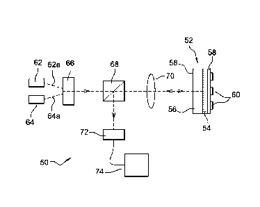
[0064] A preferred embodiment of SPR sensing system 50 adapted for
performing the present method is schematically illustrated in Fig.4. This SPR
sensing system 50 is particularly adapted for biosensing applications due to
the
structure of its sensor 52 that comprises ligands or other targeting moieties
attached to the sensing surface 54. Depending on the targeting moieties, the
biosensor can be designed to analyse a variety of samples, e.g. measure the
CA 02765972 2011-12-19
WO 2010/146160 17 PCT/EP2010/058647
concentration of specific chemical or biochemical molecules in a solution such
as
blood, urine or water etc. The present sensing system 50 is compact and can
thus
easily be installed and transported for various applications, e.g. to measure
medical parameters at a point-of-care, to detect and quantify food and water
contamination or to detect explosives, contaminants or toxicants in the
atmosphere, etc.
[0065] Biosensor 52 has a sensor surface 54 forming the sensing layer and
built
as a periodic gold nanograting configured to enable generation of SPR, in
particular localized and delocalized SPR. As it is known, localized SPR
corresponds to surface-bond electromagnetic waves that are confined to defined
regions at the sensor surface, whereas delocalized SPR corresponds to surface-
bond electromagnetic waves that are not confined to these defined regions at
the
sensor surface. The sensor surface 54 lies on top of a transparent substrate
56
that is capable of transmitting light so as to allow working in reflection
mode from
the sensor's backside 58.
[0066] Various possible methods of manufacturing such sensor 52 are known in
the art. In one embodiment, the transparent substrate 56 consists of a
transparent
glass or a transparent polymer, for example transparent polystyrene. The
sensor
surface layer 54 is preferably manufactured by depositing a layer of poly
acrylacid
(ppAA) over a glass substrate 56 and a subsequent layer of polystyrene beads
(PS). The ppAA and PS layers are etched by 02 plasma to form a grating
structure
comprising regularly spaced pillars of ppAA separated by a sub-micrometric
distance. Gold is then deposited over the pillars to fill-in the gaps between
neighbouring pillars, and the remainder of the PS mask is removed, obtaining a
periodic gold nanograting. Other possible materials for the dielectric pillars
are
polystyrene or poly-methyl-methacrylate, etc. Instead of a glass substrate,
one
may use a polymeric substrate, e.g. made from PS or PMMA or other transparent
material, that allows direct etching of the substrate to form the pillars.
[0067] It will be understood that the geometrical and physical properties of
the
patterned structure of the sensor surface determine the surface plasmon
resonance conditions (resonance coupling) at the sensor surface, in particular
the
surface plasmon frequencies and a maximum SPR excitation frequency, and thus
determine the optical effect of SPR excitation at the sensor surface.
CA 02765972 2011-12-19
WO 2010/146160 18 PCT/EP2010/058647
[0068] Reference sign 58 indicates a very thin layer of antifouling material,
e.g. of
antifouling hydrogel, namely poly-ethylene glycol. As it is known, the
antifouling
material acts in an anti-adhesive manner to prevent or reduce undesired
interactions, such as the non-specific absorption of chemical or biochemical
molecules etc. at the sensor surface. This reduces noise signals that could
have
been caused by the interaction or the binding of undesired chemical or
biochemical molecules at the sensor surface.
[0069] On top of this antifouling layer 58 is a microarray 60 of targeting
moieties,
i.e. ligand or molecules attached in an organised manner to the antifouling
layer
that will bind to or immobilize specific biomolecules or other analytes of
interest in
the liquid sample contacted with the sensor surface. These targeting moieties
may
comprise antigens/antibiodies, enzymes, proteins, oligonucleotides etc. The
target
moieties can be easily attached to the antifouling layer by microspotting that
allows
a wide variety of configurations of the array, varying the size of the spots
and the
kinds of targeting moieties. Such microspotting technique is e.g. described in
the
article "Fabrication and characterization of protein arrays for stem cell
patterning"
by Laura Ceriotti et al, pubished in Soft Matter 2009, 5, 1-12.
[0070] Referring now more generally to the optical setup of the SPR system 50,
it
comprises a test light beam source 62 and a reference light beam source 64.
The
test light beam source 62 and reference light beam source 64 comprise
preferably
each a light-emitting diode (LED) or a laser. The test light beam source 62 is
configured to emit a test light beam 62a having one or more frequencies
corresponding to one or more permitted frequencies of SPR at the sensor
surface
so as to excite resonance. Preferably, the test light beam source 62 is
configured to emit a test light beam 62a in at least one spectral emission
band
covering at least one permitted spectral band of SPR at the sensor surface 54,
similar to LED1 in Fig.1.
[0071] By contrast, reference light beam source 64 is configured to emit a
reference light beam 64a of one or more frequencies that do not correspond to
permitted frequencies of SPR at the sensor surface 54 so as not to excite SPR.
Preferably, the reference light beam source 64 is configured to emit a
reference
light beam 64a in at least one spectral emission band remote from the
permitted
frequencies of SPR at the sensor surface 54. For example, the reference light
CA 02765972 2011-12-19
WO 2010/146160 19 PCT/EP2010/058647
beam source 64 may be configured to emit reference light covering a spectral
band the spectral limits of which are at a spectral position far at least the
double of
the Full Width at Half Maximum (FWHM) of the Surface Plasmon Resonance from
the Surface Plasmon Resonance peak.
[0072] When there is more than one SPR-peak (as e.g. in case there is
localized
and delocalized peaks), the distance between the resonance peak and reference
beam is calculated with respect to the closest SPR-peak. Also, one may
consider
selecting the reference beam far by at least 2 to 4 times the closest SPR-
peak.
[0073] It shall be appreciated that in operating at such spectral position for
the
reference beam it is possible to avoid any excitation of surface plasmonic
resonances.
[0074] Conventionally in SPR testing the principle is to be able to sense
small
refractive index changes. And here this means detecting changes relative to
the
refractive index of the medium containing the biomolecules or other agents to
be
detected.
[0075] But it shall be kept in mind that, as explained above, the spectral
position
of the surface plasmon resonances (localized and delocalized) is uniquely
determined by the structure of the nanograting and the refractive index of the
medium containing the biomolecules or other agents to be detected.
[0076] When the recognition at the sensor surface occurs, the SPR will shift
in the
spectrum by a quantity, which is comprised between the limit of detection of
the
system (minimum detectable spectral shift) and the FWHM of each surface
plasmon resonance.
[0077] Therefore, selecting a reference beam having a spectral band remote
from
the SPR and namely having its closest bandwidth-end at least at the double of
the
FWHM of the SPR peak, avoids exciting the surface plasmons at the SPR-peak
otherwise monitored through the test beam.
[0078] Referring now more specifically to the present variant having a
nanostructured surface SPR sensor surface, one may note that the spectral
position of the resonance structure used as a sensitive probe depends on the
structural parameters of the nanostructured surface and, in particular, on the
size
and the shape of the polymeric pillars. As a matter of fact, the resonance
CA 02765972 2011-12-19
WO 2010/146160 20 PCT/EP2010/058647
corresponds to a charge oscillation mode having the maximum electric field
within
the surface area of the pillar and near its top. In this instance, this is due
to the
peculiar cone truncated pillar shape and the corresponding thin circular edge
formed by gold on top.
[0079] The effect of such a conformation is twofold:
- the electric field is localized and enhanced just on top of the pillar;
that is
to say, where the probability of attaching the analyte molecules is the
highest;
- the conical shape and the refractive index of the substrate allowing this
plasmonic mode be easily coupled with plasmonic oscillations of gold on the
substrate side, makes such a field enhancement particularly effective when
excited from the backside.
[0080] At a sufficient spectral distance from this kind of resonance (the
double of
its FWHM is a good spacing) the high sensitivity related to such a peculiar
field
configuration is lost because: either the electric field spatial distribution
is changed,
having its maxima values in different places on both (front or back) surfaces
or in
the inner of the pillar (where no analyte molecules can be detected); or no
matching of the modes through the pillars is allowed, then the excitation of
an
enhanced field is not transmitted to the sensitive region.
[0081] Accordingly, in selecting a reference beam located at twice the FWHM of
the SPR peak under monitoring it is possibly to observe optical properties of
the
sensor surface which are purposively not related to any plasmon resonance
supported by the surface.
[0082] Although used herein because investigation is made at fixed angle of
incidence, one may vary the incidence angle of the reference beam to
illuminate
the sample under non-SPR motivating conditions.
[0083] As can be seen in FIG.4, the test light beam 62a and the reference
light
beam 64a are directed towards the sensor surface 54 via an optical setup,
which
can comprise for example an optical coupler 66, a beam splitter 68 and a lens
system 70. The optical coupler 66 is configured to control the alternative or
simultaneous transmission of the test light beam 62a and reference light beam
64a
towards the sensor surface 54. The lens system 70 is used for transmitting the
test
and/or reference light beams 62a, 64a to homogenously illuminate the sensor
CA 02765972 2011-12-19
WO 2010/146160 21 PCT/EP2010/058647
surface 54. It will be noted that the test light beam 62a and/or the reference
light
beam 64a can illuminate the sensor surface 10 at various angles of incidence,
but
in the present setup the angle of incidence is fixed. Due to the setup, both
the
reference and test light beams arrive with the same, fixed angle of incidence
on
the sensing layer.
[0084] As it appears from FIG.4, the test and reference light beams 62a, 64a
are
directed towards the sensor surface 54 through the transparent substrate56,
where they reflect on the side of the sensor surface 54 that interfaces with
the
transparent substrate 56. It will be noted that the light reflected on this
internal side
of the sensor surface 52 does not interfere with or scatter in the sample lies
over
the microarray. Internal reflection, i.e. from the backside 58, is thus
advantageous
in that it avoids any interference of light transmission due to the sample and
microarray structure. The light beams reflected on the sensor surface are then
directed via the beam splitter 68to a photosensor 72. The photosensor 72 is
adapted to measure the intensity or a quantity indicative thereof, such as the
spectral intensities, the absorbance, the reflectance, the reflectivity etc.
It will be
noted that the photosensor 72 is also adapted to monitor the intensity of the
test
light beam 62a, in particular of a time-or frequency-averaged value thereof,
in one-
or two-dimensions, thereby enabling e.g. simultaneous detection of many local
changes in the light-SPR coupling condition at the sensor surface 54.
Advantageously, the photosensor 72 can be adapted to provide a one-or two-
dimensional image of a measured or monitored intensity. It will be understood
that
such one-or two-dimensional operating of the photosensor 72 enables e.g. the
simultaneous detection of many different molecules of interest that interact
at or
bind to the sensor surface 54. Such a one-or two-dimensional SPR imaging
photosensor 72 enables the high-throughput analysis of chemical or biochemical
events at the sensor surface 54 and also permits reducing the average cost-per-
assay of the sensing system.
[0085] In a preferred embodiment, the photosensor 72 preferably comprises a
time-and/or spectrum¨resolved camera based on a charge-coupled device (CCD)
or on a photodiode array for measuring the intensity of the test light beam
62a and
for detecting intensity changes of the reflected reference light beam 64a.
Preferably, this camera is adapted to resolve a measured signal over short
time
CA 02765972 2011-12-19
WO 2010/146160 22 PCT/EP2010/058647
intervals, which are typically in the millisecond range. More preferably, this
camera
can also output signals indicative of successive measurements of time-and/or
frequency-averaged intensities of the test light beam 62a and/or of the
reference
light beam 64a. The camera can also be adapted to measure the spectral
intensities of the reflected test light beam 62a and to output a signal
indicative of a
measured average spectral intensity, i.e. indicative of a mean value of
measured
spectral intensities, or any signal indicative thereof.
[0086] The signals corresponding to the monitored intensities of the reference
light beams and the signals corresponding to the measured intensities of the
test
light beam are then transmitted to a processor 74. Advantageously, the
processor
74 enables detecting an occurrence of SPR excitation by detecting a graded
reduction in the measured reflected intensity of test light, and in particular
by
detecting a dip in the spectrum of the measured intensity of the reflected
test light
beam 62a caused by the absorption of light at the sensor surface 542. It will
be
understood that a measured intensity of the test light beam 62a can also be
indicative of a measured time- or frequency- weighted average intensity of the
test
light beam or of a change thereof.
[0087] Moreover, the photosensor 72 is adapted to detect a change in the
monitored reflected intensity of the reference light beam 64a, in particular a
change in a monitored time-or frequency-weighted average intensity, a change
in
monitored a spectral intensity, a change in the monitored absorbance, a change
in
the monitored reflectance or reflectivity etc., of the reference light beam
64a as
reflected by the sensor surface 52 so as to determine the occurrence of an
artefact.
[0088] The processor 74 is configured to process, in particular to filter the
measurements of the photosensor 72. This processor 74 is preferably
programmed to correct the measured intensities of the test light beam 62a in
the
manner described herein before and to estimate a change in the refractive
index at
the sensor surface in the above described manner.