Note : Les descriptions sont présentées dans la langue officielle dans laquelle elles ont été soumises.
CA 02766262 2011-12-21
WO 2010/149726 PCT/EP2010/058958
1
DEVICES, A COMPUTER PROGRAM PRODUCT AND A METHOD FOR DATA
EXTRACTION
Technical field
The present invention generally relates to extraction of data originating from
a
physiological phenomenon in a subject, in particular when the vascular system
of the
subject is in connection with an extracorporeal fluid system. The present
invention is e.g.
applicable in arrangements for extracorporeal blood treatment.
Background art
Vital signs are measures of various physiological statistics often taken by
health
professionals in order to assess body functions. Vital signs of a subject,
e.g. heart rate.
blood pressure, oxygen saturation, electrocardiography (ECG), respiratory rate
and
autonomous regulation, such as blood pressure and body temperature, may be
measured,
monitored and interpreted to detect various disorders of the patient, for
instance respiratory
and heart related disorders. Typical equipment used for retrieving the vital
signs includes a
thermometer, a pulse oximeter, a capnograph and a pulse watch. Though a pulse
may often
be taken manually, a stethoscope may be required for a subject with a weak
pulse.
With external vital sign monitors, such as a thermometer, a stethoscope, a
photoplethysmograph (PPG), a pulse oximeter or a capnograph, it is possible to
measure
pulse, oxygen saturation and information on respiration, such as breathing
rate and carbon-
dioxide concentration in breath of patient.
Patients with kidney function insufficiency often suffer from various other
disorders,
for instance sleep apnea, periodic breathing and hyperventilation, making
monitoring of
vital signs of renal patients particularly important. Sleep apnea for
instance, is a common
disorder in the general population where 2% - 25% suffer from it, and it
correlates with
increased rate of several co-morbidities, such as hypertension, coronary
artery disease,
arrhythmias, heart failure and stroke. The prevalence of apnea is even higher
in the dialysis
population where 30% to 80% of dialysis patients suffer from this problem. The
reason for
this is not clear, but it is believed that hypervolemia and high levels of
uremic toxins may
worsen the disorder. In addition, many dialysis patients (40%) are diagnosed
with heart
conditions such as angina pectoris, left ventricular hypertrophy, stroke or
congestive heart
failure. These patients and other subjects may also suffer from reflex-
controlled
phenomena, such as vomiting, coughing and hiccups. Hence, there is a
particular need to
monitor vital signs of patients with kidney function insufficiency.
The origin behind the vital signs are for instance physiological pulse
generators, such
as the breathing system, the autonomous system for blood pressure regulation
and the
CA 02766262 2011-12-21
WO 2010/149726 PCT/EP2010/058958
2
autonomous system for body temperature regulation, which give rise to cyclic
physiological phenomena which are known to cause variations in the blood
pressure of a
patient.
Blood pressure regulation is part of the complex regulatory system which
controls
arterial blood pressure and is dependent on sensory inputs related to cardiac
output,
peripheral resistance to blood flow at the arterioles, the viscosity of the
blood, the volume
of blood in the arterial system, the elasticity of the arterial walls, etc.
Changes in blood
pressure are brought about by the control exerted on the same physiological
mechanisms.
The signals from which information regarding the vital signs are extracted and
the
sensors being used may vary and instruments for providing this information is
often
limited in purpose and functionality. Additionally, measurements of vital
signs are often
time consuming and require involvement and attention from staff competent in
handling
each instrument.
It is known, for instance from US5243990, of blood pressure monitors, even
ones
that are included in dialysis machine systems, that allow measurement of the
patient's pulse
and blood pressure values (e.g. systolic and diastolic pressure) at specified
intervals.
To get a good picture of body functions, it is often desirable to monitor a
plurality of
vital signs, requiring a number of specialised sensors or monitors connected
to the body of
a patient, which is costly, cumbersome and distracting.
It is also known that coughing and sneezing may influence physiological
measurements obtained from instruments. Coughing may for instance introduce
errors in
the PPG signal e.g. measured with a pulse oximeter.
Hence, there is a need for alternative and/or improved ways of monitoring
vital signs
for detecting, presenting, tracking and/or predicting disorders, such as
disorders related to
the respiratory, vascular and autonomous system of the subject.
Furthermore, in extracorporeal blood treatment, blood is taken out of a
patient,
treated and then reintroduced into the patient by means of an extracorporeal
blood flow
circuit. Generally, the blood is circulated through the circuit by one or more
pumping
devices. The circuit is connected to a blood vessel access of the patient,
typically via one or
more access devices, such as needles or venous catheters, which are inserted
into the blood
vessel access. Such extracorporeal blood treatments include hemodialysis,
hemodiafiltration, hemo filtration, plasmapheresis, etc.
In extracorporeal blood treatment, it is vital to minimize the risk for
malfunctions in
the extracorporeal blood flow circuit, since these may lead to a potentially
life-threatening
condition of the patient. Serious conditions may arise if the extracorporeal
blood flow
circuit is disrupted, e.g. by an access device for blood extraction (e.g. an
arterial needle)
coining loose from the blood vessel access, causing air to be sucked into the
circuit, or by
an access device for blood reintroduction (e.g. a venous needle) coming loose
from the
blood vessel access, causing the patient to be drained of blood within
minutes. Other mal-
3
functions may be caused by the blood vessel access becoming blocked or
obstructed, or
by the access device being positioned too close to the walls of the blood
vessel.
In WO 97/10013, the monitoring involves filtering a measured pressure signal
to
remove the frequency components that originate from a blood pump, and then
detecting
the heart signal by analysing the filtered pressure signal. The amplitude of
the filtered
pressure signal is then taken as an indication of the integrity of the fluid
connection. This
monitoring technique requires proper filtering and might thus fail if there is
a significant
frequency overlap between the heart signal and the pulses from the blood pump.
Hence, there is also a need for alternative and/or improved ways of monitoring
the
.. integrity of a fluid connection between an extracorporeal circuit and a
vascular system of
a subject.
Summary
It is an object of the invention to at least partly overcome one or more of
the above-
.. identified limitations of the prior art.
One object of the invention is to provide an alternative or complementary
technique for monitoring vital signs of a human or animal subject.
Another object of the invention is to provide an alternative or complementary
technique for monitoring the integrity of the fluid connection between the
extracorporeal
.. and vascular systems, and also preferably with an improved robustness
and/or an
increased certainty of detecting a malfunction in the fluid connection.
These and other objects, which will appear from the description below, are at
least
partly achieved by means of devices, a method and a computer program product.
According to the present invention, there is provided an extracorporeal fluid
system with a device for processing a measurement signal obtained by a
pressure sensor
in the extracorporeal fluid system when the extracorporeal fluid system is
connected to a
vascular system of a subject, said device comprising:
means for receiving the measurement signal obtained by at least one of said
pressure sensors; and
means for processing the measurement signal for identification of pressure
data
comprising one or more pulses originating from a physiological phenomenon in
said
subject, said physiological phenomenon being a breathing system of said
subject, and
that said means monitors the integrity of a fluid connection between said
extracorporeal
fluid system and said vascular system based on said pressure data.
According to the present invention, there is provided a method of monitoring
the
integrity of a fluid connection between an extracorporeal fluid system and
CA 2766262 2018-01-12
3a
a vascular system by processing a measurement signal obtained by pressure
sensor in
said extracorporeal fluid system connected to said vascular system of a
subject, said
method comprising:
receiving the measurement signal obtained from at least one of said pressure
sensors; and
processing the measurement signal for identification in the measurement
signal, of
pressure data comprising one or more pulses originating from a physiological
phenomenon in said subject, wherein said physiological phenomenon being a
breathing
system of said subject and that said monitoring of the integrity of a fluid
connection
.. between said extracorporeal fluid system and said vascular system is based
on said
pressure data.
Preferred embodiments are described hereunder.
According to the present invention, there is provided an extracorporeal fluid
system (Si)
with a device (25) for processing a measurement signal obtained by a pressure
sensor
.. (4a-4c) in the extracorporeal fluid system (Si) when the extracorporeal
fluid system (S1)
is connected to a vascular system (S2) of a subject, said device (25)
comprising:
means (28) for receiving the measurement signal; and
means (29) for processing the measurement signal for identification of
pressure
data comprising one or more pulses originating from a physiological phenomenon
in said
subject, said physiological phenomenon excluding the heart of said subject.
According to the present invention, there is also provided a method for
processing a
measurement signal obtained by a pressure sensor (4a-4c) in an extracorporeal
fluid system
(S1) connected to a vascular system (S2) of a subject, said method comprising:
receiving the measurement signal; and
processing the measurement signal for identification of pressure data
comprising
one or more pulses originating from a physiological phenomenon in said
subject, said
physiological phenomenon excluding the heart of said subject.
CA 2766262 2018-01-12
3b
Embodiments of the invention are based on the insight that these objects may
be
achieved by processing measurement signals from pressure sensors in an
extracorporeal
fluid system in contact with a vascular system of a subject, which signals
previously have
not been considered possible to extract and/or interpret and which signals now
have been
found to contain valuable information. Thus, embodiments of the invention
enable
monitoring of vital signs of a human or animal subject by processing a
measurement
signal obtained in a pressure measurement, the measurement signal being
retrieved from
a fluid system external of the subject, i.e. an extracorporeal fluid system,
and connected
to a vascular system of the subject. Correspondingly, embodiments of the
invention
enable monitoring of the integrity of a fluid connection between the
extracorporeal fluid
system and the vascular system of a subject, by processing such a measurement
signal.
Embodiments of the invention may, e.g., be used in connection with blood
treatment such as dialysis in various forms.
A first aspect of the invention is a device for processing a measurement
signal
obtained by a pressure sensor in an extracorporeal fluid system connected to a
vascular
CA 2766262 2018-01-12
CA 02766262 2011-12-21
WO 2010/149726 PCT/EP2010/058958
4
system of a subject, said device comprising: means for receiving the
measurement signal;
and means for processing the measurement signal for identification of pressure
data
originating from a first physiological phenomenon in said subject, said
physiological
phenomenon excluding the heart of said subject.
A second aspect of the invention is a method for processing a measurement
signal
obtained by a pressure sensor in an extracorporeal fluid system connected to a
vascular
system of a subject, said method comprising: receiving the measurement signal;
and
processing the measurement signal for identification of pressure data
originating from a
first physiological phenomenon in said subject, said physiological phenomenon
excluding
the heart of said subject.
A third aspect of the invention is a computer program product comprising
instructions for causing a computer to perform the method according to the
second aspect.
A fourth aspect of the invention is a device for processing a measurement
signal
obtained by a pressure sensor in an extracorporeal fluid system connected to a
vascular
system of a subject, said device comprising: an input for receiving the
measurement signal;
and a signal processor connected to said input and configured to process the
measurement
signal for identification of pressure data originating from a first
physiological phenomenon
in said subject, excluding the heart of said subject.
According to these aspects, pressure data from a first physiological
phenomenon in
the subject, excluding the heart of the subject, is identified in the
measurement signal. The
first physiological phenomenon may be reflexes in the subject, voluntary or
non-voluntary
muscle contractions in the subject, the breathing system in the subject, the
autonomous
system of the subject for blood pressure regulation, or the autonomous system
of the
subject for body temperature regulation.
The first physiological phenomenon generates one or more pressure waves that
propagate from the vascular system via the fluid connection into the
extracorporeal fluid
system to the pressure sensor, which is in direct or indirect hydrostatic
contact with the
fluid (e.g. blood) in the extracorporeal fluid system. The pressure sensor
generates a
pressure pulse for each pressure wave. A -pulse" is thus a set of data samples
that define a
local increase or decrease (depending on implementation) in signal magnitude
within the
time-dependent measurement signal. It is to be understood that the pressure
sensor may
receive pressure waves from other pulse generators, e.g. the heart of the
subject and/or a
mechanical pulse generator in the extracorporeal fluid system, and that these
pressure
waves also generate pressure pulses in the measurement signal.
Generally, the identified pressure data represents one or more pulses in the
measurement signal that originate from the first physiological phenomenon.
However, the
pressure data may take many different forms.
In one variant, the pressure data is a parameter value which is extracted
directly from
the measurement signal. As noted above, the measurement signal may not only
include one
CA 02766262 2011-12-21
WO 2010/149726 PCT/EP2010/058958
or more relevant pulses from the first physiological phenomenon, but may also
include
other pulse signals such as pulses from the heart of the subject, pulses from
a mechanical
pulse generator in the extracorporeal fluid system, as well as pulses from
other
physiological phenomena in the subject. However, in certain embodiments, it
may be
5 possible to calculate a parameter value that represents the relevant
pulses from the first
physiological phenomenon in the measurement signal.
In another variant, the pressure data is a time-dependent monitoring signal,
which is
obtained by processing the measurement signal to improve/facilitate
identification of the
relevant pulses from the first physiological phenomenon, either in the time
domain or in
the frequency domain. For example, the processing may result in a significant
suppression
or even elimination of unwanted or interfering signals in the measurement
signal. Such
unwanted signals may include pulses from the mechanical pulse generator and/or
pulses
from the heart of the subject and/or pulses from other physiological phenomena
in the
subject. After this processing, one or more relevant pulses have been
extracted from or
"isolated in" the measurement signal. As used herein, "to isolate relevant
pulses" indicates
that the measurement signal is processed to such an extent that the pulses
that originate
from the first physiological phenomenon can be detected and analyzed in the
identified
pressure data. The measurement signal may be processed to at least
significantly exclude
the heart pulses and/or to at least significantly exclude other unwanted
signals, such as the
pulses that originate from the mechanical pulse generator. For example, the
measurement
signal may be low-pass filtered to remove frequencies above about 0.4, 0.45,
0.5, 0.55, 0.6,
0.65, 0.7, 0.75 or 0.8 Hz. In another example, the measurement signal may be
band-pass
filtered in at least one of the frequency ranges about 0.15 Hz to about 0.4
Hz, about 0.04
Hz to about 0.15Hz, and about 0.001Hz to about 0.1 Hz. In yet another example,
the
measurement signal is high-pass filtered to at least remove frequencies below
about 3-5
Hz, and preferably below about 3.5-4 Hz, e.g. to isolate pulses originating
from fast muscle
contractions, movements and sounds from abdomen and bowels, the subject
speaking, etc.
It is to be understood that "to isolate relevant pulses" need not exclude that
the monitoring
signal includes pulses from one or more further physiological phenomena, other
than the
heart, in the subject. However, in certain embodiments, the monitoring signal
may indeed
be generated substantially with signal components only from the first
physiological
phenomenon.
In yet another variant, the pressure data is a parameter value which is
extracted from
the above-mentioned monitoring signal.
After its identification, the pressure data may be processed or used for the
purpose of
detecting and/or presenting and/or tracking and/or predicting a disordered
condition of the
subject. Alternatively or additionally, the pressure data may be processed or
used for the
purpose of determining the integrity of the fluid connection.
CA 02766262 2017-02-17
=
6
Embodiments of the present invention apply to processing of measurement
signals
both off-line and on-line, i.e. both during, e.g. concurrently, and subsequent
to a
treatment, such as dialysis, as well as separated from such a treatment. The
measurement
signal may comprise raw data or pre-processed data, for instance filtered for
signal noise
reduction. Embodiments of the present invention are applicable to conditions
involving
particular sources of signal noise and artefacts, such as a running pump. The
processing
may for instance involve pre-processing including general signal filtration,
removal of
particular signal noise (typically measurement noise) and signal artefacts,
such as from a
running pump, and signal analysis. Embodiments of the present invention are
also
flexible in advantageously allowing continuous as well as intermittent
measurements.
As an advantage of embodiments of the present invention, continuous or
intermittent measurements of respiration and autonomic regulation, such as
blood
pressure regulation and temperature regulation, may be provided directly from
the
extracorporeal circulation during e.g. dialysis treatment. Thus, a plurality
of vital signs
can be monitored simultaneously and continuously using a time-dependent
pressure
signal from the extracorporeal circulation, and the need to attach a number of
specialised
sensors or monitors the body of the subject is reduced.
Embodiments of the invention may be beneficial for unattended patients, e.g.
patients performing dialysis at home or nocturnal patients with limited
staffing.
Embodiments of the invention also enable monitoring of the integrity of the
fluid
connection between the extracorporeal fluid system and the vascular system
irrespective
of any frequency overlap between the heart pulses and the pulses from
mechanical pulse
generators in the extracorporeal fluid system. For example, the monitoring may
be based
on pulses originating from a physiological phenomenon, other than the heart,
that are
shifted in frequency and/or time from pulses originating from the mechanical
pulse
generators.
Still other objectives, features, aspects and advantages of the present
invention will
appear from the following detailed description, as well as from the drawings
and
appendixes.
Brief Description of the Drawings
Embodiments of the inventive concepts will now be described in more detail
with
reference to the accompanying schematic drawings.
Fig. 1 is a schematic view of a general fluid containing system in which
inventive
data processing may be used for filtering a pressure signal.
Fig. 2 is a plot of breathing signals generated/extracted from the measurement
signal and from a reference instrument (capnograph) as a function of time.
Fig. 3 is another plot of breathing signals from the measurement signals as a
function of time.
CA 02766262 2011-12-21
WO 2010/149726 PCT/EP2010/058958
7
Fig. 4 is flow chart of a signal identification process according to one
embodiment of
the invention.
Fig. 5 is a plot of a breathing signal as a function of time.
Fig. 6 is a plot of an exemplifying breathing disorder response.
Fig. 7 is a plot of breathing related parameters identified in connection with
a
breathing disorder.
Fig. 8 is a plot of a blood pressure turbulence event (BPT) in a) a healthy
subject and
b) a subject with unhealthy cardiovascular response.
Fig. 9 is a plot of breathing signals in connection with an event of blood
pressure
turbulence.
Fig. 10 is a plot of breathing signals as measured at the venous and arterial
pressure
side, respectively, in connection with an event of blood pressure turbulence.
Fig. 11 is a schematic view of a system for hemo dialysis treatment including
an
extracorporeal blood flow circuit.
Detailed Description of Exemplary Embodiments
In the following, embodiments will be described with reference to fluid
containing
systems in general, and in relation to an extracorporeal blood flow circuit in
particular.
Thereafter, physiological phenomena and embodiments for extracting signals
indicative of
such physiological phenomena will be described. Then, exemplary embodiments
for
detecting disorders based on such extracted signals are described, as well as
exemplary
embodiments for monitoring the integrity of a fluid connection based on such
extracted
signals.
Throughout the following description, like elements are designated by the same
reference signs.
GENERAL
Fig. 1 illustrates a general fluid arrangement in which a fluid connection C
is estab-
lished between a first fluid containing system Si and a second fluid
containing system S2.
The fluid connection C may or may not transfer fluid from one system to the
other. A first
pulse generator 3 is arranged to generate a series of pressure waves in the
fluid within the
first system Si, and a second pulse generator 3 is arranged to generate
single, occasional
or a series of pressure waves in the fluid within the second system S2. A
single pressure
wave may represent a sneezing, occasional pressure waves may represent one or
more
coughs, and a series of pressure waves may represent regular or non-regular
breathing. one
or more pressure sensors 4a-4c are arranged to measure the fluid pressure in
the first
system Si. As long as the fluid connection C is intact, pressure waves
generated by the
second pulse generator 3' will travel from the second system S2 to the first
system Si, and
thus second pulses originating from the second pulse generator 3' will be
detected by the
CA 02766262 2011-12-21
WO 2010/149726 PCT/EP2010/058958
8
pressure sensor(s) 4a-4c in addition to first pulses originating from the
first pulse generator
3. It is to be noted that either one of the first and second pulse generators
3, 3' may include
more than one pulse-generating device. Further, any such pulse-generating
device may or
may not be part of the respective fluid containing system Si, S2. The first
fluid system
may be an extracorporeal fluid circuit, such as an extracorporeal blood flow
circuit of the
type which is used for dialysis, and the second fluid system may be a vascular
system, such
as the blood circuit, of a subject. The second pulse generator 3' may also be
referred to as a
physiological phenomenon, and it may be a physiological pulse generator,
cyclic or non-
cyclic, repetitive or non-repetitive, autonomous or non-autonomous. The second
pulse
generator 3' may be a physiological phenomenon from the group consisting of
reflex
actions, voluntary muscle contractions, non-voluntary muscle contractions, a
breathing
system of said subject, an autonomous system of said subject for blood
pressure regulation
and an autonomous system of said subject for body temperature regulation. A
reflex action,
also known as a reflex, is to be construed as an involuntary and nearly
instantaneous
movement in response to a stimulus.
The fluid arrangement of Fig. 1 further includes a surveillance device 25
which is
connected to the pressure sensors 4a-4c. Thereby, the surveillance device 25
acquires one
or more measurement signals that may or may not be time-dependent to provide a
real time
representation of the fluid pressure in the first system Si. The surveillance
device 25
monitors the behaviour of a physiological phenomenon of a subject and may
issue an alarm
or warning signal, and/or alert a control system of the first system SI, to
take appropriate
action. The surveillance device 25 may or may not process the measurement
signal(s)
continuously (i.e. on-line). The measurement signal(s) may also comprise a set
or batch of
measurement signals, extracted for subsequent analysis (i.e. off-line).
The surveillance device 25 optionally monitors the integrity of the fluid
connection
C, based on the principle that the presence of second pulses indicates that
the fluid
connection C is intact, whereas absence of second pulses indicates that the
fluid connection
C is compromised. The absence of second pulses may bring the surveillance
device 25 to
issue an alarm or warning signal, and/or alert a control system of the first
or second fluid
containing systems Si, S2 to take appropriate action.
The surveillance device 25 may thus be configured to continuously process the
time-
dependent measurement signal(s) to determine whether second pulses are present
or not.
Typically, the determination involves analyzing the measurement signal(s), or
a pre-
processed version thereof, in the time domain to calculate a value of an
evaluation para-
meter (i.e. a parameter value) which is indicative of the presence or absence
of second
pulses in the measurement signal(s). Depending on implementation, the
surveillance device
25 may use digital components or analogue components, or a combination
thereof, for
receiving and processing the measurement signal(s).
CA 02766262 2011-12-21
WO 2010/149726 PCT/EP2010/058958
9
In the context of the present disclosure, "absence" of a pulse may imply that
the
pulse has disappeared, or at least that it has decreased sufficiently compared
to the pulse
deemed to be "present". The assessment of presence or absence may involve
calculating an
evaluation parameter value based on the measurement signal(s) and comparing
the
.. parameter value to a threshold value.
Fig. 11 shows an example of an extracorporeal blood flow circuit 20 of the
type
which is used for dialysis. The extracorporeal blood flow circuit 20 comprises
an access
device for blood extraction in the form of an arterial needle 1, and an
arterial tube segment
2 which connects the arterial needle 1 to a blood pump 3 which may be of
peristaltic type,
as indicated in Fig. 11, or any other suitable type, such as a membrane pump.
At the inlet
of the pump 3 there is a pressure sensor 4a (hereafter referred to as arterial
sensor) which
measures the pressure before the pump 3 in the arterial tube segment 2 (in the
form of an
"arterial pressure signal"). The blood pump 3 forces the blood, via a tube
segment 5, to the
blood-side of a dialyser 6. Many dialysis machines are additionally provided
with a
pressure sensor 4b that measures the pressure between the blood pump 3 and the
dialyser 6.
The blood is lead via a tube segment 10 from the blood-side of the dialyser 6
to a venous
drip chamber or deaeration chamber 11 and from there back to the patient via a
venous
tube segment 12 and an access device for blood reintroduction in the form of a
venous
needle 14. A pressure sensor 4c (hereafter referred to as venous sensor) is
provided to
measure the pressure on the venous side of the dialyser 6 (in the form of a
"venous
pressure signal"). In the illustrated example, the pressure sensor 4c measures
the pressure
in the venous drip chamber. Both the arterial needle or catheter 1 and the
venous needle or
catheter 14 are connected to the patient through a blood vessel access. The
blood vessel
access may be of any suitable type, e.g. a fistula, a Scribner-shunt, a graft,
etc. For
simplicity, the following discussion presumes that the blood vessel access is
a fistula.
In relation to the general arrangement in Fig. 1, the extracorporeal blood
flow circuit
20 corresponds to the first fluid containing system S , the blood pump 3 (as
well as any
further pulse source(s) within or associated with the circuit 20, such as a
dialysis solution
pump, valves, etc) corresponds to the first pulse generator 3, the blood
system of the
patient corresponds to the second fluid containing system S2, and a
physiological
phenomenon of the patient corresponds to the second pulse generator 3 which
thus is
located within or associated with the blood system of the patient. The fluid
connection C
corresponds to one or both of the fluid connections between the blood vessel
access and
the access device 1, 14.
In Fig. 11, a control unit 23 is provided, i.a. to control the blood flow in
the circuit 20
by controlling the revolution speed of the blood pump 3. The extracorporeal
blood flow
circuit 20 and the control unit 23 may form part of an apparatus for
extracorporeal blood
treatment, such as a dialysis machine. Although not shown or discussed further
it is to be
CA 02766262 2011-12-21
WO 2010/149726 PCT/EP2010/058958
understood that such an apparatus performs many other functions, e.g.
controlling the flow
of dialysis fluid, controlling the temperature and composition of the dialysis
fluid. etc.
In Fig. 11, the surveillance device 25 comprises a data acquisition part 28
for pre-
processing the incoming signal(s), e.g. including an AID converter with a
required mini-
5 mum sampling rate and resolution, one or more signal amplifiers, one or
more filters to
remove undesired components of the incoming signal(s), such as offset, high
frequency
noise and supply voltage disturbances.
In the examples given herein, the data acquisition part 28 comprises a DAQ
card
USB-6210 from National Instruments with a sampling rate of 1 kHz and
resolution of 16
10 bits, an operation amplifying circuit AD620 from Analog Devices, a high-
pass filter with a
cut-off frequency of 0.03 Hz (i.a., for removal of signal offset) together
with a low-pass
filter with a cut-off frequency of 402 Hz (i.a., for removal of high frequency
noise). To
obtain a short convergence time, a low¨order filter is used for the filters.
Furthermore, the
data acquisition part 28 may include an additional fixed band-pass filter with
upper and
lower cut-off frequencies to suppress disturbances outside the frequency
interval of
interest.
The pre-processed data is provided as input to a main data processing part 29,
which
executes the inventive signal analysis.
Embodiments of the present invention utilize the fact that physiological
phenomena
arising in the body of a subject cause variations in the blood pressure of the
subject. It has
been found that these variations are, in turn, conducted via the fluid
connection(s), the tube
segments, the fluid (blood/air) in the tube segments, any intermediate fluid
chamber (e.g.
drip chamber 11) and the fluid therein, to one or more pressure transducers in
the
extracorporeal blood flow circuit. By signal analysis it is then possible to
extract these
pressure variations, and then subsequently, extract rate, amplitude, phase and
shape of
signals that represent the phenomena. This information may e.g. be useful to
medical staff
in observing breathing rate and depth of breath of a subject.
Implementation of the signal analysis may be done by executing a software
algorithm in a computer, e.g. by digital filters, by mechanical filters, e.g.
restrictors and
compliance volumes, or by electronics, e.g. analogue filters or digital
circuits dedicated for
the purpose.
Hence, measurement data on e.g. respiration, blood pressure and temperature
regulation may advantageously be provided on-line and continuously during
extra-
corporeal circulation. The measurement data may be determined from sensor
information
obtainable from most extra-corporeal treatment systems without need for extra
disposables
or making an extra blood access.
Hence, embodiments of the present invention enable the provision of vital
signs, e.g.
respiration rate and amplitude, and autonomous regulation, of a patient, in
particular during
dialysis treatment.
CA 02766262 2011-12-21
WO 2010/149726 PCT/EP2010/058958
11
Embodiments of the present invention may be implemented as an apparatus, a
computer-implemented method and a computer program product for identifying
physiological signals with other origins than the heart of the subject. This
is achieved by
analysis of signals acquired from a tube/vessel in direct hydrostatic contact
with the body
of a subject via e.g. a needle or catheter inserted into the blood vessel
access of a subject.
The physiological signal relevant to the invention may for instance originate
from
reflexes, voluntary muscle contractions, non-voluntary muscle contractions,
breathing of a
subject or come from signals related to the autonomic regulation of the
subject's body. The
frequency ranges of some of these phenomena are normally:
- Breathing: approx. 0.15-0.4 Hz, with frequencies centred around approx. 0.25
Hz:
- Blood pressure regulation due to the autonomous system: approx. 0.04¨
0.14 Hz,
with frequencies centred around approx. 0.1 Hz;
- Temperature regulation due to the autonomous system: approx. 0.001-0.1,
with
frequencies centred around approx. 0.05 Hz.
For the sake of simplicity, the following description will refer to the
dialysis field
without excluding a broader scope of applications. It will be assumed that the
system
signals that are subjected to the analysis are delivered by pressure sensors
at the venous
and/or the arterial side of the blood line (cf. sensors 4c and 4a,
respectively, in Fig.11)
during a dialysis treatment. However, it may be anticipated that other type of
sensors, e.g.
optical sensors, such as a photo-plethysmograpy sensor (PPG), displacement
sensors, such
as strain gauges and accelerometers, may be used as long as these convey
equivalent
information about relevant physiological signals from the patient.
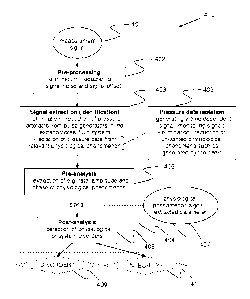
Fig. 4 is a flow chart that illustrates steps of a signal analysis process 400
executed
by the surveillance device 25 according to an embodiment of the present
invention. It is
initiated by receiving a measurement signal 401, e.g. from the venous or
arterial pressure
sensors, comprising a number of pressure induced signal components. The signal
analysis
process may be divided into a pre-processing part 402, a signal extraction
part 403 and an
analysis part 404. The pre-processing part 402 includes elimination or
reduction of signal
noise, e.g. measurement noise, and signal offset, as detailed in the section
above relating to
the data acquisition part 28. The signal extraction part 403 involves
elimination or
reduction of pressure artefacts originating from pulse generators in the
extracorporeal fluid
system and isolation of pressure data originating from a relevant
physiological
phenomenon. In the context of the present disclosure, "pressure data
isolation" 405 denotes
a process of generating a time-dependent signal (also denoted monitoring
signal herein)
which is free or substantially free from pressure modulations caused by any
unwanted
physiological phenomena. Such unwanted physiological phenomena may vary
between
different applications, but generally include at least heart beats. The
elimination of signal
noise and signal offset (cf. part 402), as well as the elimination of pressure
artefacts, may
be included in algorithms for pressure data isolation. For instance, the
measurement signal
CA 02766262 2011-12-21
WO 2010/149726 PCT/EP2010/058958
12
may be band pass filtered or low pass filtered to isolate a breathing signal,
in a way such
that signal noise and/or signal offset and/or pressure artefacts are
eliminated from the
measurement signal. The elimination of pressure artefacts may thus be
performed before,
after or during the pressure data isolation.
In a pre-analysis step 406 of the analysis part 404, one or more specific
signal
analysis algorithm(s) are applied for extraction of e.g. rate, amplitude and
phase of the
relevant physiological phenomenon. In a post-analysis step 408, based on one
or more
predetermined criteria, the output 407 of the signal analysis algorithm(s) is
analysed, e.g.
by pattern recognition, for signs of various disorders of physiological or
system character,
for instance indicated by detection of a disorder in step 409 and detection of
the integrity of
the fluid connection in step 410. The result of step 409 may be presented,
e.g. displayed, to
medical staff and may be useful in observing for instance breathing rate and
breathing
depth of a patient to detect, track or predict disorders and possibly take a
corrective action.
In the following, the physiological phenomena will be explained in more
detail, e.g.
reflexes, voluntary muscle contractions, non-voluntary muscle contractions,
the breathing
system, the autonomous system for blood pressure regulation and the autonomous
system
for body temperature regulation for a human or animal. These phenomena may
also be
referred to as physiological pulse generators due to the blood pressure
variations they
generate.
Normally, the arterial blood pressure is modulated by 4 mmHg to 6 mmHg in a
wavelike manner during respiration. Deep respiration may result in blood
pressure
variation of 20 mmHg.
The breathing induced modulation of the arterial blood pressure in the subject
has
several reasons:
- "Cross-talk" between different parts of the sympathetic control system of
the brain.
Signals of the respiratory centre spill over to the centre controlling the
vasomotor
status causing blood pressure variations, the vasomotor referring to actions
upon a
blood vessel which alter its diameter by contraction and dilatation.
- Breathing modulates the heart rate which modulates cardiac output and
blood
pressure.
- Modulation of cardiac output due to variations of the pressure in the
thoracic cavity
during breathing. At inspiration the left ventricle of the heart is supplied
with a
smaller blood volume since more blood is contained in the blood vessels in the
chest at the expense of the pump volume of the heart. Blood pressure will then
change as the cardiac output varies.
- Excitation of baroreceptors of the heart due to respiration. This will
cause
modulation of blood pressure since the sympathetic system will respond to the
stretch of the baroreceptors by changing the blood pressure.
CA 02766262 2011-12-21
WO 2010/149726 PCT/EP2010/058958
13
- The hydro-static pressure change due to the rise and fall of the
chest during
respiration of a subject in supine position. At inspiration the centre of
gravity is
elevated which causes increased pressure.
Fig. 2 illustrates the synchronous breathing signals from the vein 201 (dotted
line)
and from the artery 202 (dashed line) generated by the signal extraction
processing (cf.
402-403 in Fig. 4) of the venous and the arterial pressure signals recorded
during a dialysis
treatment by the venous and arterial pressure sensors (cf. 4c, 4a in Fig. 11).
A "breathing
signal", as used herein, denotes a signal representing/reflecting the
repetitive cycles of
inhalation and exhalation of a subject. The third curve 203 (solid line) shows
a reference of
the breathing signal provided by an external capnography device based on
measurement of
CO2 of the respiration flow. Fig. 3 is a similar plot of the breathing signals
in Fig. 2 and
shows that the amplitude of the breathing signals 201, 202 extracted from the
venous and
arterial pressure signals, respectively, change in concordance with the depth
of breath
given by the capnography signal 203.
Vasomotor oscillations appear in the blood pressure in cycles with a length of
about
7 seconds to about 26 seconds and an amplitude of about 10 mmHg to about 40
mmHg.
The phenomenon is caused by self-oscillation of the sympathetic control system
for the
blood pressure with the baroreceptors as input signals.
The autonomous system is also involved in temperature control of the body via
regulation of the vasomotor response to temperature changes. At low
temperatures e.g. the
arterioles are contracted to conserve energy of the body, which cause higher
blood
pressure. Similar to the vasomotor oscillations caused by the blood pressure
control
system, the temperature control system also give rise to cyclic variations in
blood pressure.
The temperature cycle rate is normally centred at around 0.05 Hz.
Fig. 5 shows the modulation 501 of a breathing signal 502 from the venous
pressure
signal measured by pressure sensor 4c of Fig. 11 due to oscillation of an
autonomous
control system in the frequency range of temperature regulation.
In the simplest case of pressure signal analysis, no pump or other source of
pressure
artefacts is present in the extracorporeal fluid circuit connected to the
subject during the
data acquisition. For instance, the pump may have been shut down.
In the general case, however, one or more pumps are running or other sources
of
cyclic or non-cyclic, repetitive or non-repetitive artefacts are present
during the data
acquisition. Information on the cyclic disturbances may be known from external
sources,
e.g. other sensors or controllers, or may be estimated or reconstructed from
system
parameters, e.g. the blood flow rate.
Cyclic pressure artefacts may originate from operating a peristaltic pump,
repetitive
actuation of valves, movements of membranes in balancing chambers. According
to the
findings in connection with the present invention, artefacts may also
originate from
mechanical resonance of system components such as swinging movements of blood
line
CA 02766262 2011-12-21
WO 2010/149726 PCT/EP2010/058958
14
energized by e.g. a pump. Frequencies of blood line movements are given by the
tube
lengths and harmonics thereof and by the beating between any frequencies
involved, i.e.
between different self-oscillations and pump frequencies. These frequencies
may differ
between the venous and arterial lines. Mechanical fixation of the blood lines
and other free
components may remedy the problem of mechanical resonance. Alternatively, an
operator
may be instructed to touch or jolt the blood lines to identify natural
frequencies associated
with the blood lines, which information may be used in the analysis for
improved removal
of components not belonging to the pressure data of interest.
Examples of non-cyclic artefacts are subject movement, valve actuation,
movements
of tubings, etc.
In the following, various techniques for signal extraction (cf. 403 in Fig. 4)
will be
briefly discussed.
SIGNAL EXTRACTION
In the following, embodiments for eliminating various artefacts will be
described.
Then, embodiments for isolating pressure data originating from a relevant
physiological
phenomenon are described.
The pressure data to be extracted is not limited to a single physiological
phenomenon
and may originate from one or more physiological phenomena, excluding the
heart.
Elimination of artefacts
Elimination of artefacts may be provided by:
- Controlling a pulse generator in the extracorporeal fluid system, such as
a pump
o By temporarily shutting down the pulse generator, or
o By shifting the frequency of the pulse generator;
- Low pass, band pass or high pass filtering;
- Spectral analysis and filtering in the frequency domain;
- Time domain filtering.
Controlling a pulse generator
Artefacts from a pulse generator, such as a pumping device, in the
extracorporeal
blood flow circuit may be avoided by temporarily shutting down the pulse
generator, or by
shifting the frequency of the pulse generator away from frequencies of one or
more
relevant physiological phenomena.
With specific reference to the use of the pressure data for integrity
detection (cf. step
410 in Fig. 4), artefacts may be eliminated by feedback control with respect
to the relevant
physiological signal, e.g. a breathing signal, from an independent source,
e.g. a capnograph
instrument. Such feedback control may thus be used to set the pump frequency
optimally
for detection of the relevant physiological signal in the pressure signal. For
example,
CA 02766262 2017-02-17
=
control unit 23 of Fig. 11 may be operated to set the pump frequency based on
an
external signal in order to facilitate the detection of the relevant
physiological signal, i.e.
the pump frequency is controlled to minimize overlap in frequency between the
pump
and the physiological phenomenon of relevance.
5
Artefact elimination by applying low pass, band pass or high pass filters
The measured signal may be fed into a filter, e.g. digital or analogue, with
suitable
frequency characteristics, such as frequency range and/or centre of frequency
range,
corresponding to a pulse generator, such as a pump, in the extracorporeal
circuit. For
10 instance, in a case where the pulse generator, such as a pump,
operates within the
frequency range of 1Hz, a suitable low pass filter may be applied in order to
obtain the
frequency of the physiological phenomenon below 1Hz. Correspondingly, a high
pass
filter may be applied to obtain a physiological phenomenon with frequency
higher than
the pulse generator.
Spectral analysis and filtering in the frequency domain
With spectral analysis, detection and elimination of amplitude peaks in a
spectrum
may for instance be performed by Fast Fourier Transform (FFT) methods.
Alternatively,
the elimination may be achieved by applying a notch filter or the like at one
or more
frequencies identified by an FFT method or the like.
Time domain filtering
Artefact elimination by filtering in the time domain is further disclosed and
exemplified in Appendix A. In addition to Appendix A, reference is also made
to
Applicant's PCT publication W02009/156175.
Isolating pressure data from a physiological phenomenon
Isolating pressure data originating from a relevant physiological phenomenon
(cf.
405 in Fig. 4) may be provided by any or a combination of:
- Low pass, band pass or high pass filtering;
- Spectral analysis and filtering in the frequency domain;
or
- Time domain filtering.
Pressure data isolation by applying low pass, band pass or high pass filters
The measurement signal may be fed into a filter, e.g. digital or analogue,
with
suitable frequency characteristics, such as frequency range and/or centre of
frequency
range, corresponding to a signal of relevant physiological phenomenon where
e.g. in case
the isolation concerns:
= CA 02766262 2017-02-17
16
- Breathing, a frequency range of approx. 0.15 - 0.4 Hz
will be allowed to pass
the filter;
- Blood pressure regulation due to the autonomous system, a
frequency range
of approx. 0.04 - 0.15 Hz will be allowed to pass the filter; and
- Temperature regulation due to the autonomous system, a frequency range of
approx. 0.001 ¨0.1 Hz will be allowed to pass the filter.
Spectral analysis and filtering in the frequency domain
With spectral analysis, detection and elimination of amplitude peaks in a
spectrum
may for instance be performed by Fast Fourier Transform (FFT) methods.
Alternatively,
the elimination may be achieved by applying a notch filter or the like at one
or more
frequencies identified by an FFT method or the like.
Pressure data isolation by time domain filtering
The signal of interest may be extracted from the pressure signal as an error
signal
of an adaptive filter. The adaptive filter is fed with both the measurement
signal and a
predicted signal profile of a cyclic disturbance. The cyclic disturbance may
originate
from any unwanted physiological phenomenon (e.g. heart pulsation).
Particularly, a
reconstructed pressure profile originating from the heart may be input to the
adaptive
filter. This and other time domain filtering techniques for removing unwanted
signal
components from a measurement signal are further disclosed and exemplified in
Appendix A. Although Appendix A is concerned with eliminating first pulses
originating
from a pulse generator in an extracorporeal circuit, such as a pumping device,
it is
equally applicable for eliminating first pulses originating from unwanted
physiological
phenomena, as long as a predicted signal profile of the first pulses may be
obtained. The
skilled person realizes that such a predicted signal profile may be obtained
in any of the
ways described in Appendix A. In addition to Appendix A, reference is also
made to
Applicant's PCT publication W02009/156175.
Some of the filtering techniques described above may automatically be achieved
by
down-sampling in the anti-aliasing filter included in a down-sampling signal
processing
algorithm. Additionally, some of the above described filtering techniques may
also be
achieved directly in hardware, e.g., in the Analogue-to-Digital conversion by
choosing an
appropriate sample frequency, i.e. due to the anti-aliasing filter which is
applied before
sampling.
DETECTING DISORDERS
This section relates to detection, presenting, tracking and prediction of
various
physiological disorders, such as sleep apnea, hyperventilation, coughing etc
(cf. 409 in Fig.
CA 02766262 2017-02-17
17
4). It is based on analysis of the physiological signal that is extracted out
of a pressure
signal acquired from an extracorporeal fluid system.
On a general level, the detection, presenting, tracking and prediction of
physiological disorders may involve calculating an evaluation parameter value
based on
the isolated pressure data resulting from the aforesaid signal extraction. The
evaluation
parameter value is then analysed as part of a process for detecting a
physiological
disorder. As used herein, "tracking" denotes a process of continuously or
intermittently
determining/trending a physiological phenomenon as reflected by the isolated
pressure
data as such or by the absolute/relative parameter values extracted from the
isolated
pressure data. As used herein, "prediction of a disorder" may involve
notifying the
disorder in advance and/or estimating a risk for the disorder to exist or to
emerge.
Different techniques for calculating such an evaluation parameter value are
further
disclosed and exemplified in Appendix B, in which the isolated pressure data
corresponds to a time-dependent monitoring signal which is obtained by
processing at
least one measurement signal to essentially eliminate the first pulses (e.g.
pump pulses)
while retaining the second pulses (e.g. heart pulses). In Appendix B, the
resulting time-
dependent monitoring signal may be subjected to a time domain analysis which
results in
an evaluation parameter value that is used for monitoring the integrity of a
fluid
connection between the vascular system of a patient and an extracorporeal
blood flow
circuit. All techniques disclosed in Appendix B with respect to the signal
processing and
evaluation of heart pulses, including the use of timing information, are
equally applicable
for evaluating other physiological phenomena, such as breathing, autonomic
regulation
of body temperature, and autonomic regulation of blood pressure, or
combinations
thereof, for the purpose of detecting various physiological disorders. In
addition to
Appendix B, reference is also made to Applicant's PCT publication
W02009/156174.
There are of course other techniques for calculating the evaluation parameter
value,
including other types of time domain analyses, as well as different types of
frequency
domain analyses, e.g. as indicated in the following.
Other factors, such as the medical history of the patient, e.g. heart status,
blood
pressure and heart rate may also be utilized for improving the performance of
the
detection and monitoring of the various physiological disorders.
The following sections describe a range of different physiological disorders
that
may be detected in arterial or venous pressure signals. Unless specifically
mentioned, it
is assumed that there is a medical interest of detecting or monitoring these
disorders for
diagnostic purposes, for safety and for surveillance.
One breathing disorder is periodic breathing disorder, which means that a
subject
breathes deeply for some time in a repetitive manner and directly after that
just slightly or
not at all. One type of periodic breathing is called Cheyne-Stokes breathing.
Fig. 6 shows
CA 02766262 2011-12-21
WO 2010/149726 PCT/EP2010/058958
18
an example of Cheyne-Stokes breathing 603, and also shows how the pressure
P(CO2) 160
in the pulmonary (lung) blood and delayed changes in the pressure P(CO2) 162
of the
fluids of the brain's respiratory centre excite the respiratory centre 605
which cause a
situation of deep respiration 604. It may be caused by a too long delay for
the transport of
blood, e.g. due to cardiac failure, from the lungs to the respiratory centre
of the brain to
allow the feed-back control to work properly. Functional problems of the
respiratory centre
due to for instance brain damage may also be a reason for periodic breathing.
The periodic breathing and the cycle thereof may according to the present
invention
be detected both in the time and frequency domain via e.g. filtering, envelop
detection, e.g.
Hilbert transform, or pattern matching.
Other breathing disorders include apnea (or apnoea) which may be classified as
stopped respiration for at least 10 seconds, and hypopnoea which may be
classified as
reduced respiration volume of >50%, but <100%, for at least 10 seconds with a
>4%
reduction of oxygen saturation of the blood. Hypopnea is a disorder which
involves
episodes of overly shallow breathing or an abnormally low respiratory rate.
This differs
from apnea in that there remains some flow of air. Hypopnea events may happen
while
asleep or while awake.
Sleep apnea may be manifested as repetition of a certain breathing pattern.
This may
be seen in the three curves of Fig. 7 representing air flow 701, movements of
thorax 702
and abdomen 703, respectively. Two main types of apnea are referred to as
central and
obstructive, denoted CA and OA in Fig. 7. N denotes normal breathing. Central
apnea is
caused by malfunction of the respiratory centre of the brain, whereas
obstructive apnea is
caused by blockage of the respiration path of the patient during sleep.
By identifying this kind of pattern in the breathing signal provided from the
pressure
signal analysis, apnea may be detected. The detection criterion for sleep
apnea or hypopnea
may e.g. be defined as equal to or more than 5 episodes of apnoea or hypopnea
per hour of
sleep.
Furthermore, patients in severe, life-threatening situations, e.g. after over-
dosing of
opiate-based medication or other Central Nervous System (CNS) depressant
drugs, may
stop respiration or reduce the respiration frequency markedly. Patients that
are not
observed constantly, e.g. patients performing home-dialysis treatments, may be
helped out
of dangerous situations if stopped breathing can be detected automatically. A
detection
criterion for hypoventilation may be rate-related, e.g. be set to the
frequency range below
normal breathing, e.g. approx. 0.15Hz, provided that this condition has
prevailed at least
for a period of certain length, e.g. approx. 30 s. Low amplitude of the
breathing signal may
also be used as an indicator of hypoventilation by itself or in combination
with the rate-
related detection criterion.
Heart conditions, such as angina pectoris, left ventricular hypertrophy,
stroke or
congestive heart failure are sometimes expressed via irregular heart rhythm,
ectopic beats
CA 02766262 2011-12-21
WO 2010/149726 PCT/EP2010/058958
19
and coughing. In case no surveillance of the heart is present, e.g. with
electro-cardiogram
(ECG), identification of coughing is often used as a clinical marker of heart
conditions in
dialysis patients. Intense coughing may also indicate infection or allergic
reaction, which is
also true for sneezing.
Coughing and sneezing may influence physiological measurements obtained from
external instruments, e.g. it is known that coughing will introduce errors in
a PPG signal
(e.g. measured with a pulse oximeter). Thus, detection of coughing or sneezing
may also
be used in correction procedures for errors and artefacts in other
physiological
measurements. For instance, it is known that coughing may induce false alarms
in a PPG-
based method for hypotension prediction. In embodiment of the present
invention,
detection of coughing and sneezing may thus also be used to reduce the number
of false
alarms in PPG-based methods for hypotension prediction.
The cough and sneezing reflex comprises a rapid inspiration of air, up to 2.5
litres,
followed by a forceful contraction of the abdominal and expiratory muscles
causing a rapid
increase of the pressure in the lungs (>100 mmHg) before the air is expelled
at high
velocity. The lung pressure variations of the two phases inspiration and
expiration cause
corresponding changes of the blood pressure, which is seen in pressure
measurements of an
extra-corporeal circuit. Coughing and sneezing may e.g. be detected as a
disruption of the
normal breathing signal by non-cyclic pressure peaks larger than certain
limits and with a
duration within a certain range or by pattern matching to standardized or
individualized
pressure profiles representing coughing or sneezing.
Patients in stressed conditions, e.g. suffering from a panic attack, may
breathe at
higher rate, which may result in hyperventilation. It may also occur as a
consequence of
various lung diseases, head injury, stroke and various respiration disorders,
e.g. central
.. neurogenic hyperventilation, apneustic respirations, ataxic respiration,
Cheyne-Stokes
respirations or Biot's respiration. Also, in the case of metabolic acidosis,
the body uses
hyperventilation as a compensatory mechanism to decrease acidity of the blood.
Dialysis
patients e.g. may suffer from acidosis which may trigger hyperventilation.
Hyperventilation is linked with an increased risk for disturbances of the
blood
.. chemistry (pCO2, pH, and p02), since it causes reduction of the carbon
dioxide
concentration of the blood to below its normal level, which, in turn, raises
the blood's pH
value, making it more alkaline. Alkaline blood chemistry may initiate
constriction of the
blood vessels which supply the brain and may prevent the transport of certain
electrolytes
necessary for the function of the nervous system.
Hyperventilation may, but does not always, cause symptoms such as numbness or
tingling in the hands, feet and lips, lightheadedness, dizziness, headache,
chest pain,
slurred speech and sometimes fainting.
CA 02766262 2011-12-21
WO 2010/149726 PCT/EP2010/058958
Hyperventilation may e.g. be indicated if the rate of the breathing signal
generated
from pressure analysis is higher than the normal upper range, e.g. approx. 0.4
Hz and in
particular approx. 0.8 Hz.
Asthmatic attacks are caused by congestion in the pulmonary tract, which
5 particularly reduces the ability of a subject to exhaust air from its
lungs. The flow and the
rate of ventilation are decreased while breathing effort is increased. The
respiration cycle is
therefore clearly disturbed, which may be detected as an abnormal breathing
rate with e.g.
relatively shorter inspiration compared to expiration. The unusually high
pressure
amplitude during the extended expiration phase may also be used for detecting
asthmatic
10 attacks.
A further disorder which may be detected in one embodiment of the present
invention is epilepsy, which is a common chronic neurological disorder
characterized by
recurrent unprovoked seizures. These seizures are transient signs and/or
symptoms of
abnormal, excessive or synchronous neuronal activity in the brain. Seizures
can cause
15 involuntary changes in body movement or function, sensation, awareness,
or behaviour.
Specifically it may include series of involuntary muscular contractions due to
sudden
stretching of the muscle. These may affect the blood pressure of the subject
(e.g. by
elevation or rhythmic modulation) which in turn may change the venous and
arterial
pressure in the extracorporeal circuit. A seizure can last from a few seconds
to status
20 epilepticus, a severe condition with a continuous seizure that will not
stop without
intervention.
It is clear that regular respiration is disrupted also when the subject is
talking or is
having a meal. The corresponding measurement/breathing signals do not show a
definite
pattern, however it may e.g. be detected by statistical pattern analysis with
multivariate
statistical methods or with additional signal extraction, external or
internal, e.g. with a
microphone or a blood volume sensor (it is known that blood volume is reduced
in
response to food intake). Detection of speech or food intake may be done so as
to prevent
that the measurement signal is used for detecting a disorder during such
speech/food
intake. Alternatively or additionally, the presence of speech can be detected
by analysing
the measurement signal in the frequency region above about 3.5-4 Hz, typically
above
about 100 Hz. For increased certainty, it may be required that corresponding
speech signals
are found in measurement signals from plural pressure sensors, e.g. the
arterial and venous
pressure sensors 4a, 4c in Fig. 4.
The signal levels in the arterial and venous pressure signals may change
rapidly due
to other physiological mechanisms. Contraction of the abdominal muscles causes
increase
of blood pressure and consequently also an intermittent rise in the signal
levels of the
arterial and venous pressure signals. A medically relevant example of this is
vomiting,
which may be identified as a deep breath followed by forceful contractions of
the
abdominal muscles and lowering of the diaphragm. Detection of severe
repetitive hiccups
CA 02766262 2011-12-21
WO 2010/149726 PCT/EP2010/058958
21
may also be of interest. These kind of reflex-controlled phenomena have
typical patterns
which allow detection by matching to standard patterns.
A disorder that is detected during dialysis, in particular nocturnal dialysis,
may be
automatically communicated to the clinical staff directly or stored in a
computer system for
off-line monitoring, diagnosing or statistical purposes. It may also be
provided as feedback
directly to the patient, medical staff and/or machine system to counteract the
disorder.
For instance, if the patient cannot constantly be observed during e.g. a
dialysis
treatment it may be beneficial to identify a deviating breathing pattern such
as coughing
apnea or epilepsy via automatic detection in the dialysis machine. The medical
staff may
be notified directly via an alert signal or indirectly as information sent by
a communication
channel, such as to a server for subsequent retrieval.
An alert or alarm may be issued on detection of a deviation from a normal
physiological pattern, such as breathing, of a patient, for instance when the
duration of an
asthmatic attack, coughing or apnea exceeds a predetermined limit or if
vomiting is
detected.
Detecting ectopic beats accompanied by Blood Pressure Turbulence (BPT)
An embodiment of the present invention further relates to a method for
detecting
ectopic heart beats accompanied by Blood Pressure Turbulence (BPT) events by
monitoring of the physiological signal generated by signal extraction
processing of the
pressure signal(s) acquired continuously from an extracorporeal circuit.
Hence, there is no
need for external instruments for blood pressure measurements to detect BPT
events, nor is
external instruments for heart monitoring needed for counting the presence of
ectopic beats
(also denoted EBC) which generate the BPT event.
The blood pressure of a subject is modulated directly after a ventricular
ectopic beat
(VEB) episode. Fig. 8 shows the blood pressure response after a VEB, i.e.
Blood Pressure
(BP) response 801 to a VEB in a healthy subject and blood pressure response
802 to a
VEB in a patient with idiopathic dilated cardiomyopathy.
Fig. 9 shows an event of BPT taking place during a dialysis treatment. A
capnography device is used for providing a reference or benchmark signal of
breathing PB,
and a reference or benchmark signal of heart pulsations PH is generated by a
pulse
oximeter. Signal extraction processing of a venous pressure signal from the
extracorporeal
circuit results in a pressure signal Pv which isolates pressure data that
originates from the
breathing system and the autonomous system for blood pressure regulation in
the patient.
A VEB, indicated with an arrow in Fig. 9, is seen as a prolonged delay between
the normal
beats of the heart. A sequence of pressure turbulence comes immediately after
the VEB
and can be identified in the isolated pressure signal Pv. To be more precise,
the isolated
pressure signal Pv will reflect breathing up until the time of the VEB. After
the VEB, the
isolated pressure signal Pv will reflect the combined effects of breathing and
BPT, and
CA 02766262 2011-12-21
WO 2010/149726 PCT/EP2010/058958
22
after the BPT event has faded away (after about 15 seconds in Fig. 9), only
breathing
remains. Fig. 9 illustrates that BPT events can be detected in the isolated
pressure signal
Pv, even in the presence of a breathing signal. It is to be understood that
the venous
pressure signal may be processed for removal of the breathing signal as well,
to isolate
only pressure data from the autonomous system for blood pressure regulation.
Fig. 10 illustrates isolated pressure signals Pv, PA obtained during a BPT-
event by
signal extraction processing of a venous pressure signal and an arterial
pressure signal,
respectively, from pressure sensors in an extracorporeal circuit. As shown,
the isolated
pressure signals comprise both breathing and BPT components. This figure
illustrates that
pressure measurements from both the venous and arterial sites may be used for
detecting
BPT-events during extra-corporeal circulation. Optionally, the BPT component
may be
isolated by removing the breathing component.
Detection of the BPT-event can be done in different ways, for instance:
- By band-pass filtering of the venous and/or the arterial pressure
signals, since
the spectral content of BPT is in the low frequency range of approx. 0.04-0.15
Hz, with frequencies typically centred around approx. 0.1 Hz.
- By correlation of one or more isolated pressure signals Pv, PA (which,
e.g., may
isolate pressure data originating from the autonomous system for blood
pressure
regulation, and possibly also the breathing system) with a standardized
pressure
profile of a BPT event. If the correlation coefficient is larger than a
certain
limit, a BPT event is detected.
- By averaging isolated pressure data (which, e.g., may originate from the
autonomous system for blood pressure regulation, and possibly also the
breathing system) after several different VEBs. The averaging may involve
combining (adding) isolated pressure signals obtained from plural pressure
sensors (e.g. the arterial and venous pressure sensors 4a, 4c in Fig. 4), or
combining (adding) sequential segments in one isolated pressure signal.
Detection of BPT events may be useful for determining occurrence and rate of
ectopic beats (i.e.. EBC) as an indicator of the patient's heart condition. It
has also been
shown that EBC may be used for detecting/predicting dialysis induced
hypotension.
By finding the timing of a VEB in another signal than the isolated pressure
signals
Pv and PA (e.g., in the heart pulsations PH), it may be possible to detect
impaired or lack of
autonomic blood pressure regulation. This may be accomplished by evaluating
the
magnitude of the BPT event that follows the VEB.
Furthermore, it has been shown that, dialysis patients with reduced BPT (i.e.,
impaired or lack of autonomic blood pressure regulation) are prone to dialysis
induced
hypotension, whereas dialysis patients with more normal BPT events are
resistant to
hypotension. Clinically, it may be advantageous to be able to classify
dialysis patients in
this manner.
CA 02766262 2011-12-21
WO 2010/149726 PCT/EP2010/058958
23
Detection of impaired or lack of all different kinds of autonomic regulations,
not
only blood pressure regulation, are of medical interest. In addition,
magnified or
overcompensated autonomic regulation (in contrast to impaired or lack of) are
also of
medical interest. The status of the autonomic regulation (i.e., impaired, lack
of, magnified
.. or overcompensated) may, e.g., be detected by comparing the actual
autonomic regulation
to a threshold (and/or a pattern) of the different status.
MONITORING THE INTEGRITY OF A FLUID CONNECTION
Embodiments of the invention further relates to an apparatus, a method and a
computer-implemented method for detecting disconnection of an extracorporeal
circuit
from a subject based on analysis of signals originating from a physiological
phenomenon,
such as breathing and/or autonomous regulation in the body of the subject.
Turning to Fig. 11, and as discussed by way of introduction, it may be vital
to
monitor the integrity of the connection of the access device 1, 14 to the
blood vessel access
with respect to malfunction in the injection and/or extraction of blood there
through. In
many dialysis machines, one or more of the pressure detectors 4a-4c are not
present. In one
embodiment of the invention, the integrity of the fluid connection between the
blood vessel
access and the venous access device 14 is monitored based on a measurement
signal from
the venous pressure sensor 4c.
Further, in Fig. 11, the surveillance/monitoring device 25 is configured to
monitor
the integrity of a venous-side fluid connection between the patient and the
extracorporeal
blood flow circuit 20, specifically by monitoring the presence of a signal
component
originating from a physiological phenomenon other than the heart of the
patient. Absence
of such a signal component is taken as an indication of a failure in the
integrity of the fluid
connection, e.g. that the venous access device 14 is dislodged from the blood
vessel access,
and brings the device 25 to activate an alarm and/or stop the blood flow, e.g.
by stopping
the blood pump 3 and activating a clamping device 13 on tube segment 12. The
surveillance device 25 is at least connected to receive a measurement signal
of the pressure
sensor 4c. The device 25 may also be connected to pressure sensors 4a, 4b, as
well as any
additional sensors included in or attached to the extracorporeal blood flow
circuit 20, such
as further pressure sensors (4a, 4b in Fig. 11) or a dedicated breathing
sensor, e.g. a
capnography instrument. As indicated in Fig. 11, the device 25 may also be
connected to
the control unit 23. Alternatively or additionally, the device 25 may be
connected to a
measurement device 26 for indicating the frequency and phase of the blood pump
3. The
device 25 is tethered or wirelessly connected to a local or remote device 27
for generating
an audible/visual/tactile alarm or warning signal. The surveillance device 25
and/or the
alarm device 27 may alternatively be incorporated as part of a dialysis
apparatus.
In the event of a dislodgement of the venous access device 14, the pathway of
all
physiological signals from the subject to any sensor of the corresponding side
of the
CA 02766262 2017-02-17
24
extracorporeal circuit 20 is disrupted. This may be detected directly after a
short delay
needed by the signal analysis algorithm to assert certainty of the conclusion.
The integrity of a fluid connection may be monitored by detecting transmission
of a
pressure wave across the fluid connection. There is thus a pressure wave
generator on
one side of the fluid connection and a detection device on the other side. In
an
embodiment, the patient's breathing system is used as the pressure wave
generator whilst
a pressure sensor is arranged on the other side of the fluid connection, e.g.
in the tube
segment which leads from a access device 1, 14 and further into the
extracorporeal
circuit 20. In further embodiments, the patient's reflexes, voluntary muscle
contractions,
non-voluntary muscle contractions, the autonomous system of the patient for
blood
pressure regulation or the autonomous system of the patient for body
temperature
regulation may be used as the pressure wave generator. In yet further
embodiments, the
detection of speech is used for monitoring the integrity of the fluid
connection. The
presence of speech can be detected by analysing the measurement signal in the
frequency
region above about 3.5-4 Hz, typically above about 100 Hz.
Thus, the integrity of the fluid connection is determined based in the
presence or
absence of pressure pulses originating from a relevant physiological
phenomenon in the
patient, excluding the heart. The assessment of presence or absence may
involve
calculating an evaluation parameter value based on the isolated pressure data
resulting
from the aforesaid signal extraction, and comparing the evaluation parameter
value to a
threshold value. Different techniques for calculating such an evaluation
parameter value
are further disclosed and exemplified in Appendix B. As noted above, all
techniques
disclosed in Appendix B with respect to the extraction, signal processing and
evaluation
of heart pulses are equally applicable to other physiological phenomena, such
as
breathing, autonomic regulation of body temperature, and autonomic regulation
of blood
pressure, or combinations thereof. In addition to Appendix B, reference is
also made to
Applicant's PCT publication W02009/0156174. It may be emphasized that the
above-
mentioned dedicated breathing sensor may be used to provide the timing
information that
may be used for calculating the evaluation parameter value as taught by
Appendix B.
In alternative embodiments, the evaluation parameter value is calculated based
on a
frequency domain analysis of the isolated pressure data, e.g. by finding an
amplitude
peak in an FFT spectrum.
In still other embodiments, also described in Appendix B, when the isolated
pressure data includes pressure artefacts from a pulse generator in the
extracorporeal
circuit, presence and absence of pressure pulses originating from the relevant
physiological phenomenon in the patient is detected via beatings, i.e.
amplitude
modulations, in the isolated pressure data formed by interference between
pressure waves
generated by the relevant physiological phenomenon and pressure waves
generated by
the pulse generator.
CA 02766262 2011-12-21
WO 2010/149726 PCT/EP2010/058958
Hence, instead of trying to isolate a signal component generated by the
relevant
physiological phenomenon in the isolated pressure data, the presence of such a
signal
component is thus identified via the secondary effect of beating. Generally,
beating is a
phenomenon which is especially noticeable when two signals with closely spaced
5 frequencies are added together. Thus, the beating signal detection is
inherently well-suited
to be used when other techniques fails, for instance when the frequency of the
relevant
physiological phenomenon lies close to a frequency component of the pulse
generator, e.g.
a pumping device in the extracorporeal circuit.
To avoid an overlapping frequency of the pump and the physiological signal,
which
10 may make detection more difficult, the appropriate physiological signal
may be chosen
depending on the actual pump rate, or the pump frequency may be changed
depending on
the frequency of the relevant/chosen physiological phenomenon. For example,
with a
peristaltic blood pump of a typical dialysis machine (-5 ml/pump stroke), the
breathing
signal would be applicable for blood flow rates substantially in the range of
>120 ml/min
15 (i.e. >0.4 Hz) and <45 ml/min (i.e. <0.15Hz). The autonomous signals
would in that case
be suitable for blood flow rates >45 ml/min. This means that more than one
physiological
signal may be suitable for detection of access device dislodgement in some
frequency
intervals. Note that a heart signal, e.g. isolated in accordance with Appendix
A and
processed in accordance with Appendix B, may be used for dislodgement
detection in
20 .. combination with any of the other physiological signals. Thus, the
surveillance device 25
may be configured to actively switch, e.g. based on the blood flow rate or the
pump
frequency, between different detection modes so as to avoid frequency
overlaps, where the
different modes may involve isolating pressure data from different
physiological
phenomena and detecting dislodgement based on absence/presence of a signal
component
25 originating from the relevant physiological phenomenon.
The acceptable detection time for dislodgement detection depends on maximum
acceptable blood loss and actual blood flow. This means that detection of
fluid connection
integrity by e.g. breathing or autonomous signals may not be applicable at
blood flows
higher than an upper limit. Assuming, for example, that the maximum blood loss
is 200 ml
.. from dislodgement of the venous access device to detection and that the
detection time by
autonomous signal is 120 seconds, the acceptable blood flow in that case must
be less than
approx. 100 ml/min.
In the above-described embodiments, all or part of the functionality of the
surveillance/monitoring device 25, including data acquisition part 28 and main
processing
part 29, may be provided by dedicated hardware and/or by special-purpose
software (or
firmware) run on one or more general-purpose or special-purpose computing
devices. In
this context, it is to be understood that each "element" or "means" of such a
computing
device refers to a conceptual equivalent of a method step; there is not always
a one-to-one
correspondence between elements/means and particular pieces of hardware or
software
= CA 02766262 2017-02-17
26
routines. One piece of hardware sometimes comprises different means/elements.
For
example, a processing unit serves as one element/means when executing one
instruction,
but serves as another element/means when executing another instruction. In
addition, one
element/means may be implemented by one instruction in some cases, but by a
plurality
of instructions in some other cases. Such a software controlled computing
device may
include one or more processing units, e.g. a CPU ("Central Processing Unit"),
a DSP
("Digital Signal Processor"), an ASIC ("Application-Specific Integrated
Circuit"),
discrete analog and/or digital components, or some other programmable logical
device,
such as an FPGA ("Field Programmable Gate Array"). The computing device may
further include a system memory and a system bus that couples various system
components including the system memory to the processing unit. The system bus
may be
any of several types of bus structures including a memory bus or memory
controller, a
peripheral bus, and a local bus using any of a variety of bus architectures.
The system
memory may include computer storage media in the form of volatile and/or non-
volatile
memory such as read only memory (ROM), random access memory (RAM) and flash
memory. The special-purpose software may be stored in the system memory, or on
other
removable/non-removable volatile/non-volatile computer storage media which is
included in or accessible to the computing device, such as magnetic media,
optical
media, flash memory cards, digital tape, solid state RAM, solid state ROM,
etc. The
computing device may include one or more communication interfaces, such as a
serial
interface, a parallel interface, a USB interface, a wireless interface, a
network adapter,
etc, as well as one or more data acquisition devices, such as an A/D
converter. The
special-purpose software may be provided to the computing device on any
suitable
computer-readable medium, including a record medium, a read-only memory, or an
electrical carrier signal.
It should be understood that various changes and modifications to the
presently
preferred embodiments described herein will be apparent to those skilled in
the art.
References within this text to a, an, one and first should be construed as one
or more.
In the following, a set of items are recited to summarize some aspects and
embodiments of the invention as disclosed in the foregoing, possibly taken in
combination with the content of the Appendixes A and B.
Item 1: A method for processing a measurement signal obtained by a pressure
sensor (4a-4c) in an extracorporeal fluid system (S1) connected to a vascular
system (S2)
of a subject, said method comprising: receiving the measurement signal; and
processing the
measurement signal for identification of pressure data originating from a
first physiological
CA 02766262 2011-12-21
WO 2010/149726 PCT/EP2010/058958
27
phenomenon in said subject, said physiological phenomenon excluding the heart
of said
subject.
Item 2: The method of item 1, wherein said physiological phenomenon is
included in
the group consisting of reflexes, voluntary muscle contractions, non-voluntary
muscle
contractions, a breathing system of said subject, an autonomous system of said
subject for
blood pressure regulation and an autonomous system of said subject for body
temperature
regulation.
Item 3: The method of item 1, wherein said physiological phenomenon is a
repetitive
physiological pulse generator.
Item 4: The method of any of items 1-3, wherein the step of processing
involves
filtering the measurement signal in the frequency domain.
Item 5: The method of any of items 1-4, wherein the step of processing
involves
filtering the measurement signal to remove frequencies above about 0.5 Hz.
Alternatively
or additionally, the step of processing may involve filtering the measurement
signal to
remove frequencies below about 3.5 Hz.
Item 6: The method of item 5, wherein the measurement signal is filtered with
respect to at least one frequency range included in the group consisting of
about 0.15 Hz to
about 0.4 Hz, about 0.04 Hz to about 0.15Hz, and about 0.001Hz to about 0.1
Hz.
Item 7: The method of any of items 1-6, wherein the extracorporeal fluid
system (Si)
is associated with a mechanical pulse generator (3), wherein the pressure
sensor (4a-4c) is
arranged in the extracorporeal fluid system (Si) to detect a first pulse
originating from the
mechanical pulse generator (3) and a second pulse originating from said
physiological
phenomenon.
Item 8: The method of item 7, said method comprising: controlling the
mechanical
pulse generator (3) so as to separate the first and second pulses in the time
and/or
frequency domain.
Item 9: The method of item 7, said method comprising: intermittently turning
off the
mechanical pulse generator (3) while obtaining the measurement signal.
Item 10: The method of item 7, said method comprising: obtaining a first pulse
profile (u(n)) which is a predicted temporal signal profile of the first
pulse, and filtering the
measurement signal in the time-domain, using the first pulse profile (u(n)),
to essentially
eliminate the first pulse while retaining the second pulse.
Item 11: The method of item 10, wherein the step of filtering comprises
subtracting
the first pulse profile (u(n)) from the measurement signal.
Item 12: The method of item 11, wherein step of subtracting comprises
adjusting a
phase of the first pulse profile (u(n)) in relation to the measurement signal,
wherein said
phase is indicated by phase information obtained from a phase sensor (26)
coupled to the
mechanical pulse generator (3), or from a control unit (23) for the mechanical
pulse
CA 02766262 2011-12-21
WO 2010/149726
PCT/EP2010/058958
28
generator (3). The step of subtracting may also comprise adjusting an
amplitude of the first
pulse profile in relation to the measurement signal.
Item 13: The method of any one of items 10-12, wherein the first pulse profile
(u(n))
is obtained in a reference measurement in said extracorporeal fluid system
(Si), wherein
the reference measurement comprises the steps of: operating the mechanical
pulse
generator (3) to generate at least one first pulse, and obtaining the first
pulse profile (u(n))
from a reference signal generated by a reference pressure sensor (4a-4c) in
the
extracorporeal fluid system (Si).
Item 14: The method of item 13, wherein the mechanical pulse generator (3) is
operated to generate a sequence of first pulses during the reference
measurement, and
wherein the first pulse profile (u(n)) is obtained by identifying and
averaging a set of first
pulse segments in the reference signal.
Item 15: The method of item 13 or 14, wherein the reference measurement is
effected
intermittently during operation of the extracorporeal fluid system (51) to
provide an
updated first pulse profile (u(n)).
Item 16: The method of any one of items 13-15, wherein the pressure sensor (4a-
4c)
is used as said reference pressure sensor.
Item 17: The method of any one of items 10-12, wherein the step of obtaining
comprises obtaining a predetermined signal profile.
Item 18: The method of item 17, wherein the step of obtaining further
comprises
modifying the predetermined signal profile according to a mathematical model
based on a
current value of one or more system parameters of the extracorporeal fluid
system (S1).
Item 19: The method of any one of items 13-16, wherein the extracorporeal
fluid
system (Si) is operated, during the reference measurement, such that the
reference signal
contains a first pulse and no second pulse.
Item 20: The method of any one of items 13-16, wherein the reference
measurement
comprises: obtaining a combined pulse profile based on a first reference
signal containing
a first pulse and a second pulse; obtaining a second pulse profile based on a
second
reference signal containing a second pulse and no first pulse, and obtaining
the predicted
signal profile by subtracting the second pulse profile from the combined pulse
profile.
Item 21: The method of item 20, further comprising the step of obtaining a
current
value of one or more system parameters of the extracorporeal fluid system
(Si), wherein
the first pulse profile (u(n)) is obtained as a function of the current value.
Item 22: The method of item 21, wherein said step of obtaining the first pulse
profile
(u(n)) comprises: identifying, based on the current value, one or more
reference profiles
(ri(n), r2(n)) in a reference database; and obtaining the first pulse profile
(u(n)) based on
said one or more reference profiles (ri(n), r2(n)).
Item 23: The method of item 22, wherein said one or more system parameters is
indicative of the rate of first pulses in the extracorporeal fluid system
(Si).
CA 02766262 2011-12-21
WO 2010/149726 PCT/EP2010/058958
29
Item 24: The method of item 23, wherein the mechanical pulse generator (3)
comprises a pumping device and the system parameter is indicative of a pump
frequency of
the pumping device.
Item 25: The method of any one of items 22-24, wherein each reference profile
(r)(n), r2(n)) in the reference database is obtained by a reference
measurement in the
extracorporeal fluid system (Si) for a respective value of said one or more
system
parameters.
Item 26: The method of item 21, wherein said step of obtaining the first pulse
profile
(u(n)) comprises: identifying, based on the current value, one or more
combinations of
energy and phase angle data in a reference database; and obtaining the first
pulse profile
(u(n)) based on said one or more combinations of energy and phase angle data.
Item 27: The method of item 26, wherein the first pulse profile (u(n)) is
obtained by
combining a set of sinusoids of different frequencies, wherein the amplitude
and phase
angle of each sinousoid is given by said one or more combinations of energy
and phase
angle data.
Item 28: The method of item 21, wherein said step of obtaining the first pulse
profile
(u(n)) comprises: inputting the current value into an algorithm which
calculates the
response of the pressure sensor (4a-4c) based on a mathematical model of the
extracorporeal fluid system (Si).
Item 29: The method of any one of items 10-28, wherein the step of filtering
comprises subtracting the first pulse profile (u(n)) from the measurement
signal, and
wherein the step of subtracting is preceded by an adjustment step, in which at
least one of
the amplitude, the time scale and the phase of the first pulse profile (u(n))
is adjusted with
respect to the measurement signal.
Item 30: The method of item 29, wherein the adjustment step comprises
minimizing
a difference between the first pulse profile (u(n)) and the measurement
signal.
Item 31: The method of items 10-28, wherein the step of filtering comprises:
supplying the first pulse profile (u(n)) as input to an adaptive filter (30);
calculating an
error signal (e(n)) between the measurement signal and an output signal ( (n))
of the
adaptive filter (30); and providing the error signal (e(n)) as input to the
adaptive filter (30),
whereby the adaptive filter (30) is arranged to essentially eliminate the
first pulse in the
error signal (e(n)). Further, the adaptive filter (30) may generate the output
signal as a
linear combination of M shifted first pulse profiles, specifically with the
linear
combination being formed by the adaptive filter (30) adjusting the amplitude
and phase of
M instances of the first pulse profile.
Item 32: The method of item 31, wherein the adaptive filter (30) comprises a
finite
impulse response filter (32) with filter coefficients that operate on the
first pulse profile
(u(n)) to generate the output signal ( (n)), and an adaptive algorithm (34)
which
CA 02766262 2011-12-21
WO 2010/149726 PCT/EP2010/058958
optimizes the filter coefficients as a function of the error signal (e(n)) and
the first pulse
profile (u(n)).
Item 33: The method of item 31 or 32, further comprising the step of
controlling the
adaptive filter (30) to lock the filter coefficients, based on a comparison of
the rate and/or
5 amplitude of the second pulses to a limit value.
Item 34: The method of any of items 1-6, wherein said pressure data is a time-
dependent monitoring signal including second pulses originating from said
physiological
phenomenon, said method further comprising: obtaining a reference pressure
signal from a
reference sensor in the extracorporeal fluid system (Si); identifying at least
one second
10 pulse in the reference pressure signal; calculating an estimated
difference in arrival time
between the reference sensor and said at least one pressure sensor (4a-4c)
based on a
difference in fluid pressure between the location of the reference sensor and
said at least
one pressure sensor (4a-4c); and processing the monitoring signal based on the
estimated
difference in arrival time.
15 Item 35: The method of item 34, further comprising the steps of
calculating a
magnitude value indicative of the magnitude of said at least one second pulse
in the
reference pressure signal, and comparing the magnitude value to a limit,
wherein the step
of calculating an estimated difference in arrival time is conditioned upon
said step of
comparing.
20 Item 36: The method of any of items 1-35, wherein said processing
involves one or
more of detecting, tracking and predicting a disordered condition of the
subject using said
pressure data.
Item 37: The method of item 36, wherein the disordered condition comprises one
or
more of sneezing, hiccups, vomiting, coughing, blood pressure turbulence,
ectopic beats,
25 lack of autonomous regulation, hypotension, disordered breathing, sleep
apnea, periodic
breathing, hyperventilation, asthmatic attacks, dyspnea, and Cheyne-Stokes
respiration.
Item 38: The method of item 36 or 37, wherein said pressure data is a time-
dependent monitoring signal including second pulses originating from said
physiological
phenomenon, said method further comprising: obtaining timing information
indicative of
30 .. the timing of the second pulses in the monitoring signal; processing the
monitoring signal
based on the timing information, to calculate a parameter value indicative of
the second
pulses; and analysing the parameter value for detection of the disordered
condition.
Item 39: The method of any one of items 1-6, further comprising monitoring the
integrity of a fluid connection between said extracorporeal fluid system (Si)
and said
vascular system (S2) based on said pressure data.
Item 40: The method of item 39, wherein the extracorporeal fluid system (51)
is
associated with a mechanical pulse generator (3), wherein the pressure sensor
(4a-4c) is
arranged in the extracorporeal fluid system (Si) to detect a first pulse
originating from the
mechanical pulse generator (3) and a second pulse originating from said
physiological
CA 02766262 2011-12-21
WO 2010/149726 PCT/EP2010/058958
31
phenomenon, and wherein said pressure data is a time-dependent monitoring
signal
including second pulses originating from said physiological phenomenon, said
method
further comprising calculating a parameter value based on signal values within
a time
window in the monitoring signal, the parameter value representing a
distribution of the
signal values; and determining the integrity of the fluid connection (C) based
at least partly
on the parameter value.
Item 41: The method of item 40, wherein said calculating comprises:
calculating the
parameter value as a statistical dispersion measure of the signal values
within the time
window.
Item 42: The method of item 41, wherein the statistical dispersion measure
includes
at least one of: a standard deviation, a variance, a coefficient of variation,
a sum of
differences, an energy, a power, a sum of absolute deviations from an average
value, and
an average of absolute differences from an average value.
Item 43: The method of item 40, wherein said calculating comprises: matching
the
signal values within the time window to a predicted temporal signal profile of
a second
pulse.
Item 44: The method of item 43, wherein the parameter value is a correlation
value
resulting from said matching.
Item 45: The method of item 43 or 44, wherein said calculating comprises:
calculating a cross-correlation between the signal values within the time
window and the
predicted temporal signal profile; and identifying a maximum correlation value
in the
cross-correlation; wherein said determining comprises: comparing the maximum
correlation value to a threshold value.
Item 46: The method of item 45, wherein said calculating comprises: obtaining
a
time point of the maximum correlation value, and validating the maximum
correlation
value by comparing the time point to a predicted time point.
Item 47: The method of any one of items 43-46, further comprising the step of
obtaining a reference pressure signal from a reference sensor (4a-4c) in the
extracorporeal
fluid system (Si), wherein the reference sensor (4a-4c) is arranged to detect
said second
pulses even if the fluid connection (C) is compromised, and calculating the
predicted
temporal signal profile based on the reference pressure signal.
Item 48: The method of item 47, further comprising the steps of calculating a
magnitude value indicative of the magnitude of the physiological pulses in the
reference
pressure signal, and comparing the magnitude value to a limit, wherein the
step of
calculating the predicted temporal signal profile based on the reference
pressure signal is
conditioned upon said step of comparing.
Item 49: The method of item 47 or 48, wherein the step of calculating the
predicted
temporal signal profile comprises adjusting for a difference in transit time
between the
reference sensor and said at least one pressure sensor (4a-4c).
CA 02766262 2011-12-21
WO 2010/149726 PCT/EP2010/058958
32
Item 50: The method of item 49, wherein said difference in transit time is
given by a
predefined value.
Item 51: The method of item 49, wherein said difference in transit time is
calculated
based on a difference in fluid pressure between the location of the reference
sensor and
said at least one pressure sensor (4a-4c).
Item 52: The method of any one of items 40-51, wherein the time window is
selected
so as to contain at least one second pulse.
Item 53: The method of item 52, wherein the length of the time window is
chosen to
exceed a maximum pulse repetition interval of said physiological phenomenon.
Item 54: The method of item 52 or 53, wherein the time window is chosen based
on
timing information indicative of the timing of the second pulses in the
monitoring signal.
Item 55: The method of any one of items 40-54, wherein a step of generating
the
monitoring signal comprises: filtering the measurement signal to remove the
first pulses;
deriving, based on timing information indicative of the timing of the second
pulses in the
measurement signal, a set of signal segments in the thus-filtered measurement
signal(s);
and aligning and adding the signal segments, based on the timing information.
Item 56: The method of any one of items 40-55, wherein said calculating
comprises:
identifying a candidate second pulse in the monitoring signal and a
corresponding
candidate time point; and validating the candidate second pulse based on the
candidate
time point in relation to timing information indicative of the timing of the
second pulses in
the monitoring signal.
Item 57: The method of any one of items 54-56, wherein the timing information
is
obtained from a pulse sensor coupled to the subject.
Item 58: The method of any one of items 54-56, wherein the timing information
is
obtained as a function of the relative timing of second pulses identified
based on preceding
parameter values.
Item 59: The method of any one of items 54-56, wherein the extracorporeal
fluid
system (Si) is an extracorporeal blood flow circuit (20) comprising an
arterial access
device (1), a blood processing device (6), and a venous access device (14),
wherein
vascular system (S2) comprises a blood vessel access, wherein the arterial
access device
(1) is connected to the vascular system (S2), wherein the venous access device
(14) is
connected to the blood vessel access to form the fluid connection (C), wherein
the
mechanical pulse generator (3) comprises a pumping device arranged in the
extracorporeal
blood flow circuit (20) to pump blood from the arterial access device (1)
through the blood
processing device (6) to the venous access device (14), wherein the monitoring
signal is
generated based on a venous measurement signal obtained from a venous pressure
sensor
(4c) located downstream of the pumping device (3), said method comprising:
obtaining an
arterial measurement signal from an arterial pressure sensor (4a) located
upstream of the
CA 02766262 2011-12-21
WO 2010/149726 PCT/EP2010/058958
33
pumping device (3), identifying at least one second pulse in the arterial
measurement
signal; and calculating the timing information from the thus-identified second
pulse(s).
Item 60: The method of any one of items 54-56, further comprising:
intermittently
turning off the mechanical pulse generator (3); identifying at least one
second pulse in the
monitoring signal; and calculating the timing information from the thus-
identified second
pulse.
Item 61: The method of any one of items 54-56, further comprising: identifying
a set
of candidate second pulses in the monitoring signal; deriving a sequence of
candidate time
points based on the set of candidate second pulses; validating the sequence of
candidate
time points against a temporal criterion; and calculating the timing
information as a
function of the thus-validated sequence of candidate time points.
Item 62: The method of item 39, wherein the extracorporeal fluid system (Si)
is an
extracorporeal blood processing system (20) comprising an access device (1,
14), wherein
the vascular system (S2) comprises a blood vessel access, and wherein a
connection
between the access device (1, 14) and the blood vessel access forms the fluid
connection
(C).
Item 63: The method of item 39, wherein said pressure data is a time-dependent
monitoring signal including second pulses originating from said physiological
phenomenon, said method further comprising: obtaining timing information
indicative of
.. the timing of the second pulses in the monitoring signal; processing the
monitoring signal
based on the timing information, to calculate a parameter value indicative of
presence or
absence of the second pulses; and determining the integrity of the fluid
connection (C)
based at least partly on the parameter value.
Item 64: The method of item 63, wherein said processing comprises: locating a
time
window in the monitoring signal, based on the timing information: and
calculating the
parameter value based on the signal values within said time window.
Item 65: The method of item 64, wherein said processing further comprises:
selecting
the length of the time window based on the timing information.
Item 66: The method of any one of items 63-65, wherein the extracorporeal
fluid
system (Si) is associated with a mechanical pulse generator (3) that generates
first pulses
in the extracorporeal fluid system (Si), and wherein a step of generating the
monitoring
signal comprises: filtering the measurement signal to remove the first pulses.
Item 67: The method of item 66, wherein the step of generating the monitoring
signal
further comprises: selecting a set of signal segments in the thus-filtered
measurement
signal; and aligning and adding the signal segments, based on the timing
information.
Item 68: The method of item 66 or 67, wherein said calculating comprises:
identifying a candidate second pulse in the monitoring signal and a
corresponding
candidate time point; and validating the candidate second pulse based on the
candidate
time point in relation to the timing information.
CA 02766262 2011-12-21
WO 2010/149726 PCT/EP2010/058958
34
Item 69: The method of any one of items 63-68, wherein the timing information
is
obtained from a pulse sensor coupled to the subject.
Item 70: The method of any one of items 63-68, wherein the timing information
is
obtained as a function of the relative timing of second pulses identified
based on preceding
parameter values.
Item 71: The method of any one of items 63-68, further comprising the step of
obtaining a reference pressure signal from a reference sensor (4a-4c) in the
extracorporeal
fluid system (Si), wherein the reference sensor (4a-4c) is arranged to detect
said second
pulses even if the fluid connection (C) is compromised, and wherein said step
of obtaining
the timing information comprises: identifying at least one second pulse in the
reference
pressure signal and obtaining an estimated difference in arrival time between
the reference
sensor and said at least one pressure sensor (4a-4c).
Item 72: The method of item 71, wherein the estimated difference in arrival
time is
given by a predefined value.
Item 73: The method of item 71, wherein the estimated difference in arrival
time is
calculated based on a difference in fluid pressure between the location of the
reference
sensor and said at least one pressure sensor (4a-4c).
Item 74: The method of item 71, further comprising the steps of calculating a
magnitude value indicative of the magnitude of said at least one second pulse
in the
reference pressure signal, and comparing the magnitude value to a limit,
wherein the step
of calculating an estimated difference in arrival time is conditioned upon
said step of
comparing.
Item 75: The method of any one of items 66-68, wherein the extracorporeal
fluid
system (Si) is an extracorporeal blood flow circuit comprising an arterial
access device
(1), a blood processing device (6), and a venous access device (14), wherein
the vascular
system (S2) comprises a blood vessel access, wherein the arterial access
device (1) is
connected to the vascular system (S2), wherein the venous access device (14)
is connected
to the blood vessel access to form the fluid connection (C), wherein the
mechanical pulse
generator (3) comprises a pumping device arranged in the extracorporeal blood
flow circuit
(20) to pump blood from the arterial access device (1) through the blood
processing device
(6) to the venous access device (14), wherein the monitoring signal is
generated based on a
venous measurement signal obtained from a venous pressure sensor (4c) located
downstream of the pumping device (3), said method comprising: obtaining an
arterial
measurement signal from an arterial pressure sensor (4a) located upstream of
the pumping
device (3), identifying at least one second pulse in the arterial measurement
signal; and
calculating the timing information from the thus-identified second pulse(s).
Item 76: The method of any one of items 66-68, further comprising:
intermittently
turning off the mechanical pulse generator (3); identifying at least one
second pulse in the
CA 02766262 2011-12-21
WO 2010/149726 PCT/EP2010/058958
monitoring signal; and calculating the timing information from the thus-
identified second
pulse.
Item 77: The method of any one of items 66-68, further comprising: identifying
a set
of candidate second pulses in the monitoring signal; deriving a sequence of
candidate time
5 points based on the set of candidate second pulses; validating the
sequence of candidate
time points against a temporal criterion; and calculating the timing
information as a
function of the thus-validated sequence of candidate time points.
Item 78: The method of item 63, wherein said obtaining further comprises:
identifying a set of candidate second pulses in the monitoring signal;
deriving a sequence
10 of candidate time points based on the set of candidate second pulses;
generating a set of
validated candidate second pulses by validating the sequence of candidate time
points
against a temporal criterion; wherein said processing comprises: calculating a
set of
average representations, each average representation being formed by aligning
and adding
signal segments of the monitoring signal that correspond to a unique
combination of
15 validated candidate second pulses; and calculating the parameter value
for each of said
average representations; and wherein said determining comprises comparing a
maximum
parameter value to a threshold value.
Item 79: The method of any one of items 63-66, wherein the parameter value
represents a distribution of signal values.
20 Item 80: The method of any one of items 39-79, further comprising the
step of
processing the measurement signal for identification of heart data originating
from heart
beats of said subject, and wherein the integrity of the fluid connection is
determined based
on said pressure data and said heart data.
25 Item 100: A computer program product comprising instructions for causing
a
computer to perform the method of any one of items 1-80.
Item 200: A device for processing a measurement signal obtained by a pressure
sensor (4a-4c) in an extracorporeal fluid system (Si) connected to a vascular
system (S2)
30 of a subject, said device comprising: an input (28) for receiving the
measurement signal;
and a signal processor (25) connected to said input (28) and configured to
process the
measurement signal according to any one of items 1-80.
Item 300: A device for processing a measurement signal obtained by a pressure
35 sensor (4a-4c) in an extracorporeal fluid system (Si) connected to a
vascular system (S2)
of a subject, said device comprising: means (28) for receiving the measurement
signal; and
means (29) for processing the measurement signal for identification of
pressure data
originating from a first physiological phenomenon in said subject, said
physiological
phenomenon excluding the heart of said subject.
CA 02766262 2011-12-21
WO 2010/149726 PCT/EP2010/058958
36
Item 301: The device of item 300, wherein said physiological phenomenon is
included in the group consisting of reflexes, voluntary muscle contractions,
non-voluntary
muscle contractions, a breathing system of said subject, an autonomous system
of said
subject for blood pressure regulation and an autonomous system of said subject
for body
temperature regulation.
Item 302: The device of item 300, wherein said physiological phenomenon is a
repetitive physiological pulse generator.
Item 303: The device of any of items 300-302, wherein the means (29) for
processing
is configured to filter the measurement signal in the frequency domain.
Item 304: The device of any of items 300-303, wherein the means (29) for
processing
is configured to filter the measurement signal to remove frequencies above
about 0.5 Hz.
Alternatively or additionally, the means (29) for processing may be configured
to filter the
measurement signal to remove frequencies below about 3.5 Hz.
Item 305: The device of item 304, wherein the means (29) for filtering is
configured
filter the measurement signal with respect to at least one frequency range
included in the
group consisting of about 0.15 Hz to about 0.4 Hz, about 0.04 Hz to about
0.15Hz, and
about 0.001Hz to about 0.1 Hz.
Item 306: The device of any of items 300-305, wherein the extracorporeal fluid
system (Si) is associated with a mechanical pulse generator (3), wherein the
pressure
sensor (4a-4c) is arranged in the extracorporeal fluid system (Si) to detect a
first pulse
originating from the mechanical pulse generator (3) and a second pulse
originating from
said physiological phenomenon.
Item 307: The device of item 306, further comprising means (23. 28, 29) for
controlling the mechanical pulse generator (3) so as to separate the first and
second pulses
in the time and/or frequency domain.
Item 308: The device of item 306, further comprising means (23. 28, 29) for
intermittently turning off the mechanical pulse generator (3) while obtaining
the
measurement signal.
Item 309: The device of item 306, further comprising means (29) for obtaining
a first
pulse profile (u(n)) which is a predicted temporal signal profile of the first
pulse, and
means (29) for filtering the measurement signal in the time-domain, using the
first pulse
profile (u(n)), to essentially eliminate the first pulse while retaining the
second pulse.
Item 310: The device of item 309, wherein the means (29) of filtering is
configured
to subtract the first pulse profile (u(n)) from the measurement signal.
Item 311: The device of item 310, wherein the means (29) for filtering is
configured
to subtract the first pulse profile (u(n)) by adjusting a phase of the first
pulse profile (u(n))
in relation to the measurement signal, wherein said phase is indicated by
phase information
obtained from a phase sensor (26) coupled to the mechanical pulse generator
(3), or from a
control unit (23) for the mechanical pulse generator (3). The means (29) for
filtering may
CA 02766262 2011-12-21
WO 2010/149726 PCT/EP2010/058958
37
also be configured to adjust an amplitude of the first pulse profile in
relation to the
measurement signal.
Item 312: The device of any one of items 309-311, further comprising reference
measurement means (29) for obtaining the first pulse profile (u(n)) in a
reference
measurement in said extracorporeal fluid system (Si), wherein the reference
measurement
means (29) is configured to, while the mechanical pulse generator (3) is
operated to
generate at least one first pulse, obtain the first pulse profile (u(n)) from
a reference signal
generated by a reference pressure sensor (4a-4c) in the extracorporeal fluid
system (Si).
Item 313: The device of item 312, wherein the mechanical pulse generator (3)
is
operated to generate a sequence of first pulses during the reference
measurement, and
wherein reference measurement means (29) is configured to obtain the first
pulse profile
(u(n)) by identifying and averaging a set of first pulse segments in the
reference signal.
Item 314: The device of item 312 or 313, wherein reference measurement means
(29)
is configured to intermittently effect the reference measurement during
operation of the
extracorporeal fluid system (Si) to provide an updated first pulse profile
(u(n)).
Item 315: The device of any one of items 312-314, wherein the pressure sensor
(4a-
4c) is used as said reference pressure sensor.
Item 316: The device of any one of items 309-311, wherein the means (29) for
obtaining a first pulse profile is configured to obtain a predetermined signal
profile.
Item 317: The device of item 316, wherein the means (29) for obtaining a first
pulse
profile is further configured to modify the predetermined signal profile
according to a
mathematical model based on a current value of one or more system parameters
of the
extracorporeal fluid system (Si).
Item 318: The device of any one of items 312-315, wherein the extracorporeal
fluid
system (Si) is operated, during the reference measurement, such that the
reference signal
contains a first pulse and no second pulse.
Item 319: The device of any one of items 312-315, wherein the reference
measurement means (29) is configured to: obtain a combined pulse profile based
on a first
reference signal containing a first pulse and a second pulse; obtain a second
pulse profile
based on a second reference signal containing a second pulse and no first
pulse; and obtain
the predicted signal profile by subtracting the second pulse profile from the
combined
pulse profile.
Item 320: The device of item 319, further comprising means (28. 29) for
obtaining a
current value of one or more system parameters of the extracorporeal fluid
system (Si),
wherein the means (29) for obtaining a first pulse profile is configured to
obtain the first
pulse profile (u(n)) as a function of the current value.
Item 321: The device of item 320, wherein the means (29) for obtaining a first
pulse
profile (u(n)) is configured to: identify, based on the current value, one or
more reference
CA 02766262 2011-12-21
WO 2010/149726 PCT/EP2010/058958
38
profiles (r)(n), r2(n)) in a reference database; and obtain the first pulse
profile (u(n)) based
on said one or more reference profiles (r)(n), r2(n)).
Item 322: The device of item 321, wherein said one or more system parameters
is
indicative of the rate of first pulses in the extracorporeal fluid system
(Si).
Item 323: The device of item 322, wherein the mechanical pulse generator (3)
comprises a pumping device and the system parameter is indicative of a pump
frequency of
the pumping device.
Item 324: The device of any one of items 321-323, wherein each reference
profile
(r)(n), r2(n)) in the reference database is obtained by a reference
measurement in the
extracorporeal fluid system (Si) for a respective value of said one or more
system
parameters.
Item 325: The device of item 320, wherein the means (29) for obtaining a first
pulse
profile (u(n)) is configured to: identify, based on the current value, one or
more
combinations of energy and phase angle data in a reference database; and
obtain the first
pulse profile (u(n)) based on said one or more combinations of energy and
phase angle
data.
Item 326: The device of item 325, wherein the means (29) for obtaining a first
pulse
profile (u(n)) is configured to obtain the first pulse profile (u(n)) by
combining a set of
sinusoids of different frequencies, wherein the amplitude and phase angle of
each sinusoid
is given by said one or more combinations of energy and phase angle data.
Item 327: The device of item 320, wherein the means (29) for obtaining a first
pulse
profile (u(n)) is configured to: input the current value into an algorithm
which calculates
the response of the pressure sensor (4a-4c) based on a mathematical model of
the
extracorporeal fluid system (Si).
Item 328: The device of any one of items 309-327, wherein the means (29) for
filtering is configured to adjust at least one of the amplitude, the time
scale and the phase
of the first pulse profile (u(n)) with respect to the measurement signal and
to subtract the
thus-adjusted first pulse profile (u(n)) from the measurement signal.
Item 329: The device of item 328, wherein the means (29) for filtering is
configured
to adjust by minimizing a difference between the first pulse profile (u(n))
and the
measurement signal.
Item 330: The device of items 309-327, wherein the means (29) for filtering is
configured to: supply the first pulse profile (u(n)) as input to an adaptive
filter (30);
calculate an error signal (e(n)) between the measurement signal and an output
signal
( a(n)) of the adaptive filter (30); and provide the error signal (e(n)) as
input to the
adaptive filter (30), whereby the adaptive filter (30) is arranged to
essentially eliminate the
first pulse in the error signal (e(n)). Further, the adaptive filter (30) may
be configured to
generate the output signal as a linear combination of M shifted first pulse
profiles, and
specifically the adaptive filter (30) may be configured to linearly combine M
instances of
CA 02766262 2011-12-21
WO 2010/149726 PCT/EP2010/058958
39
the first pulse profile, which are properly adjusted in amplitude and phase by
the adaptive
filter (30).
Item 331: The device of item 330, wherein the adaptive filter (30) comprises a
finite
impulse response filter (32) with filter coefficients that operate on the
first pulse profile
(u(n)) to generate the output signal ( (n)), and an adaptive algorithm (34)
which
optimizes the filter coefficients as a function of the error signal (e(n)) and
the first pulse
profile (u(n)).
Item 332: The device of item 330 or 331, further comprising means (29) for
controlling the adaptive filter (30) to lock the filter coefficients, based on
a comparison of
the rate and/or amplitude of the second pulses to a limit value.
Item 333: The device of any of items 300-305, wherein said pressure data is a
time-
dependent monitoring signal including second pulses originating from said
physiological
phenomenon, said device further comprising: means (28) for obtaining a
reference pressure
signal from a reference sensor in the extracorporeal fluid system (Si); means
(29) for
identifying at least one second pulse in the reference pressure signal; means
(29) for
calculating an estimated difference in arrival time between the reference
sensor and said at
least one pressure sensor (4a-4c) based on a difference in fluid pressure
between the
location of the reference sensor and said at least one pressure sensor (4a-
4c); and means
(29) for processing the monitoring signal based on the estimated difference in
arrival time.
Item 334: The device of item 333, further comprising means (29) for
calculating a
magnitude value indicative of the magnitude of said at least one second pulse
in the
reference pressure signal, and comparing the magnitude value to a limit,
wherein the
calculating an estimated difference in arrival time is conditioned upon the
comparing.
Item 335: The device of any of items 300-334, wherein the means (29) for
processing
is configured to perform one or more of detecting, tracking and predicting a
disordered
condition of the subject using said pressure data.
Item 336: The device of item 335, wherein the disordered condition comprises
one or
more of sneezing, hiccups, vomiting, coughing, blood pressure turbulence,
ectopic beats,
lack of autonomous regulation, hypotension, disordered breathing, sleep apnea,
periodic
breathing, hyperventilation, asthmatic attacks, dyspnea, and Cheyne-Stokes
respiration.
Item 337: The device of item 335 or 336, wherein said pressure data is a time-
dependent monitoring signal including second pulses originating from said
physiological
phenomenon, said device further comprising: means (29) for obtaining timing
information
indicative of the timing of the second pulses in the monitoring signal; means
(29) for
processing the monitoring signal based on the timing information, to calculate
a parameter
value indicative of the second pulses; and means (29) for analysing the
parameter value for
detection of the disordered condition.
CA 02766262 2011-12-21
WO 2010/149726 PCT/EP2010/058958
Item 338: The device of any one of items 300-305, further comprising means
(29) for
monitoring the integrity of a fluid connection between said extracorporeal
fluid system
(Si) and said vascular system (S2) based on said pressure data.
Item 339: The device of item 338, wherein the extracorporeal fluid system (Si)
is
5 associated with a mechanical pulse generator (3), wherein the pressure
sensor (4a-4c) is
arranged in the extracorporeal fluid system (Si) to detect a first pulse
originating from the
mechanical pulse generator (3) and a second pulse originating from said first
physiological
phenomenon, and wherein said pressure data is a time-dependent monitoring
signal
including second pulses originating from said physiological phenomenon, said
device
10 further comprising means (29) for calculating a parameter value based on
signal values
within a time window in the monitoring signal, the parameter value
representing a
distribution of the signal values; and means (29) for determining the
integrity of the fluid
connection (C) based at least partly on the parameter value.
Item 340: The device of item 339, wherein the means (29) for calculating a
15 parameter value is configured to: calculate the parameter value as a
statistical dispersion
measure of the signal values within the time window.
Item 341: The device of item 340, wherein the statistical dispersion measure
includes
at least one of: a standard deviation, a variance, a coefficient of variation,
a sum of
differences, an energy, a power, a sum of absolute deviations from an average
value, and
20 an average of absolute differences from an average value.
Item 342: The device of item 339, wherein the means (29) for calculating a
parameter value is configured to: match the signal values within the time
window to a
predicted temporal signal profile of a second pulse.
Item 343: The device of item 342, wherein the parameter value is a correlation
value
25 resulting from said matching.
Item 344: The device of item 342 or 343, wherein the means (29) for
calculating a
parameter value is configured to: calculate a cross-correlation between the
signal values
within the time window and the predicted temporal signal profile; and identify
a maximum
correlation value in the cross-correlation; wherein the means (29) for
determining the
30 integrity is configured to: compare the maximum correlation value to a
threshold value.
Item 345: The device of item 344, wherein the means (29) for calculating a
parameter value is configured to: obtain a time point of the maximum
correlation value,
and validate the maximum correlation value by comparing the time point to a
predicted
time point.
35 Item 346: The device of any one of items 342-345, further comprising
means (29) for
obtaining a reference pressure signal from a reference sensor (4a-4c) in the
extracorporeal
fluid system (Si), and means (29) for calculating the predicted temporal
signal profile
based on the reference pressure signal, wherein the reference sensor (4a-4c)
is arranged to
detect said second pulses even if the fluid connection (C) is compromised.
CA 02766262 2011-12-21
WO 2010/149726 PCT/EP2010/058958
41
Item 347: The device of item 346, further comprising means (29) for
calculating a
magnitude value indicative of the magnitude of the physiological pulses in the
reference
pressure signal, and comparing the magnitude value to a limit, wherein the
operation of the
means (29) for calculating the predicted temporal signal profile based on the
reference
pressure signal is conditioned upon said comparing.
Item 348: The device of item 346 or 347, wherein the means (29) for
calculating the
predicted temporal signal profile is configured to adjust for a difference in
transit time
between the reference sensor and said at least one pressure sensor (4a-4c).
Item 349: The device of item 348, wherein said difference in transit time is
given by
a predefined value.
Item 350: The device of item 348, wherein said difference in transit time is
calculated based on a difference in fluid pressure between the location of the
reference
sensor and said at least one pressure sensor (4a-4c).
Item 351: The device of any one of items 339-350, wherein the time window is
selected so as to contain at least one second pulse.
Item 352: The device of item 351, wherein the length of the time window is
chosen
to exceed a maximum pulse repetition interval of said first physiological
phenomenon.
Item 353: The device of item 351 or 352, wherein the time window is chosen
based
on timing information indicative of the timing of the second pulses in the
monitoring
signal.
Item 354: The device of any one of items 339-353, further comprising means
(29) for
generating the monitoring signal which is configured to generate the
monitoring signal by:
filtering the measurement signal to remove the first pulses; deriving, based
on timing
information indicative of the timing of the second pulses in the measurement
signal, a set
of signal segments in the thus-filtered measurement signal(s); and aligning
and adding the
signal segments, based on the timing information.
Item 355: The device of any one of items 339-354, wherein the means (29) for
calculating a parameter value is configured to: identify a candidate second
pulse in the
monitoring signal and a corresponding candidate time point; and validate the
candidate
second pulse based on the candidate time point in relation to timing
information indicative
of the timing of the second pulses in the monitoring signal.
Item 356: The device of any one of items 353-355, further comprising means
(28,
29) for obtaining the timing information from a pulse sensor coupled to the
subject.
Item 357: The device of any one of items 353-355, further comprising means
(29) for
obtaining the timing information as a function of the relative timing of
second pulses
identified based on preceding parameter values.
Item 358: The device of any one of items 353-355, wherein the extracorporeal
fluid
system (Si) is an extracorporeal blood flow circuit (20) comprising an
arterial access
device (1), a blood processing device (6), and a venous access device (14),
wherein
CA 02766262 2011-12-21
WO 2010/149726 PCT/EP2010/058958
42
vascular system (S2) comprises a blood vessel access, wherein the arterial
access device
(1) is connected to the vascular system (S2), wherein the venous access device
(14) is
connected to the blood vessel access to form the fluid connection (C), wherein
the
mechanical pulse generator (3) comprises a pumping device arranged in the
extracorporeal
blood flow circuit (20) to pump blood from the arterial access device (1)
through the blood
processing device (6) to the venous access device (14), wherein the monitoring
signal is
generated based on a venous measurement signal obtained from a venous pressure
sensor
(4c) located downstream of the pumping device (3). The device may further
comprise
means (28) for obtaining an arterial measurement signal from an arterial
pressure sensor
(4a) located upstream of the pumping device (3); means (29) for identifying at
least one
second pulse in the arterial measurement signal; and means (29) for
calculating the timing
information from the thus-identified second pulse(s).
Item 359: The device of any one of items 353-355, further comprising: means
(23,
28, 29) for intermittently turning off the mechanical pulse generator (3);
means (29) for
identifying at least one second pulse in the monitoring signal; and means (29)
for
calculating the timing information from the thus-identified second pulse.
Item 360: The device of any one of items 353-355, further comprising: means
(29)
for identifying a set of candidate second pulses in the monitoring signal;
means (29) for
deriving a sequence of candidate time points based on the set of candidate
second pulses;
means (29) for validating the sequence of candidate time points against a
temporal
criterion; and means (29) for calculating the timing information as a function
of the thus-
validated sequence of candidate time points.
Item 361: The device of item 338, wherein the extracorporeal fluid system (Si)
is an
extracorporeal blood processing system (20) comprising an access device (1,
14), wherein
.. the vascular system (S2) comprises a blood vessel access, and wherein a
connection
between the access device (1, 14) and the blood vessel access forms the fluid
connection
(C).
Item 362: The device of item 338, wherein said pressure data is a time-
dependent
monitoring signal including second pulses originating from said physiological
phenomenon, said device further comprising: means (29) for obtaining timing
information
indicative of the timing of the second pulses in the monitoring signal; means
(29) for
processing the monitoring signal based on the timing information, to calculate
a parameter
value indicative of presence or absence of the second pulses; and means (29)
for
determining the integrity of the fluid connection (C) based at least partly on
the parameter
value.
Item 363: The device of item 362, wherein the means (29) for processing the
monitoring signal is configured to: locate a time window in the monitoring
signal, based on
the timing information; and calculate the parameter value based on the signal
values within
said time window.
CA 02766262 2011-12-21
WO 2010/149726 PCT/EP2010/058958
43
Item 364: The device of item 363, wherein the means (29) for processing the
monitoring signal is further configured to: select the length of the time
window based on
the timing information.
Item 365: The device of any one of items 362-364, wherein the extracorporeal
fluid
system (Si) is associated with a mechanical pulse generator (3) that generates
first pulses
in the extracorporeal fluid system (Si), and wherein the device further
comprises means
(29) for generating the monitoring signal by filtering the measurement signal
to remove the
first pulses.
Item 366: The device of item 365, wherein the means (29) for generating the
monitoring signal is further configured to: select a set of signal segments in
the thus-
filtered measurement signal; and align and add the signal segments, based on
the timing
information.
Item 367: The device of item 365 or 366, wherein the means (29) for processing
the
monitoring signal is configured to: identify a candidate second pulse in the
monitoring
signal and a corresponding candidate time point; and validate the candidate
second pulse
based on the candidate time point in relation to the timing information.
Item 368: The device of any one of items 362-367, wherein the means (28, 29)
for
obtaining timing information is configured to obtain the timing information
from a pulse
sensor coupled to the subject.
Item 369: The device of any one of items 362-367, wherein the means (29) for
obtaining timing information is configured to obtain the timing information as
a function
of the relative timing of second pulses identified based on preceding
parameter values.
Item 370: The device of any one of items 362-367, further comprising means
(28) for
obtaining a reference pressure signal from a reference sensor (4a-4c) in the
extracorporeal
fluid system (Si), wherein the reference sensor (4a-4c) is arranged to detect
said second
pulses even if the fluid connection (C) is compromised, and wherein the means
(29) for
obtaining timing information is configured to: identify at least one second
pulse in the
reference pressure signal and obtain an estimated difference in arrival time
between the
reference sensor and said at least one pressure sensor (4a-4c).
Item 371: The device of item 370, wherein the estimated difference in arrival
time is
given by a predefined value.
Item 372: The device of item 370, wherein the estimated difference in arrival
time is
calculated based on a difference in fluid pressure between the location of the
reference
sensor and said at least one pressure sensor (4a-4c).
Item 373: The device of item 370, further comprising means (29) for
calculating a
magnitude value indicative of the magnitude of said at least one second pulse
in the
reference pressure signal, and comparing the magnitude value to a limit,
wherein the
calculating of an estimated difference in arrival time is conditioned upon
said comparing.
CA 02766262 2011-12-21
WO 2010/149726 PCT/EP2010/058958
44
Item 374: The device of any one of items 365-367, wherein the extracorporeal
fluid
system (Si) is an extracorporeal blood flow circuit comprising an arterial
access device
(1), a blood processing device (6), and a venous access device (14), wherein
the vascular
system (S2) comprises a blood vessel access, wherein the arterial access
device (1) is
connected to the vascular system (S2), wherein the venous access device (14)
is connected
to the blood vessel access to form the fluid connection (C), wherein the
mechanical pulse
generator (3) comprises a pumping device arranged in the extracorporeal blood
flow circuit
(20) to pump blood from the arterial access device (1) through the blood
processing device
(6) to the venous access device (14), wherein the monitoring signal is
generated based on a
venous measurement signal obtained from a venous pressure sensor (4c) located
downstream of the pumping device (3), said device comprising: means (28) for
obtaining
an arterial measurement signal from an arterial pressure sensor (4a) located
upstream of the
pumping device (3); means (29) for identifying at least one second pulse in
the arterial
measurement signal; and means (29) for calculating the timing information from
the thus-
identified second pulse(s).
Item 375: The device of any one of items 365-367, further comprising means
(23, 28,
29) for intermittently turning off the mechanical pulse generator (3); means
(29) for
identifying at least one second pulse in the monitoring signal; and means (29)
for
calculating the timing information from the thus-identified second pulse.
Item 376: The device of any one of items 365-367, further comprising means
(29) for
identifying a set of candidate second pulses in the monitoring signal; means
(29) for
deriving a sequence of candidate time points based on the set of candidate
second pulses;
means (29) for validating the sequence of candidate time points against a
temporal
criterion; and means (29) for calculating the timing information as a function
of the thus-
validated sequence of candidate time points.
Item 377: The device of item 362, wherein the means (29) for obtaining timing
information is configured to: identify a set of candidate second pulses in the
monitoring
signal; derive a sequence of candidate time points based on the set of
candidate second
pulses; generate a set of validated candidate second pulses by validating the
sequence of
candidate time points against a temporal criterion; wherein the means (29) for
processing
the monitoring signal is configured to: calculate a set of average
representations, each
average representation being formed by aligning and adding signal segments of
the
monitoring signal that correspond to a unique combination of validated
candidate second
pulses; and calculate the parameter value for each of said average
representations; and
.. wherein the means (29) for determining the integrity is configured to
compare a maximum
parameter value to a threshold value.
Item 378: The device of any one of items 362-365, wherein the parameter value
represents a distribution of signal values.
CA 02766262 2011-12-21
WO 2010/149726 PCT/EP2010/058958
Item 379: The device of any one of items 338-378, further comprising means
(29) for
processing the measurement signal for identification of heart data originating
from heart
beats of said subject, and wherein the means (29) for determining the
integrity of the fluid
connection is configured to determine the integrity based on said pressure
data and said
5 heart data.
CA 02766262 2011-12-21
WO 2010/149726 PCT/EP2010/058958
46
APPENDIX A
This Appendix is incorporated as an integral part of the International patent
application and
makes reference to Figs A1-Al2 to describe a method and device for processing
a time-
dependent measurement signal.
Brief Description of the Drawings
Exemplifying embodiments of the invention will now be described in more detail
with reference to the accompanying schematic drawings.
Fig. Al is a schematic view of a general fluid containing system in which the
inventive data processing may be used for filtering a pressure signal.
Fig. A2 is a flow chart of a monitoring process according to an embodiment of
the
invention.
Fig. A3(a) is a plot of a pressure signal as a function of time, and Fig.
A3(b) is a plot
of the pressure signal after filtering.
Fig. A4 is a schematic view of a system for hemodialysis treatment including
an
extracorporeal blood flow circuit.
Fig. A5(a) is a plot in the time domain of a venous pressure signal containing
both
pump frequency components and a heart signal, and Fig. A5(b) is a plot of the
corresponding signal in the frequency domain.
Fig. A6 is a plot of a predicted signal profile originating from a peristaltic
pump in
the system of Fig. A4.
Fig. A7 is a flow chart of a process for obtaining the predicted signal
profile.
Fig. A8 is a plot to illustrate an extrapolation process for generating the
predicted
signal profile.
Fig. A9(a) is a plot to illustrate an interpolation process for generating the
predicted
signal profile. and Fig. A9(b) is an enlarged view of Fig. A9(a).
Fig. A10(a) represents a frequency spectrum of a pressure pulse originating
from a
pumping device at one flow rate, Fig. A10(b) represents corresponding
frequency spectra
for three different flow rates, wherein each frequency spectrum is given in
logarithmic
scale and mapped to harmonic numbers. Fig. A10(c) is a plot of the data in
Fig. A10(b) in
linear scale, and Fig 10(d) is a phase angle spectrum corresponding to the
frequency
spectrum in Fig. A10(a).
Fig. Al 1 is schematic view of an adaptive filter structure operable to filter
a
measurement signal based on a predicted signal profile.
Fig. Al2(a) illustrates a filtered pressure signal (top) and a corresponding
heart
signal (bottom), obtained from a venous pressure sensor, and Fig. Al2(b)
illustrates a
filtered pressure signal (top) and a corresponding heart signal (bottom),
obtained from an
arterial pressure sensor.
CA 02766262 2011-12-21
WO 2010/149726 PCT/EP2010/058958
47
Detailed Description of Exemplifying Embodiments
In the following, exemplifying embodiments of the invention will be described
with
reference to fluid containing systems in general. Thereafter, the embodiments
and
implementations of the invention will be further exemplified in the context of
systems for
extracorporeal blood treatment.
Throughout the following description, like elements are designated by the same
reference signs.
GENERAL
Fig. Al illustrates a fluid containing system in which a fluid connection C is
established between a first fluid containing sub-system Si and a second fluid
containing
sub-system S2. The fluid connection C may or may not transfer fluid from one
sub-system
to the other. A first pulse generator 3 is arranged to generate a series of
pressure waves in
the fluid within the first sub-system Si, and a second pulse generator 3' is
arranged to
generate a series of pressure waves in the fluid within the second sub-system
S2. A
pressure sensor 4a is arranged to measure the fluid pressure in the first sub-
system Si.
Pressure waves generated by the second pulse generator 3' will travel from the
second sub-
system S2 to the first sub-system 51, via the connection C, and thus second
pulses
originating from the second pulse generator 3' will be detected by the
pressure sensor 4a in
addition to first pulses originating from the first pulse generator 3. It is
to be noted that
either one of the first and second pulse generators 3, 3' may include more
than one pulse-
generating device. Further, any such pulse-generating device may or may not be
part of the
respective sub-system 51, S2.
The system of Fig. Al further includes a surveillance device 25 which is
connected
to the pressure sensor 4a, and possibly to one or more additional pressure
sensors 4b, 4c, as
indicated in Fig. Al. Thereby, the surveillance device 25 acquires one or more
pressure
signals that are time-dependent to provide a real time representation of the
fluid pressure in
the first sub-system Si.
Generally, the surveillance device 25 is configured to monitor a functional
state or
functional parameter of the fluid containing system, by isolating and
analysing one or more
second pulses in one of the pressure signals. As will be further exemplified
in the
following, the functional state or parameter may be monitored to identify a
fault condition,
e.g. in the first or second sub-systems 51, S2, the second pulse generator 3-
or the fluid
connection C. Upon identification of a fault condition, the surveillance
device 25 may
issue an alarm or warning signal and/or alert a control system of the first or
second sub-
systems Si, S2 to take appropriate action. Alternatively or additionally, the
surveillance
device 25 may be configured to record or output a time sequence of values of
the
functional state or parameter.
CA 02766262 2011-12-21
WO 2010/149726 PCT/EP2010/058958
48
Depending on implementation, the surveillance device 25 may use digital
components or analog components, or a combination thereof, for receiving and
processing
the pressure signal. The device 25 may thus be a computer, or a similar data
processing
device, with adequate hardware for acquiring and processing the pressure
signal in
.. accordance with different embodiments of the invention. Embodiments of the
invention
may e.g. be implemented by software instructions that are supplied on a
computer-readable
medium for execution by a processor 25a in conjunction with a memory unit 25b
in the
computer.
Typically, the surveillance device 25 is configured to continuously process
the time-
dependent pressure signal(s) to isolate any second pulses. This processing is
schematically
depicted in the flow chart of Fig. A2. The illustrated processing involves a
step 201 of
obtaining a first pulse profile u(n) which is a predicted temporal signal
profile of the first
pulse(s), and a step 202 of filtering the pressure signal d(n), or a pre-
processed version
thereof, in the time-domain, using the first pulse profile u(n), to
essentially eliminate or
cancel the first pulse(s) while retaining the second pulse(s) contained in
d(n). In the context
of the present disclosure, n indicates a sample number and is thus equivalent
to a (relative)
time point in a time-dependent signal. In step 203, the resulting filtered
signal e(n) is then
analysed for the purpose of monitoring the aforesaid functional state or
parameter.
The first pulse profile is a shape template or standard signal profile,
typically given
.. as a time-sequence of data values, which reflects the shape of the first
pulse in the time
domain. The first pulse profile is also denoted "predicted signal profile" in
the following
description.
By "essentially eliminating" is meant that the first pulse(s) is(are) removed
from the
pressure signal to such an extent that the second pulse(s) can be detected and
analysed for
the purpose of monitoring the aforesaid functional state or parameter.
By filtering the pressure signal in the time-domain, using the first pulse
profile, it is
possible to essentially eliminate the first pulses and still retain the second
pulses, even if
the first and second pulses overlap or nearly overlap in the frequency domain.
Such a
frequency overlap is not unlikely, e.g. if one or both of the first and second
pulses is made
up of a combination of frequencies or frequency ranges.
Furthermore, the frequency, amplitude and phase content of the first pulse or
the
second pulse may vary over time. Such variations may be the result of an
active control of
the first and/or second pulse generator 3, 3, or be caused by drifts in the
first and/or
second pulse generator 3, 3 or by changes in the hydrodynamic properties of
the sub-
systems 51, S2 or the fluid connection C. Frequency variations may occur,
e.g., when the
second pulse generator 3' is a human heart, and the second sub-system S2 thus
is the blood
system of a human. In healthy subjects under calm conditions, variations in
heart rhythm
(heart rate variability, HRV) may be as large as 15%. Unhealthy subjects may
suffer from
severe heart conditions such as atrial fibrillation and supraventricular
ectopic beating,
CA 02766262 2011-12-21
WO 2010/149726 PCT/EP2010/058958
49
which may lead to an HRV in excess of 20%, and ventricular ectopic beating,
for which
HRV may be in excess of 60%. These heart conditions are not uncommon among,
e.g.,
dialysis patients.
Any frequency overlap may make it impossible or at least difficult to isolate
the
second pulses in the pressure signal by conventional filtering in the
frequency domain, e.g.
by operating a comb filter and/or a combination of band-stop or notch filters,
typically
cascade coupled, on the pressure signal to block out all frequency components
originating
from the first pulse generator 3. Furthermore, frequency variations make it
even harder to
successfully isolate second pulses in the pressure signal, since the frequency
overlap may
vary over time. Even in the absence of any frequency overlap, frequency
variations make it
difficult to define filters in the frequency domain.
Depending on how well the first pulse profile represents the first pulse(s) in
the
pressure signal, it may be possible to isolate the second pulses by means of
the inventive
filtering in the time-domain even if the first and second pulses overlap in
frequency, and
even if the second pulses are much smaller in amplitude than the first pulses.
Still further, the inventive filtering in the time domain may allow for a
faster
isolation of second pulses in the pressure signal than a filtering process in
the frequency
domain. The former may have the ability to isolate a single second pulse in
the pressure
signal whereas the latter may need to operate on a sequence of first and
second pulses in
the pressure signal. Thus, the inventive filtering may enable faster
determination of the
functional state or functional parameter of the fluid containing system.
The effectiveness of the inventive filtering is exemplified in Fig. A3, in
which Fig.
A3(a) shows an example of a time-dependent pressure signal d(n) containing
first and
second pulses with a relative magnitude of 10:1. The first and second pulses
have a
frequency of 1 Hz and 1.33 Hz, respectively. Due to the difference in
magnitude, the
pressure signal is dominated by the first pulses. Fig. A3(b) shows the time-
dependent
filtered signal e(n) that is obtained after applying the inventive filtering
technique to the
pressure signal d(n). The filtered signal e(n) is made up of second pulses and
noise. It
should be noted that there is an absence of second pulses after about 4
seconds, which may
be observed by the surveillance device (25 in Fig. Al) and identified as a
fault condition of
the fluid containing system.
Reverting to Fig. A2, the inventive data processing comprises two main steps:
a
determination of the first pulse profile u(n) (step 201) and a removal of one
or more first
pulses from a measurement signal d(n) using the first pulse profile u(n) (step
202).
There are many ways to implement these main steps. For example, the first
pulse
profile (standard signal profile) may be obtained in a reference measurement,
based on a
measurement signal from one or more of the pressure sensors 4a-4c in the first
sub-system
51, suitably by identifying and possibly averaging a set of first pulse
segments in the
measurement signal(s). The first pulse profile may or may not be updated
intermittently
CA 02766262 2011-12-21
WO 2010/149726 PCT/EP2010/058958
during the actual monitoring of the aforesaid functional state or parameter.
Alternatively, a
predetermined (i.e. predefined) standard signal profile may be used, which
optionally may
be modified according to a mathematical model accounting for wear in the first
pulse
generator, fluid flow rates, tubing dimensions, speed of sound in the fluid,
etc. Further, the
5 removal may involve subtracting the first pulse profile from the
measurement signal at
suitable amplitude and phase. The phase may be indicated by phase information
which
may be obtained from a signal generated by a phase sensor coupled to the first
pulse
generator 3, or from a control signal for the first pulse generator 3.
The inventive filtering may also be combined with other filtering techniques
to
10 further improve the quality of the filtered signal e(n). In one
embodiment, the filtered
signal e(n) could be passed through a bandpass filter with a passband in the
relevant
frequency range for the second pulses. If the second pulses originate from a
human heart,
the passband may be located within the approximate range of 0.5-4 Hz,
corresponding to
heart pulse rates of 30-240 beats per minute. In another embodiment, if the
current
15 frequency range (or ranges) of the second pulses is known, the passband
of the bandpass
filter could be actively controlled to a narrow range around the current
frequency range.
For example, such an active control may be applied whenever the rates of first
and second
pulses are found to differ by more than a certain limit, e.g. about 10%. The
current
frequency range may be obtained from the pressure signal, either by
intermittently shutting
20 off the first pulse generator 3, or intermittently preventing the first
pulses from reaching the
relevant pressure sensor 4a-4c. Alternatively, the current frequency range may
be obtained
from a dedicated sensor in either the first or the second sub-systems Si, S2,
or based on a
control unit (not shown) for the second pulse generator 3'. According to yet
another
alternative, the location and/or width of the passband could be set, at least
in part, based on
25 patient-specific information, i.e. existing data records for the
patient, e.g. obtained in
earlier treatments of the same patient. The patient-specific information may
be stored in an
internal memory of the surveillance device (25 in Fig. Al), on an external
memory which
is made accessible to the surveillance device, or on a patient card where the
information is
e.g. transmitted wirelessly to the surveillance device, e.g. by RFID (Radio
Frequency
30 IDentification).
These and other embodiments will be explained in further detail below, within
the
context of a system for extracorporeal blood treatment. To facilitate the
following
discussion, details of an exemplifying extracorporeal blood flow circuit will
be first
described.
MONITORING IN AN EXTRACORPOREAL BLOOD FLOW CIRCUIT
Fig. A4 shows an example of an extracorporeal blood flow circuit 20 of the
type
which is used for dialysis. The extracorporeal blood flow circuit 20 (also
denoted
"extracorporeal circuit") comprises components 1-14 to be described in the
following.
CA 02766262 2011-12-21
WO 2010/149726 PCT/EP2010/058958
51
Thus, the extracorporeal circuit 20 comprises an access device for blood
extraction in the
form of an arterial needle 1, and an arterial tube segment 2 which connects
the arterial
needle 1 to a blood pump 3 which may be of peristaltic type, as indicated in
Fig. A4. At the
inlet of the pump there is a pressure sensor 4b (hereafter referred to as
"arterial sensor")
which measures the pressure before the pump in the arterial tube segment 2.
The blood
pump 3 forces the blood, via a tube segment 5, to the blood-side of a dialyser
6. Many
dialysis machines are additionally provided with a pressure sensor 4c
(hereafter referred to
as "system sensor") that measures the pressure between the blood pump 3 and
the dialyser
6. The blood is lead via a tube segment 10 from the blood-side of the dialyser
6 to a venous
drip chamber or deaeration chamber 11 and from there back to the patient via a
venous
tube segment 12 and an access device for blood reintroduction in the form of a
venous
needle 14. A pressure sensor 4a (hereafter referred to as "venous sensor") is
provided to
measure the pressure on the venous side of the dialyser 6. In the illustrated
example, the
pressure sensor 4a measures the pressure in the venous drip chamber. Both the
arterial
needle 1 and the venous needle 14 are connected to the patient by means of a
blood vessel
access. The blood vessel access may be of any suitable type, e.g. a fistula, a
Scribner-
shunt, a graft, etc. Depending on the type of blood vessel access, other types
of access
devices may be used instead of needles, e.g. catheters. The access devices 1,
14 may
alternatively be combined into a single unit.
In relation to the fluid containing system in Fig. Al, the extracorporeal
circuit 20
corresponds to the first sub-system Si, the blood pump 3 (as well as any
further pulse
source(s) within or associated with the extracorporeal circuit 20, such as a
dialysis solution
pump, valves, etc) corresponds to the first pulse generator 3, the blood
system of the
patient corresponds to the second sub-system S2, and the fluid connection C
corresponds to
at least one of the venous-side and arterial-side fluid connections between
the patient and
the extracorporeal circuit 20.
In Fig. A4, a control unit 23 is provided, i.a., to control the blood flow in
the
extracorporeal circuit 20 by controlling the revolution speed of the blood
pump 3. The
extracorporeal circuit 20 and the control unit 23 may form part of an
apparatus for
extracorporeal blood treatment, such as a dialysis machine. Although not shown
or
discussed further it is to be understood that such an apparatus performs many
other
functions, e.g. controlling the flow of dialysis fluid, controlling the
temperature and
composition of the dialysis fluid, etc.
The system in Fig. A4 also includes a surveillance/monitoring device 25, which
is
connected to receive a pressure signal from at least one of the pressure
sensors 4a-4c and
which executes the inventive data processing. In the example of Fig. A4, the
surveillance
device 25 is also connected to the control unit 23. Alternatively or
additionally, the device
25 may be connected to a pump sensor 26 for indicating the revolution speed
and/or phase
of the blood pump 3. It is to be understood that the surveillance device 25
may include
CA 02766262 2011-12-21
WO 2010/149726 PCT/EP2010/058958
52
inputs for further data, e.g. any other system parameters that represent the
overall system
state (see e.g. discussion with reference to Fig. A7 below). The device 25 is
tethered or
wirelessly connected to a local or remote device 27 for generating an
audible/visualltactile
alarm or warning signal. Alternatively or additionally, either device 25, 27
may include a
display or monitor for displaying the functional state or parameter resulting
from the
analysis step (203 in Fig. A2), and/or the filtered signal e(n) resulting from
the filtering
step (202 in Fig. A2), e.g. for visual inspection.
In Fig. A4, the surveillance device 25 comprises a data acquisition part 28
for pre-
processing the incoming signal(s), e.g. including an AID converter with a
required
minimum sampling rate and resolution, one or more signal amplifiers, and one
or more
filters to remove undesired components of the incoming signal(s), such as
offset, high
frequency noise and supply voltage disturbances.
After the pre-processing in the data acquisition part 28, the pre-processed
pressure
signal is provided as input to a main data processing part 29, which executes
the inventive
data processing. Fig. A5(a) shows an example of such a pre-processed pressure
signal in
the time domain, and Fig. A5(b) shows the corresponding power spectrum, i.e.
the pre-
processed pressure signal in the frequency domain. The power spectrum reveals
that the
detected pressure signal contains a number of different frequency components
emanating
from the blood pump 3. In the illustrated example, there is a frequency
component at the
base frequency (f0) of the blood pump (at 1.5 Hz in this example), as well as
its harmonics
2f0, 3f0 and 4f0. The base frequency, also denoted pump frequency in the
following, is the
frequency of the pump strokes that generate pressure waves in the
extracorporeal circuit
20. For example, in a peristaltic pump of the type shown in Fig. A4, two pump
strokes are
generated for each full revolution of the rotor 3a. Fig. A5(b) also indicates
the presence of
a frequency component at half the pump frequency (0.50 and harmonics thereof,
in this
example at least fo, 1.5f0, 2f0 and 2.5f0. Fig. A5(b) also shows a heart
signal (at 1.1 Hz)
which in this example is approximately 40 times weaker than the blood pump
signal at the
base frequency fo.
The main data processing part 29 executes the aforesaid steps 201-203. In step
202,
the main data processing part 29 operates to filter the pre-processed pressure
signal in the
time domain, and outputs a filtered signal or monitoring signal (e(n) in Fig.
A2) in which
the signal components of the blood pump 3 have been removed. The monitoring
signal still
contains any signal components that originate from the patient (cf. Fig.
A3(b)), such as
pressure pulses caused by the beating of the patient's heart. There are a
number of sources
to cyclic physiological phenomena that may generate pressure pulses in the
blood stream
of the patient, including the heart, the breathing system, or the vasomotor,
which is
controlled by the autonomic nervous system. Thus, the monitoring signal may
contain
pressure pulses resulting from a combination of cyclic phenomena in the
patient. Generally
speaking, the signal components in the monitoring signal may originate from
any type of
CA 02766262 2011-12-21
WO 2010/149726 PCT/EP2010/058958
53
physiological phenomenon in the patient, or combinations thereof, be it cyclic
or non-
cyclic, repetitive or non-repetitive, autonomous or non-autonomous.
Depending on implementation, the surveillance device 25 may be configured
apply
further filtering to the monitoring signal to isolate signal components
originating from a
single cyclic phenomenon in the patient. Alternatively, such signal component
filtering is
done during the pre-processing of the pressure signal (by the data acquisition
part 28). The
signal component filtering may be done in the frequency domain, e.g. by
applying a cut-off
or bandpass filter, since the signal components of the different cyclic
phenomena in the
patient are typically separated in the frequency domain. Generally, the heart
frequency is
about 0.5-4 Hz, the breathing frequency is about 0.15-0.4 Hz, the frequency of
the
autonomous system for regulation of blood pressure is about 0.04-0.14 Hz, the
frequency
of the autonomous system for regulation of body temperature is about 0.04 Hz.
The surveillance device 25 could be configured to monitor the breathing
pattern of
the patient, by identifying breathing pulses in the monitoring signal. The
resulting
information could be used for on-line surveillance for apnoea,
hyperventilation,
hypoventilation, asthmatic attacks or other irregular breathing behaviours of
the patient.
The resulting information could also be used to identify coughing, sneezing,
vomiting or
seizures. The vibrations resulting from coughing/sneezing/vomiting/seizures
might disturb
other measurement or surveillance equipment that is connected to the patient
or the
extracorporeal circuit 20. The surveillance device 25 may be arranged to
output
information about the timing of any coughing/sneezing/vomiting/seizures, such
that other
measurement or surveillance equipment can take adequate measures to reduce the
likelihood that the coughing/sneezing/vomiting/seizures results in erroneous
measurements
or false alarms. Of course, the ability of identifying
coughing/sneezing/vomiting/seizures
may also have a medical interest of its own.
The surveillance device 25 could be configured to monitor the heart rate of
the
patient, by identifying heart pulses in the monitoring signal.
The surveillance device 25 could be configured to collect and store data on
the time
evolution of the heart rate, the breathing pattern, etc, e.g. for subsequent
trending or
statistical analysis.
The surveillance device 25 may be configured to monitor the integrity of the
fluid
connection between the patient and the extracorporeal circuit 20, in
particular the venous-
side fluid connection (via access device 14). This could be done by monitoring
the
presence of a signal component originating from, e.g., the patient's heart or
breathing
system in the monitoring signal. Absence of such a signal component may be
taken as an
indication of a failure in the integrity of the fluid connection C, and could
bring the device
25 to activate an alarm and/or stop the blood flow, e.g. by stopping the blood
pump 3 and
activating a clamping device 13 on the tube segment 12. For monitoring the
integrity of the
venous-side fluid connection, also known as VNM (Venous Needle Monitoring),
the
CA 02766262 2011-12-21
WO 2010/149726 PCT/EP2010/058958
54
surveillance device 25 may be configured to generate the monitoring signal
based on a
pressure signal from the venous sensor 4a. The device 25 may also be connected
to
pressure sensors 4b, 4c, as well as any additional pressure sensors included
in the
extracorporeal circuit 20.
The extracorporeal circuit 20 may have the option to operate in a
hemodiafiltration
mode (HDF mode), in which the control unit 23 activates a second pumping
device (HDF
pump, not shown) to supply an infusion solution into the blood line upstream
and/or
downstream of the dialyser 6, e.g. into one or more of tube segments 2,5, 10
or 12.
OBTAINING THE PREDICTED SIGNAL PROFILE OF FIRST PULSES
This section describes different embodiments for predicting or estimating the
signal
profile of first pulses in the system shown in Fig. A4. The predicted signal
profile is typically
given as a series of pressure values over a period of time normally
corresponding to at least
one complete pump cycle of the blood pump 3.
Fig. A6 illustrates an example of a predicted signal profile for the system in
Fig. A4.
Since the blood pump 3 is a peristaltic pump, in which two rollers 3b engage a
tube segment
during a full revolution of the rotor 3a, the pressure profile consists of two
pump strokes. The
pump strokes may result in different pressure values (pressure profiles), e.g.
due to slight
differences in the engagement between the rollers 3b and the tube segment, and
thus it may be
desirable for the predicted signal profile to represent both pump strokes. If
a lower accuracy
of the predicted signal profile can be tolerated, i.e. if the output of the
subsequent removal
process is acceptable, the predicted signal profile might represent one pump
stroke only.
On a general level, the predicted signal profile may be obtained in a
reference
measurement, through mathematical simulation of the fluid system, or
combinations thereof.
Reference measurement
A first main group of methods for obtaining the predicted signal profile is
based on
deriving a time-dependent reference pressure signal ("reference signal") from
a pressure
sensor in the system, typically (but not necessarily) from the same pressure
sensor that
provides the measurement signal (pressure signal) that is to be processed for
removal of first
pulses. During this reference measurement, the second pulses are prevented
from reaching the
relevant pressure sensor, either by shutting down/deactivating the second
pulse generator 3' or
by isolating the pressure sensor from the second pulses. In the system of Fig.
A4, the
reference measurement could be carried out during a priming phase, in which
the
extracorporeal circuit 20 is detached from the patient and a priming fluid is
pumped through
the blood lines. Alternatively, the reference measurement could be carried in
a simulated
treatment with blood or any other fluid. Optionally, the reference measurement
could involve
averaging a plurality of pressure profiles to reduce noise. For example, a
plurality of relevant
signal segments may be identified in the reference signal, whereupon these
segments are
CA 02766262 2011-12-21
WO 2010/149726 PCT/EP2010/058958
aligned to achieve a proper overlap of the pressure profiles in the different
segments and then
added together. The identifying of relevant signal segments may be at least
partially based on
timing information which indicates the expected position of each first pulse
in the reference
signal. The timing information may be obtained from a trigger point in the
output signal of the
5 pump sensor 26, in a control signal of the control unit 23, or in the
pressure signal from
another one of the pressure sensors 4a-4c. For example, a predicted time point
of a first
pulse in the reference signal can be calculated based on a known difference in
arrival time
between the trigger point and the pressure sensor that generates the reference
signal. In
variant, if the reference signal is periodic, relevant signal segments may be
identified by
10 identifying crossing points of the reference signal with a given signal
level, wherein the
relevant signal segments are identified to extend between any respective pairs
of crossing
points.
In a first embodiment, the predicted signal profile is directly obtained in a
reference
measurement before the extracorporeal circuit 20 is connected to the patient,
and is then used
15 as input to the subsequent removal process, which is executed when the
extracorporeal circuit
20 is connected to the patient. In this embodiment, it is thus assumed that
the predicted signal
profile is representative of the first pulses when the system is connected to
the patient.
Suitably, the same pump frequency/speed is used during the reference
measurement and
during the removal process. It is also desirable that other relevant system
parameters are
20 maintained essentially constant.
Fig. A7 is a flow chart of a second embodiment. In the second embodiment, a
reference
library or database is first created based on the reference measurement (step
701). The
resulting reference library is typically stored in a memory unit, e.g. RAM,
ROM, EPROM,
HDD, Flash, etc (cf. 25b in Fig. Al) of the surveillance device (cf. 25 in
Fig. Al). During the
25 reference measurement, reference pressure signals are acquired for a
number of different
operational states of the extracorporeal circuit. Each operational state is
represented by a
unique combination of system parameter values. For each operational state, a
reference profile
is generated to represent the signal profile of the first pulses. The
reference profiles together
with associated system parameter values are then stored in the reference
library, which is
30 implemented as a searchable data structure, such as a list, look-up
table, search tree, etc.
During the actual monitoring process, i.e. when first pulses are to be
eliminated from
the measurement signal, current state information indicating the current
operational state of
the fluid containing system is obtained from the system, e.g. from a sensor, a
control unit or
otherwise (step 702). The current state information may include a current
value of one or
35 more system parameters. The current value is then matched against the
system parameter
values in the reference library. Based on the matching, one or more reference
profiles are
selected (step 703) and used for preparing the predicted signal profile (step
704).
CA 02766262 2011-12-21
WO 2010/149726 PCT/EP2010/058958
56
Generally, the aforesaid system parameters represent the overall system state,
including
but not limited to the structure, settings, status and variables of the fluid
containing system or
its components. In the system of Fig. A4, exemplary system parameters may
include:
Pump-related parameters: number of active pumps connected directly or
indirectly (e.g. in a fluid preparation system for the dialyser) to the
extracorporeal circuit, type of pumps used (roller pump, membrane pump, etc),
flow rate, revolution speed of pumps, shaft position of pump actuator (e.g.
angular or linear position). etc
Dialysis machine settings: temperature, ultrafiltration rate, mode changes,
valve
position/changes, etc
Disposable dialysis equipment/material: information on pump chamber/pump
segment (material, geometry and wear status), type of blood line (material and
geometry), type of dialyser, type and geometry of access devices, etc
Dialysis system variables: actual absolute pressures of the system upstream
and
downstream of the blood pump, e.g. venous pressure (from sensor 4a), arterial
pressure (from sensor 4b) and system pressure (from sensor 4c), gas volumes
trapped in the flow path, blood line suspension, fluid type (e.g. blood or
dialysis
fluid), etc
Patient status: blood access properties, blood properties such as e.g.
hematocrit,
plasma protein concentration, etc
It is to be understood that any number or combination of system parameters may
be
stored in the reference library and/or used as search variables in the
reference library during
the monitoring process.
In the following, the second embodiment will be further explained in relation
to a
number of examples. In all of these examples, the pump revolution frequency
("pump
frequency"), or a related parameter (e.g. blood flow rate) is used to indicate
the current
operational state of the fluid containing system during the monitoring
process. In other words,
the pump frequency is used as search variable in the reference library. The
pump frequency
may e.g. be given by a set value for the blood flow rate output from the
control unit, or by an
output signal of a sensor that indicates the frequency of the pump (cf. pump
sensor 26 in Fig.
A4). Alternatively, the pump frequency could be obtained by frequency analysis
of the
pressure signal from any of the sensors 4a-4c during operation of the fluid
system. Such
frequency analysis could be achieved by applying any form of harmonics
analysis to the
pressure signal, such as Fourier or wavelet analysis. As indicated in Fig.
A5(b), the base
frequency fo of the pump can be identified in a resulting power spectrum.
In a first example, the reference library is searched for retrieval of the
reference profile
that is associated with the pump frequency that lies closest to the current
pump frequency. If
no exact match is found to the current pump frequency, an extrapolation
process is executed
to generate the predicted signal profile. In the extrapolation process, the
retrieved reference
CA 02766262 2011-12-21
WO 2010/149726 PCT/EP2010/058958
57
profile is scaled in time to the current pump cycle, based on the known
difference ("pump
frequency difference") between the current pump frequency and the pump
frequency
associated with the retrieved reference profile. The amplitude scale may also
be adjusted to
compensate for amplitude changes due to pump frequency, e.g. based on a known
function of
amplitude as a function of pump frequency. Fig. A8 illustrates a reference
profile ri(n)
obtained at a flow rate of 470 ml/min, and predicted signal profile u(n) which
is obtained by
scaling the reference profile to a flow rate of 480 ml/min. For comparison
only, a reference
profile ractuadn) obtained at 480 ml/min is also shown, to illustrate that
extrapolation process
indeed may yield a properly predicted signal profile.
In a second example, the reference library is again searched based on cun-ent
pump
frequency. If no exact match is found to the current pump frequency, a
combination process is
executed to generate the predicted signal profile. Here, the reference
profiles associated with
the two closest matching pump frequencies are retrieved and combined. The
combination
may be done by re-scaling the pump cycle time of the retrieved reference
profiles to the
current pump frequency and by calculating the predicted signal profile via
interpolation of the
re-scaled reference profiles. For example, the predicted signal profile u(n)
at the current pump
frequency v may be given by:
u(n) = g(v - r(n) + (1- g(v - vi)) = ri(n),
wherein r(n) and ri(n) denotes the two retrieved reference profiles, obtained
at a pump
frequency vi and vj, respectively, after re-scaling to the current pump
frequency v, and g is a
relaxation parameter which is given as a function of the frequency difference
(v - vi), wherein
v, < v < vj and 0 < g < 1. The skilled person realizes that the predicted
signal profile u(n) may
be generated by combining more than two reference profiles.
Fig. A9(a) illustrates a predicted signal profile u(n) at a current flow rate
of 320 ml/min
for a measurement signal obtained from the venous sensor 4a in the system of
Fig. A4. The
predicted signal profile u(n) has been calculated as an average of a reference
profile ri(n)
obtained at a flow rate of 300 ml/min from the venous sensor and a reference
profile r2(n)
obtained at a flow rate of 340 ml/min from the venous sensor. For comparison
only, a
reference profile raciliai(n) obtained at 320 ml/min is also shown, to
illustrate that the
combination process indeed may yield a properly predicted signal profile. In
fact, the
differences are so small that they are only barely visible in the enlarged
view of Fig. A9(b).
The first and second examples may be combined, e.g. by executing the
extrapolation
process of the first example if the pump frequency difference is less than a
certain limit, and
otherwise executing the combination process of the second example.
In a third embodiment, like in the second embodiment shown in Fig. A7, a
number of
reference signals are acquired in the reference measurement, wherein each
reference signal is
obtained for a specific combination of system parameter values. The reference
signals are
CA 02766262 2011-12-21
WO 2010/149726 PCT/EP2010/058958
58
then processed for generation of reference spectra, which are indicative of
the energy and
phase angle as function of frequency. These reference spectra may e.g. be
obtained by Fourier
analysis, or equivalent, of the reference signals. Corresponding energy and
phase data are then
stored in a reference library together with the associated system parameter
values (cf. step 701
in Fig. A7). The implementation of the reference library may be the same as in
the second
embodiment.
During the actual monitoring process, i.e. when first pulses are to be
eliminated from
the measurement signal, a current value of one or more system parameters is
obtained from
the fluid containing system (cf. step 702 in Fig. A7). The current value is
then matched
against the system parameter values in the reference library. Based on the
matching, a specific
set of energy and phase data may be retrieved from the reference library to be
used for
generating the predicted signal profile (cf. step 703 in Fig. A7). Generally,
the predicted
signal profile is generated by adding sinusoids of appropriate frequency,
amplitude and phase,
according to the retrieved energy and phase data (cf. step 704 in Fig. A7).
Generally speaking, without limiting the present disclosure, it may be
advantageous to
generate the predicted signal profile from energy and phase data when the
first pulses (to be
removed) contain only one or a few base frequencies (and harmonics thereof),
since the
predicted signal profile can be represented by a small data set (containing
energy and phase
data for the base frequencies and the harmonics). One the other hand, when the
power
spectrum of the first pulses is more complex, e.g. a mixture of many base
frequencies, it may
instead be preferable to generate the predicted signal profile from one or
more reference
profiles.
Fig. Al 0(a) represents an energy spectrum of a reference signal acquired at a
flow rate
of 300 ml/min in the system of Fig. A4. In this example, the reference signal
essentially
consists of a basic pump frequency at 1.2 Hz (f0, first harmonic) and a set of
overtones of this
frequency (second and further harmonics). Compared to the power spectrum of
Fig. A5(b),
the pressure signals used for generating the graphs in Fig. A10(a)-10(d) do
not contain any
significant frequency component at 0.5f0 and its harmonics. The graph in Fig.
A10(a) displays
the relative energy distribution, wherein the energy values have been
normalized to the total
energy for frequencies in the range of 0-10 Hz. Fig. A10(b) represents energy
spectra of
reference signals acquired at three different flow rates in the system of Fig.
A4. The energy
spectra are given in logarithmic scale versus harmonic number (first, second,
etc). As shown,
an approximate linear relationship can be identified between the logarithmic
energy and
harmonic number for the first four to five harmonic numbers. This indicates
that each energy
spectrum may be represented by a respective exponential function. Fig. Al 0(c)
illustrates the
data of Fig. A10(b) in linear scale, wherein a respective polynomial function
has been fitted to
the data. As indicated in Figs A10(a)-A10(c), the energy spectra may be
represented in
different formats in the reference library, e.g. as a set of energy values
associated with discrete
CA 02766262 2011-12-21
WO 2010/149726 PCT/EP2010/058958
59
frequency values or harmonic numbers, or as an energy function representing
energy versus
frequency/harmonic number.
Fig. A10(d) illustrates a phase angle spectrum acquired together with the
energy
spectrum in Fig. A10(a), i.e. for a flow rate of 300 ml/min. The graph in
Fig.A10(d) illustrates
phase angle as a function of frequency, and a linear function has been fitted
to the data. In an
alternative representation (not shown), the phase spectrum may be given as a
function of
harmonic number. Like the energy spectra, the phase spectra may be represented
in different
formats in the reference library, e.g. as a set of phase angle values
associated with discrete
frequency values or harmonic numbers, or as a phase function representing
phase angle
versus frequency/harmonic number.
From the above, it should be understood that the energy and phase data that
are stored
the reference library can be used to generate the predicted signal profile.
Each energy value in
the energy data corresponds to an amplitude of a sinusoid with a given
frequency (the
frequency associated with the energy value), wherein the phase value for the
given frequency
indicates the proper phase angle of the sinousoid. This method of preparing
the predicted
signal profile by combining (typically adding) sinusoids of appropriate
frequency, amplitude
and phase angle allows the predicted signal profile to include all harmonics
of the pump
frequency within a desired frequency range.
When a predicted signal profile is to be generated, the reference library is
first searched
based on a current value of one or more system parameters, such as the current
pump
frequency. If no exact match is found in the reference library, a combination
process may be
executed to generate the predicted signal profile. For example, the two
closest matching pump
frequencies may be identified in the reference library and the associated
energy and phase
data may be retrieved and combined to form the predicted signal profile. The
combination
may be done by interpolating the energy data and the phase data. In the
example of Figs
A10(a)-A10(d), an interpolated energy value may be calculated for each
harmonic number,
and similarly an interpolated phase value could be calculated for each
harmonic number. Any
type of interpolation function could be used, be it linear or non-linear.
In the first, second and third embodiments, the reference signals and the
measurement
signals are suitably obtained from the same pressure sensor unit in the fluid
containing
system. Alternatively, different pressure sensor units could be used, provided
that the pressure
sensor units yield identical signal responses with respect to the first pulses
or that the signal
responses can be matched using a known mathematical relationship.
To further improve the first, second and third embodiments, the process of
generating
the predicted signal profile may also involve compensating for other
potentially relevant
factors that differ between the reference measurement and the current
operational state. These
so-called confounding factors may comprise one or more of the system
parameters listed
above, such as absolute average venous and arterial pressures, temperature,
blood
CA 02766262 2011-12-21
WO 2010/149726 PCT/EP2010/058958
hematocrit/viscosity, gas volumes, etc. This compensation may be done with the
use of
predefined compensation formulas or look-up tables.
In further variations, the second and third embodiments may be combined, e.g.
in that
the reference library stores not only energy and phase data, but also
reference profiles, in
5 association with system parameter value(s). When an exact match is found
in the library, the
reference profile is retrieved from the library and used as the predicted
signal profile,
otherwise the predicted signal profile is obtained by retrieving and combining
(e.g.
interpolating) the energy and phase data, as in the third embodiment. In a
variant, the
predicted signal profile u(n) at the current pump frequency v is obtained by:
u(n) = r(n) - rf,(n) + rf(n),
wherein r(n) denotes a reference profile that is associated with the closest
matching
pump frequency v, in the reference library, 1,(n) denotes a reference profile
that is
reconstructed from the energy and phase data associated with the closest
matching pump
frequency v, in the reference library, and 1(n) denotes an estimated reference
profile at the
current pump frequency v. The estimated reference profile 1(n) may be obtained
by applying
predetermined functions to estimate the energy and phase data, respectively,
at the current
pump frequency v based on the energy and phase data associated with the
closest matching
.. pump frequency v,. With reference to Figs A10(b)-A10(c), such a
predetermined function
may thus represent the change in energy data between different flow rates.
Alternatively, the
estimated reference profile 1(n) may be obtained by retrieving and combining
(e.g.
interpolating) energy and phase data for the two closest matching pump
frequencies v, and vj
as in the third embodiment.
In a further variant, the reference measurement is made during regular
operation of the
fluid containing system, instead of or in addition to any reference
measurements made before
regular operation (e.g. during priming or simulated treatments with blood).
Such a variant
presumes that it is possible to intermittently shut off the second pulse
generator, or to
intermittently prevent the second pulses from reaching the relevant pressure
sensor. This
approach is more difficult in the extracorporeal circuit 20 of Fig. A4 if the
reference signals
and the measurement signals are obtained from the one and the same pressure
sensor.
However, this approach can e.g. be applied if the fluid system includes one
pressure sensor
that is substantially isolated from the second pulses. In such a situation,
the reference profile
(or reference spectra) may be obtained from the isolated sensor, and used for
generating the
predicted signal profile (optionally after adjustment/modification for
differences in
confounding factors), which is then used for removing first pulses from a
measurement signal
that contains both first and second pulses. For example, the pressure signal
from the system
sensor 4c in the circuit 20 of Fig. A4 may be essentially isolated from the
second pulses that
CA 02766262 2011-12-21
WO 2010/149726 PCT/EP2010/058958
61
originate from the patient, and this pressure signal may thus be used in a
reference
measurement.
As explained above, the extracorporeal circuit 20 in Fig. A4 may be switched
into a
HDF mode, in which an additional HDF pump is activated to supply an infusion
liquid into
the blood line of the extracorporeal circuit 20. Such a change of operating
mode may cause a
change in the signal characteristics of the first pulses in the measurement
signal. Thus, it may
necessary to account for this change, by ensuring that the reference library
includes
appropriate reference data (reference profiles and/or energy and phase angle
data) associated
with this operational state.
Alternatively, it may be desirable to isolate the pressure pulses originating
from the
HDF pump. This could be achieved by obtaining a reference profile from the
pressure signal
of the arterial sensor 4b (Fig. A4). The arterial pressure signal includes
pressure pulses
originating from the patient and from the blood pump 3, whereas pressure
pulses originating
from the HDF pump are significantly damped by the patient and the blood pump
3,
respectively, and thus barely reach the arterial sensor 4b. On the other hand,
the pressure
signals of the venous sensor 4a and the system sensor 4c contain pressure
pulses originating
from both the patient, the blood pump 3 and the HDF pump. Thus, the arterial
pressure signal
may be used for obtaining the predicted signal profile of the combined
pressure pulses
originating from the blood pump 3 and the patient as they should look in the
pressure signal
from the venous sensor 4a or the system sensor 4c. The predicted signal
profile may then be
used for isolating the pressure pulses originating from the HDF pump in the
pressure signal
from the venous sensor 4a or the system sensor 4c. In this example, the
patient and the
extracorporeal circuit 20 could be regarded as a first sub-system (Si in Fig.
Al) and the HDF
pump and the associated infusion tubing could be regarded as a second sub-
system (S2 in Fig.
Al), which are connected via a fluid connection. Thus, in this example, the
inventive data
processing is not applied to isolate pulses originating from a cyclic
physiological phenomenon
in the patient, but pulses originating from another pump in the fluid system.
It should be
realized that in other arrangements, the reference profile may be obtained
from the pressure
signal of the venous sensor 4a (Fig. A4), and used for processing the pressure
signal of the
arterial sensor 4b or system sensor 4c.
Simulations
As an alternative to the use of reference measurements, the predicted signal
profile may
be obtained directly through simulations, i.e. calculations using a
mathematical model of the
fluid containing system, based on current state information indicating the
current operational
state of the system. Such current state information may include a current
value of one or more
of the above-mentioned system parameters. The model may be based on known
physical
relationships of the system components (or via an equivalent representation,
e.g. by
representing the system as an electrical circuit with fluid flow and pressure
being given by
CA 02766262 2011-12-21
WO 2010/149726 PCT/EP2010/058958
62
electrical current and voltage, respectively). The model may be expressed,
implicitly or
explicitly, in analytical terms. Alternatively, a numerical model may be used.
The model
could be anything from a complete physical description of the system to a
simple function. In
one example, such a simple function could convert data on the instantaneous
angular velocity
of the pump rotor 3a to a predicted signal profile, using empirical or
theoretical data. Such
data on the instantaneous angular velocity might be obtained from the pump
sensor 26 in Fig.
A4.
In another embodiment, simulations are used to generate reference profiles for
different
operational states of the system. These reference profiles may then be stored
in a reference
library, which may be accessed and used in the same way as described above for
the second
and third embodiments. It is also to be understood that reference profiles
(and/or
corresponding energy and phase angle data) obtained by simulations may be
stored together
with reference profiles (and/or corresponding energy and phase angle data)
obtained by
reference measurement.
REMOVAL OF FIRST PULSES
There are several different ways of removing one or more first pulses from the
measurement signal, using the predicted signal profile. Here, two different
removal
processes will be described: Single Subtraction and Adaptive Filtering. Of
course, the
description of removal processes and their implementations is not
comprehensive (neither
of the different alternatives nor of the implementations), which is obvious to
a person
skilled in the art.
Depending on implementation, the predicted signal profile may be input to the
removal
process as is, or the predicted signal profile may be duplicated to construct
an input signal of
suitable length for the removal process.
Single Subtraction
In this removal process, a single predicted signal profile is subtracted from
the
measurement signal. The predicted signal profile may be shifted and scaled in
time and
scaled in amplitude in any way, e.g. to minimize the error of the removal.
Different
minimization criterions may be used for such an auto-scaling, e.g., minimizing
the sum of
the squared errors, or the sum of the absolute errors. Alternatively or
additionally, the
predicted signal profile is shifted in time based on timing information that
indicates the
expected timing of the first pulse(s) in the measurement signal. The timing
information
may be obtained in the same way as described above in relation to the
averaging of
pressure segments in the reference signal.
One potential limitation of this removal process is that the relationship
between
different frequencies in the predicted signal profile is always the same,
since the process
only shifts and scales the predicted signal profile. Thus, it is not possible
to change the
CA 02766262 2011-12-21
WO 2010/149726 PCT/EP2010/058958
63
relationship between different harmonic frequencies, neither is it possible to
use only some
of the frequency content in the predicted signal profile and to suppress other
frequencies.
To overcome this limitation, adaptive filtering may be used since it uses a
linear filter
before subtraction, e.g. as described in the following.
Adaptive Filtering
Fig. All is a schematic overview of an adaptive filter 30 and an adaptive
filter
structure which is designed to receive the predicted signal profile u(n) and a
measurement
signal d(n), and to output an error signal e(n) which forms the aforesaid
monitoring signal
in which the first pulses are removed.
Adaptive filters are well-known electronic filters (digital or analog) that
self-adjust
their transfer function according to an optimizing algorithm. Specifically,
the adaptive
filter 30 includes a variable filter 32, typically a finite impulse response
(FIR) filter of
length M with filter coefficients w(n).
Even if adaptive filters are known in the art, they are not readily applicable
to cancel
the first pulses in the measurement signal d(n). In the illustrated
embodiment, this has been
achieved by inputting the predicted signal profile u(n) to the variable filter
32, which
processes the predicted signal profile u(n) to generate an estimated
measurement signal
a(n), and to an adaptive update algorithm 34, which calculates the filter
coefficients of the
variable filter 32 based on the predicted signal profile u(n) and the error
signal e(n). The
error signal e(n) is given by the difference between the measurement signal
d(n) and the
estimated measurement signal d(n).
Basically, the adaptive filtering also involves a subtraction of the predicted
signal
profile u(n) from the measurement signal d(n), since each of the filter
coefficients operates
to shift and possibly re-scale the amplitude of the predicted signal profile
u(n). The
estimated measurement signal //(n), which is subtracted from the measurement
signal d(n)
to generate the error signal e(n), is thus formed as a linear combination of M
shifted
predicted signal profiles u(n), i.e. a linear filtering of u(n).
The adaptive update algorithm 34 may be implemented in many different ways,
some
of which will be described below. The disclosure is in no way limited to these
examples,
and the skilled person should have no difficulty of finding further
alternatives based on the
following description.
There are two main approaches to adaptive filtering: stochastic and
deterministic.
The difference lies in the minimization of the error signal e(n) by the update
algorithm 34,
where different minimization criteria are obtained whether e(n) is assumed to
be stochastic
or deterministic. A stochastic approach typically uses a cost function J with
an expectation
in the minimization criterion, while a deterministic approach typically uses a
mean. The
squared error signal e2 (n) is typically used in a cost function when
minimizing e(n), since
this results in one global minimum. In some situations, the absolute error
le(n)1 may be
CA 02766262 2011-12-21
WO 2010/149726 PCT/EP2010/058958
64
used in the minimization, as well as different forms of constrained
minimizations. Of
course, any form of the error signal may be used, however convergence towards
a global
minimum is not always guaranteed and the minimization may not always be
solvable.
In a stochastic description of the signal, the cost function may typically be
according
to,
J (n) = E e(n) },
and in a deterministic description of the signal the cost function may
typically be
according to,
J (n)= e2 (n) .
The first pulses will be removed from the measurement signal d(n) when the
error
signal e(n) (cost function J(n)) is minimized. Thus, the error signal e(n)
will be cleaned
from first pulses while retaining the second pulses, once the adaptive filter
30 has
converged and reached the minimum error.
In order to obtain the optimal filter coefficients w(n) for the variable
filter 32, the
cost function J needs to be minimized with respect to the filter coefficients
w(n). This may
be achieved with the cost function gradient vector 'V , which is the
derivative of J with
respect to the different filter coefficients WO, wm,/. Steepest Descent is
a recursive
method (not an adaptive filter) for obtaining the optimal filter coefficients
that minimize
the cost function J. The recursive method is started by giving the filter
coefficients an
initial value, which is often set to zero, i.e., w(0) = 0. The filter
coefficients is then updated
according to,
w(n +1) = w(n) + ¨1p[¨ V J(n)],
2
where w is given by,
w = [wo ]T M X1 .
Furthermore, the gradient vector V./ points in the direction in which the cost
is
growing the fastest. Thus, the filter coefficients are corrected in the
direction opposite to
the gradient, where the length of the correction is influenced through the
step size
parameter ,u. There is always a risk for the Steepest Descent algorithm to
diverge, since the
algorithm contains a feedback. This sets boundaries on the step size parameter
in order to
CA 02766262 2011-12-21
WO 2010/149726 PCT/EP2010/058958
ensure convergence. It may be shown that the stability criterion for the
Steepest Descent
algorithm is given by,
2
0<ji<
Amax
5
where kmax is the largest eigenvalue of R. the correlation matrix of the
predicted
signal profile u(n), given by
r(0) r(1) = = = r(M ¨1)
r(1) r(0) r(M ¨ 2)
R =E[F(n) t7T (n)1=
=
r(M ¨1) r(M ¨ 2) === r(0)
where 17(n) is given by,
W(n) =[u(n) u(n ¨1) ... u(n ¨ M +1)1T M X1 .
If the mean squared error (MSE) cost function (defined by J =Ejle(n)12 is
used,
it may be shown that the filter coefficients are updated according to,
w(n +1) = w(n) + gE[ tT(n) e(n) 1,
where e(n) is given by,
e(n) = d(n)¨ 11T (n)vv(n) .
The Steepest Descent algorithm is a recursive algorithm for calculation of the
optimal filter coefficients when the statistics of the signals are known.
However, this
information is often unknown. The Least Mean Squares (LMS) algorithm is a
method that
is based on the same principles as the Steepest Descent algorithm, but where
the statistics
is estimated continuously. Thus, the LMS algorithm is an adaptive filter,
since the
algorithm can adapt to changes in the signal statistics (due to continuous
statistic
estimations), although the gradient may become noisy. Because of the noise in
the
gradient, the LMS algorithm is unlikely to reach the minimum error Jõ,,õ,
which the
Steepest Descent algorithm does. Instantaneous estimates of the expectation
are used in the
LMS algorithm, i.e., the expectation is removed. Thus, for the LMS algorithm,
the update
equation of the filter coefficients becomes
CA 02766262 2011-12-21
WO 2010/149726 PCT/EP2010/058958
66
w(n + 1) = w(n) + /.1 (n) e(n) .
The convergence criterion of the LMS algorithm is the same as for the Steepest
Descent algorithm. In the LMS algorithm, the step size is proportional to the
predicted
.. signal profile ti(n), i.e., the gradient noise is amplified when the
predicted signal profile is
strong. One solution to this problem is to normalize the update of the filter
coefficients
with
(n) r = T() (n) =
The new update equation of the filter coefficients is called the Normalized
LMS, and
is given by
w(n +1) = w(n) + ____________ r (n) e(n) ,
a +11t7(n)
where 0 < ji < 2, and a is a positive protection constant.
There are many more different alternatives to the LMS algorithm, where the
step size
is modified. One of them is to use a variable adaptation step.
w(n + 1) = w(n) + a(n) (n) e(n) ,
where a(n) for example may be,
a(n) ¨ ..
n + c
where c is a positive constant. It is also possible to choose independent
adaptation
steps for each filter coefficient in the LMS algorithm, e.g., according to,
w(n + 1) = w(n) + A IT(n) e(n) ,
where A is given by,
al 0 0 = = = 0
0 a 0 = = = 0
A = 0 0 a, = = = 0 .
. . . . .
0 0 0 = = = a
m _
CA 02766262 2011-12-21
WO 2010/149726 PCT/EP2010/058958
67
If instead the following cost function
J (n) =Efle(n)11
is used, then the update equation becomes
w(n +1) = w(n) + a si gn[e (n)] (n) .
This adaptive filter is called the Sign LMS, which is used in applications
with
extremely high requirements on low computational complexity.
Another adaptive filter is the Leaky LMS, which uses a constrained
minimization
with the following cost function
J (n) = E e(n) 12}+ alw(n) r=
This constraint has the same effect as if white noise with variance a was
added to
the predicted signal profile u(n). As a result, the uncertainty in the input
signal u(n) is
increased, which tends to hold the filter coefficients back. The Leaky LMS is
preferably
used when R, the correlation matrix of u(n), has one or more eigenvalues equal
to zero.
However, in systems without noise, the Leaky LMS makes performance poorer. The
update equation of the filter coefficients for the Leaky LMS is given by,
w(n +1) = (1¨pa) vv(n) + /1 t 7 (n) e(n) .
Instead of minimizing the MSE cost function as above, the Recursive Least
Squares
(RLS) adaptive filter algorithm minimizes the following cost function
J (n) = e (Or ,
where X, is called forgetting factor, 0 <?. 1, 1, and the method is called
Exponentially
Weighted Least Squares. It may be shown that the update equations of the
filter
coefficients for the RLS algorithm are, after the following initialization
w(0) = 0,õ1
P(0) = im.m
where /m,m is the identity matrix MxM, given according to
CA 02766262 2011-12-21
WO 2010/149726 PCT/EP2010/058958
68
2-1P(n ¨1)17(n)
k(n)=
1+ /1:1 t7T (n)P(n ¨1)17(n)
(n) = d(n)¨ WT (11 -1) t7(n)
w(n) = w(n ¨1) + k(n)(n)
P(n) = P(n ¨1) ¨ k(n)t7j (n) P(n ¨1) ,
where 6 is a small positive constant for high signal-to-noise ratio (SNR), and
a large
positive constant for low SNR, 6 0.01.52, and ,(n) corresponds to e(n) in the
preceding
algorithms. During the initialization phase the following cost function
J(n)= Ln e(i)12 82n Ow(n)112,
is minimized instead, due to the use of the initialization P(0) = I. The
RLS
algorithm converges in approximately 2M iterations, which is considerably
faster than for
the LMS algorithm. Another advantage is that the convergence of the RLS
algorithm is
independent of the eigenvalues of R, which is not the case for the LMS
algorithm.
Several RLS algorithms running in parallel may be used with different k and 6,
which may be combined in order to improve performance, i.e., X, = 1 may also
be used in
the algorithm (steady state solution) with many different 6:s.
It should be noted that both the LMS algorithm and the RLS algorithm can be
implemented in fixed-point arithmetic, such that they can be run on a
processor that has no
floating point unit, such as a low-cost embedded microprocessor or
microcontroller.
To illustrate the effectiveness of the removal process using an adaptive
filter, the top
graph in Fig. Al 2(a) illustrates the error signal e(n) output by the adaptive
filter structure
in Fig. All, using an RLS algorithm as adaptive update algorithm 32, operating
on a
measurement signal from the venous sensor 4a in Fig. A4, at a flow rate of 430
ml/min.
The adaptive filter structure is provided with a predicted signal profile
obtained in a
reference measurement at the same flow rate. The RLS algorithm, designed with
M=15,
converges after about 2M, which equals 3 seconds with the current sampling
frequency of
10 Hz. The top graph thus shows the measurement signal after elimination of
the first
pulses. The bottom graph in Fig. Al2(a) is included for reference, and shows
the
measurement signal from the venous sensor 4a while the blood pump 3 is
stopped. Clearly,
the adaptive filtering is operable to provide, after a convergence period, a
monitoring
signal that properly represents the second pulses.
CA 02766262 2017-02-17
=
69
Fig. Al2(b) corresponds to Fig. Al2(a), but is obtained for a measurement
signal
from the arterial sensor 4b in Fig. A4.
Irrespective of implementation, the performance of the adaptive filter 30
(Fig. All)
may be further improved by switching the adaptive filter 30 to a static mode,
in which
the update algorithm 34 is disabled and thus the filter coefficients of the
filter 32 (Fig.
All) are locked to a current set of values. The switching of the adaptive
filter 30 may be
controlled by an external process that analyses the second pulses in the error
signal e(n),
typically in relation to first pulse data. The first pulse data may be
obtained from the
measurement signal, a reference signal (see above), a dedicated pulse sensor,
a control
unit for the first pulse generator, etc. The adaptive filter 30 may be
switched into the
static mode if the external process reveals that the rate of second pulses
starts to approach
the rate of the first pulses and/or that the amplitude of the second pulses is
very weak (in
relation to an absolute limit, or in relation to a limit given by the
amplitude of the first
pulses). The adaptive filter may remain in static mode for a predetermined
time period, or
until released by the process.
The invention has mainly been described above with reference to a few
embodiments.
For example, the measurement and reference signals may originate from any
conceivable type of pressure sensor, e.g. operating by resistive, capacitive,
inductive,
magnetic or optical sensing, and using one or more diaphragms, bellows,
Bourdon tubes,
piezo-electrical components, semiconductor components, strain gauges, resonant
wires,
accelerometers, etc.
Although Fig. Al indicates that the pressure sensor 4a-4c is connected to the
first
sub-system Sl, it may instead be connected to measure the fluid pressure in
the second
sub-system S2. Further, the fluid containing system need not be partitioned
into first and
second sub-systems Si, S2 connected via a fluid connection C, but could
instead be a
unitary fluid containing system associated with a first pulse generator and a
second pulse
generator, wherein the each pressure sensor is arranged in the fluid
containing system to
detect a first pulse originating from the first pulse generator and a second
pulse
originating from the second pulse generator.
Further, the inventive technique is applicable for monitoring in all types of
extracorporeal blood flow circuits in which blood is taken from the systemic
blood
circuit of the patient to have a process applied to it before it is returned
to the patient.
Such blood flow circuits include circuits for hemodialysis, hernofiltration,
hemodiafiltration,
plasmapheresis, apheresis, extracorporeal membrane oxygenation, assisted blood
circulation, and extracorporeal liver support/dialysis. The inventive
technique is likewise
CA 02766262 2011-12-21
WO 2010/149726 PCT/EP2010/058958
applicable for monitoring in other types of extracorporeal blood flow
circuits, such as
circuits for blood transfusion, infusion, as well as heart-lung-machines.
The inventive technique is also applicable to fluid systems containing other
liquids
than blood.
5 Further, the inventive technique is applicable to remove pressure pulses
originating
from any type of pumping device, not only rotary peristaltic pumps as
disclosed above, but
also other types of positive displacement pumps, such as linear peristaltic
pumps,
diaphragm pumps, as well as centrifugal pumps. In fact, the inventive
technique is
applicable for removing pressure pulses that originate from any type of pulse
generator, be
10 it mechanic or human.
Likewise, the inventive technique is applicable to isolate pressure pulses
originating
from any type of pulse generator, be it human or mechanic.
The inventive technique need not operate on real-time data, but could be used
for
processing off-line data, such as a previously recorded measurement signal.
CA 02766262 2011-12-21
WO 2010/149726 PCT/EP2010/058958
71
APPENDIX B
This Appendix is incorporated as an integral part of the International patent
application and
makes reference to Figs Bl-B20 to describe methods and devices for monitoring
the
integrity of a fluid connection.
Brief Description of the Drawings
Embodiments of the inventive concepts will now be described in more detail
with
reference to the accompanying schematic drawings.
Fig. B1 is a schematic view of a general fluid arrangement in which the
inventive
concepts may be used for monitoring the integrity of a fluid connection.
Fig. B2 is a flow chart of a monitoring process according to a first inventive
concept.
Fig. B3(a) is a plot of the measurement signal as a function of time, Fig.
B3(b) is a
plot of the measurement signal in Fig. B3(a) after filtering, and Fig. B3(c)
illustrates a
statistical dispersion measure calculated for a sequence of time windows in
the signal in
Fig. B3(b).
Fig. B4(a) illustrates a matching procedure between a measurement signal and a
predicted signal profile, Fig. B4(b) illustrates the position of best match,
and Fig. B4(c) is a
correlation curve resulting from the matching procedure in Fig. B4(a).
Fig. B5(a) is a plot of a signal segment containing a second pulse, and Fig.
B5(b) is
plot of an evaluation segment generated by averaging ten signal segments.
Fig. B6 is a flow chart of a monitoring process according to a second
inventive
concept.
Fig. B7(a)-7(d) illustrate processing of candidate pulses identified in a
measurement
signal.
Fig. B8 is a flow chart of part of a monitoring process according to the
second
inventive concept.
Fig. B9 is a flow chart of a monitoring process that combines the first and
second
inventive concepts.
Fig. B10 is a schematic view of a system for hemodialysis treatment including
an
extracorporeal blood flow circuit.
Fig. B11(a) is a plot in the time domain of a venous pressure signal
containing both
pump frequency components and a heart signal, and Fig. B11(b) is a plot of the
corresponding signal in the frequency domain.
Fig. B12 is a flow chart of an exemplifying monitoring process.
Fig. B13 is a block diagram of a data analyser for executing the process of
Fig. B12.
Figs B14(a) and B14(b) are plots in the time domain of a pressure signal after
processing in a beating detection module in the data analyser of Fig. B13,
with and without
a heart signal.
CA 02766262 2011-12-21
WO 2010/149726 PCT/EP2010/058958
72
Figs B15(a) and B15(b) are enlarged view of the plots in Figs B14(a) and
B14(b).
Figs B16(a) and B16(b) are plots of envelopes extracted from the data in Figs
B15(a)
and B15(b).
Fig. B17 is a plot of the sum of derivatives as a function of time, calculated
from
envelopes with and without a heart signal.
Fig. B18 is a plot of variance as a function of time, calculated from
envelopes with
and without a heart signal.
Fig. B19 is a diagram illustrating the performance of a beating detection
module, for
different relative magnitudes between the blood pulse and the heart pulse.
Fig. B20 is a schematic view of an arrangement of analog devices for detection
of a
beating component in a pressure signal.
Detailed Description of Inventive Concepts and Embodiments
In the following, inventive concepts and associated embodiments will be
described
with reference to fluid containing systems in general. Thereafter, the
inventive concepts
will be further exemplified in the context of systems for extracorporeal blood
treatment.
Throughout the following description, like elements are designated by the same
reference signs.
GENERAL
Fig. B1 illustrates a general fluid arrangement in which a fluid connection C
is
established between a first fluid containing system Si and a second fluid
containing system
S2. The fluid connection C may or may not transfer fluid from one system to
the other. A
first pulse generator 3 is arranged to generate a series of pressure waves in
the fluid within
the first system Si, and a second pulse generator 3' is arranged to generate a
series of
pressure waves in the fluid within the second system S2. A pressure sensor 4c
is arranged
to measure the fluid pressure in the first system Si. As long as the fluid
connection C is
intact, pressure waves generated by the second pulse generator 3' will travel
from the
second system S2 to the first system 51, and thus second pulses originating
from the
second pulse generator 3' will be detected by the pressure sensor 4c in
addition to first
pulses originating from the first pulse generator 3. It is to be noted that
either one of the
first and second pulse generators 3, 3' may include more than one pulse-
generating device.
Further, any such pulse-generating device may or may not be part of the
respective fluid
containing system 51, S2.
The fluid arrangement of Fig. B1 further includes a surveillance device 25
which is
connected to the pressure sensor 4c, and possibly to one or more further
pressure sensors
4a, 4b, as indicated in Fig. Bl. Thereby, the surveillance device 25 acquires
one or more
measurement signals that are time-dependent to provide a real time
representation of the
fluid pressure in the first system Si. The surveillance device 25 monitors the
integrity of
CA 02766262 2011-12-21
WO 2010/149726 PCT/EP2010/058958
73
the fluid connection C, based on the principle that the presence of second
pulses indicates
that the fluid connection C is intact, whereas absence of second pulses
indicates that the
fluid connection C is compromised. The absence of second pulses may bring the
surveillance device 25 to issue an alarm or warning signal, and/or alert a
control system of
the first or second fluid containing systems Si, S2 to take appropriate
action.
The surveillance device 25 is thus configured to continuously process the time-
dependent measurement signal(s) to determine whether second pulses are present
or not.
Typically, the determination involves analyzing the measurement signal(s), or
a pre-
processed version thereof, in the time domain to calculate a value of an
evaluation
parameter which is indicative of the presence or absence of second pulses in
the
measurement signal(s). Depending on implementation, the surveillance device 25
may use
digital components or analog components, or a combination thereof, for
receiving and
processing the measurement signal(s).
In the context of the present disclosure, "absence" of a pulse may imply that
the
pulse has disappeared, or at least that it has decreased sufficiently in
magnitude compared
to the pulse deemed to be "present". The assessment of presence or absence may
involve
calculating an evaluation parameter value based on the measurement signal(s)
and
comparing the parameter value to a threshold value.
FIRST INVENTIVE CONCEPT
Fig. B2 is a flow chart that illustrates steps of a monitoring process
according to a
first inventive concept. A measurement signal is received (step 201) and
subjected to a
filtering process (step 202) that essentially removes the first pulses from
the measurement
signal, while leaving at least part of the second pulses intact. The filtered
measurement
signal is then subjected to a time domain analysis (step 203), in which a
value of an
evaluation parameter is calculated based on signal values within a time window
in the
filtered measurement signal, which is denoted "evaluation segment" in the
following. The
calculation is typically designed such that the evaluation parameter
represents the
distribution of signal values within the evaluation segment. Based on the
resulting value of
the evaluation parameter, it is decided (step 204) whether the fluid
connection is intact or
not, typically by comparing the resulting value to a threshold value.
For continuous surveillance, a time sequence of evaluation parameter values is
calculated based on a time sequence of evaluation segments obtained from the
measurement signal. These evaluation segments may be overlapping or non-
overlapping in
time. In one embodiment, individual sections of the measurement signal are
acquired,
filtered and analyzed, one after the other. Each evaluation segment may
correspond to one
such section of the measurement signal; the time window is thus applied
already when the
measurement signal is acquired. In another embodiment, the measurement signal
is
CA 02766262 2011-12-21
WO 2010/149726 PCT/EP2010/058958
74
continuously acquired and filtered, whereupon evaluation segments are
extracted from the
filtered signal and analyzed.
Fig. B3(a) shows an example of a time-dependent measurement signal containing
first and second pulses with a relative magnitude of 10:1. The first and
second pulses have
a frequency of 1 Hz and 1.33 Hz, respectively. Fig. B3(b) shows the time-
dependent
measurement signal after removal of the first pulses, leaving only second
pulses and noise.
It should be noted that there is an absence of second pulses after about 4
seconds. Fig.
B3(c) illustrates a variance measure calculated for a sequence of non-
overlapping time
windows in the filtered measurement signal in Fig. B3(b), each time window
being about
0.75 seconds. Clearly, by using the variance measure as an evaluation
parameter, it is
possible to detect the absence of the second pulse at the time point of about
4 seconds. An
exemplifying threshold value is indicated by a dotted line.
The first inventive concept has the potential of providing a comparatively
robust
measure of the integrity of the fluid connection C. By analyzing the temporal
distribution
of signal values within the evaluation segment, an improved tolerance to noise
and
disturbing signals may be obtained.
Furthermore, compared to techniques that rely on frequency domain analysis of
the
measurement signal for detecting the presence of second pulses, the first
inventive concept
may provide an improved tolerance to variations in the pulse repetition
interval of the
second pulse generator 3, since the first inventive concept relies on a time
domain
analysis. Such variations may occur, e.g., when the second pulse generator 3
is a human
heart, and the second system S2 thus is the blood system of a human.
Variations in heart
rhythm (heart rate variability, HRV) will cause the peak from the heart in the
frequency
domain to be smeared out, making it harder to detect. In healthy subjects
under calm
conditions, HRV may be as large as 15%. Unhealthy subjects may suffer from
severe heart
conditions such as atrial fibrillation and supraventricular ectopic beating,
which may lead
to an HRV in excess of 20%, and ventricular ectopic beating, for which HRV may
be in
excess of 60%. These heart conditions are not uncommon among, e.g., dialysis
patients.
As long as the time window is selected such that each evaluation segment
contains at
least one second pulse, the presence/absence of second pulses will affect the
evaluation
parameter, if properly chosen. A fixed-length time window may be used, with
the length of
the time window being chosen with respect to a maximum pulse repetition rate
of the
second pulse generator 3'. The length of the time window may be set by
constraints in the
second pulse generator 3' or by a selected performance limit of the
surveillance method.
Alternatively, the length of the time window and/or the location of the time
window in the
filtered measurement signal may be selected based on a predicted timing of the
second
pulse(s) to be detected. The acquisition and use of such a predicted timing
("timing
information") will be further exemplified below with reference to the second
inventive
concept.
CA 02766262 2011-12-21
WO 2010/149726 PCT/EP2010/058958
Still further, the time domain analysis according to the first inventive
concept may
allow for faster detection than a frequency domain analysis, since the former
may have the
ability to detect a single second pulse in the evaluation segment whereas the
generation of
a frequency spectrum requires a greater number of second pulses in the
evaluation
5 segment. Thus, frequency domain analysis may be associated with a greater
time lag than
time domain analysis.
The evaluation parameter may be calculated as a statistical dispersion measure
of the
signal values within the evaluation segment. Non-limiting examples of
potentially useful
statistical dispersion measures include standard deviation (G), variance (02),
coefficient of
10 variation (01) and variance-to-mean (02/1.1). Other examples include a
sum of differences,
e.g. given by
n
ExiXj_i., Or
i=2 i=1 j=i
or an energy or power measure, such as
11
x, 2 .
i=1
15 with n being the number of signal values x in the evaluation segment.
Yet other
examples include a measure based on a sum of absolute differences from an
average value
m, with the average value m being calculated for the signal values in the
evaluation
segment using any suitable function, such as arithmetic mean, geometric mean,
median,
etc. It is to be noted that all of the above suggested dispersion measures
also include
20 normalized and/or weighted variants thereof.
As an alternative or supplement to calculating a statistical dispersion
measure, the
evaluation parameter may result from a matching procedure, in which the
evaluation
segment is matched to one or more predicted signal profiles of a second pulse.
Preferably,
but not necessarily, each predicted signal profile represents a single second
pulse.
25 Typically, the matching procedure involves convolving or cross-
correlating the evaluation
segment and the predicted signal profile, and the evaluation parameter value
is a resulting
correlation value, typically the maximum correlation value.
A matching procedure based on cross-correlation is further exemplified in Figs
B4(a)-B4(c). The matching procedure is used to distinguish between the
hypotheses
Ho: x(n) = w(n)
H1: x(n) = s(n) + w(n)
with x(n) being the evaluation segment, w(n) being an error signal
representing
disturbances introduced by noise/signal interference/measurement errors, etc,
and s(n)
being the predicted signal profile of the second pulse. If H1 is deemed more
likely than Flo,
CA 02766262 2011-12-21
WO 2010/149726 PCT/EP2010/058958
76
then a second pulse has been identified and the fluid connection C is deemed
intact. If Ho is
deemed more likely than H1, then a second pulse cannot be identified and the
fluid
connection C may be compromised.
Fig. B4(a) is a graph showing an example of a predicted signal profile s(n)
and an
evaluation segment x(n). In this particular example, the evaluation segment
has a signal-to-
noise ratio (SNR) of 4.8 dB, i.e. the energy of the signal profile s(n) is 3
times the energy
of the error signal w(n). During the cross-correlation, the signal profile
s(n) is slid in a
number of time steps along the time axis, as indicated by arrow in Fig. B4(a),
and the
integral of the product s(n).x(n) is calculated for each time step. The cross-
correlation thus
results in a time sequence of correlation values, with the maximum correlation
value
indicating the time point of best match between x(n) and s(n). Fig. B4(b)
illustrates the
relative position between x(n) and s(n) at the time point for best match, and
Fig. B4(c)
illustrates the resulting correlation values as a function of said time steps.
The magnitude
of the maximum correlation value, optionally calculated as a weighted average
within a
range around the maximum correlation value (c.), may thus be used to
distinguish
between the above hypotheses.
As indicated in Fig. B4(c), the matching procedure not only identifies the
presence of
a second pulse, it also provides an indication of the location of the second
pulse in the
evaluation segment, given by the time point (tp) for the maximum correlation
value (cmax).
This time point may be used to assess the reliability of the determined
maximum
correlation value, by comparing this time point to a predicted time point.
Such a predicted
time point may be obtained from aforesaid timing information, as will be
further explained
below in relation to the second inventive concept.
The predicted signal profile may be generated as an average of a number of
recordings of second pulses. For example, it may be generated by averaging a
number of
evaluation segments, before and/or during the monitoring process.
To improve the signal quality of the predicted profile, with or without
averaging, the
measurement signal may be acquired while the first pulse generator is stopped,
whereby
the measurement signal is free of first pulses. Thus, the first pulse
generator may be
intermittently stopped during the monitoring process for calculation of an
updated signal
profile of the second pulses.
In another variant, the predicted signal profile is obtained from one or more
reference
signals originating from a reference pressure sensor (e.g. any one of pressure
sensors 4a-4c
in Fig. B1) in the first system. Such a reference pressure sensor is suitably
arranged to
detect second pulses even if the fluid connection is compromised, e.g. via a
second fluid
connection between the first and second fluid containing systems. The
reference pressure
sensor may be installed to be isolated from the first pulses, such that the
reference signal is
essentially free of first pulses. Alternatively, if the reference signal
includes both first and
second pulses, the reference signal may be subjected to a filtering process
(e.g. according
CA 02766262 2011-12-21
WO 2010/149726 PCT/EP2010/058958
77
to step 202 in Fig. B2) to remove the first pulses while leaving the second
pulses intact in
the reference signal. An example of such a reference pressure sensor is an
arterial pressure
sensor in an extracorporeal blood flow circuit, to be further described below.
In such an
extracorporeal blood flow circuit, the measurement signal(s) may originate
from one or
more venous pressure sensors, e.g. if the monitoring process aims at
monitoring the
integrity of the venous-side fluid connection between the extracorporeal blood
flow circuit
and a patient.
In one specific implementation, the reference signal is obtained continuously
or
intermittently during the monitoring process, and the predicted signal profile
is
continuously or intermittently calculated based on the reference signal. Thus,
in the context
of the above-mentioned extracorporeal blood flow circuit, the integrity of the
venous-side
fluid connection may be monitored by continuously matching evaluation segments
from
the venous pressure sensor against a predicted signal profile obtained from
the arterial
pressure sensor. It is even conceivable that the predicted signal profile is
updated for each
evaluation segment (denoted "synchronous monitoring" in the following). The
matching
procedure may benefit from the use of timing information, as will be further
explained
below in relation to the second inventive concept. Alternatively, the
predicted signal
profile may be pre-generated, e.g. by averaging recordings of second pulses
from a number
of fluid arrangements, similar to the one that is being monitored (cf. Fig.
B1). Optionally,
such a pre-generated signal profile may be adapted to specifics of the fluid
arrangement to
be monitored, by applying a mathematical model taking into account arrangement-
specific
parameters, such a type of fluid connection, flow rate, fluid characteristics,
etc.
Alternatively, the predicted signal profile may be obtained entirely by
mathematical
modelling based on arrangement-specific parameters. According to yet another
alternative,
a standard profile is used as predicted signal profile, e.g. a bell-shaped
function such as a
Gaussian distribution function.
In order to improve the detection of second pulses, it is conceivable to
subject the
filtered measurement signal/evaluation segment to a signal enhancement
process, which
removes high-frequency components (cf. error signal w(n)), before calculation
of the
evaluation parameter value. Such a signal enhancement process may involve
subjecting the
filtered measurement signal/evaluation segment to a low-pass filtering.
However, a more
significant improvement in SNR of the evaluation segment may be achieved by
averaging
several consecutive second pulses in the filtered measurement signal, again
based on the
above-mentioned predicted timing of the second pulse(s) (i.e. timing
information). Such a
.. signal enhancement process would thus involve using the predicted timing to
identify a set
of second pulse segments in the filtered measurement signal, aligning the
second pulse
segments in the time domain based on the predicted timing, and generating an
average
representation by summing the aligned signal values for each time value in the
time
domain. Optionally, the average representation is normalized by the number of
second
CA 02766262 2011-12-21
WO 2010/149726 PCT/EP2010/058958
78
pulse segments to generate a true average. The average representation may then
be used as
the above-mentioned evaluation segment, or the evaluation segment may be
extracted from
a time window within the average representation.
The signal enhancement process is further exemplified in Figs B5(a)-B5(b).
Fig.
.. B5(a) is a time domain representation of a filtered measurement signal x(n)
= s(n) + w(n)
with a SNR of -9 dB, i.e. the energy of the error signal w(n) is 8 times the
energy of the
signal profile s(n), making time domain analysis for detection of the second
pulse difficult,
if not impossible. Fig. B5(b) is a time domain representation after averaging
of 10 different
second pulse segments similar to the one in Fig. B5(a). Clearly, the SNR has
been
improved significantly, allowing a second pulse to be detected using time
domain analysis.
It is to be understood that the monitoring process of Fig. B2 may operate on
more
than one measurement signal, if the fluid arrangement to be monitored includes
more than
one pressure sensor (cf. 4a, 4b in Fig. B1). In such a configuration, the
above-described
signal enhancement process may involve using aforesaid timing information to
identify
.. and average second pulse segments from at least two filtered measurement
signals
originating from different pressure sensors. Thus, the second pulse segments
may be
extracted from plural time windows in each measurement signal, and/or from one
or more
time windows in different measurement signals.
The filtering process according to step 202 in Fig. B2 aims at removing the
first
.. pulses from the measurement signal to such an extent that the second pulses
can be
detected by the subsequent time domain analysis (step 203). For example, a
comb filter
and/or a combination of band-stop or notch filters, typically cascade coupled,
may be
operated on the measurement signal to block out all frequency components
originating
from the first pulse generator 3. Alternatively, such blocking may be achieved
by the use
.. of one or more adaptive filters and notch-equivalent filters, e.g. as
disclosed in aforesaid
WO 97/10013. In yet another alternative embodiment, the measurement signal is
processed
in the time domain to cancel the first pulses. In such an embodiment, a
standard signal
profile of the first pulses may be obtained, which is then subtracted from the
measurement
signal at suitable amplitude and phase. The phase is indicated by phase
information which
may be obtained from a signal generated by a phase sensor coupled to the first
pulse
generator 3, or from a control signal for the first pulse generator 3. The
standard signal
profile may be obtained from one or more of the pressure sensors 4a-4c in the
first fluid
containing circuit Si, suitably by identifying and averaging a set of first
pulse segments in
the measurement signal(s) similarly to the above-mentioned signal enhancement
process.
The standard signal profile may or may not be updated intermittently during
the
monitoring process. Alternatively, a predetermined standard signal profile is
used, which
optionally may be modified according to a mathematical model accounting for
wear in the
first pulse generator, fluid flow rates, tubing dimensions, speed of sound in
the fluid, etc. It
should be noted that by filtering the measurement signal in the time domain,
instead of the
CA 02766262 2011-12-21
WO 2010/149726 PCT/EP2010/058958
79
frequency domain, it is possible to eliminate the first pulses and still
retain the second
pulses, even if the first and second pulses overlap in the frequency domain.
SECOND INVENTIVE CONCEPT
Fig. B6 is a flow chart that illustrates steps of a monitoring process
according to a
second inventive concept. In this process, a measurement signal is received
(step 601) and
timing information is obtained, from the measurement signal or otherwise (step
602). The
timing information is indicative of the timing of second pulses in the
measurement signal.
Subsequently, the measurement signal is processed (step 603) based on the
timing
information, to calculate a value of an evaluation parameter which is
indicative of the
presence or absence of a second pulse in the measurement signal. Based on the
resulting
value of the evaluation parameter, it is decided (step 604) whether the fluid
connection is
intact or not, typically by comparing the resulting value to a threshold
value.
Thus, in the second inventive concept, timing information indicates the
expected
position of a second pulse in the measurement signal. This additional
information may
allow the second pulse to be identified from other types of signal features,
e.g.
different/simpler evaluation parameters, and/or it may allow for an increased
reliability in
detecting presence/absence of second pulses.
Furthermore, as explained above, the provision of timing information allows
for
signal enhancement by identifying and averaging second pulse segments in one
or more
measurement signals. The signal enhancement may increase the SNR of the
measurement
signal, allowing for the use of a rudimentary measure as evaluation parameter,
such as
signal amplitude, local maximum, local average, etc. This may serve to improve
the
processing speed and/or allow for less sophisticated detection equipment.
It is to be understood that the second inventive concept can be combined with
any of
the features of the first inventive concept. For example, the measurement
signal may be
filtered to remove first pulses, and the evaluation parameter may be
calculated for an
evaluation segment given by signal values within a time window in the filtered
measurement signal. Also, any one of the evaluation parameters suggested in
relation to
the first inventive concept is equally applicable to the second inventive
concept. It is to be
noted, however, that the filtering of the measurement signal is not an
essential feature of
the second inventive concept, since the use of timing information may allow
second pulses
to be detected in the measurement signal even in the presence of first pulses.
The second inventive concept may also improve the detection speed, since the
timing
information may provide a predicted time point for the second pulse in the
measurement
signal/filtered measurement signal/evaluation segment. Thereby, the number of
signal
values that need to be processed for calculation of the evaluation parameter
value may be
reduced. For example, the aforesaid matching procedure may be simplified,
since the
correlation between the predicted signal profile and the evaluation segment
need only be
CA 02766262 2011-12-21
WO 2010/149726 PCT/EP2010/058958
calculated for the predicted time point, or a confined time range around this
predicted time
point. Correspondingly, the calculation of a statistical dispersion measure or
the above-
mentioned rudimentary measure may be simplified, since the provision of timing
information makes it possible to reduce the size of the time window for
extracting the
5 evaluation segment, while still ensuring that each evaluation segment
includes at least one
second pulse. For example, the size of the time window may be reduced if the
timing
information indicates a shortened pulse interval between the second pulses,
and/or the time
window may be centred on the predicted time point of each second pulse.
Still further, the second inventive concept allows for assessing the
reliability of a
10 calculated evaluation parameter value, by comparing a time point
associated with the
evaluation parameter value with a predicted time point given by the timing
information.
For example, the time point for a maximum correlation value obtained in the
aforesaid
matching procedure may be compared with a predicted time point for a second
pulse. If
these time points deviate too much, the monitoring process may determine that
a second
15 pulse is absent, even though the magnitude of the correlation value
might indicate presence
of a second pulse.
The timing information may be obtained in any one of a plurality of different
ways.
For example, the timing information may be extracted from the output signal of
a pulse
sensor coupled to the second fluid containing system. The output signal may
indicate
20 individual second pulses or an average time between second pulses. In
either case, a
predicted time point for a second pulse in the measurement signal can be
calculated based
on the output signal of the pulse sensor and a known difference in arrival
time between the
pulse sensor and the pressure sensor(s) that generates the measurement
signal(s). The pulse
sensor may sense the pressure waves that are generated in the fluid by second
pulse
25 .. generator, or it may directly reflect the pulse generation process in
the second pulse
generator, e.g. via a control signal for the second pulse generator or a pulse
rate meter
mechanically coupled to the second pulse generator. In one application, to be
further
exemplified below, the second fluid containing system is a blood system of a
human, and
the pulse generator is a human heart. In such an application, the timing
information may be
30 provided by any conventional pulse sensor such as a pulse watch, a pulse
oximeter, an
electrocardiograph, etc.
Alternatively, the timing information may be obtained based on the relative
timing of
previously detected second pulses in the measurement signal, e.g. given by the
time points
associated with previously calculated evaluation parameter values. For
example, the time
35 difference between the two most recently detected second pulses may be
used to predict
the time point for subsequent second pulse(s).
Alternatively, the timing information may be obtained from one or more
reference
signals originating from a reference pressure sensor in the first system. Such
a reference
pressure sensor is suitably arranged to detect second pulses even if the fluid
connection is
CA 02766262 2011-12-21
WO 2010/149726 PCT/EP2010/058958
81
compromised, e.g. via a second fluid connection between the first and second
fluid
containing systems.
An example of such a reference pressure sensor is an arterial pressure sensor
in an
extracorporeal blood flow circuit, to be further described below. In such an
extracorporeal
blood flow circuit, the measurement signal(s) may originate from one or more
venous
pressure sensors, e.g. if the monitoring process aims at monitoring the
integrity of the
venous-side fluid connection between the extracorporeal blood flow circuit and
a patient.
The reference signal may be processed for detection of at least one second
pulse, using any
suitable technique, including the time domain techniques disclosed herein. The
time point
of the detected second pulse in the reference signal can then be converted to
a predicted
time point in the measurement signal/filtered measurement signal/evaluation
segment
using a known/measured difference in pulse arrival/transit time between the
reference
sensor and the pressure sensor(s) used for monitoring. Thus, in one
embodiment, the
difference in transit time is given by a fixed and predefined value.
In another embodiment, the difference in transit time between a blood line on
the
arterial side and a blood line on the venous side in the extracorporeal blood
flow circuit is
determined based on the actual arterial and venous pressures (absolute,
relative, or
average), which may be derived from any suitable sensor in the extracorporeal
blood flow
circuit (including the venous and arterial pressure sensors). The transit time
decreases if the
pressure increases, i.e., high pressure equals short transit time. During
operation of the
extracorporeal blood flow circuit, the venous pressure should be higher than
the arterial
pressure, and thus the transit time should be shorter in the venous blood line
compared to
the transit time in the arterial blood line. The difference in transit time
may be determined
based on, e.g., a physical model or a look-up table. The model/table may not
only include
information about pressure (absolute, relative, or average), but also
information about
material (elasticity, plasticity, etc), geometry (length, diameter, wall
thickness, etc).
temperature (both fluids and ambient temperature), mechanical factors (clamp,
tension,
actuators, kinking/occlusion, etc), fluid properties (viscosity, water/blood,
chemical
composition, etc), etc. The thus-determined difference in transit time may
then be used to
relate a time point of a detected second pulse in the reference signal from
the arterial
pressure sensor to a predicted time point in the measurement signal/filtered
measurement
signal/evaluation segment originating from the venous pressure sensor.
In a variant, an improved estimation of the timing information may be obtained
by
aligning and adding the filtered measurement signal/evaluation segment
(derived from the
venous pressure signal) with a correspondingly filtered reference signal
(derived from the
arterial pressure signal), to thereby calculate an average time-dependent
signal with
improved SNR. The aligning may be based on the aforesaid difference in transit
time,
given by the actual arterial and venous pressures (absolute, relative, or
average). By
CA 02766262 2011-12-21
WO 2010/149726
PCT/EP2010/058958
82
identifying one or more second pulse(s) in the average time-dependent signal,
an improved
estimation of the timing information is obtained.
Alternatively or additionally, to potentially improve the precision of the
timing
information, the timing information may be obtained by intermittently stopping
the first
pulse generator, while identifying at least one second pulse in the reference
signal or the
measurement signal.
Optionally, the process of obtaining timing information based on an identified
second pulse, be it in the reference signal or the measurement signal, may
involve
validating the identified second pulse (a candidate pulse) against a temporal
criterion. Such
.. a temporal criterion may, e.g., indicate an upper limit and/or a lower
limit for the time
difference between the time point for the candidate pulse and one or more
previously
identified (and suitably validated) second pulses. These limits may be fixed,
or they may
be set dynamically in relation to a preceding time difference. Any candidate
pulse that
violates the temporal criterion may be removed/discarded from use in obtaining
the timing
information.
In yet another alternative, the timing information is obtained from a
measurement
signal using an iterative approach. In this iterative approach, the
measurement signal is
processed to calculate a time-sequence of evaluation parameter values, e.g.
based on the
first inventive concept. These evaluation parameter values identify a sequence
of candidate
pulses and associated candidate time points, which is validated against a
temporal criterion.
Such a temporal criterion may, e.g., indicate an upper limit and/or a lower
limit for the
time difference between the candidate time points. The temporal criterion may
be given by
constraints in the second pulse generator 3'. Any candidate time points that
violate the
temporal criterion may be removed/discarded, and the timing information may be
obtained
from the remaining time points.
Different validation methods may be used depending on the availability of
previous
timing information, i.e. information about time points of preceding second
pulses. Such
previous timing information may be given by any one of the methods described
in the
foregoing, or resulting from a previous iteration of the iterative approach.
Fig. B7(a) illustrates a sequence of candidate pulses (denoted by X), as well
as a
sequence of preceding second pulses (denoted by Y), laid out on a time axis.
In a first
validation step, predicted time points (arrows 1 in Fig. B7(b)) are calculated
based on the
previous timing information (e.g. second pulses Y). In a second validation
step, a first
temporal criterion is applied to remove/discard any candidate pulses that lie
too far from
the predicted time points, as also shown in Fig. B7(b). In a third validation
step, a second
temporal criterion is applied to retain only the candidate pulse with the
largest evaluation
parameter value among any candidate pulses that lie too close to each other,
as shown in
Fig. B7(c).
CA 02766262 2011-12-21
WO 2010/149726 PCT/EP2010/058958
83
A different validation method may be used if previous timing information is
not
available. Fig. B8 is a flow chart for such a validation method. The initial
step 801 of
identifying candidate pulses is followed by a first validation step 802, in
which a first
temporal criterion is applied to retain only the candidate pulse with the
largest evaluation
parameter value among any candidate pulses that lie too close to each other.
Fig. B7(d)
shows an exemplifying result of applying the first validation step 802 to the
sequence of
candidate pulses in Fig. B7(a). Then, in step 803, different combinations of
the remaining
candidate pulses are formed. In step 804, an average representation is
calculated for each
such combination, by aligning and summing corresponding signal segments of the
measurement signal/filtered measurement signal. The combinations may be formed
based
on a second temporal criterion that defines an upper limit and/or a lower
limit for the time
difference between the candidate pulses. In a second validation step 805, an
evaluation
parameter value is calculated for each such average representation, and the
maximum
evaluation parameter value is extracted. Finally, in step 806, it is decided
whether the fluid
connection is intact or not, by comparing the maximum evaluation parameter
value to a
threshold value. If the maximum evaluation parameter value exceeds the
threshold value, it
may be concluded that a second pulse is present and that the fluid connection
is intact. It
may be noted that there is no need to explicitly extract the timing
information in the
validation method in Fig. B8, since the use of the timing information is
embedded in the
final step 806 of determining the integrity of the fluid connection.
It should also be noted that different evaluation parameters and/or threshold
values
may be used in steps 801 and 806. It is also conceivable to use a combination
of two or
more of the above alternative methods for obtaining the timing information.
Fig. B9 is a flow chart of an embodiment that combines features of the first
and
second inventive concepts. Specifically, a measurement signal is obtained and
filtered
according to steps 201 and 202 of the first inventive concept. Then, in step
202, the
filtered measurement signal is processed for signal enhancement, based on
timing
information. As discussed above in relation to Fig. B5, step 202 typically
involves
identifying, aligning and summing a set of second pulse segments in the
filtered
measurement signal, to create an average signal representation. An evaluation
parameter
value is then calculated based on the enhanced signal representation according
to step
203/603 of the first/second inventive concept, and it is decided whether the
fluid
connection is intact or not (steps 204/604). The method also involves
receiving a
measurement signal (which may be the same measurement signal as in step 201,
or the
aforesaid reference signal) according to step 601 of the second inventive
concept. Then,
the measurement/reference signal is filtered to remove the first pulse, if
required, according
to step 202 of the first inventive concept. Finally, the timing information is
obtained
according to step 602 of the second inventive concept.
CA 02766262 2011-12-21
WO 2010/149726 PCT/EP2010/058958
84
COMBINATIONS OF MONITORING TECHNIQUES
As explained in the foregoing, the technique for monitoring the integrity of
the fluid
connection can be based on either of the first and second inventive concepts,
or a
combination thereof. It is also possible to combine such an inventive
monitoring technique
with one or more conventional monitoring techniques, which e.g. involve the
use of an air
detector, or a comparison of average pressure levels with threshold values as
described by
way of introduction. Other conventional monitoring techniques are disclosed in
aforesaid
WO 97/10013 and US2005/0010118.
It might also be desirable to combine the inventive monitoring techniques with
other
techniques that are specially designed to handle adverse operating conditions.
One such
operating condition may arise when the first and second pulses overlap in the
frequency
domain. As discussed above in relation to step 202 of Fig. B2, such an
operating condition
could be handled by filtering the measurement signal in the time domain.
However, the
monitoring precision may be increased further by combining the inventive
monitoring
.. technique with a phase-locking technique or a beating detection method, to
be described in
the following.
The phase-locking technique involves controlling the first/second pulse
generator 3,
3' so as to synchronize the pulse rate of the first and second pulse
generators 3, 3' while
applying a phase difference between the first and second pulses. Thereby, the
first and
second pulses will be separated in time, and can be detected using the time
domain analysis
according to the first and/or second inventive concepts. The phase difference
may be
approximately 180 , since this may maximize the separation of the first and
second pulses
in the time domain. The phase-locking technique may be activated when it is
detected that
the frequency of the second pulse generator approaches a frequency of the
first pulse
generator, or vice versa.
The beating detection method is an alternative or complementary monitoring
technique which involves evaluating the presence or absence of a beating
signal in the
measurement signal to determine the integrity of the fluid connection. The
beating signal
manifests itself as an amplitude modulation of the measurement signal and is
formed by
.. interference between pressure waves generated by the first pulse generator
and pressure
waves generated by the second pulse generator. Instead of trying to identify
second pulses
in the measurement signal, the presence of second pulses is identified via the
secondary
effect of beating. Generally, beating is a phenomenon which is especially
noticeable when
two signals with closely spaced frequencies are added together. Thus, the
beating signal
detection is inherently well-suited to be used when the first and second
pulses are closely
spaced in the frequency domain. The beating signal may or may not be detected
by
analysing the measurement signal in the time domain. Suitably, the beating
detection
involves obtaining one or more specific frequencies related to the first pulse
generator, and
creating at least one filtered measurement signal in which all but one of said
specific
CA 02766262 2011-12-21
WO 2010/149726 PCT/EP2010/058958
frequencies are removed. The beating signal may then be detected by
determining an
envelope of the filtered measurement signal. The beating detection method is
the subject of
Applicant's co-pending Swedish patent application No. 0800890-6 and U.S.
provisional
patent application No. 61/045642, both filed on April 17, 2008.
5 It is to be understood that in any one of the above combinations, the
different
monitoring techniques may be carried out in series, in any order, or in
parallel.
PERFORMANCE IMPROVEMENTS
The performance of the different methods for monitoring the integrity of a
fluid
10 connection as described herein may be improved by applying any of the
following
variations.
Hypothesis Test
The determination of the integrity of the fluid connection between the first
and
15 second fluid containing systems could be represented by a hypothesis
test. In this
hypothesis test, the above-mentioned evaluation parameter value fi is compared
to a
threshold. The output of the hypothesis is a decision, which may be "intact
fluid
connection" (H1) if fl>yi, "compromised fluid connection" (Ho) if fl<yo, or
"uncertain
decision" if yo5,8<yi, wherein yo and yi are different thresholds.
Magnitude Dependent Monitoring Technique
The monitoring technique may be dynamically adjusted based on the magnitude of
the first and/or second pulses in the measurement signal and/or in the
reference signal. The
dynamic adjustment may affect the process for obtaining timing information
and/or the
process for obtaining the parameter value based on the measurement signal.
For example, if the magnitude (e.g. amplitude) of second pulses in the
reference
signal are found to be smaller than the magnitude (e.g. amplitude) of second
pulses in the
measurement signal, or smaller than a predetermined absolute limit, the timing
information
may be obtained based on the measurement signal, whereas the timing
information
otherwise is obtained based on the reference signal (or vice versa). Thus,
with reference to
Fig. B9, step 601 is adjusted based on the magnitude of second pulses.
In another example, if the magnitude (amplitude) of the second pulses in the
reference signal again are found to be too small, the monitoring method may
switch to
another method for detecting presence or absence of second pulses in the
measurement
signal, e.g. a method that operates without timing information (e.g. by
omitting steps 601,
602, 202 and 202- in Fig. B9).
In the above examples, if the magnitude of first and second pulses are
covariant
entities, the dynamic adjustment may alternatively be based on the magnitude
of first
pulses, or the magnitude of a combination of first and second pulses.
CA 02766262 2011-12-21
WO 2010/149726 PCT/EP2010/058958
86
Monitoring Technique Based on Patient Data Records
When the second fluid containing system (S2 in Fig. B1) is a blood system of a
patient, the monitoring method may be configured to access and use patient-
specific
information, i.e. existing data records for the patient, e.g. obtained in
earlier treatments of
the same patient. The patient-specific information may be stored in an
internal memory of
the surveillance device (25 in Fig. B1), on an external memory which is made
accessible to
the surveillance device, or on a patient card where the information is e.g.
transmitted
wirelessly to the surveillance device, e.g. by RFID (Radio Frequency
IDentification). For
example, the surveillance device may compare the filtered measurement signal,
or a
parameter derived therefrom, to the patient-specific information. If large
differences are
identified, a warning may be issued and/or the monitoring technique may be
modified (or
chosen according to a predetermined table). Furthermore, the patient-specific
information
may be used by the surveillance device to optimize the monitoring technique by
e.g.
determining personal threshold values for use in the foregoing
algorithms/processes. The
patient-specific information may also be used by the surveillance device to
determine if an
alternative monitoring technique or combinations of monitoring techniques
should be used.
Use of Information from Regular Stops of First Pulse Generator
In one embodiment, the first pulse generator is regularly (intermittently or
periodically) stopped, and the measurement signal and/or reference signal is
analysed for
determination of amplitude, frequency and phase of second pulses. This
resulting
information may then be used to achieve detection by the above-mentioned phase-
locking
technique.
Alternatively or additionally, if the magnitude (e.g. amplitude) of the second
pulse(s)
detected during such a stop is smaller than a certain limit (chosen with a
margin for safe
detection), an alert on "uncertain detection" may be issued. Alternatively, if
the magnitude
is smaller than another limit, the first pulse generator may be actively
controlled to be
stopped at specific time intervals, where the information obtained during each
stop may be
used to modify the monitoring technique. For example, the thus-obtained
information may
be used to change (or add) threshold values in the foregoing
algorithms/processes, or to
determine if an alternative monitoring technique or combinations of monitoring
techniques
should be used. In another example, if the thus-obtained information indicates
the pulse
rate of second pulses, a dedicated bandpass filter (e.g. centred on the thus-
obtained pulse
rate) may be operated on the measurement signal/filtered measurement
signal/evaluation
segment to further improve the input to the process for obtaining timing
information (cf.
step 602 in Fig. B6) and/or the process for obtaining the parameter value
based on the
measurement signal (cf. step 203/603 in Figs B2 and B9). In one embodiment,
such a
CA 02766262 2011-12-21
WO 2010/149726 PCT/EP2010/058958
87
bandpass filter is applied if the rates of first and second pulses are found
to differ by more
than a certain limit, e.g. about 10%.
In another embodiment, the first pulse generator is selectively controlled so
as to
reduce the flow rate through the fluid arrangement. By reducing the flow rate,
it is possible
to accept a longer response time of the monitoring process to a fault
condition, while such
a longer response time may serve to improve the precision of the monitoring
process in
detecting fault conditions.
MONITORING OF AN EXTRACORPOREAL BLOOD FLOW CIRCUIT
In the following, for the purpose of illustration only, an implementation of
the first
and second inventive concepts for monitoring the integrity of a fluid
connection is
described in the context of extracorporeal blood treatment. The following
example
involves a combination with the above-mentioned beating detection method. This
is only
an example, and the monitoring process could be equally implemented without
the beating
detection method and/or in combination with any one of the other monitoring
techniques
discussed above.
It should also be understood that the following implementation of the first
and
second inventive concepts, as well as the beating detection method, is not
limited to
extracorporeal blood treatment, but is generally applicable for monitoring the
integrity of a
fluid connection between first and second fluid containing systems.
Fig. B10 shows an example of an extracorporeal blood flow circuit 20 of the
type
which is used for dialysis. The extracorporeal blood flow circuit 20 comprises
components
1-14 to be described in the following. Thus, the extracorporeal blood flow
circuit 20
comprises an access device for blood extraction in the form of an arterial
needle 1, and an
.. arterial tube segment 2 which connects the arterial needle 1 to a blood
pump 3 which may
be of peristaltic type, as indicated in Fig. B10. At the inlet of the pump
there is a pressure
sensor 4a (hereafter referred to as arterial sensor) which measures the
pressure before the
pump in the arterial tube segment 2. The blood pump 3 forces the blood, via a
tube
segment 5, to the blood-side of a dialyser 6. Many dialysis machines are
additionally
provided with a pressure sensor 4b that measures the pressure between the
blood pump 3
and the dialyser 6. The blood is lead via a tube segment 10 from the blood-
side of the
dialyser 6 to a venous drip chamber or deaeration chamber 11 and from there
back to the
patient via a venous tube segment 12 and an access device for blood
reintroduction in the
form of a venous needle 14. A pressure sensor 4c (hereafter referred to as
venous sensor) is
provided to measure the pressure on the venous side of the dialyser 6. In the
illustrated
example, the pressure sensor 4c measures the pressure in the venous drip
chamber. Both
the arterial needle 1 and the venous needle 14 are connected to the patient by
means of a
blood vessel access. The blood vessel access may be of any suitable type, e.g.
a fistula, a
CA 02766262 2011-12-21
WO 2010/149726 PCT/EP2010/058958
88
Scribner-shunt, a graft, etc. Depending on the type of blood vessel access,
other types of
access devices may be used instead of needles, e.g. catheters.
As discussed by way of introduction, it may be vital to monitor the integrity
of the
fluid connection to the blood vessel access with respect to malfunction in the
injection
and/or extraction of blood theretlu-ough. In many dialysis machines, one or
more of said
pressure detectors 4a-4c are not present. However, there will be at least one
venous
pressure sensor. The following description is focused on monitoring the
integrity of the
fluid connection between the blood vessel access and the venous needle based
on a
measurement signal from the venous pressure sensor. The monitoring process
involves a
so-called direct detection method, which may implement one of the first and
second
inventive concepts, and its different embodiments, as discussed above. Thus,
in relation to
the general arrangement in Fig. Bl, the extracorporeal blood flow circuit 20
corresponds to
the first fluid containing system Si, the blood pump 3 (as well as any further
pulse
source(s) within or associated with the extracorporeal blood flow circuit 20,
such as a
dialysis solution pump, valves, etc) corresponds to the first pulse generator
3, the blood
system of the patient corresponds to the second fluid containing system S2,
and the heart of
the patient corresponds to the second pulse generator 3'.
In Fig. B10, a control unit 23 is provided, i.a., to control the blood flow in
the circuit
by controlling the revolution speed of the blood pump 3. The extracorporeal
blood flow
20 circuit 20 and the control unit 23 may form part of an apparatus for
extracorporeal blood
treatment, such as a dialysis machine. Although not shown or discussed further
it is to be
understood that such an apparatus performs many other functions, e.g.
controlling the flow
of dialysis fluid, controlling the temperature and composition of the dialysis
fluid. etc.
Further, in Fig. B10, a surveillance/monitoring device 25 is configured to
monitor the
integrity of the venous-side fluid connection between the patient and the
extracorporeal
blood flow circuit 20, specifically by monitoring the presence of a signal
component
originating from the patient's heart in a blood pressure signal. Absence of
such a signal
component is taken as an indication of a failure in the integrity of the fluid
connection, and
brings the device 25 to activate an alarm and/or stop the blood flow, e.g. by
stopping the
blood pump 3 and activating a clamping device 13 on tube segment 12. The
surveillance
device 25 is at least connected to receive a measurement signal of the
pressure sensor 4c.
The device 25 may also be connected to pressure sensors 4a. 4b, as well as any
additional
pressure sensors included in the extracorporeal blood flow circuit 20. As
indicated in Fig.
B10, the device 25 may also be connected to the control unit 23. Alternatively
or
additionally, the device 25 may be connected to a measurement device 26 for
indicating
the frequency and phase of the blood pump 3. The device 25 is tethered or
wirelessly
connected to a local or remote device 27 for generating an
audible/visual/tactile alarm or
warning signal. The surveillance device 25 and/or the alarm device 27 may
alternatively be
incorporated as part of a dialysis apparatus.
CA 02766262 2011-12-21
WO 2010/149726 PCT/EP2010/058958
89
In Fig. B10, the surveillance device 25 comprises a data acquisition part 28
for pre-
processing the incoming signal(s), e.g. including an AID converter with a
required
minimum sampling rate and resolution, one or more signal amplifiers, one or
more filters
to remove undesired components of the incoming signal(s), such as offset, high
frequency
noise and supply voltage disturbances.
In the examples given herein, the data acquisition part 28 comprises a DAQ
card
USB-6210 from National Instruments with a sampling rate of 1 kHz and
resolution of 16
bits, an operation amplifying circuit AD620 from Analog Devices, a high-pass
filter with a
cut-off frequency of 0.03 Hz (i.a., for removal of signal offset) together
with a low-pass
filter with a cut-off frequency of 402 Hz (i.a., for removal of high frequency
noise). To
obtain a short convergence time, a low¨order filter is used for the high-pass
filter.
Furthermore, the data acquisition part 28 may include an additional fixed band-
pass filter
with upper and lower cut-off frequencies of 0.5 Hz and 2.7 Hz, respectively,
which
corresponds to heart pulse rates between 30 and 160 beats per minute. This
filter may be
used to suppress disturbances outside the frequency interval of interest.
After the pre-processing in the data acquisition part 28, the signal from the
pressure
sensor 4c is provided as input to a data analysis part 29, which executes the
actual
monitoring process. Fig. B11(a) shows an example of such a pre-processed
pressure signal
in the time domain, and Fig. B11(b) shows the corresponding power spectrum,
i.e. the
pressure signal in the frequency domain. The power spectrum reveals that the
detected
pressure signal contains a number of different frequency components emanating
from the
blood pump 3. In the illustrated example, there is a frequency component at
the base
frequency (f0) of the blood pump (at 1.5 Hz in this example), as well as its
harmonics 2f0,
3f0 and 4f0. The base frequency, also denoted pumping frequency in the
following, is the
frequency of the pump strokes that generate pressure waves in the
extracorporeal blood
flow circuit. For example, in a peristaltic pump of the type shown in Fig.
B10, two pump
strokes are generated for each full revolution of the rotor. Fig. B11(b) also
indicates the
presence of a frequency component at half the pumping frequency (0.5f0) and
harmonics
thereof, in this example at least fo, 1.5f0, 2f0 and 2.5f0. Fig. B11(b) also
shows a heart
signal (at 1.1 Hz) which in this example is approximately 40 times weaker than
the blood
pump signal at the base frequency f0.
Fig. B12 is a flow chart for a data analysis or monitoring process according
to an
embodiment of the present invention. The illustrated process implements a
combination of
detection methods to monitor the integrity of the fluid connection between the
extracorporeal blood flow circuit 20 and the blood system of a human. One
detection
method ("direct detection") involves using a time domain analysis for
detecting a heart
pulse in the pressure signal. Another detection method ("beating detection")
involves
detecting an amplitude modulation (beating signal) in the pressure signal, the
amplitude
modulation being caused by interference between pressure waves originating
from the
CA 02766262 2011-12-21
WO 2010/149726 PCT/EP2010/058958
patient's heart and the blood pump. These detection methods will be described
in further
detail below, but first the overall operation of the process will be briefly
outlined.
The monitoring process starts by inputting a signal segment of the pressure
signal
(step 401), as well as information on the base frequency (fo) of the blood
pump (step 402).
5 This frequency information may be obtained from processing of the
pressure signal itself.
Alternatively, it may be obtained from a signal generated by a dedicated
measurement
device (cf. 26 in Fig. B10), or from a signal indicative of a set value or
actual value used
by the control unit (cf. 23 in Fig. B10). It is to be understood that step 402
need not be
executed for every iteration of the monitoring process.
10 The direct detection method involves steps 403-405, in which the signal
segment is
processed so as to remove first pulses originating from the blood pump, e.g.
by blocking
one or more of the frequency components (see 0.5th, fo, 1.5th, 2th, 2.5th, 3th
and 4th in Fig.
B11) related to the blood pump. Typically, step 403 (corresponding to step 202
in Fig. B2)
is designed to effectively "clean" the signal segment from all frequency
components
15 emanating from the blood pump. In step 404 (corresponding to step 203 in
Fig. B2), the
signal segment is analysed in the time domain to identify any remaining signal
pulse
emanating from the patient's heart. If such a heart pulse is detected in step
405
(corresponding to step 204 in Fig. B2), the monitoring is returned to step
401, in which a
new pressure signal segment is inputted for processing. As mentioned above,
this new
20 signal segment may or may not partially overlap the preceding signal
segment. If no heart
component is detected in step 405, the monitoring proceeds to beating
detection. The lack
of a heart pulse may result from a malfunction of the venous-side fluid
connection, e.g. by
the venous needle detaching from the blood vessel access, or by the heart
pulse being too
weak to be detected. Alternatively, the heart beat frequency may essentially
coincide with
25 any of the frequency components of the blood pump, causing the heart
pulse to be
accidentally eliminated in the filtering step 403.
In an alternative implementation, the direct detection method steps 403-405
correspond to steps 602-604 according to the second inventive concept
discussed above in
relation to Fig. B6.
30 In either implementation, the direct detection method may utilize timing
information,
which may be obtained as described above in relation to the second inventive
concept.
The beating detection method involves steps 406-408, in which the signal
segment is
processed so as to identify a beating signal caused by interference between
pressure waves
originating from the heart and the blood pump, respectively. The beating
signal is
35 perceived as periodic variations in signal amplitude with a frequency
equal to the
difference in frequency between these two pressure waves. Thus, instead of
searching for
the heart pulse itself in the pressure signal, the beating detection looks at
indirect effects of
the heart pulse on the pressure signal in the time domain.
CA 02766262 2011-12-21
WO 2010/149726 PCT/EP2010/058958
91
In step 406, the signal segment is processed to remove all frequencies except
for one
or more selected frequency bands. Each such selected frequency band is a band
surrounding only one of the frequency components (see 0.5f0, fo, 1.5f0, 2f0,
2.5f0, 3f0 and
4f0 in Fig. B11) related to the blood pump. This selective bandpass filtering
may be
effected to facilitate the detection of the beating signal. The pressure wave
from the heart is
generally much smaller (typically 20-200 times) than the pressure wave from
the blood
pump, so a potential beating wave will be weak and possibly difficult to
detect. Typically,
all frequencies outside one such selected frequency band are removed from the
signal
segment, whereupon the resulting filtered signal segment is analysed in the
time domain
for detection of a beating signal (step 407). If the blood pump is known to
produce a
number of frequency components (as shown in Fig. B11), step 406 results in a
set of
filtered signal segments, each including only frequencies around one of these
frequency
components. These filtered signal segments may be generated in parallel and
then analysed
in step 407. Alternatively, filtered signal segments may be generated in
sequence, based on
a given order of blood pump frequency components. Each filtered signal segment
may be
passed on to step 407 for analysis before another filtered signal segment is
generated, such
that the generating of filtered signal segments is interrupted as soon as a
beating signal is
detected.
In yet another embodiment, the heart pulse rate is known. In such a situation,
step
406 may be limited to generating only one filtered signal segment, which
includes only
frequencies around the frequency component that lies closest to the known
heart
frequency. The heart pulse rate is suitably obtained in similar way as the
timing
information.
The selective bandpass filtering of step 406 may use a fixed width of the
frequency
band(s), which is set in view of a desired performance of the beating
detection method,
typically the maximum frequency spacing between a heart pulse and a pump
frequency
component that should result in a beating signal. For example, the frequency
bands used by
the beating detection method may be small compared to the spacing of the pump
frequency
components, if the beating detection method is used in combination with
another detection
method (e.g. the direct detection method) which is capable of detecting
presence/absence
of a heart signal in specific frequency regions in between these frequency
components. In
other situations, the frequency bands may have about the same total width as
the spacing of
the pump frequency components, or the frequency bands of adjacent pump
frequency
components may even overlap. In another embodiment, the width of the frequency
band(s)
may be adaptively set as a function of a previously determined heart
frequency. For
example, the width may be reduced as the heart frequency approaches one of the
pump
frequency components. As mentioned above, the heart frequency may e.g. be
obtained
from a separate pulse rate meter, another pressure sensor, or in a preceding
iteration of the
monitoring process.
CA 02766262 2011-12-21
WO 2010/149726 PCT/EP2010/058958
92
However, it is to be understood that the selective bandpass filtering around
different
frequency components of the blood pump is included to facilitate beating
detection, but
may be dispensed with.
If a beating signal is detected in step 408, the monitoring is returned to
step 401, in
which a new pressure signal segment is inputted for processing. If no beating
signal is
detected in step 408, the monitoring proceeds to activate an alarm that
indicates a
malfunction, or at least a warning that such a malfunction may have occurred
(step 409).
Concurrently with activating the alarm/warning, the process may proceed to
step 410 in
which the pumping frequency is changed, whereupon the monitoring process may
return to
step 401 to continue to monitor the integrity of the fluid connection between
the blood
vessel access and the venous needle. If a heart component/beating signal is
discovered
during subsequent iteration(s) of the monitoring process, the alarm/warning
may be shut
off. Alternatively, to minimize the number of false alarms, the alarm/warning
may be
activated only if the monitoring process fails to detect the heart signal both
before and after
such a change in pumping frequency.
In one embodiment of step 410, the pump is kept operative, but its pumping
frequency is changed. In one variant, the pumping frequency is lowered in
order to reduce
the blood flow and thereby minimize any blood loss caused by the potential
malfunction
that has been detected. In another variant, the pumping frequency is actively
shifted such
that its frequency components are non-coincident with its previous frequency
components.
For example, the base frequency could be shifted by a fraction of the spacing
between the
frequency components originating from the pump. In the example of Fig. Bll.
this would
mean a fraction of 0.5f0. Typically, the shift represents a reduction in the
pumping
frequency.
In another embodiment of step 410, the pump is shut-down (i.e. fo = 0) to
remove the
interference from the blood pump while also minimizing any blood loss caused
by the
potential malfunction that has been detected. In a variant of such an
embodiment, step 410
also involves identifying the frequency of the heart while the blood pump is
shut-down,
and then re-starting the blood pump with a pumping frequency shifted from the
thus-
identified heart frequency. The heart frequency may be identified from the
pressure signal,
e.g. using the spectral signal analysis of step 404.
Fig. B13 is a block diagram of the data analysis part (cf. 29 in Fig. B10)
which is
configured to carry out the monitoring process shown in Fig. B12. In the
illustrated
embodiment, the data analysis part includes a storage block 50, a pump
frequency
determination block 51, a direct detection block 52, a beating detection block
53, and
switching blocks 54, 55 for connecting the output of the direct detection
block 52 and the
beating detection block 53 to an alarm device. Although not shown, a control
block may be
provided to synchronize the operation of the blocks 50-55.
CA 02766262 2011-12-21
WO 2010/149726 PCT/EP2010/058958
93
The data analysis part 29 may be implemented by software running on a
processing
device, such as a general- or special-purpose computer device or a programmed
microprocessor. The storage block 50 may be a volatile or non-volatile memory
of such a
computer device, whereas the other blocks 51-55 may be implemented by software
instructions. However, it is conceivable that some or all blocks are fully or
partially
implemented by dedicated hardware, such as an FPGA, an ASIC, or an assembly of
discrete electronic components (resistors, capacitors, operational amplifier,
transistors,
etc), as is well-known in the art.
The storage block 50 is operated to store the incoming pressure signal as a
sequence
of data samples. The other blocks 51-53 are then operated to receive or
retrieve segments
of the stored pressure signal from the storage block 50. The storage block 50
thus buffers
the incoming pressure signal, allowing overlapping or non-overlapping signal
segments to
be individually processed and analysed. The storage block 50 may, e.g., be
implemented as
a plurality of linear buffers or as a circular buffer.
Block 51 is configured to determine the frequency of the blood pump based on a
signal segment. An example of an algorithm used by such a block will be
further described
below.
Block 52 implements the direct detection steps 403-405 (Fig. B12), based on an
estimated pumping frequency provided by the pump frequency determination block
51. If
the outcome of the determination step 405 is negative, i.e. no heart component
is found,
switching block 54 is operated to activate block 53. If a heart component is
found,
switching block 54 may be operated to provide a positive status indication to
the alarm
device, and a new signal segment may be received or retrieved by blocks 51,
52.
Block 53 implements the beating detection steps 406-408 (Fig. B12), again
based on
the estimated pumping frequency. If the outcome of determination step 408 is
negative, i.e.
no beating signal is detected, switching block 55 is operated to provide a
negative status
indication to the alarm device, which issues an alarm. If a beating signal is
found,
switching block 55 may be operated to provide a positive status indication to
the alarm
device, and a new signal segment may be received or retrieved by the blocks
51, 52.
In Fig. B13, the data analysis part also includes an input 56 for receiving a
signal
indicative of the pumping frequency (e.g. from the measurement device 26 or
the control
unit 23 in Fig. B10). As discussed in relation to step 410 (Fig. B12),
frequency information
obtained from this signal may supplement or replace the frequency determined
by block
51.
Fig. B13 also indicates the provision of an input 57 for a measurement signal
indicative of the patient's heart frequency, e.g. to provide timing
information to block 52
or to be used by block 53 when executing step 406.
An exemplifying operation for each of the blocks 51-53 will now be described,
starting with the pump frequency determination block 51.
CA 02766262 2011-12-21
WO 2010/149726 PCT/EP2010/058958
94
The pump frequency determination block 51 is configured to calculate a power
spectrum from a pressure signal segment, and identify the base pumping
frequency in the
power spectrum. The power spectrum can be calculated in any known way, e.g. by
operating a DFT (Discrete Fourier Transform) or an FFT (Fast Fourier
Transform) on the
pressure signal segment. The base pumping frequency may be identified as the
frequency
of the largest peak in the power spectrum, or at least among one of the
largest peaks.
If the resolution of the power spectrum is low, special measures may be
employed to
increase the accuracy of the estimated frequency. The resolution is dependent
on the
sampling frequency fc and the number of samples N in the signal segment as
fc/N. In one
example, signal segments of 20 seconds are sampled at 10 Hz, with a resolution
of 0.05Hz.
This accuracy may be inadequate for the processing in the direct detection
block 52 and/or
beating detection block 53. To increase the accuracy, the signal segment may
be bandpass
filtered in a narrow range around the estimated frequency obtained from the
power
spectrum, resulting in a comparatively noiseless and sinusoid-like signal
segment. A
precise estimation of the base frequency can then be obtained by determining
the period of
the filtered signal segment in the time domain, e.g. by adapting a sinusoid to
the filtered
signal and identifying the time difference between zero-crossings.
The direct detection block 52 may comprise components for cancelling the
signal
pulses that emanate from the blood pump, and any further interfering pulse
sources (i.e. the
"first pulses" discussed above in relation to the first and second inventive
concepts).
Furthermore, the direct detection block 52 may comprise components that obtain
the
aforesaid timing information, as well as components that carry out the time
domain
analysis according to the first and/or second aspects for identification of
heart pulses in the
pressure signal.
The beating detection block 53 is configured to filter the signal segment with
respect
to a set of passbands, each containing one frequency component of the blood
pump. Each
resulting filtered signal segment is essentially a sinusoid. If the frequency
of the heart lies
within one of these passbands, then the corresponding filtered signal segment
will have a
waveform not to be found in any of the other filtered signal segments.
Fig. B14(a) shows a 20 second signal segment which has been filtered with a
narrow
bandpass surrounding the base frequency of the blood pump at 1.5029 Hz. The
filtered
signal also contains a heart pulse, which has a frequency shift of 0.037 Hz
with respect to
the base frequency. The relative magnitude between the blood pump and heart
pulse is
40:1. Fig. B14(b) shows a corresponding filtered signal segment without a
heart signal.
Although being very small, it is possible to distinguish a difference between
the signal
segments, where the presence of the heart causes an overlying variation in
signal amplitude
in Fig. B14(a) which is lacking in Fig 14(b). Fig. B15(a) and 15(b) are
enlarged views of
the signal peaks in Figs B14(a) and B14(b), respectively, showing a clear
difference
between the filtered signal segments with and without a heart pulse.
CA 02766262 2011-12-21
WO 2010/149726 PCT/EP2010/058958
In one embodiment, the beating detection block 53 is configured to detect the
beating
signal based on an envelope obtained from the filtered signal segment.
In one such variant, the beating detection block 53 obtains the envelope by
extracting
an array of peak values from the signal segment. The extracted peak values may
be given
5 by extracting signal values of individual peaks identified in the signal
segment. To improve
noise robustness, each extracted peak value may instead be calculated as an
average or sum
of the signal values forming each peak in the signal segment, e.g. including
signal values
within 10-25% of the peak value or within a given time range around the peak
value. The
obtained envelope (peak value array) is then processed for calculation of an
evaluation
10 parameter. Figs B16(a) and B16(b) show peak value arrays extracted from
Figs B15(a) and
B15(b), respectively.
In another variant, block 53 obtains the envelope by applying a linear, time-
invariant
filter known as a Hilbert transformer to the signal segment x. This operation
results in a
transformed signal segment X , which is a 90 phase-shifted version of the
signal segment.
15 The envelope b(n) can then be obtained from
b(n) = Vx2 (n)+ 2(n) , with n being the different positions in the signal
segment.
For improved processing efficiency, block 53 may obtain an approximate
envelope
20 L(n) from the signal segment x based on the relation
L(n) =lx(n)1+ ¨2x(n +1) ¨ x(n ¨1)1.
The obtained envelope, be it approximate or not, is then processed for
calculation of
25 an evaluation parameter.
In either variant, the obtained envelope may be low-pass filtered to further
remove
envelope noise, before being processed for calculation of the evaluation
parameter.
In either variant, the resulting value of the evaluation parameter may be
compared to
a threshold value for determining presence or absence of a beating signal.
30 In one example, the evaluation parameter is the absolute sum of
derivatives of the
values of the envelope, given by:
N-I
(b(n +1) ¨ b(n 1
n=0
35 with b(n) being the envelope value at position n, and N being the number
of values in the
envelope.
Fig. B 17 illustrates a result of moving a 20 second window over a 5 minute
pressure
signal, one second at the time, and calculating the absolute sum of
derivatives on an
CA 02766262 2011-12-21
WO 2010/149726 PCT/EP2010/058958
96
envelope obtained for each 20-second signal segment. The upper curve is
calculated for
filtered signal segments containing a heart signal, and the lower curve is
calculated for
filtered signal segments without a heart signal. Clearly, a threshold value
can be defined to
distinguish between the presence and absence of a heart signal.
The upper curve exhibits a waveform due to the fact that the signal segment
contains
part of a full beating signal period. Thus, over time, the signal segments
will contain
different parts of the beating signal. Since the gradient is small around the
peaks and
valleys of the envelope and larger therebetween, the calculated sum of
derivatives will vary
correspondingly over time. It should be realized that, for a given length
(time window) of
the signal segment, the detectability of the gradients will decrease with
decreasing
frequency difference between heart and blood pump, since this lowers the
beating
frequency and flattens the envelope. A wider time window will improve the
detectability
until the point where the amplitude of the beating becomes smaller than the
noise.
In another example, the evaluation parameter is the variance of the values of
the
envelope. Fig. B18 is a plot corresponding to Fig. B17, but illustrating the
variance as a
function of time, with (upper) and without (lower) a heart signal. Clearly, a
threshold value
can be defined to distinguish between the presence and absence of a heart
signal.
In yet another example, which may reduce influence of envelope noise, the
evaluation parameter is an averaged sum of derivatives, e.g. given by
(b(n +1) ¨ b(n ¨1))
2
In another embodiment, the beating detection block 53 determines the presence
or
absence of a beating signal based on pattern recognition processing. For
example, all or
part of the signal segment or the envelope may be matched against one or more
predetermined signal patterns that are representative of a beating signal. In
one example,
the obtained envelope (optionally low-pass filtered) may be cross-correlated
or otherwise
convolved with each of a set of sinus waves of different frequencies. Each
cross-
correlation/convolution results in a correlation curve, from which a maximum
correlation
value can be obtained. The resulting set of maximum correlation values may
then be
compared to a threshold value for determining presence/absence of a beating
signal, where
a high enough maximum correlation value may be taken as an indication of such
presence.
In an alternative implementation, the beating detection block 53 operates on
signal
segments that are long in relation to the period of the beating signal, and
processes these
signal segments to detect the beating signal in the frequency domain, e.g. by
operating a
Fourier transformation on the envelope.
All of the above examples of determining presence of a beating signal may
involve
the further step of assessing the reliability of the determined beating
signal. This
CA 02766262 2017-02-17
=
97
assessment may involve determining the beating frequency of the beating signal
and
checking if this beating frequency is reasonable. Depending on how the beating
signal is
identified, the beating frequency may be determined by processing the obtained
envelope
in the time/frequency domain, or by identifying the frequency of the sinus
wave that
yields the maximum correlation value. The beating frequency may be checked in
absolute terms and/or in relation to one or more beating frequencies
determined in
preceding iterations of the monitoring process (Fig. B12), where large enough
deviations
from the preceding beating frequency/frequencies may be taken as an indication
that the
determined beating signal is unreliable. The assessment may result in a
reliability score
that indicates the reliability of the determined beating signal. Alternatively
or
additionally, the reliability assessment may include the step of controlling
the pump to
change its pumping frequency and checking if a corresponding change occurs in
the
beating signal. For example, the pumping frequency may be shifted slightly, or
the pump
may be intermittently shut-down. The outcome of the reliability assessment may
affect
the execution of steps 409-410, e.g. whether an alarm/warning is activated,
whether
further iterations of the monitoring process is required before activating the
alarm/warning, whether the pumping frequency is to be changed, etc.
Tests have shown that different evaluation parameters may be preferable in
different situations. For example, the use of variance may increase the
detectability when
looking for a beating signal around one of the harmonics, whereas the use of
absolute
sum of derivatives or averaged sum of derivatives may be better when looking
for a
beating signal around the base frequency. Pattern recognition may be resorted
to when
other detection methods fail. Thus, the beating detection block 53 may be
configured to
use one or any combination of these evaluation parameters.
Fig. B19 is an example of frequency and amplitude ranges in which a heart
pulse is
detectable using the beating detection block 53. The dotted lines indicate the
frequency
range of a normal heart, and the dark horizontal bands indicate the
frequencies at which a
heart pulse could be detected in a system using a pumping frequency of 1.13
Hz. The five
rows of horizontal bands represent different relative magnitudes between the
blood pump
and heart pulses, ranging from 20:1, 40:1, 60:1, 80:1 and 100:1 from the
bottom row to
the top row.
The invention has mainly been described above with reference to a few
embodiments.
For example, the pressure signal may originate from any conceivable type of
pressure sensor, e.g. operating by resistive, capacitive, inductive, magnetic
or optical
sensing, and using one or more diaphragms, bellows, Bourdon tubes, piezo-
electrical
components, semiconductor components, strain gauges, resonant wires, etc.
CA 02766262 2011-12-21
WO 2010/149726 PCT/EP2010/058958
98
Further, the illustrated embodiments are applicable for surveillance of all
types of
extracorporeal blood flow circuits in which blood is taken from a patient's
circulation to
have a process applied to it before it is returned to the circulation. Such
blood flow circuits
include hemodialysis, hemofiltration, hemodiafiltration, plasmapheresis,
apheresis,
extracorporeal membrane oxygenation, assisted blood circulation, and
extracorporeal liver
support/dialysis.
Further, the inventive monitoring techniques are applicable to any type of
pumping
device that generates pressure pulses in the first fluid containing system,
not only rotary
peristaltic pumps as disclosed above, but also other types of positive
displacement pumps,
such as linear peristaltic pumps, diaphragm pumps, as well as centrifugal
pumps.
Still further, the inventive monitoring techniques are applicable also for
monitoring
the integrity of the fluid connection between the blood vessel access and the
arterial needle
based on a measurement signal from one or more arterial pressure sensors. Such
a
monitoring technique may provide a faster detection of malfunction than the
conventional
air detector, and more reliable detection of malfunction than conventional
comparison of
average pressure levels to threshold values. In such an application, the
aforesaid reference
signal may be derived from one or more venous pressure sensors in the
extracorporeal
blood flow circuit.
Also, it is to be understood that the monitoring technique is equally
applicable to
single-needle dialysis.
The inventive monitoring techniques are also applicable when the measurement
signal originates from a pressure sensor arranged to sense the pressure in the
human blood
system. In such an embodiment, the first fluid containing system (51) is the
human blood
system, the second fluid containing system (S2) is the extracorporeal blood
flow circuit,
and the fluid connection (C) may be formed by a connection between an access
device and
a blood vessel access. The first pulses thus originate from the human heart,
and the second
pulses originate from the pumping device in the extracorporeal blood flow
circuit (and/or
any other pulse generator within or associated with the extracorporeal blood
flow circuit),
and the integrity of the fluid connection is determined by applying the first
and/or second
inventive concepts to detect the presence/absence of the second pulses in the
measurement
signal.
Furthermore, the monitoring process is not limited to digital signal
processing. Fig.
B20 illustrates an exemplary combination of analog devices for detection of a
beating
component in a pressure signal. The individual devices are known per se, and
alternative
implementations are readily available to the skilled person. The exemplary
combination of
analog devices includes a bandpass filter 151 which is adapted to filter an
incoming
pressure signal to isolate a signal component at the base frequency (f0) of
the pumping
device. A frequency multiplier 152 is arranged to receive the filtered
pressure signal and is
controllable to generate a corresponding output signal at a selected multiple
(0.5, 1, 2.5, 3
CA 02766262 2011-12-21
WO 2010/149726 PCT/EP2010/058958
99
etc) of the base frequency. The output signal from the multiplier 152 is input
as a control
signal to a controllable bandpass filter 153, which is adapted to receive and
filter the
incoming pressure signal. The filter 153 is thereby controlled to process the
pressure signal
by removing all frequencies except for a frequency band around the frequency
of the
control signal from the multiplier 152 (cf. step 406 in Fig. B12). The
processed pressure
signal is input to a peak detector 154 which thereby generates an envelope
signal, which in
turn is fed to a high-pass filter 155 which removes any DC component from the
envelope
signal. Optionally, a low-pass filter (not shown) may be included to remove
high-
frequency noise from the envelope signal. Finally, the envelope signal is
received by an
amplitude detector 156 which is adapted to determine presence/absence of a
beating signal.
The amplitude detector may include, in sequence, a full wave rectifier 156a, a
low-pass
filter 156b and a comparator 156c which is fed with a reference signal. If the
amplitude of
the input signal to the comparator 156c exceeds the reference signal, the
comparator 156c
may output a signal indicating presence of a beating signal, otherwise not, or
vice versa.
The above-described inventive concepts may also be applicable to monitoring
the
integrity of fluid connections for transferring other liquids than blood.
Likewise, the fluid
connections need not be provided in relation to a human, but could be provided
in relation
to any other type of fluid containing system.
In one example, the fluid connection is provided between a blood processing
circuit
and a container/machine, wherein blood is pumped from one container/machine
through a
blood processing device in the blood processing circuit and back to the
container/machine,
or to another container/machine downstream of the blood processing device. The
blood
processing device could be any known device configured to modify and/or
analyse the
blood.
In a further example, the fluid connection is provided between a dialyser and
a
reprocessing system, which reprocesses the dialyser by pumping water,
optionally together
with suitable chemicals through the dialyser. An example of a dialyser
reprocessing system
is known from US2005/0051472.
In another example, the fluid connection is provided between a dialysate
supply and
a dialysate regeneration system, which circulates dialysate from the dialysate
supply
through a dialysate regeneration device and back to the supply. An example of
a dialysate
regeneration device is known from WO 05/062973.
In yet another example, the fluid connection is provided in an arrangement for
priming an extracorporeal blood flow circuit by pumping a priming fluid from a
supply via
the blood flow circuit to a dialyser. The priming fluid may e.g. be dialysis
solution, saline,
purified water, etc.
In a still further example, the fluid connection is provided in an arrangement
for
cleaning and disinfecting the dialysis solution flow path of a dialysis
machine, which
CA 02766262 2011-12-21
WO 2010/149726 PCT/EP2010/058958
100
pumps a cleaning fluid via a flow path to a dialyser/dialyser tubing. The
cleaning fluid may
e.g. be hot water, a chemical solution, etc.
In a further example, the fluid connection is provided in an arrangement for
purifying
water, which pumps water from a supply through a purifying device. The
purifying device
may use any known water purification technique, e.g. reverse osmosis,
deionization or
carbon absorption.
In another example, the fluid connection is provided in an arrangement for
providing
purified water to a dialysis machine, e.g. to be used in the preparation of
dialysis solution
therein.
In all of these examples, and in other applications related to medical
treatment of
human or animal patients, it may be vital to monitor the integrity of the
fluid connection.
Such monitoring can be accomplished according to the inventive concepts
disclosed
herein.