Note : Les descriptions sont présentées dans la langue officielle dans laquelle elles ont été soumises.
CA 02768735 2012-01-19
WO 2011/009213 PCT/CA2010/001151
TESTING OF BIOFILM FOR ANTI-MICROBIAL AGENT SUSCEPTIBILITY
FIELD OF THE INVENTION
This invention relates to improved methods and devices for the analysis of
biofilms,
and to determining microbial sensitivity or susceptibility to anti-microbial
or anti-biofilm
reagents, preferably combinations of anti-biofilm reagents, such as
antibiotics or biocides. In
a preferred embodiment of the invention, methods and devices include selecting
appropriate
individual and combinations of anti-biofilm agents with enhanced efficacy for
determining
susceptibility of one or more microorganisms to one or more anti-biofilm
agents.
In accordance with the present invention, determining susceptibility provides
clinical
information and guidance appropriate for the treatment of biofilm-mediated
disease,
including but not limited to Pseudomonas aeruginosa, specifically lung
infections in cystic
fibrosis (CF) patients.
This invention provides methods and devices for the selection of appropriate
anti-
biofilm agents with enhanced efficacy for the treatment of CF. The invention
also provides
methods and devices for selecting an antibiotic or combination of antibiotics
for the treatment
of CF in a specific patient.
BACKGROUND OF THE INVENTION
Standardized susceptibility testing, which is based on the minimum inhibitory
concentration (MIC), has guided drug discovery and clinical antibiotic
selection for decades.
The crux of the MIC test is to identify the lowest concentration of an
antimicrobial agent that
is required to inhibit planktonic bacterial growth in a liquid culture' The
standardized MIC
assay-which is used worldwide-has a good track record of predicting treatment
outcome
for a variety of acute infections. However, there are certain circumstances in
which the
prognostic ability of these assays is limited, particularly with chronic
infections hypothesized
to have a biofilm etiology.
During biofilm formation, microbes aggregate with each other or may adhere to
a
surface, encasing themselves in a self-produced matrix of extracellular
polymers. This occurs
in a tightly regulated response to environmental cues2 and results in
physiological and genetic
diversification of the cells in the biofilm3-6. This cellular diversity is
linked to an increase in
antimicrobial resistance and tolerance of the microbial population. Because of
this, bioflms
are thought to be responsible for many chronic or device-related infections
that are
recalcitrant to personalized antibiotic therapy based on MIC testin 6 9,10
py g 0. Thsimplest
1
CA 02768735 2012-01-19
WO 2011/009213 PCT/CA2010/001151
example of this is single-species biofilms formed by nontypeable Haemophillus
influenzae in
the inner ear of children with chronic otitis media with effusion (OME)".
Antibiotic therapies
for OME guided by standardized MIC testing generally show short-term
therapeutic benefit,
but little long-term efficacy12. Antibiotic susceptibility testing of
nontypeable H. influenzae
biofilms predicts different antibiotic combinations than MIC testing for the
treatment of
OME13, and some of these drug regimens are currently being studied for
treatment of this
chronic disease. Thus, there is an increasing need for laboratory technologies
to accurately
assess the susceptibility of biofilms to antimicrobial agents during
diagnostic testing.
In addition to this demand, biofilm susceptibility test methods are also
required to
develop biocides that can eliminate microbial biofilms from hard surfaces in a
wide range of
industrial and agricultural settings. Moreover, there is a growing demand for
simple biofilm
models in basic microbiological research. To address these needs, several in
vitro biofilm
models have been developed.
The most widespread systems used to grow biofilms in laboratories are flow
cells,
drip flow reactors, spinning-disk and tube biofilm reactors. These models have
several
advantages in common, including growth of biofilms to high population
densities, high
biomass yields, continuous culture conditions and controlled fluid dynamics.
However, these
systems are hampered by an inability to produce more than a few biofilm
samples at one
time. Moreover, as these reactors depend on continuous flow, they require
large volumes of
culture medium to operate and are somewhat prone to contamination or leakage.
To enable
small-volume, high-throughput experimental approaches, two batch culture
methods have
been developed, namely, (i) cultivation of biofilms directly in microtiter
plates14-16 and (ii)
growth of biofilms on peg lids''. A simple, yet versatile, apparatus for
cultivating biofilms on
peg lids is the Calgary Biofilm Device (CBD) (or minimum biofilm eradication
concentration
(MBEC) assay.
The characterization of microorganisms has traditionally employed methods of
batch
culture studies, where the organisms exist in a dispersed or planktonic state.
Over the past 25
years, it has been recognized that the major component of the bacterial
biomass in many
environments are sessile bacteria, e.g., in biofilms, and that the growth of
organisms in
biofilms is physically and physiologically different than growth of the same
organisms in
batch culture. These differences contribute to observed alterations in both
the pathogenesis
of these organisms and their susceptibilities to antimicrobial agents. The
antibiotic resistance
is generally attributed to the production of a protective exopolysaccharide
matrix and
alterations in microbial physiology.
2
CA 02768735 2012-01-19
WO 2011/009213 PCT/CA2010/001151
P. aeruginosa, which is a gram-negative rod, and its associated biofilm
structure has
far-reaching medical implications and is the basis of many pathological
conditions. P.
aeruginosa is an opportunistic bacterium that is associated with a wide
variety of infections,
e.g., chronically colonizes the lung of patients with cystic fibrosis.
Pseudomonas aeruginosa
growing as biofilms are highly resistant to antibiotics and are resistant to
phagocytes.
The inventors have developed assays with a specific purpose of identifying
anti-
biofilm agents and anti-biofilm agent combinations that are effective in
eliminating and
controlling any gram-negative bacterium, including but not limited to E. coli,
Burkholderia
spp., Acinetabacter spp., Proteus spp, Salmonella spp. Stenotrophomonas spp,
Vibrio spp,
Yersinia spp, Campylobacteria spp., and Pseudomonas spp. biofilms. Such a
product
improves the selection of antimicrobial drug therapy for patients with a
disease or condition
mediated by a gram-negative bacterium.
It is now widely known that bacteria in the form of biofilms are more
resistant to
antibacterial reagents than planktonic bacteria. Yet testing for the presence
of bacteria and the
testing the efficacy of antibiotics against bacteria has traditionally
involved testing for
planktonic bacteria. Studies have shown a greater than hundred-fold resistance
to antibiotics
of biofilms when compared to the same bacteria in a planktonic (free floating)
state. This
resistance is multi-factorial due to many phenotypic adaptations as part of
the biofilm mode
of growth, including but not limited to the mucopolysaccharide coating that is
developed, and
a physiological alteration in the microorganism.
Selecting antibiotics and combinations of antibiotics for treating biofilm
infections
continues to rely on minimal inhibitory concentration (MIC) assays despite the
recognized
lack of efficacy of these tests. Some have suggested the use of biofilm
inhibitory
concentrations (BIC) (Moskowitz, et al.; J. Clin. Microbiology, 42:1915-1922
(May 2004)),
but the evidence suggests that both BIC and MIC address planktonic bacteria,
not sessile
bacteria.
In contrast, the present invention uses sonication or re-growing biofilm on a
separate
recovery plate in its processing so that the complete, intact biofilm can be
obtained and
assayed. Also, the processes of the present invention include growing the
biofilm under
dynamic or flowing conditions, and neutralizing the anti-microbials, both of
which
individually and collectively fortify any assay results.
Therefore a need exists for improved processing and assaying devices and
methods
for selecting effective compositions against gram-negative bacteria, including
anti-biofilm
3
CA 02768735 2012-01-19
WO 2011/009213 PCT/CA2010/001151
compositions that are effective against gram-negative biofilm mediated
conditions and
infections.
SUMMARY OF THE INVENTION
The invention comprises improved methods and devices for the selection of one
or
more active agents, either alone or in combination, effective against biofilm
formed by one or
more gram-negative bacteria. In preferred embodiments of the invention, the
devices and
methods may be used in the treatment of a biofilm infection. In the most
preferred
embodiments of the invention, the methods and devices may be used in the
diagnosis and
treatment of cystic fibrosis.
The biofilm may be any gram-negative biofilm, including but not limited to
those
formed from E. coli, Burkholderia spp, Acinetobacter spp, Proteus spp,
Salmonella spp.
Stenotrophomonas spp and Pseudomonas spp, Vibrio spp, Yersinia spp,
Campylobacteria
spp.; other additional bacteria, fungi, or algae, viruses, and parasites; or a
microorganism that
is incorporated within a biofilm as it is formed; or mixed biofilms, e.g.,
containing more than
one bacterial, viral, fungal, parasitic, or algal biofilm. In preferred
embodiments of the
invention, the Pseudomonas species is Pseudomonas aeruginosa. As shown by the
examples,
the methods and devices of the present invention are generic for any gram-
negative bacterium
species and biofilm, including combinations of gram-negative bacterium species
and
biofilms.
The devices and methods of the present invention also include developing a
treatment
protocol. In preferred embodiments, the treatment protocol can be tailored to
a specific
patient and or may form the basis of developing a personalized medical
treatment or
approach.
The devices and methods of the present invention are effective in treating any
gram-
negative species. The devices and methods are also effective in treating
diseases and/or
medical conditions caused or mediated by a gram-negative bacterium. The
invention also
provides a clinically significant assay tailored to growing a particular
biofilm or biofilms, and
to determining the appropriate active agent or agents effective against that
biofilm. In
preferred embodiments of the invention, the assay provides the minimum biofilm
eradication
concentration (MBEC), the minimum inhibitory concentration (MIC), or the
minimum
biocidal concentration (MBC), or combinations thereof. In the most preferred
embodiments
of the invention, the susceptibility assay and devices provide MBEC, MBC, and
MIC values
in combination, that is in a single assay protocol.
4
CA 02768735 2012-01-19
WO 2011/009213 PCT/CA2010/001151
The invention also provides an easy, economical, and clinically significant
assay that
can be conducted over a wide interval between tests, e.g., every six months,
so that the
clinician can determine if there is a change in the patient's condition that
warrants a change in
the treatment. In these embodiments of the invention, a biological specimen
from a patient is
tested using an assay device of the present invention, the appropriate
treatment is determined,
then, after a predetermined interval (e.g., several months), a biological
specimen from the
patient is tested using an assay device of the present invention, and any
changes to the
treatment protocol are determined.
The present invention provides a panel of individual and/or combined active
agents
for selecting a composition containing one or more active agents with efficacy
against one or
more gram-negative biofilms. These agents or combination of agents may be
useful in
treating patient-specific infectious organisms. The present invention provides
a method and
apparatus for the selection of combinatorial antibiotic treatment of biofilm
associated
infectious diseases. As used herein combinatorial refers to combining a first
active agent
with at least one second active agent. The active agents may be an antibiotic,
a
pharmaceutical, a biological, a chemical, or any other agent that provides a
beneficial result
in the treatment of a gram-negative bacterium and/or a disease or condition
mediated by the
gram-negative bacterium.
The devices and methods of the present invention may also be useful in
determining
and developing a pharmaceutical composition specific for anti-microbial
therapeutic use on
an individual patient. In preferred embodiments of the invention, the devices
and methods
are used to determine and develop a treatment protocol for a patient suffering
from a disease
or infection caused by a gram-negative biofilm, e.g., a Pseudomonas species
and/or a patient
suffering from CF. The devices and methods of the present invention also
provide an
alternative to existing treatments that contribute to well-publicized
antibiotic resistance.
The devices and methods of the present invention may also be used to identify
genetic
shift, antibiotic resistance, and genetic variations in the process of
developing the appropriate
treatment protocol tailored for the particular patient. In these embodiments
of the invention,
the devices and methods of the present invention are used over a defined time
interval,
including but not limited to daily, every month, every two months, every six
months, and/or
annually. In these embodiments of the invention, the treatment protocol may be
confirmed or
changed according to the results of any subsequent assay.
The invention also provides an in vitro assay tailored to the presence of a
biofilm,
namely an assay based on determining the minimum biofilm eradication
concentration
5
CA 02768735 2012-01-19
WO 2011/009213 PCT/CA2010/001151
(MBEC). In preferred embodiments of the invention, the devices and methods
provide any
combination of MBEC, minimum inhibitory concentration (MIC), and minimum
biocidal
concentration (MBC) values.
The devices and methods of the present invention are improved over prior art
devices
in one or more of the following: the device and process involve testing intact
biofilm; using
sonication to remove the intact biofilm; the devices and process apply to a
wider range of
gram-negative biofilms, the anti-biofilm agent covers a wider range of agents,
including
biocides, etc.; the devices and methods are high-throughput and therefore more
efficient and
cost effective; growing the biofilm is improved, involving increased
understanding and
application of process conditions to enhance biofilm growth; and the devices
and methods
may be adapted or configured to test the susceptibility of two or more
bacteria on a single
plate (or device assembly) and/or with one or more anti-biofilm agents.
The invention also includes the use of an integrated device or assembly,
multiple or
plural assemblies, multiple or plural sub-assemblies, or combinations thereof.
Batch culture of biofilms on peg lids is a versatile method that can be used
for
microtiter determinations of biofilm antimicrobial susceptibility. The present
invention
teaches this versatile method and a set of parameters (e.g., surface
composition, the rate of
rocking or orbital motion, temperature, cultivation time, inoculum size,
atmospheric gases
and nutritional medium) that can be adjusted to grow single- or multispecies
biofilms on peg
surfaces. Mature biofilms formed on peg lids can then be fitted into
microtiter plates
containing test agents. After a suitable exposure time, biofilm cells are
disrupted into a
recovery medium using sonication. Microbiocidal end points can be determined
qualitatively
using optical density measurements or quantitatively using viable cell
counting. Once
equipment is calibrated and growth conditions are at an optimum, the procedure
typically
involves about five hours of work over four to six days. This method allows
antimicrobial
agents and exposure conditions to be tested against biofilms on a high-
throughput scale.
Originally described by Ceri et al. ", growth of biofilms on peg follows a
core
protocol with several discrete parameters that can be adjusted to facilitate
biofilm growth for
a variety of bacterial and fungal species. In comparison with biofilm
cultivation directly in
microtiter plates, a key advantage of peg lids is the ability to detach pegs
for pre-exposure
control measurements and for microscopy. Although peg lids are a more
expensive substrate
for biofilm cultivation than microtiter plates, this approach eliminates
concerns that
aggregation may be linked to sedimentation of the microorganisms in test
wells. Peg lid
biofilm reactors are not prone to contamination. For instance, the microtiter
plate method of
6
CA 02768735 2012-01-19
WO 2011/009213 PCT/CA2010/001151
cultivation has been used to culture biofilms of different organisms in each
row of the device
without any detectable cross-contamination between wells. An assessment of
biofilm growth
on peg lids indicates that this method of batch culture produces biofilms of
reproducible cell
density.
Experiments designed to examine biofilm antimicrobial susceptibility using peg
lids
have two phases, namely, (i) calibration of the equipment and biofilm growth
conditions and
(ii) high-throughput screening (Fig. 1). Calibration of the peg lid biofilm
reactor and
optimization of growth conditions for the test organism may involve some
effort; however,
once the instruments are set up, susceptibility determinations are rapid. The
following
instructions provide a technique to assess biofilm sensitivity to a twofold
dilution gradient of
two antimicrobial agents. This approach uses a challenge plate configuration
analogous to the
standard broth microdilution MIC test'. In practice, however, the challenge
plate can have
any configuration, and additional single and combination antimicrobial agents
could be tested
as desired. After exposure, the surviving biofilm microbes are recovered and
microbicidal
end points can be determined qualitatively by looking for visible growth in
the recovery
medium after a suitable period of incubation. Alternatively, immediately after
exposure, the
surviving microbes can be plated out for viable cell counts (VCCs) and
survival can be
assessed quantitatively using mathematical analysis. If the experimental
design requires
biofilm resistance and tolerance to be distinguished from one another, then
quantitative
susceptibility testing should be performed using two different exposure time
periods. In
short, the suggested protocol may be followed, and on the basis of
experimental results,
certain parameters can be optimized to suit a specific experimental design or
organism.
These and other aspects of the invention will be made apparent in the figures,
description, and claims that follow.
BRIEF DESCRIPTION OF THE DRAWINGS
Figure 1 is a flow chart and timeline for biofilm cultivation and
susceptibility testing
in accordance with the present invention.
Figure 2 shows an example of a biofilm growth and formation process of the
present
invention.
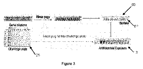
Figure 3 shows an example of a biofilm susceptibility assay of the present
invention.
Figure 4 shows an example of a process for recovering intact biofilm in
accordance
with the present invention.
7
CA 02768735 2012-01-19
WO 2011/009213 PCT/CA2010/001151
Figure 5 shows an example of a process for establishing MBEC and MIC
determinations in accordance with the present invention.
Figure 6 shows the configuration of a challenge plate used in Example 10.
Figure 7 is a chart of the MIC, MFC, and MBEC values determined biofilms.
Figure 8 (a-c) illustrates reading qualitative end points from patterns in
recovery
plates and interpreting biofilm survival data from kill curves.
DETAILED DESCRIPTION OF THE INVENTION
The invention comprises improved methods and devices for the selection of one
or
more active agents, either alone or in combination, effective against one or
more biofilms
alone or in combination. The invention may further comprise optimizing the
method and/or
devices for growing the biofilm. The invention may further comprise
susceptibility testing
one or more microorganisms or mixtures of microorganisms, providing measures
of
resistance and/or tolerance of the microrganism(s).
In some embodiments of the invention, the methods and devices involve setting
up
and/or calibrating a biofilm growth device, said biofilm growth device
comprising a lid
comprising at least one peg; optimizing the methods and devices to promote
biofilm growth,
and susceptibility testing one or more microorganisms.
The methods and devices of the previous two paragraphs may further comprise
one or
more of the following, alone or in various combinations: optimizing the device
and growing
conditions specific for a particular microorganism; growing multi-species
biofilms;
susceptibility testing multi-species biofilms; growing biofilm to an amount
greater than about
104 cells; optimizing and/or changing the surface attributes of the peg or
substrate to promote
biofilm growth and/or cell adherence; evaluating microbial growth in the
biofilm reactor,
including but not limited to using viable cell count (VCC), determining the
number of cells
growing in the planktonic inoculum; determining the number of cells growing in
the peg
biofilms, and assessing the biofilm growth for nonequivalent or asymmetric
growth patterns;
pre-exposure control measurements; promoting reproducible cell density;
biofilm recovery
using sonication; qualitative biofilm recovery; quantitative biofilm recovery;
optimizing
sonication time; establishing end points, including but not limited to
measurements for
tolerance, measurements for resistance, MIC, MBEC, MBIC, MBC, and MLC;
providing a
growth medium suitable for the specific microorganism(s); providing a recovery
medium
suitable for the specific microorganism(s); providing susceptibility testing
suitable for the
specific microorganism(s); selecting a processing temperature suitable for a
specific
8
CA 02768735 2012-01-19
WO 2011/009213 PCT/CA2010/001151
microorganism's growth, recovery, and/or susceptibility testing; selecting a
cultivation time
suitable for a specific microorganism's growth, recovery, and/or
susceptibility testing;
selecting an inoculum amount suitable for a specific microorganism's growth,
recovery,
and/or susceptibility testing; and selecting a nutritional medium suitable for
a specific
microorganism's growth, recovery, and susceptibility testing.
One skilled in the art will recognize that the devices and parameters for
growing,
recovering, and/or susceptibility testing one or more biofilms may involve
tailoring the
device and parameters for a specific biofilm(s), and further, that this
tailoring may include a
wide variety of variables. Some of these variables are noted above; other
variables are shown
in the Examples. These and other variables are included within the scope of
the present
invention.
An embodiment of the invention includes establishing optimized devices and
process
parameters for each of the 65+ microorganisms shown in Example 29.
In preferred embodiments of the invention, the devices and methods may be used
in
susceptibility testing one or more biofilms alone or in combination; and/or in
the treatment of
infections or conditions mediated by one or more gram-negative bacteria. In
the most
preferred embodiments of the invention, the devices and methods may be used as
a diagnostic
tool to determine various compositions, including the optimum composition, for
treating one
or more biofilms and/or one or more disease or conditions mediated by the
biofilm. In some
embodiments of the invention, the methods and devices provide diagnostic or
clinical
susceptibility testing, and in the most preferred embodiments, provide any
combination of
MBEC, MBC, and MIC values in a single experiment.
The invention also provides methods and devices for selecting one or more
biofilm
agents, alone or in combination, for the treatment of one or more gram-
negative bacteria,
alone or in combination; one or more gram-positive bacteria, alone or in
combination; one or
more diseases or conditions mediated by a gram-negative bacteria; one or more
fungal
organisms, alone or in combination; one or more diseases or conditions
mediated by a gram-
positive bacteria; and one or more diseases and/or conditions mediated by
fungal
microorganisms. As used herein, alone or in combination includes a single
microorganism
on a single peg or in a single well; a single microorganism alone on a single
peg or in a single
well on a lid or bottom having a different microorganism in a different peg or
well (i.e.,
multiple microorganisms on a single lid or bottom); and multiple
microorganisms on a single
peg or well. Thus, in accordance with the present invention, the devices and
methods may
include single species or multiple species; the multiple species may include a
combination
9
CA 02768735 2012-01-19
WO 2011/009213 PCT/CA2010/001151
lid, plate, or susceptibility test, "combination" referring to a single
species separated from
another species, but on a single lid or plate. Combination lids, plates, and
tests also refers to
pre-determined groups of microorganisms grown or tested on a single lid or
plate, e.g., a
gram (-) lid or a gram (+) lid, or mixtures thereof Multiple species also
includes a "mixed"
lid, plate, or susceptibility test, "mixed" referring to more than one species
in the same peg or
well (e.g., a mixed biofilm).
The invention also provides methods and devices for treating one or more
diseases or
conditions mediated by one or more gram-negative bacteria.
As used herein, gram-negative biofilm or bacteria refers to any bacterium or
biofilm
formed by that bacterium that is termed gram-negative by one skilled in the
art. Typically,
gram-negative refers to the inability of a type of bacterium to resist
decolorization with
alcohol after being treated with crystal violet (Stedman's Medical Dictionary,
28th Ed., 2006).
Exemplary gram-negative bacterium families include, but are not limited to E.
coli,
Burkholderia spp, Acinetobacter spp, Proteus spp, Salmonella spp.
Stenotrophomonas spp
and Pseudomonas spp, Vibrio, Yersinia, Campylobacteria. Exemplary strains
within these
families include but are not limited to A. lwoffii, A. radioresistens, A.
baumanii, A.
heamolyticus, A. calcoaceticus anitatus; E. coli, E coli strain 0157:H7; B.
cepacia; P.
aeruginosa, Proteus mirabilis, Proteus vulgaris, Stenotrophomonas maltophilia,
Yersinia
enterocoloticia, Campylobacter jejuni, and Vibrio cholerae.
The invention also provides methods and devices for selecting an antibiotic or
combination of antibiotics for the treatment of CF in a specific patient. The
present invention
also includes methods and devices for treating a patient or subject having a
disease or
condition mediated or caused by a biofilm. In these embodiments of the
invention, a
biological sample from a patient or subject is processed with an apparatus or
portion of an
assembly adapted and/or configured to promote biofilm growth. The biofilm may
then be
processed with an apparatus or portion of an assembly adapted and/or
configured to expose
the biofilm to one or more antimicrobial agents or one or more anti-biofilm
agents.
The methods and devices or assemblies of the present invention comprise
optionally
calibrating the equipment; growing the biofilm, preferably including
optimizing the apparatus
or a portion thereof, and/or optimizing the growth conditions; removing intact
biofilm from
the growth assembly; and subjecting the biofilm to antimicrobial
susceptibility testing,
preferably including optimizing the apparatus or a portion thereof, and/or
optimizing the
exposure conditions in a manner specific for the particular organism(s).
CA 02768735 2012-01-19
WO 2011/009213 PCT/CA2010/001151
Exemplary biofilm formation devices, biofilm susceptibility devices, and
biofilm
testing assemblies are described in U.S. Serial No. 11/996,478 (filed 19
August 2008).
An embodiment of the invention includes an assembly comprising one or more
plates
pre-loaded with one or more pre-selected anti-biofilm agents against a
specific biofilm or
biofilms, said plates may be used to identify efficacious individual or
combined active agents
for treating biofilm-mediated diseases or conditions.
In some embodiments of the invention, the method may also include one or more
of
the following: growing multiple or plural biofilms under conditions that
promote the
production of substantially uniform biofilms; screening the biological sample
against a large
group of active agents; selecting a subgroup of active agents; loading an
assay device with
multiple or plural active agents in the subgroup; growing biofilm from a
specific patient's or
subject's sample; screening the biofilm from the specific patient or subject
against the
subgroup of active agents; reading the results; determining the appropriate
active agent or
combination of active agents suitable for the particular biofilm; conducting a
turbidity assay
if the microorganism produces visible turbidity when growing (e.g.
Pseudomonas); and
conducting a plating assay if the microorganism does not grow with visible
turbidity.
An embodiment of the invention includes methods for selecting specific
combinations
of antibiotics that have efficacy against isolates of one or more gram-
negative bacteria as a
biofilm by screening a broad range of clinical isolates of a species against
an extensive panel
of antibiotics alone or in combination to identify combinations with efficacy
against biofilm
grown organisms.
An embodiment of the invention includes determining the active agent or
antibiotic(s)
of choice for the treatment of a biofilm infection by challenging the biofilm
of the patient's
specific isolate against the diagnostic plate specific for the species that
forms the biofilm.
An embodiment of the invention includes rehydrating a species specific plate
of
preloaded antibiotics as the challenge plate to identify antibiotics with
efficacy against the
specific pathogen. Plates may be frozen (no rehydration required), or
lyophilized, freeze dried
or vacuum dried.
An embodiment of the invention includes a well plate containing frozen or
lyophilized
antibiotic combinations that can be re-hydrated to be used in an antibiotic
susceptibility
assay.
An embodiment of the invention includes growing biofilm obtained from a
biological
specimen obtained from a patient, and using the biofilm in a susceptibility
assay. In this
embodiment of the invention, the susceptibility assay provides which active
agent or
11
CA 02768735 2012-01-19
WO 2011/009213 PCT/CA2010/001151
combination of active agents is best suited to eradicate a gram-negative
Pseudomonas species
or a Pseudomonas aeruginosa biofilm. In this embodiment, the susceptibility
assay may also
provide which active agent or combination of active agents is best suited to
treat a disease or
condition mediated by the gram-negative biofilm. An embodiment of the
invention includes
challenging a biofilm against selected combinations of an anti-microbial or an
anti-biofilm
agent, thereby identifying the most appropriate combination. Some embodiments
of the
invention further include using the identified antimicrobial agent or agents
to treat a patient,
to treat a microorganism, and/or to change an existing treatment regimen or
antimicrobial
agent to a more medically beneficial regimen or agent(s).
An embodiment of the invention includes providing MBEC values in the diagnosis
and treatment of any gram-negative microorganism capable of biofilm formation,
and using
those values to treat or develop a treatment protocol for any gram-negative
microorganism-
mediated disease, infection, or condition. The invention may further include
providing MIC
and/or MBC values.
In a further aspect of the invention, after growing the biofilm on adherent
sites on a
lid or plate, the methods and devices may include dislodging the biofilm from
the biofilm
adherent sites and further incubating the biofilm. Dislodging the biofilm from
the biofilm
adherent sites may include dislodging the biofilm from each biofilm adherent
site into a
separate well of a microtiter plate or base. In preferred embodiments of the
invention, the
biofilm is dislodged using any process that results in intact biofilm being
removed from the
adherent sites. The inventors have found that using centrifugation removes
only a portion of
the microorganism, and therefore any resulting assay may be incomplete or
inaccurate.
Preferably, the plural biofilm adherent sites are formed in plural rows, with
plural
sites in each row; and the container includes plural channels, with one
channel for each row
of plural biofilm adherent sites. Devices or assemblies so configured permit
high throughput
analysis of the biofilm.
An embodiment of the invention also includes a pharmaceutical composition
suitable
for treating one or more gram-negative bacteria, and/or one or more diseases
or conditions
caused by the gram-negative bacteria. In these embodiments of the invention,
the
pharmaceutical composition includes one or more active agents specifically
chosen as
effective. In these embodiments of the invention, the active agent(s) are
selected by
processing a biological sample from a patient through biofilm growth and
susceptibility
testing devices of the present invention. These device(s) grow biofilm from
the bacteria
found in the patient's sample, then subject the biofilm to a panel containing
at least one active
12
CA 02768735 2012-01-19
WO 2011/009213 PCT/CA2010/001151
agent. The optimum active agent or combination of active agents may then be
selected for
use in treating the patient.
A biofilm reactor, as used herein, comprises a lid having one or more
substrates,
wherein said lid is configured to engage a bottom plate. The substrate may be
variously
configured, but is typically a peg or the like. The first bottom plate may be
variously
configured, including but not limited to a typical microtiter plate having a
well configured to
receive an individual peg; or a trough having one or more channels configured
to receive at
least one peg. It is intended that the lid and first bottom plate are
configured to promote
biofilm growth. In preferred embodiments of the invention, the lid/bottom
assembly that
comprises a biofilm reactor exhibits reduced or eliminated contamination.
It is intended that the lid and/or pegs may be configured to engage at least
one second
bottom plate. It is intended that the lid and second bottom plate may be
variously configured
to provide and/or promote susceptibility testing.
Peg lids, as used herein, refers to the lid noted above, suitable for growing
and testing
one or more biofilms. Suitable, as used here, refers to various structures and
characteristics,
including but not limited to a peg detachable from the lid, breakable or
removable pegs, pegs
that have been scored so that they may be removed from the lid; pegs that are
positioned in
the lid with a permanent or removable adhesive backing; a coated or uncoated
substrate or
peg; and/or a substrate or peg comprising or coated with any of a wide
assortment of
materials that promote biofilm growth and/or recovery. The preferred peg lid
comprises
polystyrene, but may be formed of any material or materials that have a
neutral electrostatic
charge. Peg lids may be constructed individually, or are commercially
available from Nunc,
Trek Diagnostics, and other manufacturers. Commercially available structures
may need to
be altered or reconfigured in accordance with the teachings of this invention
to provide
biofilm growth, promote biofilm growth, provide and/or promote biofilm
adherence; provide
and/or promote biofilm recovery; and provide and/or promote biofilm
susceptibility testing.
Exemplary parameters and controls for biofilm cultivation on peg lids. Reactor
set up and growth conditions. Microbes depend on diverse environmental and
nutritional cues
to attach to a surface and to initiate biofilm formation. Fastidious criteria
for microbial
surface attachment can be met by mixing and matching a set of reactor parts
and by coating
pegs with conditioning films. Other considerations include the rate of motion
of the
inoculated reactor, incubation temperatures and time periods, inoculum size,
atmospheric
gases, composition of the growth medium and frequency of medium exchange. We
have
conducted a comprehensive review of the growth conditions reported in the
literature for
13
CA 02768735 2012-01-19
WO 2011/009213 PCT/CA2010/001151
growing biofilms on peg lids, and all of these discrete parameters can be
thought of as
adjustable steps in a core protocol (Example 28). To date, variations in this
core procedure
have been used to cultivate biofilms representing >65 different microbial
species (Example
28), of which several have been grown in multispecies biofilms.
There are several considerations when growing biofilms on peg lids:
Surface attributes may be important for getting microbes to attach to a
substratum.
Lids can be made from different materials, such as raw polystyrene (the MBEC
assay) or
from chemically modified plastics, such as those used for solid-surface enzyme-
linked
immunosorbant assays (Nunc Immuno-TSP). It is also possible to coat peg lids
with
conditioning films to facilitate the adhesion of fastidious microorganisms
that might not
otherwise stick to the surface. Such coatings might include L-lysine20, BSA,
trichloroacetic
acid treated with ethylene oxide21, human saliva22,23 and polycyclic aromatic
hydrocarbons,
such as phenanthrene24 .
In addition to microtiter plates, peg lids can be fit into troughs and these
platforms can
be used for biofilm cultivation on an orbital shaker or a rocking table. A
disadvantage of
using the trough method is that a rocking table is not a customary piece of
equipment in many
microbiological laboratories. However, not all microbial species will form
biofilms with
consistent peg-to-peg cell densities on an orbital shaker (or vice versa), and
therefore, the
choice of platform is dictated by the requisite growth conditions for the
microorganism25. We
would recommend the use of the microtiter plate method for biofilm cultivation
as a first
choice over the trough format of the assay because of its increased
simplicity.
Incubation temperatures and time periods not only depend on the growth optima
of
the test organism but also can be influenced by temperature-dependent changes
in production
of extracellular polymers or adhesins. For example, certain Escherichia coli
strains produce
cellulose and curli fimbriae at 23 C but not at 37 C, and thus a temperature
shift can affect
the adherence of E. coli to a surface26.
Similar to MIC testing, inoculum size for biofilm cultivation is measured
using
McFarland standards; however, as this is based on optical density (OD)
measurements, these
standards can represent different numbers of cells for different organisms.
Relatively lower
starting inoculum sizes have been linked to increased biofilm production for
some bacteria,
such as for Pseudomonas aeruginosa PAOI (ref. 27) (Example 28). By contrast, a
relatively
larger inoculum size seems to be essential for biofilm formation by other
species, such as for
Rhizobium leguminosarum biovar viciae (ref. 28).
14
CA 02768735 2012-01-19
WO 2011/009213 PCT/CA2010/001151
Atmospheric gases can be controlled in air-tight environmental chambers or
incubators to facilitate biofilm formation by facultative and obligate
anaerobes
Electron acceptors, host factors, carbon and nitrogen sources, as well as
inorganic
ions, such as magnesium and phosphorous, can be environmental cues that affect
microbial
adhesion and growth on surfaces2. It is because of this biological fact that
there can be no
universal medium for growing a biofilm, even among microorganisms that can be
routinely
cultured on rich laboratory media. Similar to larger scale reactors, the best
experimental
approach is to choose a medium that most closely resembles the environment of
interest.
We recommend the experimenter start with conditions suited, appropriate, or
advantageous to the specific species being tested. Exemplary conditions are
listed in
Example 28 to cultivate the organism of interest. If the test organism has not
been grown on
peg lids before, a good starting point is to use a nutritional medium that is
known to support
growth of the microbe in vitro. Optimization of biofilm growth for more
fastidious organisms
can be achieved by experimenting with different reactor assemblies, medium
formulations,
surface coatings and other parameters as seen fit, and then by testing them
empirically. For
instance, it might be possible to test several growth media for their ability
to promote biofilm
formation on pegs by inoculating different media with the same test organism.
These inocula
could be arranged in separate wells of a microtiter plate and a single peg lid
could be used as
the substratum. The number of cells in biofilms could then be quantified using
the methods
presented in the core protocol.
Media for susceptibility testing and cell recovery. Growth media for
susceptibility
testing. The guidelines set by the Clinical Laboratory Standards Institute
(CLSI) and the
European Committee on Antimicrobial Susceptibility Testing indicate that in
most cases,
standardized bacterial MIC testing should be performed in cation-adjusted
Mueller-Hinton
broth'. Standardization of the test medium has been essential for
interlaboratory
reproducibility of MIC testing. No such standard medium exists, however, for
biofilm
susceptibility testing, and this is likely because of the different
nutritional requirements for
getting biofilms to grow under laboratory conditions. When choosing a medium
for microbial
exposure, it is most important that it is chemically compatible with the test
agent and that it
contains no components that might detrimentally affect biofilm growth. For
example,
polysorbate-80, an additive routinely used to prevent the adsorption of some
antibiotics to
plastic surfaces, can inhibit biofilm formation by some Staphylococcus30 and
Pseudomonas31
sp., and therefore, should not be used during biofilm susceptibility testing.
A good
CA 02768735 2012-01-19
WO 2011/009213 PCT/CA2010/001151
experimental strategy is to choose a test medium that most closely resembles
the environment
in which the biofilm is likely to encounter the antimicrobial agent that is
being tested.
Sonication and biofilm cell recovery. Biofilm cells can be recovered using low-
frequency (60 Hz) vibrations to disrupt cells into a rich medium that contains
1% Tween-20
or a comparable surfactant. As an exception, recovery of biofilm Clostridium
difficile
(Example 28) requires 0.5% Tween-80 supplemented with 0.1% taurocholate in the
recovery
medium. This process is carried out at room temperature (20-25 C) and the
recovered cells
are immediately serially diluted and plated onto agar in less than the
doubling time of the test
organism. This ensures that there are no artificial increases in biofilm cell
numbers due to
processing time. Vibrations can be generated using a water table sonicator,
wherein the peg
lid, which is inserted into a microtiter plate containing the recovery medium,
is placed on the
steel insert tray of this device. Ali et al.32 recommend a sonication time of
10 min, as shorter
time periods led to incomplete cell recovery and longer time periods (i.e., 15
min) did not
result in significantly increased cell recovery from pegs. For example,
Listeria innocua
grown in the CBD using the parameters listed in Example 28 yielded 5.4 0.1,
5.9 0.1 and
6.0 + 0.1 logio colony forming units per peg (CFU per peg) with 5, 10 and 15
min of
sonication, respectively. Ali et al.32 recommended this intermediate
sonication time as
recovery was high and consistent; moreover, an intermediate sonication time
reduced the
possibility of damage to injured cells that extended time periods might cause,
especially after
susceptibility testing. Nonetheless, one might find it worthwhile testing this
optimum
sonication time for different instruments and organisms. A simple strategy
here would be to
follow the protocol and to test different sonication time periods for an
effect on the mean
VCC determined from batch culture biofilm growth controls for the desired test
organism.
Inactivating antimicrobial agents. In general, there are three optional
methods to
inactivate antimicrobials: (i) membrane filtration, (ii) dilution of the agent
to a sub-inhibitory
level and (iii) the addition of a neutralizing agent33. In the core protocol
for biofilm
susceptibility testing, we opt to dilute the antimicrobial agent back to sub-
inhibitory levels by
rinsing the biofilms twice before disrupting the cells into the recovery
medium. If the
experimental design is modified to include a comparison of biofilm and
planktonic cell
susceptibility, then biofilms and planktonic cells could be treated with a
neutralizing agent. In
this way, similar inactivating regimens can be used to carry out a fair
comparison of biofilm
and planktonic cell susceptibilities. If carry-over of low antimicrobial
concentrations prevents
accurate VCCs, or if the susceptibility data are to be used for a regulatory
submission34, then
use of a neutralizing agent in addition to rinsing is warranted. Neutralizing
agents, in general,
16
CA 02768735 2012-01-19
WO 2011/009213 PCT/CA2010/001151
are filter sterilized and then added to the sterile recovery medium at an
appropriate molar
concentration that typically exceeds the working concentration of the
antimicrobial agent. A
list of neutralizing agents for antibiotics, biocides and some metal ions can
be found
elsewhere33,34. It is important to confirm that the neutralizer works and does
not harm the
recovered microorganisms, and for a discussion of this we suggest that one
should consult the
guidelines published by the American Society for Testing and Materials (ASTM
International)35
In addition to susceptibility testing, peg lid biofilm reactors of the present
invention
serves as the starting point for a variety of downstream applications; e.g.
additional or
alternative modifications, applications and limitations:
1) Biofilm biomass may be stained on lids with crystal violet40,41
y peg ,which is
adapted from the O'Toole and Kolter15,16 method of staining biofilms grown in
the wells of
microtiter plates.
2) Biofilm structure on pegs may be determined by microscopy7'25'42,43;
however,
biofilms cultivated on pegs are subject to complex fluid dynamics and,
although gross
morphological changes in structure may be discerned, flow cell models might be
more
suitable for testing this. Batch culture systems, such as the peg lid biofilm
reactor described
here, do not provide continuous flow or replacement of media and therefore may
significantly
influence the structure of the biofilm. Thus, the intricate microcolony
structure of biofilms
obtained using flow cells might be altered or absent from peg lid biofilms.
Even so, it is
possible to visualize 3-D patterns in peg lid biofilm killing by antibiotics
that are similar to
those produced in flow cells.
3) Low-speed centrifugation can be used to disrupt cells from pegs into a
recovery
medium44
4) RT-PCR and promoter-reporter constructs can be used to measure the gene
expression in biofilms; however, the tiny amount of biomass produced on each
peg makes the
peg lid biofilm reactor ill-suited for proteomics.
5) Cell viability may be assessed using a variety of methods, including
quantitative
PCR22,23 and tetrazolium salts42,43
6) Challenge plate configurations can be set up to screen libraries of
compounds for
anti-biofilm activity, to perform checkerboard assays to identify
antimicrobial antagonism or
synergy19 and to perform multiple combination susceptibility testingl3.
7) Isogenic mutants at similar biofilm cell densities can be compared to
determine
differences in antimicrobial sensitivity due to gene deletion or
overexpression4s,46
17
CA 02768735 2012-01-19
WO 2011/009213 PCT/CA2010/001151
8) It is possible to modify this technique to determine MIC data and VCCs for
planktonic cells shed from the surface of the biofilm, while simultaneously
determining
biofilm susceptibility. An important limitation of this approach is that the
starting number of
cells for planktonic susceptibility testing cannot be determined, and thus,
log-killing of
planktonic cells cannot be calculated. Nonetheless, MIC measurements made
using this type
of experimental design in some instances approximate those made using a
standard CLSI
MIC test17. Moreover, planktonic cells that are shed from the surface of the
peg biofilms and
isolated from the wells of the challenge plate have a different sensitivity to
antibiotics and
metal ions than the biofilms from which they were derived47'48. This
nonstandardized method
to test planktonic cell susceptibility is not presented here, and instead we
direct researchers to
a discussion of this approach elsewhere17'47'49
Peg lids (MBEC P&G or HTP assays, Innovotech or Nunc Immuno-TSP, Nunc, cat.
no. 445497) The MBEC and Nunc Immuno-TSP peg lids are manufactured in
different ways.
The MBEC peg lid is designed as a substratum for biofilm growth. These lids
are made from
polystyrene, bare an overall neutral electrostatic charge and have a plastic
backing, as well as
are engineered with break points that facilitate detachment of individual
pegs. The MBEC
P&G assay is packaged with a microtiter plate, whereas the HTP assay comes
with a trough
that serves as the inoculum reservoir. In contrast, the Nunc Immuno-TSP lids
were designed
as supports for solid-surface enzyme-linked immunosorbant assays, but can also
be used for
biofilm cultivation. These lids have a chemically modified polystyrene
surface, bare an
overall positive electrostatic charge and lack the plastic backing and break
points that
facilitate peg detachment. These lids are packaged with a trough that can be
used as an
inoculum reservoir, but this can be swapped for a microtiter plate at ones
discretion. Nunc
Immuno-TSP lids will need to be modified for biofilm assays as described in
the Examples.
Building and sterilizing peg lid reactors If not carried out by the
manufacturer (e.g.,
Nunc-TSP lid), trim an adhesive backing (e.g., Costar plate sealers) and fit
it to the top of the
peg lid. This will maintain sterility of the device once pegs have been
removed for control
measurements. It is also possible to swap the troughs that come with the Nunc-
TSP lids for
microtiter plates at this point. If peg lids are nonsterile when purchased
from the
manufacturer, if an adhesive backing has been applied before use or if the
reactors have
opened and parts have been swapped, assemble the device, seal it in an air-
tight plastic bag
and sterilize it using ethylene oxide (Anprolene), according to the directions
of the supplier.
Sterile agar and broth growth media specific for the microorganism to be
cultured.
There are no standardized media for biofilm cultivation or susceptibility
assays; however,
18
CA 02768735 2012-01-19
WO 2011/009213 PCT/CA2010/001151
there must be no variation in the chosen medium composition from one
experiment to the
next. This will ensure reproducibility of intra- and interlaboratory results.
One is cautioned to
strictly control the composition of the growth medium when comparing data sets
generated
by technicians in the same and different laboratories
A device of the present invention may comprise a biofilm growth assembly 1, a
biofilm challenge assembly 2, a rinsing assembly 3, and a biofilm dislodging
and re-growth
assembly 4. Used in concert, the assemblies provide MIC, MBC, and MBEC values
in a
single experiment.
In accordance with the present invention, the biofilm growth assembly 1 may
include
a base or plate 20 configured to receive a lid 10. Lid 10 may be configured to
include one or
more projections 12 that extend into a space defined by base 20. In most
preferred
embodiments of the invention, the biofilm growth assembly 1 is rocked, moved,
or the like so
that the growth fluid in the assembly flows or moves across projections 12. In
preferred
embodiments of the invention, base 20 is an incubation base and is configured
to provide
each projection with substantially equivalent exposure to the source of
microorganisms and
its nutrient/growth broth.
In accordance with the present invention, the biofilm challenge assembly 2
comprises
a second base or plate 21 configured to receive a lid 60 having projections 61
typically
covered by biofilm. Projections 61 extend into one or more wells configured in
plate 21. A
typical second base 21 is a standard 96 well microtiter plate, although one
skilled in the art
will readily recognize that other configurations may be used. Second base 21
includes one or
more anti-biofilm agents in the wells. In accordance with the present
invention, second plate
21 may be removed and used for determining the MIC value of the non-biofilm
(e.g.,
planktonic) microorganism (see Figure 5).
In accordance with the present invention, the biofilm rinsing assembly 3
comprises a
third base or plate 40 configured to receive a lid 60 having projections 61
typically covered
by biofilm. Projections 61 extend into one or more wells configured in plate
40. A typical
third plate 40 is a standard 96 well microtiter plate, although one skilled in
the art will readily
recognize that other configurations may be used. Third plate 40 includes one
or more rinsing
and/or neutralizing agents in the wells.
After rinsing, lid 60 may then be joined with a fourth base 50, also referred
to as a
recovery plate. Lid 60 and fourth base 50 form the biofilm disruption assembly
4. The
recovery plate contains recovery media, and, in accordance with the present
invention,
assembly 4 may be subjected to sonication and biofilm re-growth (confirming
that the biofilm
19
CA 02768735 2012-01-19
WO 2011/009213 PCT/CA2010/001151
has not been removed). In preferred embodiments of the invention, the recovery
medium
includes one or more neutralizing agents. As shown in the examples, assaying
the projections
on lid 60 after it has been exposed to recovery media provides an MBEC value
of the
microorganism, and plating from the recovery plate provides an MBC value.
The device includes biofilm lid 10 composed of tissue grade plastic or other
suitable
material (e.g. stainless steel, titanium) with projections 12 extending
downwardly from the lid
10. The projections 12 may be biofilm adherent sites to which a biofilm may
adhere, and may
be configured into any pattern or shape suitable for use in conjunction with a
channel or well-
containing bottom, such as base 20. The pattern of projections 12 preferably
mirror the
pattern of channels and/or wells in convention plates, e.g. a 96-microtiter or
well plate
commonly used in assay procedures. In most preferred embodiments of the
invention, the
projections 12 are preferably formed in at least eight rows 14 of at least
twelve projections
each. Other numbers of rows or numbers of projections in a row may be used,
but this is a
convenient number since it matches the 96 well plates commonly used in
biomedical devices.
Additional or some of the projections as shown may be used to determine the
initial biofilm
concentration after incubation. The exemplary projections 12 shown are about
1.5 cm long
and 2 mm wide, but may be any size and/or shape.
The lid 10 has a surrounding lip 16 that fits tightly over a surrounding wall
28 of the
vessel 20 to avoid contamination of the inside of the vessel during
incubation.
Base 20 serves two important functions for biofilm development. The first is a
reservoir for liquid growth medium containing the bacterial population which
will form a
biofilm on projections 12. The second function is having a configuration
suitable for
generating shear force across the projections. While not intending to be
limited to any
particular theory of operation, the inventors believe that shear force formed
by fluid passing
across the projections promotes optimal biofilm production and formation on
the projections.
Shear force on the projections 12 may be generated by rocking the vessel 20
with lid
10 on a tilt table 30. The inventors have found that using a rocking table
that tilts to between
about 7 and about 11 is suitable for most applications. In preferred
embodiments of the
invention, the rocking table should be set on about 9 . It is intended that
the invention should
not be limited by the use of an actual degree of tilt, but that any tilt used
for any particular
machine be appropriate for growing biofilm in accordance with the present
invention.
The projections 12 may be suspended in the channels or wells so that the tips
of the
projections 12 may be immersed in liquid growth medium flowing in the
channels. The
ridges 26 channel the liquid growth medium along the channels 24 past and
across the
CA 02768735 2012-01-19
WO 2011/009213 PCT/CA2010/001151
projections 12, and thus generate a shear force across the projections.
Rocking the vessel 10
causes a repeated change in direction of flow, in this case a repeated
reversal of flow of liquid
growth medium, across the projections 10, which helps to ensure a biofilm of
equal
proportion on each of the projections 12 of the lid 10. Rocking the vessel so
that liquid flows
backward and forward along the channels provides not only an excellent biofilm
growth
environment, but also simulates naturally occurring conditions.
Each projection 12 and each channel 24 preferably has substantially the same
shape
(within manufacturing tolerances) to ensure uniformity of shear flow across
the projections
during biofilm formation. In preferred embodiments of the invention, channels
24 should all
be configured or connected so that they share the same liquid nutrient and
bacterial mixture
filling the basin 22. The inventors have found that substantially uniform
channel
configuration and access to the same source of microorganisms promotes the
production of
an equivalent biofilm on each projection, equivalent at least to the extent
required for testing
anti-biofilm agents. Biofilms thus produced are considered to be uniform.
Results have been
obtained within P<0.05 for random projections on the plate.
Sensitivity of a biofilm may be measured by treating the biofilm adherent
sites with
one or more anti-biofilm agents, i.e., challenging the biofilm, and then
assaying the biofilm.
This may be accomplished by placing the lid 60 (having a biofilm formed on the
projections)
into a second base 21 adapted to receive lid 10 and projections 12. In
preferred embodiments
of the invention, lid 60 engages second base 21 in a manner sufficient to
prevent
contamination of the assembly. As used herein, a manner sufficient to prevent
contamination
refers to the configuration and assembly of mating structures so that the
contents of the
closed assembly are free of outside contamination.
In accordance with the present invention, one skilled in the art may use any
arrangement or scheme for challenging a group of biofilms. For example, all of
the wells of
the challenge plate may include the same anti-biofilm agent; plural or
multiple wells may
include different doses of the same anti-biofilm agent; plural or multiple
wells in a single row
may include the same dose or different doses of anti-biofilm agent; plural or
multiple rows
may include the same dose or different doses of anti-biofilm agent; plural or
multiple wells or
plural or multiple rows may include more than one anti-biofilm agent; or
plural or multiple
wells or plural or multiple rows may include more than one anti-biofilm agent,
varying the
dose by well, by row, and/or by anti-biofilm agent. It is intended that the
configuration and
arrangement of wells, type and number of anti-biofilm agents, and dose in each
well should
be variable as desired by one skilled in the art to achieve a specific
purpose, e.g., testing one
21
CA 02768735 2012-01-19
WO 2011/009213 PCT/CA2010/001151
or more biofilms with one or more anti-biofilm agents using as many variables
as reasonable
to the intended purpose.
For example, projections 12 that have been incubated in the same channel 24 of
the
vessel 20 may be treated with a different anti-bacterial reagent. In this
manner, consistent
results may be obtained since the growth conditions in any one channel will be
very similar
along the entire channel and thus for each projection 12 suspended in that
channel. This helps
improves the reliability of treatment of different projections 12 with
different anti-bacterial
reagents. The examples show different arrangements suitable for use with the
assemblies of
the present invention.
As noted above, a device of the present invention may be loaded with one or
more
anti-biofilm agents. An incomplete and exemplary list of possible anti-biofilm
agents
include, but are not limited to: Antibiotics. Including, but not limited to
the following classes
Aminoglycosides; Antipseudomonals, including Cephalosporins; beta.-Lactams;
Antibiotics;
Urinary Tract Antiseptics, such as Methenamine, Nitrofurantoin,
Phenazopyridine and other
napthpyridines; Penicillins, Tetracyclines; Tuberculosis Drugs, such as
Isoniazid, Rifampin,
Ethambutol, Pyrazinamide, Ethinoamide, Aminosalicylic Acid, Cycloserine; Anti-
Fungal
Agents, such as Amphotericin B, Cyclosporine, Flucytosine Imidazoles and
Triazoles
Ketoconazole, Miconazaole, Itraconazole, Fluconazole, Griseofulvin; Topical
Anti Fungal
Agents, such as Clotrimazole, Econazole, Miconazole, Terconazole,
Butoconazole,
Oxiconazole, Sulconazole, Ciclopirox Olamine, Haloprogin, Tolnaftate,
Naftifine, Polyene,
Amphotericin B, and Natamycin.
Several different conventional methods may be used to count the bacteria. It
may be
done by incubating the sonicated bacteria, taking serial dilutions and
visually counting the
colony forming units, or automated methods may be used, as for example using
an optical
reader to determine optical density. It has been found however that the
optical reader of
turbidity is too imprecise for practical application, and it is preferred that
vital dye technology
be applied to automate the measurement of viability, by treating the biofilm
with a vital dye,
and measuring the intensity of light given off by the dyed biofilm. In the
case of using vital
dye technology, the biofilm need not be further incubated. One skilled in the
art will
recognize that other dyes for cell mass may be used; these dyes may be later
extracted and
read for OD (a measure of remaining cell biomass). In a further embodiment,
the assay may
be carried out by sonicating the cells until they lyse and release ATP and
then adding
luciferase to produce a mechanically readable light output. In a still further
embodiment, the
assay may be carried out directly on the biofilm on the projections using a
confocal
22
CA 02768735 2012-01-19
WO 2011/009213 PCT/CA2010/001151
microscope, although it should be considered that this is difficult to
automate. In the
examples that follow, the results are obtained from a manual count following
serial dilution.
The concentration (MBEC) of anti-bacterial reagent at which the survival of
bacteria
or biofilm falls to zero may be assessed readily from the assay. Likewise, the
MIC may also
be determined from the assay.
The inventors have found that in some instances a biofilm will not form
without the
inclusion of host components in the biofilm. Host components may therefore be
added to the
growth medium in the vessel during incubation of the bacteria to form the
biofilm. Host
components that may be added include serum protein and cells from a host
organism. For the
testing of the effect of different host cells and components, the ends of the
channels 24 may
be sealed by walls to prevent growth medium in one channel from flowing into
another, thus
isolating the bacteria growth in each channel from other channels. The device
thus described
may also be used to test coatings used to inhibit biofilm growth and to test
coatings which
may enhance biofilm formation. In an initial step, the projections 12 may be
coated with a
coating to be tested, and then the biofilm grown on the projections. The
biofilm may then be
assayed, or treated with anti-bacterial reagent and then assayed. The assay
may be in situ or
after dislodging of the biofilm. Different coatings may be tested on different
rows of pegs.
Enhanced biofilm formation may be used to create large viable biofilms for
biofermentation.
Definitions
As used herein, assembly refers to an integrated collection of elements or
components
designed or configured to work in concert. A typical assembly of the present
invention
includes a lid and its corresponding base or plate. In some embodiments of the
invention, an
element of one assembly may function or work with a separate assembly. For
example, the
lid of assembly 1 may be used as the lid in assembly 2, i.e., with a different
base. In
preferred embodiments of the invention, a lid may engage a base in a
removable, sealingly
fashion. In other embodiments of the invention, a lid may engage a base in a
closed,
sealingly fashion; in these embodiments, it may be desirable to adapt other
elements of the
assembly so that they are removable, e.g., one or more removable projections.
As used herein, challenge plate refers to any base having one, multiple, or
plural
configurations of wells, troughs, or the like, said plate being used to expose
one or more
biofilms to one or more anti-biofilm agents. A typical challenge plate may be
used to
determine biofilm growth in an environment that includes one or more anti-
biofilm agents.
In a later step of a process of the present invention, the challenge plate may
be used to
23
CA 02768735 2012-01-19
WO 2011/009213 PCT/CA2010/001151
determine the MIC value of any planktonic microorganism. An exemplary
challenge plate is
shown in Figures 3 and 5.
The challenge plate may be used to screen antimicrobial libraries, multiple
combination susceptibility testing, and gene deletion or over-expression.
As used herein, recovery plate refers to any base having one, multiple, or
plural
configurations of wells or the like, said plate being used to rinse biofilm
after it has been
exposed to an anti-biofilm agent, neutralize any anti-biofilm agent, to
collect any disrupted
biofilm after the assembly has been sonicated, or combinations thereof. In a
later step of a
process of the present invention, the recovery plate may be used to determine
the MBEC
value of any biofilm formed in the process. An exemplary recovery plate is
shown in Figures
4 and 5.
As used herein, neutralizing agent refers to any composition suitable for
reducing or
counteracting any toxicity caused by an anti-biofilm agent. A neutralizing
agent is
appropriate if it is effective for the anti-biofilm agent(s) being used and
for a particular
biofilm. The choice of neutralizing agent is within the skill of the art.
Several neutralizing
agents and compositions are shown in the Examples. As described in the
Examples, a
recovery medium is a composition that includes one or more neutralizing
agents.
As used herein, active agent or anti-biofilm agent refers to one or more
agents that are
effective in reducing, degrading, or eliminating a biofilm or biofilm-like
structures. The
present invention includes but is not limited to active agents that are
already well known, e.g.,
antibiotics, anti-microbials, and biocides. One or more active agents may act
independently;
one or more active agents may act in combination or synergistically; one or
more active
agents may be used sequentially or serially.
As used herein, a panel or library of active agents refers to a collection of
multiple or
plural active agents grouped according to a pre-determined strategy. For
example, a first
library may include one or more active agents that show some degree of
potential in being
effective against a particular biofilm. A second library may begin with a
subset of the first
library, and is designed to narrow the choices effective active agents, or to
provide more
information about a particular subset of active agents. A panel or library may
also include a
proprietary or non-proprietary group of active agents grouped according to a
pre-determined
strategy, e.g., variable doses.
As used herein a composition containing an active agent may include one or
more
active agents, and may further include one or more additional agents,
including but not
limited to bacteriocins or other anti-bacterial peptides or polypeptides, one
or more
24
CA 02768735 2012-01-19
WO 2011/009213 PCT/CA2010/001151
disinfectants or the like, one or more surfactants or the like, one or more
carriers,
physiological saline or the like, one or more diluents or the like, and one or
more
preservatives or the like.
As used herein, sample refers to a biological or fluid sample taken from a
patient,
animal, or environment; sample expressly includes any source or potential
source of
microorganism. A patient's isolate is derived by standard laboratory methods
and prepared
for assay using by standard laboratory practice (CLSI). As used herein,
biofilm challenge
involves the placement of the biofilm culture, grown on a substrate as noted
above, into the
wells of the challenge plate, thereby exposing planktonic and/or biofilm to a
range of
concentrations or a spectra of anti-biofilm agents. In preferred embodiments
of the
invention, the concentration of anti-biofilm agent(s) is selected for its
possible effectiveness
against the target organism. Incubation time and conditions and medium used
will vary with
isolate.
As used herein, efficacy is based on the ability of the active agent or active
agents to
have activity of the biofilm and is defined on the basis of MIC (minimal
inhibitory
concentration), MBC (minimal biocidal concentration), and MBEC (minimal
biofilm
eradication concentration). The standard assay for testing the antibiotic
susceptibility of
bacteria is the minimum inhibitory concentration (MIC), which tests the
sensitivity of the
bacteria in their planktonic phase. The MIC is of limited value in determining
the true
antibiotic susceptibility of the bacteria in its biofilm phase. The MBEC, on
the other hand,
allows direct determination of the bacteria in the biofilm phase, and involves
forming a
biofilm in a biofilm growth device or plate, exposing the biofilm to one or
more test
antibiotics or active agents for a defined period, transferring the biofilm to
a second plate
having fresh bacteriologic medium, and incubating the biofilm overnight. The
MBEC value
is the lowest active agent dilution that prevents re-growth of bacteria from
the treated biofilm.
As used herein, treatment protocol refers to a dose of one of more active
agents, the
composition of the active agent, and how often it should be administered to a
patient. With
the devices and methods of the present invention, the treatment protocol can
be tailored to a
specific human or animal, a specific biofilm or biofilms, and/or a specific
disease or
condition. For some diseases and conditions, e.g., CF, it may be desirable to
perform
separate assays at different times to optimize the course of treatment,
particularly optimizing
treatment or the concentration of active agent(s) over time. For example, it
is believed that a
CF patient's condition changes over time as both the patient and the infection
change; it
would be a beneficial result to monitor those changes and alter any treatment
as required.
CA 02768735 2012-01-19
WO 2011/009213 PCT/CA2010/001151
As used herein beneficial result refers to any degree of efficacy against a
microorganism or biofilm. Examples of benefits include but are not limited to
reduction,
elimination, eradication, or decrease in a biofilm or a microorganism that
forms a biofilm;
and the capability of treating a microorganism hidden or protected by a
biofilm. Exemplary
examples of a beneficial result in the manner in which a patient is treated
includes but is not
limited to the ability or capability of treating a specific patient, of the
ability to tailor a
treatment protocol for a particular patient at a particular time; and of the
increased ability of
being able to choose a particular active agent or agents. A beneficial result
may also include
any diagnostic, medical, or clinical benefit or improvement that assists the
doctor or the
patient in determining the appropriate active agent(s) and/or treatment
protocol. For
example, beneficial results are obtained when a panel of possible active
agents can be tested
rapidly, with greater efficiency, and/or with a greater number of
combinations.
The potential patient benefits are improvement in quality of life; and the
delay in the
progression of disease.
The potential doctor benefits are improved patient outcomes; greater
confidence in
susceptibility testing; reduction of treatment failures; and quantification of
combination
antibiotic choices.
The potential diagnostic laboratory benefits are reduced susceptibility
testing caused
by treatment failures and greater confidence in susceptibility testing.
The benefits to the Healthcare system are reduced costs of drug treatment and
hospitalization; delay in lung transplantation costs; and reduced resistance
development due
to the use of inappropriate drugs
As used herein susceptibility testing or similar phrases refers to determining
if and by
how much an active agent affects the growth or condition of a microorganism in
a biofilm. In
the devices and methods of the present invention, susceptibility testing is
distinguished from
prior art methods by using high through-put devices, typically a peg lid
device or assembly,
by forming a biofilm in a non-static environment, and by generating biofilms
through a flow
system.
Susceptibility testing, as noted above, may be used to determine one or more
of
several endpoints, e.g., MBEC, MBIC, MBC, etc. In accordance with the present
invention,
susceptibility testing does not include MIC or planktonic testing alone;
rather, susceptibility
testing includes biofilm testing alone, or in combination with planktonic
testing.
As used herein, high throughput refers to the capability of growing and/or
assaying a
high number of biofilms and/or a high number of anti-biofilm agents at the
same time or in
26
CA 02768735 2012-01-19
WO 2011/009213 PCT/CA2010/001151
the same procedure. Typically, high throughput translates into structural
elements in one or
more of the assemblies in order to increase speed or quantities of materials
being grown or
tested, e.g., a 96 well assay plate, a top adapted to and configured to engage
the 96 well plate,
a top with pegs corresponding to the wells, and a biofilm growth plate with
channels so that
you can process a large number of individual biofilms at the same time.
TIMING
Reagent preparation: 3 h (plus 2 days of drying time for agar media)
Equipment setup: 3 h
Optimization of growth conditions and test for equivalent biofilm growth:
Step 1A, Colony suspension method: < 15 min (plus 2 overnight incubations)
Step 2B: 10 min (plus 24 h incubation)
Steps 3-8: 15 min (plus 24 h incubation)
Box 2: 90 min (plus 24 h incubation)
Data entry and calculations: 30 min
High-throughput screening: (for each device )
Steps IA: < 15 min (plus 2 overnight incubations)
Step 2B: 10 min (plus 24 h incubation)
Steps 3-8: 15 min (plus 24 h incubation)
Steps 9-15: 45 min
Steps 16-31: 75 min (plus 24 h incubation)
Steps 32-38: < 15 min
Step 39B: 90 min (plus 24 h incubation)
Data entry and calculations: 60 min
As used herein, a pharmaceutical composition is any composition suitable for
use in
treating a disease or condition involving or mediated by a microorganism. A
pharmaceutical
composition of the present invention comprises one or more active agents
selected by using a
susceptibility device described herein. The pharmaceutical composition may
also include any
other ingredient(s) which one skilled in the art might determine is
appropriate or beneficial.
Other such ingredients include but are not limited to one or more adjuvants,
one or more
carriers, one or more excipients, one or more stabilizers, one or more
permeating agents (e.g.,
agents that modulate movement across a cell membrane), one or more imaging
reagents, one
or more effectors; and/or physiologically-acceptable saline and buffers.
Generally, adjuvants
are substances mixed with an immunogen in order to elicit a more marked immune
response.
27
CA 02768735 2012-01-19
WO 2011/009213 PCT/CA2010/001151
The composition may also include pharmaceutically acceptable carriers.
Pharmaceutically
acceptable carriers include, but are not limited to, saline, sterile water,
phosphate buffered
saline, and the like. Other buffering agents, dispersing agents, and inert non-
toxic substances
suitable for delivery to a patient may be included in the compositions of the
present
invention. The compositions may be solutions suitable for administration, and
are typically
sterile, non-pyrogenic, and free of undesirable particulate matter. The
compositions may be
sterilized by conventional sterilization techniques.
As used herein, breakpoint value refers to an active agent's concentration in
the serum
of a patient that produces a positive clinical response. Bacteria that are
susceptible to an
active agent(s) are killed at or above the breakpoint value. In the
embodiments of the
invention that include a combination of active agents, the breakpoint value is
that for the
combination.
Qualitative end points. According to CLSI standards, the MIC is defined as the
lowest concentration of an antimicrobial agent that prevents visible growth in
the challenge
medium after a set period of incubation. By contrast, the MBEC is defined as
the lowest
concentration of antimicrobial agent that prevents visible growth from
occurring in the
recovery medium used to collect biofilm cells17. The MBEC can be determined
using the
protocol presented here, and is measured after the recovery medium has been
incubated for a
suitable period of time, the length of which depends on the growth rate of the
microorganism.
Quantitative end points. According to guidelines set by the CLSI, the minimum
bactericidal concentration (MBC) is defined as the lowest concentration of an
antimicrobial
agent required to kill 99.9% of the starting planktonic bacterial population
(or 3.0 on the loglo
scale). This definition can be extended to both planktonic and biofilm cells
and these end
points will be denoted as the MBCP and MBCB, respectively. Consistent with
these guide-
lines and the definitions provided by Fothergill37 for the testing of
planktonic yeast cells,
fungal minimum lethal concentrations for planktonic cells (MLCP) and biofilms
(MLCB) can
be defined as the lowest concentration of an antimicrobial agent required to
kill 99.5% of the
starting fungal population (which also approximates 3.0 on the loglo scale).
The technique
presented here allows the MBCB or MLCB to be calculated. Another measure of
biofilm
susceptibility that has been proposed is the minimum biofilm inhibitory
concentration
(MBIC)38,39. Here, the MBIC is defined as the lowest concentration of an
antimicrobial at
which there is no time-dependent increase in biofilm MVCC when an early
exposure time is
compared with a later exposure time. The MBIC thus corresponds to the
intersecting point of
28
CA 02768735 2012-01-19
WO 2011/009213 PCT/CA2010/001151
two concentration-dependent killing curves, and therefore, it is possible to
distinguish
between biofilm resistance and tolerance on the basis of VCC (Fig. 8).
Susceptibility may be determined by comparing the breakpoint susceptibility of
an
organism with either the attainable blood or urine level of the antimicrobial
agent. The
following table lists the interpretive criteria as indicated in the CLSI
document M100-S9 or
M100-S16.
Interpretive Breakpoints
Antimicrobial Agent Susceptible Intermediate Resistant
Amikacin < 16 32 > 64
Aztreonam < 8 16 > 32
Cefepime <8 16 >_ 32
Ceftazidime < 8 16 > 32
Chloramphenicol < 8 16 > 32
Ciprofloxacin < 1 2 > 4
Colistin < 2 - > 4
Gentamicin < 4 8 > 16
Meropenem < 4 8 >_ 16
Piperacillin/tazobactam < 16/4 32/4-64/4 > 128/4
Trimethoprim/sulfamethoxazole < 2/38 - > 4/76
Tobramycin < 4 8 > 16
Amikacin/aztreonam < 16/8 32/16 > 64/32
Amikacin/cefepime < 16/8 32/16 > 64/32
Amikacin/ceftazidime < 16/8 32/16 > 64/32
Amikacin/ciprofloxacin < 16/1 32/2 > 64/4
Amikacin/colistin < 16/2 - > 64/4
Amikacin/meropenem < 16/4 32/8 > 64/16
Amikacin/piperacillin/tazobactam < 16/16/4 32/32/4-32/64/4 > 64/128/4
Amikacin/trimethoprim/sulfamethoxazole < 16/2/38 - > 64/4/76
Chloramphenicol/ceftazidime < 8/8 16/16 > 32/32
Chloramphenicol/meropenem < 8/4 16/8 > 32/16
Ciprofloxacin/aztreonam < 1/8 2/16 > 4/32
Ciprofloxacin/colistin < 1/2 - > 4/4
Ciprofloxacin/meropenem < 1/4 2/8 > 4/16
Ciprofloxacin/piperacillin/tazobactam < 1/16/4 2/32/4-2/64/4 > 4/128/4
Ciprofloxacin/trimethoprim/sulfamethoxazole < 1/2/38 - > 4/4/76
Gentamicin/aztreonam < 4/8 8/16 > 16/32
Gentamicin/cefepime < 4/8 8/16 > 16/32
Gentamicin/ceftazidime < 4/8 8/16 > 16/32
Gentamicin/ciprofloxacin < 4/1 8/2 > 16/4
Gentamicin/colistin < 4/2 - > 16/4
Gentamicin/meropenem < 4/4 8/8 > 16/16
29
CA 02768735 2012-01-19
WO 2011/009213 PCT/CA2010/001151
Gentamicin/piperacillin/tazobactam < 4/16/4 8/32/4-8/64/4 > 16/128/4
Gentamicin/trimethoprim/sulfamethoxazole < 4/2/38 - > 16/4/76
Tobramycin/aztreonam < 4/16 8/32 > 16/64
Tobramycin/cefepime < 4/8 8/16 > 16/32
Tobramycin/ceftazidime < 4/8 8/16 > 16/32
Tobramycin/ciprofloxacin < 4/1 8/2 > 16/4
Tobramycin/colistin < 4/2 - > 16/4
Tobramycin/meropenem < 4/4 8/8 > 16/16
Tobramycin/piperacillin/tazobactam < 4/16/4 8/32/4-8/64/4 > 16/128/4
Tobramycin/trimethoprim/sulfamethoxazole < 4/2/38 - > 16/4/76
Trimethoprim/sulfamethoxazole/aztreonam < 2/38/16 - > 4/76/64
Trimethoprim/sulfamethoxazole/ceftazidime < 2/38/8 - > 4/76/32
Trimethoprim/sulfamethoxazole/meropenem _< 2/38/4 - >_ 4/76/16
Trimethoprim/sulfamethoxazole/piperacillin/ < 2/38/16/4 - > 4/76/128/4
Terminology. Resistance is defined as the ability of a microorganism to
continue
growing in the presence of an antimicrobial agent. The MIC and MBIC are
measures of
planktonic cell and biofilm resistance, respectively. By contrast, tolerance
is defined as the
ability of a microorganism to survive, but not grow, in the presence of an
antimicrobial agent.
The MBEC, MBC and MLC are measures of tolerance. Figure 8 provides an example
of how
to interpret these measurements.
McFarland standards Originally described in 1907, McFarland standards are used
as
a reference to adjust the turbidity of bacteria in suspension53. This
calibration is based on OD
and is widely used in susceptibility testing to ensure that consistent
starting numbers of
microorganisms are used from one experiment to the next. McFarland OD
standards
prepared from latex beads can be purchased from one of several suppliers, or
alternatively,
these can be prepared in the laboratory. To do this, prepare a 1.0% (wt/vol)
solution of
anhydrous barium chloride (BaC12, 0.048 moll -1) and a 1.0% (vol/vol) solution
of sulfuric
acid (H2SO4, 0.18 mol 1 -1). Alternatively a 1.175% solution of barium
chloride dihydrate
(BaC12.2H2O) could be used instead of the anhydrous BaC12 salt. One skilled in
the art may
determine the appropriate volumes of these solutions that may be mixed to
obtain the desired
McFarland standard reference. Prepare standards in clear, screw-capped glass
tubes that are
of the same diameter as those used for preparing the bacterial suspension for
inoculation. Seal
the tubes tightly with Parafilm to prevent evaporation. Use a vortex mixer to
suspend the
barium sulfate (BaSO4) precipitates in the McFarland standards before each
use. Note that
commercial standards containing latex beads should not be vortexed and
instead, these can be
mixed by inverting the tubes several times. Standards can be stored in the
dark at room
temperature (20-25 C) for up to 6 months, after which they should be
discarded. H2SO4 is
CA 02768735 2012-01-19
WO 2011/009213 PCT/CA2010/001151
toxic and corrosive. Always wear gloves and safety clothing when handling this
acid. The
growth phase of inoculating microbes is of paramount importance in
susceptibility testing.
Bacteria have an extended and variable lag phase after stationary-phase
growth, which could
impact biofilm cultivation in a different way on a day-to-day basis. The
McFarland standard
strategy for inoculation used in the core protocol calibrates starting cell
number based on OD
of the colonies picked from a fresh agar plate or broth culture and
circumvents this problem.
This strategy is consistent with CLSI protocols for standardized MIC testing.
When testing for asymmetrical biofilm formation across the peg lid, one-way
ANOVA may be used to compare the loglo-transformed, dilution factor-corrected
plate
counts for 48 of the pegs in the device (wells 1-6 from rows A to H of the peg
lid. The VCCs
are grouped by row of the peg lid, and one-way ANOVA is tested at the 5% level
of
confidence using a statistical software package such as MiniTab 15 (Minitab,
State College,
PA, USA). If P < 0.05, then the null hypothesis that the mean biofilm cell
density in each row
of the peg lid is equivalent is rejected. This indicates that the growth
conditions might need to
be adjusted or that the equipment, such as the rocking table or the orbital
shaker, might need
to be calibrated. If P > 0.05, then there is no significant difference between
cell density in the
different regions of the device and this indicates that the growth conditions
are suitable for
biofilm susceptibility testing.
Most bacterial and fungal media can be purchased from suppliers or they can be
prepared from ingredients according to existing protocols in the literature.
If prepared from
powdered forms, dissolve media in ddH2O and adjust the pH as required.
Autoclave (121 C
for 30 min, 23 p.s.i.) or filter sterilize all media before use. Dry the
surface of agar media
leaving Petri dishes to sit at room temperature for 2 d; alternatively, after
agar medium has
set, dry the surface of the agar in an incubator or a biological safety
cabinet for 30 min, with
the lid of the Petri dish kept ajar. It is essential that the agar surface be
sufficiently dry to
obtain accurate counts by a spot plating technique. Once prepared, most
microbiological
media can be stored at 4 C for up to several months. Agar plates should be
stored bottom-up
to prevent moisture from accumulating on the agar surface.
The solution for rinsing biofilms and for making serial dilutions of recovered
biofilm
cells is an important choice. Salinity of the rinse solution can affect cell
viability and thus it
may be necessary to use PBS for some microorganisms and ddH2O for others.
Certain buffers
might also affect susceptibility testing and, hence, compatibility of the
rinse solution with the
test agents must be carefully considered. For instance, biofilms tested
against CuSO4 should
not be rinsed with PBS, as phosphates may be carried over to the exposure step
and copper
31
CA 02768735 2012-01-19
WO 2011/009213 PCT/CA2010/001151
phosphates, which are biologically less available forms of Cu, can readily
form even in those
media specifically formulated for metal susceptibility testing50. Autoclave
the rinse solution
to sterilize it. A sterile rinse solution may be stored at room temperature
for up to 6 months.
Examples
The following protocol is used for the examples, except where noted. An
example of a
protocol of the present invention is shown in Figure 1.
Antibiotic and other antimicrobial stock solutions should be prepared in
advance at 5
X the highest concentration to be used in the challenge plate. For example, de-
ionized water
or an appropriate solvent is used to prepare stock solutions of antibiotics at
120 g ml of
active agent. Consult Clinical Laboratory Standards Institute (CLSI) document
M100-S8 for
details of which solvents and diluents to use.
Stock solutions of antibiotics and other antimicrobial agents The solubility
of
antimicrobial agents can vary considerably and thus chemistry dictates the
choice of solvent.
Many drugs and antimicrobial agents are water soluble, but some will require
solvents other
than water (consult the manufacturer's instructions or The Merck Index51).
Prepare stock
agents at 5x concentrations, the highest concentration to be tested against
biofilms. Split
stock solutions of antibiotics into aliquots and store at - 70 C. Most
antibiotics are stable at
this temperature for at least 6 months (see Andrews52 for information on
specific antibiotics).
Solutions of metal ions and industrial biocides can be stored at room
temperature in air-tight
containers for at least 1 month, although this might vary by compound (consult
the
manufacturers' guidelines). Many stock antibiotic and metal ion solutions can
be filter
sterilized using a 0.22 gm membrane syringe filter; however, it is important
to ascertain that
the compound will not adsorb to the membrane filter (consult the
manufacturer's guidelines).
In some cases, filter sterilization is neither required nor recommended, such
as in the cases of
most disinfectants, biocides and antimicrobial peptides'. Instead, prepare
these agents using
semi-sterile technique: autoclave the utensils used to handle the compounds
and filter sterilize
the solvent before dissolving the antimicrobial agent in it. Many antibiotics
are standardized
by biological assays performed by the manufacturer. The specific activity of
the antibiotic
(expressed in U mg - 1) must be used to correct for the amount of antibiotic
used to make up
the stock solution. This standardization ensures day-to-day reproducibility of
susceptibility
data.
32
CA 02768735 2012-01-19
WO 2011/009213 PCT/CA2010/001151
It is sometimes appropriate to employ a neutralizing agent for determining
minimum
bactericidal and fungicidal concentrations. These agents reduce toxicity from
the carry-over
of biologically active compounds from challenge to recovery media. For
example, (3-
2+
lactamase may be used to neutralize penicillin, or L-cysteine may be used to
neutralize Hg
or some other heavy metal cations. The following experiments use a universal
neutralizer in
biocide susceptibility assays comprising 1.0 g L-histidine, 1.0 g L-cysteine,
and 2.0 g
reduced glutathione. Make up to 20 ml in double distilled water. Pass through
a syringe with
a 0.20 to 0.22 gm filter to sterilize. This solution may be stored at -20 C.
Make up 1 liter of
the appropriate growth medium (e.g., cation adjusted MHB). Supplement this
medium with
20.0 g per liter of saponin and 10.0 g per liter of Tween-80. Adjust with
dilute NaOH to the
correct pH (7.0 0.2 at 20 C). Add 500 l of the universal neutralizer to
each 20 ml of the
surfactant supplemented growth medium used for recovery plates.
An overview of this experimental protocol is provided in Figure 1. The number
of
days required to complete this protocol is dependent on the growth rate of the
microorganism
being examined. The protocol has been divided into 6 sequential steps, each of
which is
detailed below.
This protocol has been developed for use with Nunc Brand, flat bottom, 96-well
microtiter plates. These microplates have a maximum volume of 300 gl per well.
The
medium and buffer volumes listed here may need to be adjusted for different
brands of
microtiter plates.
Example 1
Step 1- growing sub-cultures of the desired microorganism.
1. If using a cryogenic stock (at -70 C), streak out a first sub-culture of
the desired
bacterial or fungal strain on an appropriate agar plate. Incubate at the
optimum growth
temperature of the microorganism for an appropriate period of time. For most
bacterial
strains, the first sub-culture may be wrapped with ParafilmTM and stored at 4
C for up to 14
days.
2. Check the first sub-culture for purity (i.e. only a single colony
morphology should be
present on the plate).
3. From the first sub-culture or from a clinical isolate, streak out a second
sub-culture on
an appropriate agar plate. Incubate at the optimum growth temperature of the
microorganism
33
CA 02768735 2012-01-19
WO 2011/009213 PCT/CA2010/001151
for an appropriate period of time. The second sub-culture should be used
within 24 h starting
from the time it was first removed from incubation.
4. Verify the purity of the second sub-culture.
It is not recommended to grow subcultures on media containing selective
agents.
Antibiotics and other antimicrobials may trigger an adaptive stress response
in bacteria and/or
may increase the accumulation of mutants in the population. This may result in
an aberrant
susceptibility determination.
Step 2 - This step, inoculating the assembly, is illustrated in Figure 2.
In summary, a fresh second sub-culture is used to create an inoculum that
matches a 1.0
McFarland Standard. This solution is diluted 1 in 30 with growth medium. 22 ml
of the 1 in
30 dilution is added to the trough of the base in an assembly of the present
invention. The
device is placed on a rocking table to assist the formation of biofilms on the
polystyrene pegs.
It is recommended that the following steps be carried out in a biological
safety cabinet
(if available). However, it is possible to use aseptic technique on a bench
top:
1. Open a sterile 96-well microtiter plate. For each high throughput assay,
fill 4
`columns' of the microtiter plate from `rows' A to F with 180 gl of a
physiological saline
solution.
2. Put 1.5 ml (plus 1.0 ml for each additional device being inoculated at the
same time)
of the desired broth growth medium into a sterile glass test tube.
3. Using a sterile cotton swab, collect the bacterial colonies on the surface
of the second
agar sub-culture. Cover the tip of the cotton swab with a thin layer of
bacteria.
4. Dip the cotton swab into the broth to suspend the bacteria. The goal is to
create a
8 a
suspension that matches a 1.0 McFarland standard (i.e. 3.0 x 10 cfu ml ). Be
careful not to
get clumps of bacteria in the solution.
5. Repeat step 2, parts 3 and 4 as many times as required to match the optical
standard.
6. Put 29 ml of the appropriate broth growth medium (e.g. TSB) into a sterile
50 ml
polypropylene or glass tube. To this, add 1.0 ml of the 1.0 McFarland standard
bacterial
suspension. This 30 fold dilution of the 1.0 McFarland standard (i.e. 1.0 x
107 cfu ml 1) serves
as the inoculum for the device.
7. Open the sterile package of the device. Pour the inoculum into a reagent
reservoir.
Using a sterile pipette, add 22 ml of the inoculum to the trough packaged with
the device.
Place the peg lid onto the trough.
34
CA 02768735 2012-01-19
WO 2011/009213 PCT/CA2010/001151
The volume of inoculum used in this step has been calibrated such that the
biofilm
covers a surface area that is immersed, entirely, by the volume of
antimicrobials used in the
challenge plate set up in Step 3 (below). Using a larger volume of inoculum
may lead to
biofilm formation high on the peg that physically escapes exposure in this
challenge step.
8. Place the device on the rocking table in a humidified incubator at the
appropriate
temperature. The table should be set to between 3 and 5 rocks per minute. The
Inventors
have found that setting the angle of the rocking table to between 9 and 16
of inclination
provides biofilm growth with the appropriate cell density. This motion must be
symmetrical.
The target is to generate a biofilm of > 105 cfu/peg, usually 24 hour
incubation is sufficient.
9. Serially dilute (ten-fold) a sample of the inoculum (do 3 or 4 replicates).
These are
controls used to verify the starting cell number in the inoculum (should
contain approx. Ix
107 cfu/mL) and to check for contaminants in the culture.
-6 -1
10. Spot plate the serial 10 fold dilutions of the inoculum from 10 to 10 on
an
appropriately labeled series of agar plates. Incubate the spot plates for an
appropriate period
of time and score for growth.
Sterility Controls (optional). Using alcohol flamed pliers, break off pegs Al,
BI, Cl
and D1 such that there will no longer be protrusions to which bacteria could
adhere. These
positions will serve as sterility controls for the assay.
Example 2
Step 3 -- Set up the antimicrobial challenge plate.
The following section describes how to set up a serial two-fold dilution
gradient of a
single antimicrobial in the challenge plate. The antimicrobial challenge plate
may be set up
in any manner desired with any combination of antimicrobials. It is important
that the final
volume in each well of the challenge plate is 200 gl in order to ensure
complete submersion
of the biofilm in the antimicrobial composition. Consult NCCLS document M100-
S8 for
details on which solvents and diluents to use.
1. Open a sterile 96-well microtiter plate in a laminar flow hood.
2. Setup a working solution of the desired antimicrobial in the appropriate
growth
medium. Do not dilute the antimicrobial by more than 20% (i.e., no more than 1
part stock
antimicrobial solution per 4 parts of growth medium). The working solution of
the
antimicrobial should be made at a concentration equal to the highest
concentration to be
tested in the challenge plate. An example of how the challenge plate can be
prepared follows.
CA 02768735 2012-01-19
WO 2011/009213 PCT/CA2010/001151
3. Add 200 gl of growth medium to `column' 1 and `column' 12 of the challenge
plate.
These will serve as sterility and growth controls, respectively.
4. Add 100 l of growth medium to `columns' 3 to 11 of the microtiter plate.
5. Add 200 l of the working solution to `column' 2 of the microtiter plate.
6. Add 100 gl of the working solution to `column' 3 and `column' 4 of the
microtiter
plate.
7. Using the multi-channel micropipette, mix the contents of `column' 4 by
pipetting up
and down. After mixing, transfer 100 l from the wells in `column' 4 to the
corresponding
wells in `column' 5.
8. Mix and transfer 100 gl from `column' 5 to `column' 6. Serially repeat this
mix and
transfer process down the length of the microtiter plate until reaching
`column' 11.
9. Mix the contents of column 11 up and down. Extract 100 gl from each well in
`column' 11 and discard.
10. Add 100 gl of growth media to the wells in `columns' 4 through 11.
11. Replace the lid on the challenge plate. Gently tap the plate to facilitate
mixing of
biocide/antibiotic and media.
Alternatively, designate the antibiotics to be tested in the assay and assign
them to
rows A through H. This example of a plate set-up will allow 8 different
antibiotics at 10
concentrations to be tested. These concentrations can be adjusted accordingly
to suit the
needs of the study, or of the biofilm(s) being tested.
Step 4 - Expose the biofilms.
This step, exposing the biofilm to one or more anti-microbials, is illustrated
in Figure
3. In summary, the assembly prepared above is removed from the gyrorotary
shaker and the
biofilms are rinsed in a physiological saline solution. The rinsed biofilms
are then immersed
in the antimicrobials of the challenge plate and incubated for the desired
exposure time.
1. Set up a sterile microtiter plate with 200 gl of physiological saline
solution in every
well. This plate will be used to rinse the pegs to remove loosely adherent
planktonic cells
from the biofilm (this is termed a `rinse plate').
2. This step will be used to determine biofilm growth on four sample pegs and
from four
wells of the planktonic cultures. Setup a sterile microtiter plate with 200 gl
of physiological
saline solution in 4 `columns' of row A for each device inoculated (i.e., 1
microtiter plate is
required for every 3 devices). Fill rows B to F with 180 gl of physiological
saline solution.
In a second microtiter plate, fill 4 `columns' from rows A to H with 180 gl of
physiological
36
CA 02768735 2012-01-19
WO 2011/009213 PCT/CA2010/001151
saline solution for each device inoculated. The first microtiter plate will be
used to do serial
dilutions of biofilm cultures, the second will be used to check the growth of
planktonic cells
in the wells of the microtiter plate that contained the inoculum.
3. Following the desired period of incubation, remove the high throughput
assembly
from the rocking table and into the laminar flow hood. Remove the peg lid from
the trough
and submerse the pegs in the wells of the rinse plate. Let the rinse plate sit
for 1 to 2 minutes
while performing step 4 below.
4. Use a micropipette to transfer 20 l of the planktonic culture (in the
corrugated trough
of the device) into the 180 l of saline in row `A' of the latter plate set up
in step 2
(immediately above). Repeat this three more times for a total of 4 x 20 pl
aliquots.
5. Discard the planktonic culture into the appropriate biohazard waste.
6. In the laminar flow hood, dip a pair of pliers into 95% ethanol. Flame the
pliers using
the ethanol lamp in the flow hood. Be cautious when using the ethanol lamp. Do
not light the
ethanol lamp and do not flame the pliers before your hands have dried
following disinfection
using 70% ethanol.
7. Using the flamed pliers, break off pegs Al, Cl, El and G1 from the lid of
the
assembly and immerse them in the 200 l of saline in row A (and each in a
different
`column') of the first plate setup in step 2.
8. Using the flamed pliers, break off pegs B1, D1, FI and Hl and discard.
8a. Biofilm inoculum check (optional): using flamed pliers remove pegs E1, Fl,
G1, and
Hl, placing each in 200 L saline in a dilution plate. Sonicate the sample pegs
E1-H1 for 5
minutes on high to dislodge the biofilm bacteria then serially dilute to 10-7
and spot plate on
TSA (or appropriate media) and incubate overnight to determine cfu/peg.
9. Insert the peg lid of the assembly into the challenge plate. Place the
challenge plate in
the appropriate incubator for the desired exposure time. Incubations may be
carried out at
alternative temperatures, taking into consideration extended times for MIC
determinations.
10. Place the microtiter plate containing the sample pegs in the tray of the
ultrasonic
cleaner (the sonicator). Sonicate on the setting `high' for 5 to 30 minutes
(the time required
depends on the microorganism being assayed). The vibrations created in the
water by the
sonicator transfer first through the water, then through the steel insert
tray, and finally to the
device to use vibrations to disrupt biofilms from the surface of the 96 pegs
into the saline.
11. Serially dilute 20 gl aliquots of the planktonic cultures (from step 4) in
the wells of
the corresponding microtiter plate. Once sonication is complete, repeat this
serial dilution
process with the biofilm cultures.
37
CA 02768735 2012-01-19
WO 2011/009213 PCT/CA2010/001151
-g
12. Spot plate the serial 10 fold dilutions of the planktonic and biofilm
cultures from 10
-3 to 0
to 10 and 10 to 10 on an appropriately labeled series of agar plates. Incubate
the spot
plates for an appropriate period of time and score for growth.
Step 5 - neutralize and recover.
This step, neutralizing the anti-microbials and recovering surviving biofilm
bacteria,
is illustrated in Figure 4. In summary, after exposure, biofilms are rinsed
twice in
physiological saline. The biofilms are then transferred to a microtiter plate
containing a
neutralizing agent and recovery medium. The biofilms are disrupted into this
by sonication
on a water table sonicator.
1. Add 200 l of the appropriate recovery medium (e.g., containing a
neutralizing agent)
to each well of a brand new 96-well microtiter plate. This plate is termed the
`recovery
plate'.
2. Prepare 2 rinse plates for every assembly used.
3. Remove the challenge plate from the incubator and place in the laminar flow
hood (or
use careful aseptic technique). Remove the peg lid and immerse the pegs in the
physiological
saline of a rinse plate. Cover the challenge plate with the sterile lid of the
rinse plate. After
approximately 1 min, transfer the peg lid from the first rinse plate into the
second rinse plate.
Cover the challenge plate and retain for an MIC determination if appropriate.
4. Transfer the peg lid from the second rinse plate into the recovery plate
setup above.
Transfer the recovery plate (containing the pegs of the device) onto the tray
of the sonicator.
Sonicate on high for 5 to 30 min. (depending on the thickness of the biofilm).
The vibrations
will disrupt biofilms from the surface of the 96 pegs into the recovery plate.
5. After sonication, remove the peg lid from the recovery plate and replace
the original
lid of the microtiter plate. The lid of the device may now be discarded into
autoclave
garbage.
6. Place the recovery plate in the incubator and incubate a minimum of 24 to
72 h,
depending on the organism being examined.
Viable cell counting
For viable cell counts of biofilms after treatment with an antimicrobial,
transfer 100
gl of the recovery media (containing the sonicated biofilms) from the recovery
plate to row A
of a serial dilution plate. This plate may contain 180 l of physiological
saline solution in
38
CA 02768735 2012-01-19
WO 2011/009213 PCT/CA2010/001151
each well of rows B to F. Serially dilute 20 l from row A using the multi-
channel pipette.
Ensure that the tips on the multi-channel pipette are changed between
transfers to each row in
the microtiter plate. Spot plate biofilm cultures (which have been serially
diluted ten-fold) on
appropriately labeled agar plates. Incubate for a minimum of 48 hours to
ensure maximum
recovery of the surviving microorganisms.
Following incubation, enumerate bacteria recovered on plates. Use the formulas
in
the following section to determine killing of the biofilm population.
To calculate death and survival (log-kill), use the following formula:
log-kill = logio(initial cfu/ml) - logio(remaining cfu/ml after exposure).
Alternatively,
log-kill = log10[1/(1 - % kill (as a decimal))]
To calculate percent kill, use the following formula:
% kill = [1 - (remaining cfu/ml) / (initial cfu/ml)] x 100
To calculate percent survival, use the following formula:
% survival = [(remaining cfu/ml after exposure) / (initial cfu/ml)] x 100
To calculate log percent survival, use the following formula:
log % survival = log, o(% survival)
Microscopy
For many microscopy techniques, it may be desirable to fix the biofilms to the
surface
of the pegs of the assembly. The following protocols may be used to prepare
biofilms for
scanning electron microscopy (SEM) and confocal laser scanning microscopy
(CLSM). In
the standard experimental procedure above, each challenge plate has eight
growth controls
(before exposure). Four of these are used for growth controls. The remaining
four may be
used for microscopy instead of being discarded.
Fixing Biofilms for Scanning Electron Microscopy (SEM)
Preparing Working Solutions
Wear protective gloves in the following steps and handle these highly toxic
chemicals
in a fume hood.
Cacodylate buffer 0.1 M : dissolve 16 g of cacodylic acid in 1 liter of double
distilled
H2O; adjust to pH 7.2.
39
CA 02768735 2012-01-19
WO 2011/009213 PCT/CA2010/001151
Glutaraldehyde 2.5% in cacodylate buffer: dissolve 2 ml of 70% glutaraldehyde
in 52
ml of cacodylate buffer (yields a 2.5% solution). It is also possible to use a
5% solution (2 ml
of glutaraldehyde into 26 ml of cacodylate buffer).
Standard protocol
This fixing technique is destructive to biofilms. However, this allows an
examination
of the cell structure of the underlying bacteria.
1. Break pegs from the MBECTM-HTP device using a pair of flamed pliers.
2. Rinse pegs in 0.9% saline for 1 min. This disrupts loosely-adherent
planktonic
bacteria.
3. Fix the pegs in 2.5% glutaraldehyde in 0.1 M cacodylic acid (pH 7.2). Pegs
are
placed in this solution at 4 C for 16 h.
4. Following this fixing step, wash the pegs once in 0.1 M cacodylic acid for
approximately 10 min.
5. Wash the pegs once in double distilled water for approximately 10 min.
6. Dehydrate the pegs in 70% ethanol for 15 to 20 minutes.
7. Air dry for a minimum of 24 h.
8. Mount specimens and examine by SEM.
Alternative protocol
This fixing technique is less destructive. It is possible to observe the
extracellular
polymeric matrix and some (albeit dehydrated) biofilm structure.
1. Break pegs from the MBECTM-HTP device using a pair of flamed pliers.
2. Rinse pegs in 0.9% saline for 2 min. This disrupts loosely-adherent
planktonic
bacteria.
3. Fix the pegs in 2.5% glutaraldehyde in 0.1 M cacodylic acid (pH 7.2). Pegs
are placed
in this solution at 20 C for 2 to 3 h.
4. Air dry for at least 120 h.
5. Mount specimens and examine by SEM.
Fixing Biofilms for Confocal Scanning Laser Microscopy (CLSM)
Glutaraldehyde 5% in phosphate buffered saline: dissolve 2 ml of 70%
glutaraldehyde in 26 ml of phosphate buffered saline (yields a 5% solution).
CA 02768735 2012-01-19
WO 2011/009213 PCT/CA2010/001151
Standard protocol
1. Break pegs from the lid using a pair of flamed pliers.
2. Rinse pegs in 0.9% saline for 1 min. This disrupts loosely-adherent
planktonic
bacteria.
3. Fix the pegs in 5% glutaraldehyde in phosphate buffered saline (pH 7.2).
Pegs are
placed in this solution at 30 C for 0.5 to 1 h.
4. Rinse pegs in 0.9% saline for 1 min.
5. Stain pegs with the appropriate fluorphores and examine using the confocal
laser
scanning microscope.
Example 3
Determine MBEC values
To determine the minimum biofilm eradication concentration (MBEC) values,
check
for turbidity (visually) in the wells of the recovery plate. Alternatively,
use a microtiter plate
reader to obtain optical density measurements at 650 nm (OD650). Clear wells
(OD650 < 0.1)
are evidence of biofilm eradication.
Example 4
Determine MIC values
To determine the minimum inhibitory concentration (MIC) values, check for
turbidity
(visually) in the wells of the challenge plate. Alternatively, use a
microtiter plate reader to
obtain optical density measurements at 650 nm (OD650). The MIC is defined as
the minimum
concentration of antibiotic that inhibits growth of the organism. Clear wells
(OD650 < 0.1) are
evidence of inhibition following a suitable period of incubation.
Example 5
Pseudomonas aeruginosa (Pa) and Staphylococcus aureus (Staph) form biofilms on
tissue and implanted surfaces resulting in persistent infections that are
frequently
unresponsive to conventional antimicrobial therapy, believed to be due in part
to biofilm-
specific resistance mechanisms. The use of MIC to select antimicrobial
therapeutics for
biofilm infections is therefore usually not suitable. An assay of the present
invention was
41
CA 02768735 2012-01-19
WO 2011/009213 PCT/CA2010/001151
used for evaluation of antimicrobial susceptibility of biofilm and planktonic
bacteria to single
and combinations of agents.
Biofilms of Pseudomonas aeruginosa (12 isolates from Cystic Fibrosis patients)
were
formed on the pins of a device lid of the present invention. Biofilm and
Planktonic bacteria
were then exposed to various antibiotic and antibiotic combinations for 24
hours (Table 1).
The assay provides qualitative sensitivity of each isolate as a biofilm and
planktonic
organism to antimicrobial agents alone or in combination.
Results:
Table 1. Pseudomonas resistance to individual antibiotics and antibiotic
combinations
Antibiotic Planktonic Biofilm Antibiotic Planktonic Biofilm
GM/AZTR 1 12 CLO/TMS 0 3
GM/CFTZ 3 12 CFTZ/AZTR 11 12
TB/AZTR 1 12 CIPRO/AK 5 12
TB/CFTZ 3 12 CIPRO/AZTR 0 3
P+T/TB 1 12 P+T 4 12
P+T/GM 1 12 CLO 2 12
AK/AZTR 2 12 AZTR 0 9
AK/P+T 2 12 CIPRO 4 12
TB/CIPRO 3 12 GM 8 12
TB/IMP 1 12 AK 8 11
GM/IMP 8 12 TB 3 12
CLO/RIF 8 12 TMS 1 6
AK/CFTZ 2 12 CFTZ 3 12
AK/IMP 4 11 IMP 12 12
Conclusions: Pseudomonas strains were sensitive to multiple antibiotics as
planktonic
forms but significantly more resistant as a biofilm. Certain antibiotics were
more effective as
combinations than as individual agents.
Example 6
In the following example, a device of the present invention was used. This
medical
device was specifically developed for testing planktonic and biofilm
susceptibility at serum
breakpoint levels of clinical isolates putatively containing Pseudomonas
aeruginosa.
42
CA 02768735 2012-01-19
WO 2011/009213 PCT/CA2010/001151
A device of the present invention was used for testing planktonic and biofilm
susceptibility of clinical isolates of the opportunistic bacterial pathogen
Pseudomonas
aeruginosa at serum breakpoint levels. Qualitative antimicrobial agent
susceptibility
information was provided simultaneously for 12 single antibiotics and 35
combination
antibiotics tested against planktonic and biofilm (sessile) growth forms of
the organism.
96 equivalent biofilms of the clinical isolate were first formed on the high
throughput
(HTP) Assay under flow conditions. In a 96 well platform (NuncTM brand) a
range of
antimicrobial agents alone and in combination are diluted in cation adjusted
Mueller-Hinton
Broth (CAMHB) at categorical breakpoint concentrations, as determined by the
Clinical and
Laboratory Standards Institute (CLSI) and British Society for Antimicrobial
Chemotherapy.
Wells were inoculated with planktonic and biofilm P. aeruginosa using the 95
peg
inoculation device. Panels were incubated at 35 C for 16-24 hours. Planktonic
susceptibility
and resistance was then determined by measuring growth in the wells in the
presence of the
antimicrobial agents.
The pegged lid containing the biofilm bacteria that have been exposed to
antimicrobial agents was then placed in a recovery panel containing only CAMHB
in its
wells. Biofilm susceptibility and resistance was determined by measuring
growth after
incubation for an additional 16-24 hours at 35 C.
The following data was obtained from 14 hospitalized anonymous CF patients:5
of
the 14 patients had antibiotics changed directly as a result of the MBEC data.
Of the 14 patients only 5 have sufficient data on clinical isolates in their
notes to
assess whether the results caused a reduction in the number and quantity of P.
aeruginosa
isolated. Of the 5 patients 2 demonstrated a reduction in the quantity of P.
aeruginosa
isolated and for 3 patients no change was observed.
8 of the 14 patients observed changes in their lung function and spirometry
after
susceptibility testing.
43
CA 02768735 2012-01-19
WO 2011/009213 PCT/CA2010/001151
Table 2: Changes which occurred as a result of knowledge of the results
obtained from
bioFILM PATM Susceptibility Kit in 14 Cystic Fibrosis (CF) patients at the CF
Clinic of
the University of Alberta Hospitals, Edmonton.
Patient
3 4 5 7 8 10 13 14 15 19 23 33 56 78
Changes in
antibiotic NO YES YES YES YES YES
prescribing
Changes in -
hospitalization
rates
Reduction in
clinical NO NO YES NO ` YES
isolates
Improvement
YES YES YES NO YES YE"T YES YES
in spirometry
Key: Insufficient data to assess any changes
Example 7
Refer to the table below for a summary of patient spirometry data. The
spirometry
data analysed in this study was pre bronchodilation forced vital capacity (pre
FVC) (the
maximal expiration to residual volume), and pre bronchodilation forced
expirational volume
in 1 second (pre FEV1).
Of the 14 patients there is sufficient spirometry data for 8 patients to
assess whether
biofilm susceptibility results had an impact on lung function.
7 of the 8 patients demonstrated an improvement in pre FVC of between 103 and
145% up to 134 days after susceptibility testing was performed. 6 of the 8
patients
demonstrated an improvement in pre FEV 1 of between 107 and 353% up to 134
days after
susceptibility testing was performed and one patient demonstrated no change.
44
CA 02768735 2012-01-19
WO 2011/009213 PCT/CA2010/001151
1 of the 8 patients demonstrated a reduction in lung function in the six
months (189
days) proceeding biofilm susceptibility testing: pre FVC decreased from 80 to
73 (9%) and
pre FEV 1 decreased from 62 to 52 (8%).
Table 3: Changes in spirometry data as a result of knowledge of the results
obtained
from biofilm susceptibility testing in 14 Cystic Fibrosis (CF) patients.
Patient
3 4 8 13 14 19 33 56
Pre biofilm
susceptibility Kit 40 42 64 80 43 88 117 76.7
Pre FVC (forced results
vital capacity) Post biofilm
susceptibility Kit 48 61 74 73 66 106 120 88
results
Pre biofilm
Pre FEV1
susceptibility Kit 30 22 63 62 18 63 86 65.4
(forced
results
expirational
Post biofilm
volume in 1
susceptibility Kit 32 63 76 52 69 72 86 75
second)
results
12 15
Pre FVC 145 116 -9 120 103 115
Percentage 0 3
change (%) 10 35
Pre FEV1 286 121 -5 114 0 115
7 3
Time elapsed between biofilm
18
susceptibility Kit testing and 62 122 76 9 85 134 91 37
spirometry data (days)
Example 8
The CF clinic at the University Hospital has tested over 100 isolates from
patients ranging
from 9 to 15 years of age with a device and methods shown in the above
examples. A
CA 02768735 2012-01-19
WO 2011/009213 PCT/CA2010/001151
biofilm susceptibility test was order by the doctor, and based on the test
results, therapy was
changed to a new combination of antibiotics.
PATIENT 1: After two weeks of new antibiotic treatment the patient improved;
three of the bacterial strains were eradicated; lung function had improved by
33% from the
lowest post operative measurement; and the patient was discharged from
hospital.
PATIENT 2: A biofilm susceptibility test was order by the doctor, and based on
the
test results, therapy was changed to a new combination of antibiotics before
transplant
surgery.
Within two days of admission donor lungs became available and the patient
underwent a successful double lung transplantation; the patient was kept on
the pre-operative
intravenous drug regimen during the recovery from surgery; in a period of over
two years the
patient has not required antibiotics for lung infection. Normally patients
receiving
transplanted lungs see a reoccurrence of symptoms within 6-8 months. The
transplant
surgeons credit this to the antibiotics received in the peri-operative period.
PATIENT 3: Patient was receiving antibiotics prior to transplantation based on
traditional susceptibility testing, but the transplant team were reluctant to
proceed based on
the patient's poor condition. A biofilm susceptibility test was order by the
doctor, and based
on the test results, therapy was changed to a new combination of antibiotics.
The infection
responded to treatment and the transplant was performed successfully; since
that time (over 2
years), the patient has had only one recurrence of a lung infection and was
treated as an out-
patient.
PATIENT 4: Patient was receiving home intravenous antibiotics for a
Pseudomonas
aeruginosa lung infection. The antibiotics had been successfully used one year
earlier. A
biofilm susceptibility test was order by the doctor. When the test results
returned, an
antibiotic not often used in CF lung infections was identified and added to
the treatment. The
patient has not had a recurrence of symptoms and has not required antibiotics
in one year.
Example 9
Escherichia coli strain ESBL 300-1 was susceptibility tested following the
susceptibility testing protocols described in the previous Examples. It was
found that E. coli
46
CA 02768735 2012-01-19
WO 2011/009213 PCT/CA2010/001151
were resistant to more antibiotics as biofilms than as planktonic. Many of the
antibiotics that
could be selected for treatment were those that would not be selected
empirically or on the
basis of the MIC test results.
Example 10
Burkholderia cepacia strain ATCC 17616 was susceptibility tested following the
susceptibility testing protocol described in the previous Examples. It was
found that B.
cepacia were resistant to most antibiotics as biofilms while many antibiotics
were effective
against planktonic forms. Many of the antibiotics that could be selected for
treatment were
those that would not be selected empirically or on the basis of the MIC test
results.
Example 11 Coating The Surface Of Pegs With Agents That Promote Microbial
Adhesion = Timing -2 H On Day 1
Not all microorganisms can stick to polystyrene and initiate biofilm
formation. A way
to circumvent this problem is to coat the plastic surface with a compound that
instigates
microbial attachment. This is functionally analogous to cell culture treatment
of plastics used
in tissue culture of Eukaryotic cell lines. Such an approach has been used,
e.g., to cultivate
biofilms of Candida tropicalis in the Calgary Biofilm Device (CBD)2o,21,42.
Pegs lacking
pretreatment with a sterile 1.0% L-lysine (or 5.0% BSA) are unevenly colonized
by as few as
10-100 yeast cells per peg In contrast, coated pegs have robust biofilms
containing >104
cells, many of which will differentiate into hyphal cells during 48 h growth
in a buffered
RPMI-1640-based nutrient medium21 (Example 28 for culture conditions). Pegs
may be
coated with various different agents that promote adhesion; one is not
restricted to the
example of the water-soluble amino acid or protein presented here. If desired,
it is possible to
prepare a mock treatment (i.e., solvent with no added agent) to assess the
effect of a surface
coating on biofilm growth or antimicrobial susceptibility.
Solutions of L-lysine or BSA may be prepared in double distilled water and are
filter
sterilized. These solutions may be stored at room temperature (25 C) for
several months.
Coated peg lids are typically used the same day that they are prepared.
1. In a biological safety cabinet, open sterile packages containing one
reagent
reservoir and one 96-well microtiter plate. Pipette 20 ml of sterile 1.0% L-
lysine (or 5.0%
BSA) into the reagent reservoir.
2. Using a multichannel pipette, transfer 200 gl of 1.0% L-lysine (or 5.0%
BSA)
solution into each well of the microtiter plate.
47
CA 02768735 2012-01-19
WO 2011/009213 PCT/CA2010/001151
3. Remove the sterile peg lid from its package and insert the lid into the
microtiter
plate containing the coating solution.
4. Incubate for 1 h at room temperature (25 C).
5. Remove the peg lid from the microtiter plate and place the lid upside down
in a
biological safety cabinet for 30 min to air dry.
6. Use the coated peg lid in the protocol for biofilm cultivation and
susceptibility
testing.
Example 12
Controls for microbial growth and biofilm formation. There are three sets of
controls that
should be carried out to evaluate microbial growth in the peg lid biofilm
reactor (Fig. 1).
First, the number of cells in the inoculum should be verified by VCC (Fig. 1,
Steps 3-8). This
ensures that a standard number of cells are used to initiate biofilm growth in
every device.
Second, and after biofilm cultivation, the number of cells growing in the
planktonic inoculum
as well as in the peg biofilms should be determined (Fig. 1, Steps 16-3 1).
This verifies that
the microbes can reproduce in the growth medium, provides a starting biofilm
cell density
that can be used for log-killing calculations and makes certain that a
consistent number of
microbes are present on pegs from lids in one batch to those in the next. In
addition, if a
viable contaminant is present either in the inoculum or on the peg lid, this
will be identified
by contaminating colonies present on the agar plates used for growth controls.
Finally, it
should be ascertained whether the growth of biofilms is nonequivalent in
different regions of
the device (Fig. 12 and Example 13). This last control only needs to be
performed once per
test isolate and ensures that the equipment and device set up do not generate
asymmetry in
biofilm growth within the reactor. Typically, this is assessed by determining
biofilm cell
viability counts for half of the pegs in the device, which are grouped by row
and compared
using a statistical test, such as one-way analysis of variance (ANOVA). We
recommend that
the experimenter carry out this last control before commencing high-throughput
biofilm
susceptibility testing.
Example 13 Test For Nonequivalent Biofilm Formation = Timing -90 min per
Isolate
On Day 2
This process only needs to be carried out once per test strain to show that,
under the growth
conditions used to cultivate the microbes, there is no asymmetry in biofilm
growth in
different regions of the peg lid biofilm reactor.
48
CA 02768735 2012-01-19
WO 2011/009213 PCT/CA2010/001151
1. In a biological safety cabinet, open sterile packages containing two 96-
well microtiter
plates and two reagent reservoirs. Pipette 25 ml of rinse solution into one
reservoir and 25 ml
of recovery medium into the other.
2. Using a multichannel pipette, transfer 200 l of rinse solution into each
well of the
first microtiter plate and 200 l of recovery medium into each well of the
second plate.
3. Rinse the biofilms formed in Step 6 by submersing the peg lid into the
wells of the
microtiter plate containing the rinse solution. Let them stand for 1 min.
4. Transfer the peg lid into the microtiter plate containing the recovery
medium. Retain
the sterile lid of the microtiter plate so that it can be used in Step 6.
Place the microtiter plate
containing the peg lid into the tray of the ultrasonic cleaner (the
sonicator). Disrupt the
biofilms by sonicating for 10 min.
5. While the cells are being disrupted into the recovery medium, return to the
biological
safety cabinet and open sterile packages containing five microtiter plates.
Set up the first four
plates to facilitate serial dilution of the recovery medium. To do this, use
the multichannel
pipette to add 180 gl of rinse solution into each well of rows B to H of these
four microtiter
plates. Set up the fifth plate to facilitate serial dilution of the planktonic
inoculum. To do this,
transfer 180 gl of rinse solution into each well of columns 1-4 in this last
microtiter plate.
6. When sonication is complete, retrieve the peg lid and recovery medium and
return it
to the biological safety cabinet. Remove and discard the peg lid in
appropriate biohazardous
waste container. Cover the microtiter plate containing the recovery medium
with the sterile
lid retained in Step 4.
7. Using a multichannel pipette, transfer 50 l of recovery medium from wells
Al A6
into wells Al-A6 of one of the microtiter plates set up for serial dilutions
in Step 5. Next,
transfer 50 gl of recovery medium from wells B1-B6 into wells A7-A12 of the
same
microtiter plate. Repeat this transfer process for each pair of wells CI-C6
and D1-D6, El-E6
and F1-F6, G1-G6 and H1-H6, each time arranging the 50 l aliquots into the
first 12 wells
of the remaining microtiter plates set up in Step 5.
8. Using a multichannel pipette, transfer a 20 tl aliquot of the now turbid
inoculum into
each of the wells Al, 131, Cl and D1 of the fifth microtiter plate prepared in
Step 5. If
biofilms were grown in a trough format device, these four aliquots are taken
from the fluid
sitting in the bottom of the trough; if biofilms were grown in microtiter
plates, these four
aliquots can be taken from any four different wells.
9. Discard the planktonic inoculum into an appropriate biohazardous waste
receptacle.
49
CA 02768735 2012-01-19
WO 2011/009213 PCT/CA2010/001151
10. Use a multichannel micropipette to serially dilute 20 gl aliquots of the
recovery
medium and the planktonic cell culture in the sterile rinse solution in the
corresponding
microtiter plates. Use the same technique described in Step 6 of the protocol.
11. Using a multichannel pipette, transfer 10 gl aliquots from every well of
rows F to A of
the microtiter plates onto appropriately labeled agar growth medium. Use the
same technique
described in Step 7 of the protocol. Incubate this spot plate using the
optimum growth
conditions of the test organism.
12. Score the spot plates by colony counting using the same approach described
in Step 8.
Determine the viable cell counts for the batch planktonic culture and each peg
biofilm using
equation (1). Group the biofilm viable cell counts by row and compare them
using one-way
ANOVA (analysis of variance). Stop the protocol here and assess the reaction
set up for the
anticipated planktonic growth and for nonequivalent biofilm formation.
Example 14
Evaluation of cell viability data
Mathematical analysis. A set of statistical calculations36 may be carried out
to determine the
number of cells in the biofilm population, and these measurements can be
expressed in CFU
per peg. The sample VCC, the sample mean VCC (MVCC) and sample standard
deviation
(SD) can be determined from the dilution factor (DF)-corrected, loglo-
transformed plate
counts using the following equations:
VCC= logio(platecount x DF) (1)
MVCC= E VCC = E Lo lo(platecount x DF) (2)
n n
SD= Y. (VCC- MVCCZ) (3)
n
where n is the number of measurements. Log-transformation is required to
normalize these
population data, and this normal distribution is an assumption of the
statistical tests used to
analyze these data. Note that as a matter of convention, a plate count of zero
will result in a
value of 1, as loglo (0) = 1. This approach is adopted as it is not
mathematically possible to
plot a zero value on a logarithmic scale.
CA 02768735 2012-01-19
WO 2011/009213 PCT/CA2010/001151
Next, the sample log-kill (LK) and sample mean log-kill (MLK) for biofilm
populations can
be calculated from this data. This is done by subtracting each of the post-
exposure VCC
values from the pooled, initial MVCCi for each strain. MVCCi calculations are
based on plate
counts for growth controls that are determined before exposure of biofilms to
antimicrobials,
and this calculation is carried out using equation (2). This approach is used
to normalize cell
death calculations to the starting number of cells as well as to average out
sampling error.
These calculations may be represented by the equations:
LK=MVCC i -VCC (4)
MLK= E LK =MVCCi-VCC (5)
n n
Note that if these calculations have been performed correctly, SD will be
equal in both the
MVCC and MLK calculations.
Example 15
Calibrating a rocking table
If it is necessary to use a trough for biofilm cultivation on peg lids, set up
a rocking
table. It is crucial that the motion of this device is both (i) symmetrical
and (ii) set between -9
and 16 of inclination. This range of motion is intended as a guideline and
may vary by
manufacturer. Nonequivalent biofilm formation is empirically tested as part of
the protocol
(Example 13), and hence, slight deviation from this recommended range might
still produce
acceptable results. It is possible to measure the motion of the rocker using a
laser pointer,
meter stick and pencil. Attach the laser pointer to the midpoint of the rocker
and set the laser
pointer and the rocker at a 90 angle to the wall. Use the meter stick to
measure (i) the
distance between the midpoint and (ii) the distance through which the laser
light travels along
the wall. Using trigonometry, quantify the angle through which the rocker
moves. If the
angle of inclination is outside the performance range or if it is
asymmetrical, it will be
necessary to adjust the rocker according to the manufacturer's directions. One
should note
that no equivalent calibration step is required for using an orbital shaker
when biofilms are
cultivated on peg lids that have been inserted into inoculated microtiter
plates.
Asymmetrical rocking motion will cause pegs on one side of the device to be
immersed to a greater depth in the inoculum than those on the other side. By
contrast, a large
51
CA 02768735 2012-01-19
WO 2011/009213 PCT/CA2010/001151
rocking angle will cause pegs on outer rows to be submerged to a greater depth
than those on
the interior of the device. Either instance can lead to nonequivalent biofilm
formation.
Shallow rocking angles may lead to poor mixing of the growth medium in the
trough and this
may inhibit biofilm formation by some microorganisms. In contrast, large
rocking angles can
cause growth medium to slosh out of the trough. This calibration step is
carried out to
identify an acceptable setting for the equipment at hand.
Example 16
Setting up an incubator for biofilm growth
Ensure that the incubator is large enough to accommodate the orbital shaker or
the rocking
table used during biofilm cultivation. There should be enough space on either
side of the
platform for it to remain in motion unimpeded by the sides or the door of the
incubator.
Humidify the incubator before use by filling a tray with water and placing it
on the shelf
above the heating element.
Example 17
Biofilm disruption using an ultrasonic cleaner ("sonicator").
Adjust the water levels to that suggested by the manufacturer immediately
before starting
sonication. This ensures efficient disruption of the biofilms into the
recovery medium. As the
water in these devices is exposed to air, evaporation can cause significant
changes in water
levels in between uses. The peg lid and the microtiter plate containing
recovery medium
must be placed in a dry, steel tray in the ultrasonic cleaner and not directly
into the water
bath. Submerging the peg lid will result in contamination.
Example 18
Growing microbial cultures and preparing standardized inocula for biofilm
cultivation
Fig. 1, Step 1: grow microbial cultures. Different methods for the growth of
starter cultures
and preparation of the inoculum can be used. If working with cryogenic stocks
from a
laboratory archive, we recommend using the method of direct colony suspension
from agar
subcultures (option A). If working with microbial strains that have been
directly isolated from
a clinical or environmental specimen or if one prefers to work with liquid
media, then a broth
culture method could be used (option B).
52
CA 02768735 2012-01-19
WO 2011/009213 PCT/CA2010/001151
(Option A) Colony suspension method = TIMING -15 min per isolate on day 3
(i) Starting from a cryogenic stock and using aseptic technique, use a sterile
inoculation loop to streak out a first subculture of the desired microbial
strain on the
appropriate agar medium. Incubate this first subculture using the optimum
growth conditions
of the test organism.
Do not grow the test organisms in an antibiotic selection medium. Antibiotics
can
initiate adaptive stress responses in microorganisms and in some instances may
lead to
accumulation of mutants in the microbial population. These events can affect
biofilm
formation and susceptibility determinations. For many microorganisms, it is
possible to grow
a first subculture, to wrap it with Parafilm and to store it for up to 7 d at
4 C.
(ii) Using standard aseptic technique, use an inoculation loop to pick a
single colony
from the first agar subculture and then streak out a second subculture on the
appropriate agar
medium. Incubate this second subculture using the optimum growth conditions of
the test
organism. Second agar subcultures should be pure monocultures and only single
colony
morphology should be present.
(iii) Pipette 1.5 ml of rinse solution into a sterile glass test tube.
(iv) Use a sterile cotton swab to collect microbial colonies from the surface
of a fresh
second subculture. Dip the cotton swab into the rinse solution to suspend the
microbes.
(v) Visually match the OD of this suspension to the appropriate McFarland
standard.
Comparison against a white background with contrasting black lines is helpful.
If the
turbidity is too high, adjust the suspension by adding sterile rinse solution;
alternatively, if the
turbidity is too low, add more microbial material. Use gentle vortex mixing to
ensure that
there are no microbial clumps in the McFarland standard suspension.
Alternatively, pipette
the mixture up and down using a 1,000 l micropipette with a tip.
(vi) Pipette 29 ml of the appropriate broth growth medium into a sterile 50 ml
conical
tube. To this medium, add 1.0 ml of the bacterial suspension that was matched
to the
McFarland standard. This standardized bacterial culture serves as the inoculum
for biofilm
cultivation. After creating the standardized inoculum, the microbial
suspension should be
used within 30 min, as the cell number will begin to increase.
(Option B) Broth culture method = TIMING -15 min per isolate on day 2
(i) Starting from a agar subculture provided by a clinical or diagnostic
laboratory or
starting from a cryogenic stock that has been streaked out on a first agar
subculture as
described in Step 1(i) above, use a sterile inoculation loop or cotton swab to
aseptically
53
CA 02768735 2012-01-19
WO 2011/009213 PCT/CA2010/001151
transfer three to five colonies from the fresh agar plate into 3-5 ml of the
appropriate broth
growth medium.
(ii) Incubate the broth in a shaker set at 225 rpm using the optimum growth
temperature of the test organism. Grow the culture to an OD that is equal to
or greater than
the turbidity of the desired McFarland standard.
(iii) Transfer 1.5 ml of this culture into a sterile glass test tube.
(iv) Visually match the OD of this suspension to the appropriate McFarland
standard.
Comparison against a white background with contrasting black lines is helpful.
If the
turbidity is too high, adjust the suspension by adding sterile rinse solution;
alternatively, if the
turbidity is too low, add more microbial material.
(v) Pipette 29 ml of the appropriate broth growth medium into a sterile 50 ml
conical
tube. To this medium, add 1.0 ml of the bacterial suspension that was matched
to the
McFarland standard. This standardized bacterial culture serves as the inoculum
for biofilm
cultivation. After creating the standardized inoculum, the microbial
suspension should be
used within 30 min, as the cell number will begin to increase.
Example 19
Biofilm cultivation = TIMING -10 min per isolate on day 1
Fig. 1, Step 21 Next cultivate biofilms. There are at least two approaches
that may be used to
grow biofilms on peg lids: biofilms may be grown on peg lids inserted into
inoculated
microtiter plates (option A) or on peg lids inserted into inoculated troughs
(option B). If pegs
lids need to be surface modified, then this should be carried out ahead of
time (see Example
11).
(A) Growing biofllms on peg lids in microtiter plates
(i) In a biological safety cabinet, open the sterile packages containing the
peg lid
reactor(s) and a reagent reservoir.
(ii) Pour the standardized inoculum prepared in Step 1 into a reagent
reservoir.
(iii) Using the multichannel pipette, add 150 l of the standardized inoculum
to each
well of the microtiter plate. Do not use more than 150 gl of inoculum in each
well. This
volume ensures that the biofilms grown on the peg surfaces will be completely
submersed in
the rinse and antimicrobial challenge plates prepared in subsequent steps of
the protocol.
(iv) Insert the peg lid into the wells of the microtiter plate. To ensure the
correct fit,
peg Al of the device should be inserted into well Al of the microtiter plate.
54
CA 02768735 2012-01-19
WO 2011/009213 PCT/CA2010/001151
(v) Seal the inoculated device with Parafilm.
(vi) Place the inoculated device on an orbital shaker in a humidified
incubator. Select
an appropriate speed of rotation and temperature that correspond to the
optimum for the test
organism and incubate the inoculated device for an appropriate period of time
(e.g., see
Example 28).
(B) Growing biofilms on peg lids in troughs
(i) In a biological safety cabinet, open the sterile packages containing the
peg lid
reactor(s).
(ii) Pipette 22 ml of the standardized inoculum prepared in Step 1 into the
corrugated
trough.
(iii) Insert the peg lid into the trough.
(iv) Seal the inoculated device with Parafilm.
(v) Place the inoculated device on a rocking table in a humidified incubator.
Select an
appropriate rocking speed and temperature that correspond to an optimum for
the test
organism. Incubate the inoculated device for an appropriate period of time
(e.g., see Example
28).
Example 20
Verify the starting cell number in the standardized inoculum = TIMING -20 min
per
isolate on day 2
(Fig. 1, Steps 3-8) 31 In a biological safety cabinet, open a sterile package
containing a
reagent reservoir. Pipette 10 ml of rinse solution into the reservoir.
41 For each standardized inoculum prepared, pipette 180 tl of sterile rinse
solution
into four columns of wells, from rows A to F, of a sterile 96-well microtiter
plate.
5( Transfer a 20 l aliquot of the standardized inoculum into row A of each
column.
61 Use a multichannel micropipette to serially dilute these 20 l aliquots in
the sterile
rinse solution. To do this, mix the contents of row A by pipetting up and
down, then transfer
20 1 from the wells of row A into the corresponding wells of row B. Repeat
this serial mix
and transfer process for each of the remaining rows B to F, each time taking a
20 l aliquot
from the most recently prepared dilution and transferring it to the next row
in the series.
Change the tips of the multichannel pipette between each mix and transfer
step. This
prevents incidental carry-over of bacteria from the lowest to highest
dilutions.
CA 02768735 2012-01-19
WO 2011/009213 PCT/CA2010/001151
71 Using a multichannel pipette, transfer 10 l aliquots from every well of
rows F to A
of the microtiter plate onto the surface of the appropriate agar growth
medium. Incubate this
`spot' plate using the optimum growth conditions of the test organism.
It is possible to carry out this step without changing tips on the
micropipette. To do this, the
10 l spots are applied in order from the highest (10 - 8, row F) to lowest
(10 -', row A)
dilutions.
81 Score the spot plates for growth by colony counting. To ensure accurate
colony
counts, score the colonies at the lowest test dilution at which it is still
possible to count them.
Determine the mean VCC and SD for the inoculum using equations (2) and (3).
The mean VCC determined from these controls should be within 1.0 loglo CFU ml
I
of the desired starting cell number. This ensures reproducible biofilm growth
from one
experiment to the next. Devices inoculated with cell numbers outside the
target range should
be discarded along with any data generated from these assays. The rationale is
to eliminate
the potential contribution of an inoculum effect to biofilm growth and to the
subsequent
antimicrobial susceptibility determinations.
Example 21
Set up an antimicrobial challenge plate = TIMING -60 min per isolate on day 1
(Fig. 1, Steps 9-15) 91 If this is the first time that biofilms have been
cultivated under these
test conditions, then check for asymmetry in biofilm formation on the peg lid
before
undertaking any subsequent step experiments for susceptibility testing
(Example 13). If
biofilm cultivation conditions are sufficient, then proceed to make up a 2 ml
working solution
for each of the desired antimicrobial agents in the appropriate growth media.
Do not dilute
the growth medium by using more than one part stock antimicrobial solution per
four parts of
growth medium. The working solution of the antimicrobial should be made at a
concentration
equal to the highest concentration to be tested in the challenge plate.
101 In a biological safety cabinet, open sterile packages containing one 96-
well
microtiter plate and three reagent reservoirs. Pipette 25 ml of growth medium
into one
reservoir and add the working solutions of the antimicrobial agents into the
others.
11 Using a multichannel pipette, transfer 200 l of growth medium into the
wells of
columns 1 and 12 of the microtiter plate. Leaving the wells in column 2 empty
for the time
being, transfer 100 gl of growth medium to all the wells of columns 3-11 in
the microtiter
plate.
56
CA 02768735 2012-01-19
WO 2011/009213 PCT/CA2010/001151
As previously noted, one can set up challenge plates in any desired
configuration and
this is not limited to the alternating, loge dilution gradient of two
antimicrobials presented
here. However, it is crucial to include growth and sterility controls in every
challenge plate.
In the present step, the wells of columns 1 and 12 serve as the sterility and
growth controls,
respectively.
121 Using a multichannel pipette, transfer 200 l of the first antimicrobial
working
solution into the wells A2, C2, E2, G2 and 100 gl into the wells A3, C3, E3,
G3 and A4, C4,
E4 and G4. Next, transfer 200 l of the second antimicrobial working solution
into the wells
B2, D2, F2, H2 and 100 l into the wells B3, D3, F3, H3 and B4, D4, F4 and H4.
This sets up
the challenge plate so that dilutions of each antimicrobial are made in
alternating rows of the
microtiter plate.
Change pipette tips between handling the different antimicrobials. This
ensures that
there is no cross-contamination of the two agents.
If the quantity of antimicrobial is limited, it is possible to use a single
channel 200 }.il
micropipette to arrange the working solution in the antimicrobial challenge
plate.
131 Serially dilute 100 l aliquots of the antimicrobial working solution in
the growth
medium. To do this, transfer 100 gl of growth medium to column 4 and then mix
the contents
by pipetting up and down. Transfer 100 l from the wells of column 4 into the
corresponding
wells of column 5. Repeat this serial mix and transfer process for each of the
remaining
columns 5-11, each time taking a 100 gl aliquot from the most recently
prepared dilution and
transferring it to the next column of wells in the series. After the contents
of column 11 have
been mixed thoroughly, extract 100 gl from each well of column 11 and discard.
141 Using a multichannel pipette, add 100 l of growth medium to every well in
columns 4-11. The final volume of every well in the challenge plate should now
be 200 l.
To prevent backward contamination of the growth medium, do not insert the
pipette
tips into the challenge media. Instead, lean the sides of the pipette tips
against the edges of
the microtiter plate wells. This allows the lumen of the tip to be suspended
in the air above
the center of each well and prevents direct contact of the tips with the
antimicrobial agents
below.
The final volume in each well of the antimicrobial challenge plate should be
200 l.
Volumes less than this may be insufficient to completely immerse the biofilms;
in contrast,
volumes greater than this might overflow when the peg lid is inserted into the
wells.
151 Replace the sterile lid on the microtiter plate and place this challenge
plate aside
for use in Step 24. Antimicrobial challenge plates should be used within 1 h
of preparation.
57
CA 02768735 2012-01-19
WO 2011/009213 PCT/CA2010/001151
Example 22
Controls for batch culture growth of microbes in the peg lid reactor and
exposure of
biofilms to antimicrobials = TIMING -30 min per isolate on day 2
(Fig. 1, Steps 16-31) 161 In the biological safety cabinet, open sterile
packages containing
three 96-well microtiter plates and two reagent reservoirs. Pipette 25 ml of
rinse solution into
one reservoir and 5 ml of recovery medium into the other. Refill the reagent
reservoirs as
required during Step 17.
171 Using a multichannel pipette, add 200 l of rinse solution to each well of
the first
microtiter plate. This first plate will be used in Step 19 to rinse the
biofilms. To the second
plate, add 200 l of recovery medium to wells Al, B1, Cl and Dl, and then add
180 gl of
rinse solution to wells in columns 1, 2, 3 and 4. This second plate will be
used in Step 21 to
verify batch culture biofilm cell counts. To the third plate, transfer 180 l
of rinse solution
into each well of columns 1-4. This last plate will be used in Step 26 to
determine batch
culture planktonic cell counts.
181 Ignite the ethanol lamp. As a general rule, it is recommended that open
flames
should not be used in biological safety cabinets. Extra care must be taken as
flames in
laminar airflow can move rapidly. In addition, take extra care to not to
ignite clothing or latex
gloves that may have come into contact with ethanol while working in the
biological safety
cabinet.
191 Rinse the biofilms formed in Step 2 by submersing the peg lid into the
wells of the
microtiter plate containing the rinse solution. Let them stand for 1 min.
201 Dip the tips of the needle nose pliers into the jar filled with 95%
ethanol and then
flame them using ethanol lamp.
211 Remove the peg lid from the rinse solution. Using needle nose pliers,
break off
pegs Al, Bl, Cl and D1 and place them into wells Al, A2, A3 and A4 of the
second
microtiter plate prepared in Step 17. When removing a peg from the lid, grab
the peg by
positioning the pliers as close to the lid as possible. Ensure that the pliers
do not touch an area
on the peg in which the biofilm is growing. Pull the peg off the lid in a
direction away from
any other pegs in proximity, ensuring that no other peg is touched in the
process. Pegs should
always be removed from outer rows toward the inside rows to prevent scraping
of adjacent
peg surfaces. The needle nose pliers should be flamed each time a new peg is
removed from
the device.
221 Break off the remaining pegs in column 1 of the device and discard them in
appropriate biohazardous waste container.
58
CA 02768735 2012-01-19
WO 2011/009213 PCT/CA2010/001151
231 Extinguish the ethanol lamp.
241 Place the peg lid into the challenge medium prepared in Step 15. Seal the
plate
with Parafilm and incubate at the required temperature for the desired
exposure time.
251 Place the microtiter plate containing the broken pegs onto the metal
insert tray of
the ultrasonic cleaner. Disrupt the biofilms into the recovery medium by
sonicating for 10
min.
261 While the biofilms are being disrupted, return to the biological safety
cabinet.
Using a multichannel pipette, transfer a 20 gl aliquot of the now turbid
inoculum into each of
the wells Al, B1, Cl and D1 of the third microtiter plate prepared in Step 17.
If biofilms
were grown in a trough format device, these four aliquots are taken from the
fluid sitting in
the bottom of the trough; if biofilms were grown in microtiter plates, these
four aliquots can
be taken from any four different wells.
271 Discard the planktonic inoculum into an appropriate biohazardous waste
receptacle.
281 Use a multichannel micropipette to serially dilute 20 pl aliquots of the
planktonic
cells suspended in row A of the microtiter plate prepared in Step 26. Use the
same technique
described in Step 6 above.
291 Retrieve the microtiter plate containing the broken pegs from the
sonicator and
return to the biological safety cabinet. Use a multichannel micropipette to
serially dilute 20 pl
aliquots from the recovery medium, which contains the disrupted biofilm cells,
in the sterile
rinse solution. Use the same technique described in Step 6 above.
301 Using a multichannel pipette, transfer 10 l aliquots from every well of
rows F to
A of the microtiter plates from Steps 28 (planktonic cells) and 29 (biofilm
cells) onto the
appropriate agar growth medium. Use the same technique described in Step 7
above. Incubate
these `spot' plates using the optimum growth conditions of the test organism.
311 Score the spot plates for growth by colony counting using the approach
described
in Step 8. Calculate the MVCC; and SD using equations (2) and (3).
Example 23
Rinse and recover biofilms = TIMING -15 min per isolate on day 1
(Fig. 1, Steps 32-38) 321 In a biological safety cabinet, open sterile
packages containing two
reagent reservoirs and three 96-well microtiter plates. Pipette 25 ml of rinse
solution into one
reagent reservoir and 25 ml of recovery medium into the other. Refill the
reagent reservoirs
as a required.
59
CA 02768735 2012-01-19
WO 2011/009213 PCT/CA2010/001151
331 Using a multichannel pipette, transfer 200 l of rinse solution into every
well of
the first two microtiter plates. Next, transfer 200 l of recovery medium into
every well of the
third microtiter plate.
341 Retrieve the challenge plate, which contains the exposed biofilms, from
the
incubator and place it in the biological safety cabinet.
351 Remove the peg lid from the challenge medium and submerse the pegs into
rinse
solution in the first rinse plate prepared in Step 33. Let them stand for 1
min.
361 Remove the peg lid from the first rinse plate and submerse the pegs into
rinse
solution in the second rinse plate prepared in Step 33. Let them stand for an
additional 1 min.
371 Remove the peg lid from the second rinse plate and submerse the pegs into
the
recovery medium in the third microtiter plate prepared in Step 33. Place the
microtiter plate
containing the peg lid onto the metal insert tray of the ultrasonic cleaner.
Disrupt the biofilms
into the recovery medium by sonicating for 10 min.
381 Discard the microtiter plates containing the challenge medium and spent
rinse
solution in appropriate biohazardous waste container. Retain the sterile lid
from the microtiter
plate containing the recovery medium.
Example 24
Determine end points from antimicrobial susceptibility testing
(Fig. 1, Step 39) 391 Determine biofilm susceptibility to antimicrobials
qualitatively (option
A) or qualitatively and quantitatively (option B).
(Option A) Qualitative end point determinations = TIMING -5 min per test
isolate on
day 2
(i) When sonication is completed, retrieve the peg lid and the recovery plate
and
return to the biological safety cabinet. Remove the peg lid from microtiter
plate and cover the
recovery medium with the lid retained in Step 38. Discard the peg lid in
appropriate
biohazardous waste container.
(ii) Seal this microtiter plate with Parafilm. Incubate this recovery plate
for a suitable
period of time using the optimum growth conditions of the test organism.
(iii) Read the OD of the microtiter plate wells at 650 nm using the microtiter
plate
reader and visually inspect the wells for growth. The MBEC corresponds to the
lowest of an
antimicrobial agent that results in no visual growth of microbes in the well
of the microtiter
plate (see Example 25).
CA 02768735 2012-01-19
WO 2011/009213 PCT/CA2010/001151
(Option B) Qualitative and quantitative determinations of survival = TIMING -
90 min
per isolate on day 2
(i) While the biofilms are being disrupted by sonication, return to the
biological safety
cabinet and open sterile packages containing eight microtiter plates. Using a
multichannel
pipette, transfer 180 l of rinse solution into rows B to F of each of these
microtiter plates.
(ii) When sonication is completed, retrieve the peg lid and the recovery plate
and
return to the biological safety cabinet. Remove the peg lid from microtiter
plate and cover the
recovery medium with the lid retained in Step 38. Discard the peg lid in
appropriate
biohazardous waste container.
(iii) Using a multichannel pipette, transfer 50 l of the recovery medium from
row A
of the recovery plate to row A of one of the microtiter plates. Repeat this
transfer process for
each row of the recovery plate, pipetting 50 l of the recovery medium into
row A of a
separate microtiter plate prepared in Step 39B(i).
(iv) Using a multichannel pipette, serially dilute 20 gl aliquots from the
recovery
medium, taken from row A of these microtiter plates, into the sterile rinse
solution. Use the
same technique described in Step 6.
(v) Using a multichannel pipette, transfer 10 l aliquots from every well of
rows F to
A of each of these microtiter plates onto the appropriate agar growth medium.
Use the same
technique described in Step 7. Incubate this spot plate using the optimum
growth conditions
of the test organism.
(vi) Seal the microtiter plate containing the recovery medium with Parafilm
and
incubate as described in Step 39A(iii).
(vii) Read the OD of the microtiter plate wells as described in Step 39A(iii)
and
determine the MBEC values.
(viii) Score the spot plates for growth by colony counting using the same
approach
described in Step 8. Determine the mean VCCs and SD for the broth culture
using equations
(2) and (3). Calculate biofilm mean log-killing and SD using equation (5).
Example 25
To illustrate the results obtained through this protocol, Biofilms of P.
aeruginosa
ATCC 27853, a control strain used routinely in standardized CLSI testing, were
grown in LB
broth using the parameters outlined in Example 28. The biofilms were then
exposed to
61
CA 02768735 2012-01-19
WO 2011/009213 PCT/CA2010/001151
gentamicin for either 2 or 20 hours; qualitative and quantitative
determinations of biofilm cell
survival were then performed. For ease of understanding, we focus on a single
antibiotic.
Mean VCC for the standardized inoculum (Steps 3-8)
In most cases, reproducible biofilm growth occurs when the inoculum is
prepared to within
1.0 loglo of the target cell number, which in this case was 7.0 loglo CFU ml -
1 (Example 28),
and this corresponded to a 30-fold dilution of a McFarland standard 1Ø Here,
we inoculated
three devices; one was used to perform a test for equivalent growth, and two
were used on a
subsequent day to carry out susceptibility determinations. We took three or
four
measurements to verify the size of these inocula, and on average, the starting
cell number was
7.4 0.2, 7.1 0.3 and 7.0 0.2 loglo CFU ml -1, respectively.
Test for nonequivalent biofilm formation (Example 13)
We enumerated bacteria on 48 pegs of one device and grouped these data by row.
Note that these counts were ascertained from pegs still attached to the peg
lid when they were
sonicated into recovery medium in a microtiter plate. VCCs were 6.5 0.2, 6.5
0.2, 6.5 f
0.1,6.4 0.4,6.2 0.6,6.1 0.6,6.2 0.5and 6.3 0.5 log10 CFU per peg for rows A
to H,
respectively. When tested by one-way ANOVA, there was no significant
difference in
population means in different rows of the device (P = 0.842)49.
Controls for batch culture growth of microbes (Steps 16-31)
Viable cell counting indicated a mean population density of 9.1 0.3, 9.0 +
0.2 and
9.2 0.2 loglo CFU ml - 1 for planktonic microbes growing in the troughs of
each of these
devices. In the case of the devices used for susceptibility testing, the
biofilm MVCC; was 6.5
f 0.2 and 6.6 0.2 logio CFU per peg for the devices used for 2 and 20 h
exposure
measurements, respectively. Note that these biofilm counts were determined
from pegs
broken from the lid before they were sonicated into the recovery medium. Thus,
biofilm cell
numbers were similar regardless of whether pegs remained attached or were
broken from the
lid before sonication (i.e., these counts were directly comparable with the
results obtained
from the test for equivalent biofilm formation above).
62
CA 02768735 2012-01-19
WO 2011/009213 PCT/CA2010/001151
Determinations of biofilm susceptibility to antimicrobials (Step 39)
Whether exposed for 2 or 20 hours to gentamicin, we determined that the MBEC
was
>512 gg ml -1 for P. aeruginosa ATCC 27853 biofilms. One should note that it
is normal for
cells to be lost from biofilms during the process of antimicrobial exposure;
however, these
cells will recommence growth during an appropriate incubation time, and this
feature of the
peg lid system allows the MBIC to be determined (Fig. 8). Here, VCC indicated
an MBIC =
128 gg ml -1 gentamicin, which corresponded to the point wherein biofilm cell
numbers no
longer increased during exposure. The MBCB was >512 gg ml -1 gentamicin when
biofilms
were only exposed for 2 h; however, the MBCB was 512 g ml - 1 gentamicin
after 20 h
exposure.
Example 26
(a) A batch culture biofilm reactor of the present invention may comprise a
plastic lid
with 96 pegs that is covered with an adhesive backing. The backing allows pegs
to be
detached from the lid without compromising the integrity of the device. The
peg lid can be fit
into a standard 96-well microtiter plate or into a grooved trough, either of
which can serve as
the inoculum reservoir for biofilm cultivation. The lid of the reactor has a
lip that fits snugly
against the microtiter plate or trough, and a plastic stop prevents insertion
of the pegs in the
reverse direction. (b) Each peg has a total surface area of -109 mm2. Biofilms
grown using
the inoculum volumes suggested in this protocol cover an area of -44 mm2. Each
peg is
engineered with a break point that allows it to be removed from the lid with
needle nose
pliers. This break point is positioned above the anticipated air-liquid-
surface interface
wherein biofilm growth is at a maximum. (c) Intralaboratory reproducibility of
P. aeruginosa
15442 biofilms cultivated in the CBD. This organism forms biofilms with an
overall mean
cell density of 5.0 0.6 loglo CFU mm 2 (or -6.6 0.6 log10 CFU per peg) in
the CBD. The
repeatability standard deviation of the log density based on eight pegs per
experiment was
0.74, variation of which is comparable to biofilms grown on coupons in the
Center for
Disease Control (CDC) biofilm reactor18. Moreover, the mean cell density from
this
evaluation is nearly identical to the 6.8 f 0.6 loglo CFU per peg (n = 297),
previously
reported in the literature for peg biofilms of P. aeruginosa ATCC 15442 grown
under similar
nutritional conditions19
63
CA 02768735 2012-01-19
WO 2011/009213 PCT/CA2010/001151
Example 27
Examples of organisms previously grown in multispecies biofilms on peg lids.
1St microorganism 2 microorganism 3r d microorganism Ref.
Aggregatibacter Viellonella sp. 23
actinomycetemcomitans
Viellonella sp. Fusobacterium 23
nucleatum
Fusobacterium nucleatum Aggregatibacter 23
actinomycetemcomitans
Actinomyces naeslundii Streptococcus oralis 22
Porphyromonas gingivalis Aggregatibacter 54
actinomycetemcomitans
Fusobacterium nucleatum 54
Viellonella sp. 54
Pseudomonas aeruginosa Burkholderia cenocepacia 55
Example 28
As noted in the specific description and in the examples above, the protocol
shown in
Figure 1 and described in Example 25 (among others) may be changed or tailored
for a
specific microorganism. Typically the changes are intended to improve biofilm
growth,
improve biofilm/bacteria adherence to the substrate or pegs, improve the
quality of the results
in the susceptibility testing, and/or to address nutritional or environmental
cues for a specific
species.
The inventors have established amended protocol parameters and apparatus
configurations for more than 65 different microbial species. These are listed
below in table 4.
64
CA 02768735 2012-01-19
WO 2011/009213 PCT/CA2010/001151
I">
...., .._ .. ~.. ._. 7 -. r
r; r
4- Z,
Z
-r
73
n - c. C
'lc N J, V ,~ ~, tJ N ~"
W ~tl yJ I.iJ 'll dal 'Jl ill ii.1 :N ~.lJ yJ ~.._
owl
'l+ Vi 'J 4 ~l ~.I 1'J IJ Jd PJ I..)
C C C
v ti ^.. ti w., r
Qa
r~~ J? i x `fib
CA 02768735 2012-01-19
WO 2011/009213 PCT/CA2010/001151
'! w r L ~.~
fi'J~1 > t J t~ 1,i _ Z) v N '~ _
-IT
Ix
.1 is ..,
,,, W
.a ~,.i (.J l ~ t ~ t J t J '.7 ,- J '9 t > F.~ rw^,. U r GW:.
V~ a
ti \
71
71
J T (4 r r- C r (4 ( r r
II.
i-~
1 '.4 7- I ,. v: Ja
66
CA 02768735 2012-01-19
WO 2011/009213 PCT/CA2010/001151
'.r:'/.. ! : s3 /. 3 /:. 3 rr,. ,* rf /:. rte..
GT. ry r. T .: ~=
r 3. rt: 2 ,r- ,.t
V v
C: t'i C' 6 - h
"i ) !Ja ~`1J :.,J t.J ' ) 'JJ 'vJ !..)J VJ '.~~ L+J s;,
-7 7
q-L
i3 C` `.tY 9 v
C
77 CT,
a :1 t~
J ry õ r' 1'
:v~
67
CA 02768735 2012-01-19
WO 2011/009213 PCT/CA2010/001151
'Denotes the supplier of the peg lid and any surface modification used to grow
the test
organism on the peg surfaces: HA = hydroxyapetite; M = MBEC assay lid; N =
Nunc Immuno-
TSP solid surface ELISA lid; PA = phenanthrene.
2 Denotes the assay format and the rate of rocking or orbital motion: R =
rocking table for
trough format (rocks per minute); 0 = orbital shaker for microtiter plate
format (revolutions per
minute); NR = not reported.
3 Denotes the cell density of the standardized inoculum for biofilm
cultivation. ON = denotes
that an overnight culture was used as the inoculum.
4Abbreviations for growth media: ADC = albumin, dextrose and catalase
enrichment; AYE _
N-(2-acetamido)-2-aminoethanesulfonic
acid (ACES)-buffered yeast extract medium; BHI = brain heart infusion broth;
BHIC = BHI
supplemented with yeast extract and L-cystine; BHIS = 25% brain-heart infusion
broth
supplemented with 2% sucrose; BMM = Brunner's minimal medium; CA-NM = cation
adjusted Mueller Hinton broth; FCS = fetal calf serum; HTM = Haemophilus test
medium; KB
= King's broth; LB = Luria-Bertani broth; M260 = American Tissue Culture
Collection
medium 260; M1490 = American Tissue Culture Collection medium 1490; MSD =
minimal
salts dextrose; MSD-YC = minimal salts dextrose enriched with yeast extract
and casamino
acids; MSVP = minimal salts vitamins pyruvate; NB = nutrient broth; R2A =
Reasoners 2A
medium; RQMB = Roswell Park Memorial Institute (RPMI) 1640 supplemented with
glutamine and buffered with MOPS and sodium bicarbonate; SA =
sucroseasparagine medium;
THB = Todd Hewitt broth; TSB = tryptic soy broth; TSB-YE, tryptic soy broth
with yeast
extract; VMM = Vincent's minimal medium; YNB = yeast nitrogen base medium
supplemented with 100 mM glucose
5A'-'denotes that mean cell counts were calculated from graphical data. NR =
not reported,
CV = denotes microbial biomass was evaluated using crystal violet staining; OD
= denotes
biofilm growth was evaluated qualitatively using optical density endpoints as
described in
this protocol; XTT biofilm growth was evaluated using the tetrazolium salt 2,3-
bis(2-
methoxy-4-nitro-5-sulfo-phenyl)-2H-tetrazolium-5-carboxanilide as a metabolic
indicator.
6n denotes the number of replicate measurements used to calculate the mean
biofilm cell
count and standard devistion. NA = not applicable; NR = not reported; gPCR =
cell number
was determined by quantitative polymerase chain reaction.
*Growth medium was changed every 24 h by transferring the peg lid to a new
microtiter
plate containing 150 R1 of fresh medium in each well.
68
CA 02768735 2012-01-19
WO 2011/009213 PCT/CA2010/001151
"Growth medium wash change after the first 3 h of incubation by transferring
the peg lid to
a new microtiter plate containing 150 gI of fresh medium in each well.
=(Unpublished results, N. D. Allan and M. E. Olson.
These microorganisms will grow optimally in static culture as part of a
multispecies biofilm
Example 29
Figure 8 (a, b, and c) illustrate an exemplary method of reading qualitative
end points
from patterns in recovery plates and interpreting biofilm survival data from
kill curves.
In Figure 8a, column 1 includes a sterility control and column 12 includes a
growth
control. Columns 2-11 may contain different dilutions of antimicrobial agent,
e.g., as shown,
x, x/2, x/4, x/8, x/16, x/64, x/128, x/256, and x/512.
Figure 8a shows interpretations of growth patterns in recovery plates for
determining
MBEC endpoints, and that it is similar in many ways to MIC testing, with three
exceptions.
First, a skipped well, e.g., a clear well in a series of wells with visible
growth, is usually
ignored; however, it might indicate uneven biofilm growth in the device or
that biofilms of a
specific species might need to be grown longer before antimicrobial exposure.
Second, scant growth in the recovery medium might indicate low numbers of
survivors in biofilms, which may be seen for some variant cell populations
that
characteristically arise during biofilm cultivation.
Finally, paradoxical growth, e.g., wherein several wells in the middle of a
dilution
series are clear, but visible growth occurs at low and high concentrations,
can occur for
biofilms of some microbial species, especially Candida.
The rows as illustrated are shown as examples of how growth patterns may be
interpreted. Row A shows that the MBEC is greater than x, indicating , if it
is relevant, that
the concentration range needs to be increased and the test repeated. Row B
shows that the
MBEC is equal to x/32. Row C shows that the MBEC is less than or equal to
x/512, and if
relevant, that the concentration range needs to be decreased and the test
repeated. Row D
shows the MBEC equal to x, and that low numbers of biofilm survivors when the
concentration is greater than x/128. Row E shows that the MBEC is greater than
x or
insufficient biofilm cultivation time, and that the test should be repeated.
Row F shows
asymmetrical biofilm formation, and that growth conditions should be optimized
before the
test is repeated, or that there is paradoxical killing of the microorganism by
the antimicrobial
agent. Row G shows that the recovery medium is likely contaminated and that
the test should
69
CA 02768735 2012-01-19
WO 2011/009213 PCT/CA2010/001151
be repeated. Row H shows that the organism did not grow in the recovery medium
and that
the test should be repeated.
Figure 8b shows the theoretical quantitative time- and concentration-dependent
mean
viable cell counts (VCC).
Figure 8c shows log-killing trends for microbial biofilms exposed to
antimicrobial
agents. MBIC is the minimum biofilm inhibitory concentration; MBCb is the
minimum
bactericidal concentration for biofilms; and MLCb is the minimum lethal
concentration for
fungal biofilms.
References
1. Wiegand, I., Hilpert, K. & Hancock, R.E.W. Agar and broth dilution methods
to
determine the minimal inhibitory concentration (MIC) of antimicrobial
substances. Nat.
Protoc. 3, 163-175 (2008).
2. Karatan, E. & Watnick, P. Signals, regulatory networks, and materials that
build and
break bacterial biofilms. Microbiol. Mol. Biol. Rev. 73, 310-347 (2009).
3. Stewart, P.S. & Franklin, M.J. Physiological heterogeneity in biofilms.
Nat. Rev.
Microbiol. 6, 199-210 (2008).
4. Boles, B.B., Theondel, M. & Singh, P.K. Self-generated diversity produces
`insurance
effects' in biofilm communities. Proc. Natl. Acad. Sci. USA. 101, 16630-16635
(2004).
5. Lewis, K. Persister cells, dormancy and infectious disease. Nat. Rev.
Microbiol. 5,
48-56 (2007).
6. Stewart, P.S. & Costerton, J.W. The antibiotic resistance of bacteria in
biofilms.
Lancet 358, 135-138 (2001).
7. Harrison, J.J., Ceri, H. & Turner, R.J. Multimetal resistance and tolerance
in
microbial biofilms. Nat. Rev. Microbiol. 5, 928-938 (2007).
8. Drenkard, E. & Ausubel, F.M. Pseudomonas biofilm formation and antibioitic
resistance are linked to phenotypic variation. Nature 416, 740-743 (2002).
9. Costerton, J.W., Stewart, P.S. & Greenberg, E.P. Bacterial biofilms: a
common cause
of persistent infections. Science 284, 1318-1322 (1999).
10. Parsek, M.R. & Singh, P.K. Bacterial biofilms: an emerging link to disease
pathogenesis. Annu. Rev. Microbiol. 57, 677-701 (2003).
11. Hall-Stoodley, L. et al. Direct detection of bacterial biofilms on the
middle-ear
mucosa of children with chronic otitis media. J. Am. Med. Assoc. 296, 202-211
(2006).
CA 02768735 2012-01-19
WO 2011/009213 PCT/CA2010/001151
12. Mandel, E.M. & Casselbrant, M.L. Antibiotics for otitis media with
effusion. Minerva
Pediatr. 56, 481-495 (2004).
13. Slinger, R. et al. Multiple combination antibiotic susceptibility testing
of nontypeable
Haemophilus influenzae biofilms. Diagn. Microbiol. Infect. Dis. 56, 247-253
(2006).
14. Pierce, C.G. et al. A simple and reproducible 96-well plate-based method
for the
formation of fungal biofilms and its application to antifungal susceptibility
testing. Nat.
Protoc. 3, 1494-1500 (2008).
15. O'Toole, G.A. & Kolter, R. Initiation of biofilm formation in Pseudomonas
fluorescens WCS365 proceeds via multiple, convergent signalling pathways: a
genetic
analysis. Mol. Microbiol. 28, 449-461 (1998).
16. O'Toole, G.A. & Kolter, R. Flagellar and twitching motility are necessary
for
Pseudomonas aeruginosa biofilm development. Mol. Microbiol. 30, 295-304
(1998).
17. Ceri, H. et al. The Calgary Biofilm Device: new technology for rapid
determination of
antibiotic susceptibilities in bacterial biofilms. J. Clin. Microbiol. 37,
1771-1776 (1999).
18. Goeres, D.M. et al. Statistical assessment of a laboratory method for
growing
biofilms. Microbiology 151, 757-762 (2005).
19. Harrison, J.J. et al. Copper and quaternary ammonium cations exert
synergistic
bactericidal and anti-biofilm activity against Pseudomonas aeruginosa.
Antimicrob. Agents
Chemother. 52, 2870-2881 (2008).
20. Harrison, J.J. et al. Metal resistance in Candida biofilms. FEMS
Microbiol. Ecol. 55,
479-491 (2006).
21. Harrison, J.J., Turner, R.J. & Ceri, H. A subpopulation of Candida
albicans and
Candida tropicalis biofilm cells are highly tolerant to chelating agents. FEMS
Microbiol.
Lett. 272, 172-181 (2007).
22. Periasamy, S., Chalmers, N.I., Du-Thumm, L. & Kolenbrander, P.E.
Fusobacterium
nucleatum ATCC 10953 requires Actinomyces naeslundii ATCC 43146 for growth on
saliva
in a three-species community that includes Streptococcus oxalis 34. Appl.
Environ.
Microbiol. 75, 3250-3257 (2009).
23. Periasamy, S. & Kolenbrander, P.E. Aggregatibacter actinomycetemcomitans
builds
mutualistic biofilm communities with Fuseobacterium nucleatum and Veillonella
species in
saliva. Infect. Immun. 77, 3542-3551 (2009).
24. Cunliffe, M. & Kertesz, M.A. Autecological properties of soil
sphingomonads
involved in the degradation of polycyclic aromatic hydrocarbons. Appl.
Microbiol.
Biotechnol. 72, 1083-1089 (2006).
71
CA 02768735 2012-01-19
WO 2011/009213 PCT/CA2010/001151
25. Harrison, J.J. et al. The use of microscopy and three-dimensional
visualization to
evaluate the structure of microbial biofilms cultivated in the Calgary Biofilm
Device. Biol.
Proced. Online 8, 194-215 (2006).
26. Gualdi, L. et al. Cellulose modulates biofilm formation by counteracting
curli-
mediated colonization of solid surfaces in Escherichia coli. Microbiology 154,
2017-2024
(2008).
27. Spoering, A. & Lewis, K. Biofilm and planktonic cells of Pseudomonas
aeruginosa
have similar resistance to killing by antimicrobials. J. Bacteriol. 183, 6746-
6751 (2001).
28. Vanderlinde, E.M. et al. Rhizobium leguminosarum biovar viciae 3841,
deficient in
27-hydroxyoctacosanoate-modified lipopolysaccharide, is impaired in
desiccation tolerance,
biofilm formation and motility. Microbiology 155, 2055-3069 (2009).
29. Goeres, D.M. et al. A method for growing a biofilm under low shear at the
air-liquid
interface using the drip flow biofilm reactor. Nat. Protoc. 4, 783-788 (2009).
30. Belley, A. et al. Oritavancin kills stationary-phase and biofilm
Staphylococcus aureus
in vitro. Antimicrob. Agents Chemother. 53, 918-925 (2009).
31. Toutain-Kidd, C.M. et al. Polysorbate 80 inhibition of Pseudomonas
aeruginsa
biofilm formation and its cleavage by the secreted lipase LipA. Antimicrob.
Agents
Chemother. 53, 136-145 (2009).
32. Ali, L., Khambaty, F. & Diachenko, G. Investigating the suitability of the
Calgary
Biofilm Device for assessing the antimicrobial efficacy of new agents.
Bioresour. Technol.
97, 1887-1893 (2006).
33. Russel, A.D., Ahonkhai, I. & Rogers, D.T. Microbiological applications of
the
inactivation of antibiotics and other antimicrobial agents. J Appl. Bacteriol.
46, 207-245
(1979).
34. Buckingham-Meyer, K., Goeres, D.M. & Hamilton, M.A. Comparative evaluation
of
biofilm disinfectant efficacy tests. J. Microbiol. Methods 70, 236-244 (2007).
35. ASTM International. E-1054-02: Standard Test Method for Evaluation of
Inactivators
ofAntimicrobial Agents in Annual Book ofASTMStandards Vol. 11.05 (ASTM
International,
West Conshohocken, PA, 2004).
36. Harrison, J.J. et al. Chromosomal antioxidant genes have metal ion-specifc
roles as
determinants of bacterial metal tolerance. Environ. Microbiol. 11, 2491-2509
(2009).
37. Fothergill, A.W. & McGough, D.A. In vitro antifungal susceptibility
testing of yeasts.
in Clinical Microbiology Procedures Handbook Vol. 1 (eds. Isenberg, H.D. &
Hindler, J.)
5.15.11-15.15.16 (ASM Press, Washington, 1995).
72
CA 02768735 2012-01-19
WO 2011/009213 PCT/CA2010/001151
38. Garcia-Castillo, M. et al. Differences in biofilm development and
antibiotic
susceptibility among Streptococcus pneumoniae isolates from cystic fibrosis
samples and
blood cultures. J Antimicrob. Chemother. 59, 301-304 (2007).
39. Peeters, E., Nelis, H.J. & Coenye, T. In vitro activity of ceftazidime,
ciprofloxacin,
meropenem, minocycline, tobramycin and trimethoprim/sulfamethoxazole against
planktonic
and sessile Burkholderia cepacia complex bacteria. J. Antimicrob. Chemother.
64, 801-809
(2009).
40. Wei, G.-X., Campagna, A.N. & Bobek, L.A. Effect of MUC7 peptides on the
growth
of bacteria and on Streptococcus mutans biofilm. I Antimicrob. Chemother. 57,
1100-1109
(2009).
41. De Keersmaecker, S.C.J. et al. Chemical synthesis of (S)-4,5-dihydroxy-2,3-
pentanedione, a bacterial signal molecule precursor, and validation of its
activity in
Salmonella typhimurium. J. Biol. Chem. 280, 19563-19568 (2005).
42. Harrison, J.J. et al. Metal ions may supress or enhance cellular
differentiation in
Candida albicans and Candida tropicalis biofilms. Appl. Environ. Microbiol.
73, 4940-4949
(2007).
43. Parahitiyawa, N.B. et al. Interspecies variation in Candida biofilm
formation studied
using the Calgary biofilm device. APMIS 114, 298-306 (2006).
44. Moskowitz, S.M., Foster, J.M., Emerson, J. & Burns, J.L. Clinically
feasible biofilm
susceptibility assay for isolates of Pseudomonas aeruginosa from patients with
cystic
fibrosis. J. Clin. Microbiol. 42, 1915-1922 (2004).
45. Harrison, J.J. et al. The chromosomal toxin gene yafQ is a determinant of
multidrug
tolerance for Escherichia coli growing in a biofilm. Antimicrob. Agents
Chemother. 53,
2253-2258 (2009).
46. Brooun, A., Liu, S. & Lewis, K. A dose-response study of antibiotic
resistance in
Pseudomonas aeruginosa biofilms. Antimicrob. Agents Chemother. 44, 640-646
(2000).
47. Harrison, J.J. et al. Persister cells mediate tolerance to metal oxyanions
in Escherichia
coli. Microbiology 151, 3181-3195 (2005).
48. Harrison, J.J., Turner, R.J. & Ceri, H. Persister cells, the biofilm
matrix and tolerance
to metal cations in biofilm and planktonic Pseudomonas aeruginosa. Environ.
Microbiol. 7,
981-994 (2005).
49. Harrison, J.J., Turner, R.J. & Ceri, H. High-throughput metal
susceptibility testing of
microbial biofilms. BMC Microbiol. 5, 53 (2005).
73
CA 02768735 2012-01-19
WO 2011/009213 PCT/CA2010/001151
50. Teitzel, G.M. & Parsek, M.R. Heavy metal resistance of biofilm and
planktonic
Pseudomonas aeruginosa. Appl. Environ. Microbiol. 69, 2313-2320 (2003).
51. O'Neil, M.J., Heckelman, P.E., Koch, C.B. & Roman, K.J. eds The Merck
Index: An
Encyclopedia of Chemicals, Drugs and Biologicals 14 ed. (Merck Research
Laboratories,
Whitehouse Station, NJ, USA, 2006).
52. Andrews, J.M. Determination of minimum inhibitory concentrations. J.
Antimicrob.
Chemother. 48(Suppl. 1): 5-16 (2001).
53. McFarland, J. An instrument for estimating the number of bacteria in
suspensions
used for calculating the opsonic index and for vaccines. J. Am. Med. Assoc.
14, 1176-1178
(1907).
54. Periasamy, S. & Kolenbrander, P.E. Mutualistic biofilm communities develop
with
Porphyromonas gingivalis and initial, early and late colonizers of enamel. J.
Bacteriol. 191,
6804-6811 (2009).
55. Tomlin, K.L., Coll, O.P. & Ceri, H. Interspecies biofilms of Pseudomonas
aeruginosa
and Burkholderia cepacia. Can. J. Microbiol. 47, 949-954 (2001).
Supplemental References for Example 28
1. Periasamy, S., Chalmers, N. I., Du-Thumm, L., and Kolenbrander, P. E.
Fusobacterium nucleatum ATCC 10953 requires Actinomyces naeslundii ATCC 43146
for
growth on saliva in a three-species community that includes Streptococcus
oxalis 34. Appl.
Environ. Microbiol. 75, 3250-3257 (2009).
2. Periasamy, S. and Kolenbrander, P. E. Aggregatibacter actinomycetemcomitans
builds mutualistic biofilm communities with Fuseobacterium nucleatum and
IYeillonella
species in saliva. Infect. Immun. 77, 3542-3551 (2009).
3. Olson, M. E. et al. Biofilm bacteria: formation and comparative
susceptibility to
antibiotics. Can. J. Yet. Res. 66, 86-92 (2002).
4. Carson, L. et at. Antibiofilm activities of 1-alkyl-3-methylimidazolium
chloride ionic
liquids. Green Chemistry 11, 492-497 (2009).
5. Harrison, J. J. et al. The use of microscopy and three-dimensional
visualization to
evaluate the structure of microbial biofilms cultivated in the Calgary Biofilm
Device. Biol.
Proced. Online 8, 194-215 (2006).
6. Harrison, J. J., Turner, R. J., and Ceri, H. A subpopulation of Candida
albicans
and Candida tropicalis biofilm cells are highly tolerant to chelating agents.
FEMSMicrobiol. Lett. 272, 172-181 (2007).
74
CA 02768735 2012-01-19
WO 2011/009213 PCT/CA2010/001151
7. Alfa, M. J. and Howie, R. Modeling microbial survival in buildup biofilm
for
complex medical devices. BMC Infect. Dis. 9, 56 (2009).
8. Parahitiyawa, N. B. et al. Interspecies variation in Candida biofilm
formation
studied using the Calgary biofilm device. APMIS 114, 298-306 (2006).
9. Harrison, J. J. et al. Metal ions may supress or enhance cellular
differentiation in
Candida albicans and Candida tropicalis biofilms. App!. Environ. Microbiol.
73, 4940-
4949 (2007).
10. Sandoe, J. A. T. et al. Measurement of ampicillin, vancomycin, linezolid
and
gentamicin activity against enterococcal biofilms. J Antimicrob. Chemother.
57, 767-770
(2006).
11. Ceri, H. et al. The Calgary Biofilm Device: New technology for rapid
determination
of antibiotic susceptibilities in bacterial biofilms. J. Clin. Microbiol. 37,
1771-1776 (1999).
12. Harrison, J. J. et al. Persister cells mediate tolerance to metal
oxyanions in
Escherichia coli. Microbiology 151, 3181-3195 (2005).
13. Harrison, J. J., Turner, R. J., and Ceri, H. High-throughput metal
susceptibility
testing of microbial biofilms. BMC Microbiol. 5, 53 (2005).
14. Harrison, J. J. et al. The chromosomal toxin gene yafQ is a determinant of
multidrug tolerance for Escherichia coli growing in a biofilm. Antimicrob.
Agents
Chemother. 53, 2253-2258 (2009).
15. Harrison, J. J. et al. Effects of the twin-arginine translocase on the
structure and
antimicrobial susceptibility of Escherichia coli biofilms. Can. J. Microbiol.
51, 671-683
(2005).
16. Slinger, R. et al. Multiple combination antibiotic susceptibility testing
of
nontypeable Haemophilus influenzae biofilms Diagn. Microbiol. Infect. Dis. 56,
247-253
(2006).
17. Mampel, J. et al. Planktonic replication is essential for biofilm
formation by
Legionella pneumophila in a complex medium under static and dynamic flow
conditions
Appl. Environ. Microbiol. 72, 2885-2895 (2006).
18. Ali, L., Khambaty, F., and Diachenko, G. Investigating the suitability of
the Calgary
Biofilm Device for assessing the antimicrobial efficacy of new agents.
Bioresource
Technology 97, 1887-1893 (2006).
19. Greendyke, R. and Byrd, T. F. Differential antibiotic susceptibility of
Mycobacterium abscessus variants in biofilms and macrophages compared to that
of
planktonic bacteria. Antimicrob. Agents Chemother. 52, 2019-2026 (2008).
CA 02768735 2012-01-19
WO 2011/009213 PCT/CA2010/001151
20. Bardouniotis, E., Ceri, H., and Olson, M. E. Biofilm formation and biocide
susceptibility testing of Mycobacterium fortuitum and Mycobacterium marium.
Curr. Microbiol. 46,28-32 (2003).
21. Bardouniotis, E., Huddleston, W., Ceri, H., and Olson, M. E.
Characterization
of biofilm growth and biocide susceptibility testing of Mycobacterium phlei
using the
MBECTM assay system FEMSMicrobiol. Lett. 203, 263-267 (2001).
22. Periasamy, S. and Kolenbrander, P. E. Mutualistic biofilm communities
develop
with Porphyromonas gingivalis and initial, early and late colonizers of
enamel. J.
Bacteriol. 191, 6804-6811 (2009).
23. Jones, S. M., Dang, T. T., and Martinuzzi, R. Use of quorum sensing
antagonists to deter the formation of crystalline Proteus mirabilis biofilms.
Int. J.
Antimicrob. Agents 34, 360-364 (2009).
24. Harrison, J. J. et al. Copper and quaternary ammonium cations exert
synergistic
bactericidal and anti-biofilm activity against Pseudomonas aeruginosa.
Antimicrob. Agents
Chemother. 52, 2870-2881 (2008).
25. Harrison, J. J., Ceri, H., Stremick, C., and Turner, R. J. Biofilm
susceptibility to
metal toxicity. Environ. Microbiol. 6, 1220-1227 (2004).
26. Harrison, J. J., Turner, R. J., and Ceri, H. Persister cells, the biofilm
matrix and
tolerance to metal cations in biofilm and planktonic Pseudomonas aeruginosa.
Environ.
Microbiol. 7, 981-994 (2005).
27. Davies, J. A. et al. The GacS sensor kinase controls phenotypic reversion
of small
colony variants isolated from biofilms of Pseudomonas aeruginosa PA14. FEMS
Microbiol. Ecol. 59, 32-46 (2007).
28. Spoering, A. and Lewis, K. Biofilm and planktonic cells of Pseudomonas
aeruginosa
have similar resistance to killing by antimicrobials. J. Bacteriol. 183, 6746-
6751 (2001).
29. Brooun, A., Liu, S., and Lewis, K. A dose-response study of antibiotic
resistance in
Pseudomonas aeruginosa biofilms. Antimicrob. Agents Chemother. 44, 640-646
(2000).
30. Workentine, M. L. et al. Pseudomonas fluorescens' view of the periodic
table.
Environ. Microbiol. 10, 238-250 (2007).
31. Lagacb, L., Jacques, M., Mafu, A. A., and Roy, D. Biofilm formation and
biocides
sensitivity of Pseudomonas marginalia isolated from a maple sap collection
system. J.
Food Prot. 69, 2411-2416 (2006).
76
CA 02768735 2012-01-19
WO 2011/009213 PCT/CA2010/001151
32. Tremaroli, V. et al. Pseudomonas pseudoalcaligenes KF707 upon biofilm
formation
on a polystyrene surface acquires a strong antibiotic resistance with minor
changes in their
tolerance to metal cations and metalloid oxyanions Arch. Microbiol. 190, 29-39
(2008).
33. Vanderlinde, E. M. et al. Rhizobium leguminosarum biovar viciae 3841,
deficient in
27-hydroxyoctacosanoate-modified lipopolysaccharide, is impaired in
desiccation tolerance,
biofilm formation and motility. Microbiology 155, 2055-3069 (2009).
34. Vanderlinde, E. M. et al. Identification of a novel ABC transporter
required for
desiccation tolerance, and biofilm formation in Rhizobium leguminosarum by.
viciae 3841.
FEMSMicrobiol. Ecol. 71, 327-340 (2010).
35. De Keersmaecker, S. C. J. et al. Chemical synthesis of (S)-4,5-dihydroxy-
2,3-
pentanedione, a bacterial signal molecule precursor, and validation of its
activity in
Salmonella typhimurium. J. Biol. Chem. 280, 19563-19568 (2005).
36. Cunliffe, M. and Kertesz, M. A. Autecological properties of soil
sphingomonads involved in the degradation of polycyclic aromatic hydrocarbons.
Appl. Microbiol. Biotechnol. 72, 1083-1089 (2006).
37. Belley, A. et al. Oritavancin kills stationary-phase and biofilm
Staphylococcus
aureus in vitro. Antimicrob. Agents Chemother. 53, 918-925 (2009).
38. Frank, K. L., Reichert, E. J., Piper, K. E., and Patel, R. In vitro
effects of
antimicrobial agents on planktonic and biofilm forms of Staphylococcus
lugdunensis clinical isolates. Antimicrob. Agents Chemother. 51, 888-895
(2007).
39. Chalmers, N. I., Palmer, R. J. J., Cisar, J. 0., and Kolenbrander, P. E.
Characterization of a Streptococcus sp. -Veillonella sp. community
micromanipulated
from dental plaque. J. Bacteriol. 190, 8145-8154 (2008).
40. Shimazu, K. et al. Identification of the Streptococcus gordonii glmM gene
encoding
phosphoglucosamine mutase and its role in bacterial cell morphology, biofilm
formation, and
sensitivity to antibiotics. FEMSImmunol. Med. Microbiol. 53, 166-177 (2008).
41. Wei, G.-X., Campagna, A. N., and Bobek, L. A. Effect of MUC7 peptides on
the
growth of bacteria and on Streptococcus mutans biofilm. J. Antimicrob.
Chemother. 57,1100-
1109 (2009).
Although a few preferred embodiments have been described, it will be
appreciated by
those skilled in the art that various changes and modifications might be made
without
departing from the scope of the invention. The terms and expressions in the
preceding
specification have been used therein as terms of description and not of
limitation, and there is
77
CA 02768735 2012-01-19
WO 2011/009213 PCT/CA2010/001151
no intention in the use of such terms and expressions of excluding equivalents
of the features
shown and described or portions thereof, it being recognized as the scope of
the invention as
defined and limited only by the claims that follow.
78