Note : Les descriptions sont présentées dans la langue officielle dans laquelle elles ont été soumises.
CA 02770642 2016-09-29
Orthodontic Methods and Devices
FIELD OF THE INVENTION
[0002] The present invention provides methods for performing dental procedures
including
orthodontic procedures and devices useful for performing such procedures.
BACKGROUND OF THE INVENTION
[0003] It was estimated that in 2007, more than 75% of U.S. population was
over 18. Today,
increasing numbers of adults are seeking orthodontic treatment to enhance the
social and
psychological status of their life. Treatment of these patients is complicated
by the fact that
the correction of their malocclusion orthodontically is limited to the dento-
alveolar element,
since any opportunity for control over their growth and development has
passed. While
simple cases can be treated by orthodontics treatment alone, the severity of
malocclusion in
many adults is beyond orthodontics treatment, and can only be addressed
through
combination with orthognatic surgery. Unfortunately, orthognatic surgery by
itself is very
expensive, and due to extensive bone cuts in upper and lower jaws can be
accompanied by
many complications. Therefore at present, there is no other treatment modality
for these
groups of patients.
[0004] It is certainly common for a patient to need an alignment of one or
more teeth and,
typically, one method of carrying out such alignment or movement of a tooth is
through the use
of braces that are installed to the teeth and which include wires and other
tension devices, such
as rubber bands and coils, to exert a continual tension on the tooth to move
the tooth or teeth in
to the desired position. One of the problems, however is that the use of
braces to move the teeth
can take a long period of time, some times 3-4 years, and the patient must
continue to wear those
braces throughout these long periods. The wearing of braces is sometime
difficult for patients,
CA 02770642 2012-02-09
WO 2011/019382
PCT/US2010/002202
particular adults, who do not like the appearance of the braces and do not
like the discomfort. In
addition it has been shown that having braces for long time can increase the
risk of root
resorption and loss of alveolar bone.
100051 One of the reasons for the lengthy period of time is that the tooth
needs to move within
the jaw bone, which includes the alveolar bone, that contains the tooth
sockets, and the cortical
plate encasing the dento-alveolar component. In effect, the tooth cannot move
until the alveolar
bone has been remodeled and that simply takes considerable time. It would
therefore be
advantageous to have a means to hasten the movement of a tooth or teeth so
that the time period
to move the tooth or teeth to a desired location is shortened.
100061 Orthodontic cases are generally divided into two categories according
to the direction
the tooth movements are made, either expansion where crowded and crooked teeth
are moved
toward the periphery of the outline of the jawbone or retraction where one or
more teeth are
removed to create more room in the jaw. To align the teeth, one or more teeth
may be moved
in the direction of spaces created. Conventional orthodontics is performed by
moving the root
of a tooth through its surrounding bone in the jaw. The bone of the jaw has a
hard outer shell,
called the cortical plate or cortical bone, and a softer interior called the
medullary bone.
100071 The medullary bone has a good blood supply and is highly populated with
pluripotential cells that can convert to osteoclasts that resorb old bone and
osteoblasts that
make new bone. Therefore, the medullary bone responds relatively dramatically
and timely
to physical insult including the forces used to move teeth. To move a tooth
orthodontically,
the root of the tooth must be moved through the bone surrounding the tooth,
the alveolar bone
consisting of the medullary bone and surrounding cortical plates that comprise
the upper and
lower jaws. The alveolar bone remodels around a tooth being moved in response
to pressure
and tension around the roots of teeth. . In the course of such bone
remodeling, bone
resorption occurs on the pressure side of the root surface in the direction in
which the tooth is
moving. Bone deposition or new bone formation occurs on the tension side of
the root surface
in the direction away from which the tooth is being moved.
[0008] The root of a typical tooth is usually so large in diameter that it
occupies most of the
space between the lingual cortical plate on the inside of the jaw and the
facial cortical plate
2
CA 02770642 2012-02-09
WO 2011/019382
PCT/US2010/002202
on the outside of the jaw. As a result, much of the root of a tooth is covered
with hard
cortical plate and with very little soft medullary bone.
[0009] A major drawback to conventional orthodontics is the long treatment
time during
which braces must be worn. Corticotomy has been used for several decades to
attempt to
shorten orthodontic treatment times. The term refers to a bony cut or
perforation that extends
through the entire thickness of the cortical plate of the alveolus and into
the underlying
medullary bone or, if no medullary bone is present under the cortical plate,
it refers to a bony
cut or perforation that extends through most of the thickness of the cortical
plate, but not its
entire thickness.
[0010] Fischer etal., Angle Orthod 2007; 77:417-420 propose that instead of
orthognatic
surgery, small cuts be made in the alveolar bone around the teeth, a process
that is known as
corticotomy. It would be desirable if this highly invasive corticotomy
procedure can be
simplified even further and replaced with minimal, shallow, small perforations
in alveolar
bone without need for soft tissue flaps (as required with corticotomies).
[0011] Corticotomy has been used in difficult adult cases as an alternative to
conventional
orthodontic treatment or orthognathic surgery. It has been claimed that by
combining a
corticotomy procedure with orthodontics, it is possible to complete treatment
in a shorter
period of time due to the ability to move teeth more rapidly. The mechanism of
this action is
not clear. Several authors have described rapid tooth movement observed in
conjunction with
corticotomy as movement by "bony block." Based on this concept, a fissure is
made through
the cortical plate that surrounds a tooth, so that this tooth will now be in a
block of bone
connected to surrounding bone only through the medullary bone. The tooth is
the "handle" by
which this block of bone can be moved. Others have related the effect of
corticotomy-
facilitated orthodontics to the repair mechanism that is observed following
injury of bone.
After bone injury, accelerated bone turnover and decreases in regional bone
density have
been described.
[0012] Scott, U.S. Patent 7,329,122 and Scott, U.S. Patent Publication No.
2008/0102415
teach using flapless corticotomy using long needles. This procedure requires
fabrication of a
guide to determine the best places for application of cortical perforations.
Scott proposes
using needles to produce deep and narrow perforations that may be damaging to
tooth roots
3
CA 02770642 2012-02-09
WO 2011/019382
PCT/US2010/002202
and surrounding tissues. To compensate for this side effect, Scott designed a
complex
template as a guide for safe application of multiple cortical plate
perforations. This
technology makes the application of these procedures very difficult and
unpractical.
[0013] Wilcko etal., U.S. Patent 6,109,916 teaches extensive cortical plate
perforations
requiring full thickness mucoperiosteal flap and bone grafting. These
procedures are rather
excessive to accelerate tooth movement. In addition they are extremely
uncomfortable, time
consuming, and expensive, involving different specialists. They also pose a
significant risk
for infection, rejection of bone graft, gingival recession, and bone loss.
Some references
describing this and similar procedures include for example, Yen S et al., J
Oral Maxillofac
Surg 61:1346-1350; 2003; lino S et al., Am J Orthod Dentofacial Orthop 131:
448.e1-448.e8;
2007; Liou et al., Am J Orthod Dentofacial Orthop 117:391-8; 2000; Hwang et
al., Am J
Orthod Dentofacial Orthop 120:209-16; 2001; Germec D etal., Angle Orthodontist
76:882-
890; 2006; Wilcko et al., World J Orthod. 4:197-205; 2003; Wilcko et al., Int
J Perio & Rest
Dent. 21: 9-19; 2001; and Fischer, Angle Orthodontist. 77-3; 2007.
[0014] Orthodontic forces induce an aseptic inflammatory response. During
early stages of
tooth movement, there is an increase in vascular permeability and cellular
infiltration of
leukocytes (Krishnan, etal., Am J Orthod Dentofacial Orthop, (2006a)
129:469.e1-469.e32;
Meikle, Eur J Orthod (2006) 28:221-240). Migrated immune cells along with
native cells
such as fibroblasts and osteoblasts produce inflammatory cytokines which
include
lymphocyte- and monocyte-derived factors, colony-stimulating factors, growth
factors, and
chemotactic factors (Krishnan et. al., J Dent Res (2009) 88(7):597-608; Ren,
et aL, Eur J
Oral Sci (2008) 116(2):89-97). High concentrations of inflammatory cytokines
such as
interleulcin-1 (IL-1), IL-2, IL-3, IL-6, IL-8,tumor necrosis factor-a (TNFa),
interferon-7
(IFNy,) and osteoclast differentiation factor have been found in the gingival
crevicular fluid
surrounding moving teeth (Alhashimi et al., J Interferon Cytokine Res (2000)
20(1):7-12;
Garlet et al., Eur J Oral Sci (2007) 115(5):355-62; Ren et al., J Periodontol
(2007)
78(3):453-8).
[0015] The role of cytokines during tooth movement is not very clear. It has
been suggested
that cytokines and other inflammatory markers such as prostaglandin E2 (Saito
et al., Am J
Orthod Dentofacial Orthop (1991) 99(3):226-40) may activate bone remodeling
4
CA 02770642 2012-02-09
WO 2011/01982
PCT/US2010/002202
characterized by bone resorption in the compression region and bone deposition
in the
tension region of the periodontal ligament (PDL) (Davidovitch et al., Dent
Clin North Am
(1988) 32(3):411-35; Garlet et al., Eur J Oral Sci (2007) 115(5):355-62). This
is in
agreement with previous studies that demonstrated that bone injury which
causes cytokine
release, leads to an accelerated bone turnover and a decrease in regional bone
density (Frost,
Henry Ford Hosp Med J(1983) 31(1):3-9; Frost, Part II. Clin Orthop Relat Res
(1989a) 248
:294-309; Frost, Part I. Clin Orthop Re/at Res (1989b) 248 :283-93; Shih, et
al., Bone (1985)
6(5):377-9; Yaffe et al., J Periodontol (1994) 65(1):79-83). One possible
mechanism through
which inflammatory cytokines may affect bone remodeling is through recruitment
of
osteoclast precursors from the circulation, their maturation and activation.
Many cytokines
that promote osteoclast formation and activation, such as IL-1, IL-6, and TNFa
(Glantschnig
et al. Cell Death Differ (2003) 10(10):1165-77; Seidenberg, et al., Pharmacol
Res (2004)
50(2):151-6; Yao etal., J Biol Chem (2008) 283(15):9917-24), have also been
found in
crevicular fluid during orthodontic tooth movement (Basaran et al., Am J
Orthod Dent ofacial
Orthop 2006; 130:E1-6; Uematsu et al., J Dent Res. 1996; 75:562-567).
[0016] The effect of cytokine expression on bone remodeling is important since
the rate of
tooth movement correlates with the efficiency of bone remodeling in the
alveolar process.
Studies of knockout mice deficient for TNFa receptors (Yoshimatsu et al., El
Bone Miner
Metab (2006) 24(1):20-7) showed a slower rate of tooth movement in response to
orthodontic
forces. Also previous reports showed that anti-inflammatory medication can
decrease the rate
of tooth movement (Arias, et al., Am J Orthod Dentofacial Orthop (2006)
130(3):364-70).
[0017] It would be advantageous to provide methods and devices for assisting
tooth
movement that provide fewer number and lesser depth of perforations. Likewise,
it would be
advantageous to provide devices and kits that facilitate performing effective
perforations so
as to assist tooth movement without the disadvantages of conventional needles.
SUMMARY OF THE INVENTION
[0018] The present invention is based in part upon the discovery that limited
and shallow
perforations of the buccal cortical plate of the maxilla increase the
expression of
inflammatory cytokines, accelerate the bone remodeling process and therefore
increase the
CA 02770642 2012-02-09
WO 2011/01982
PCT/US2010/002202
rate of tooth movement. The present invention is also based in part upon the
discovery that
deep cortical perforations are not required to induce inflammation capable of
accelerating
tooth movement. The methods of the present invention do not require any
template to prevent
side effects associated with deep and narrow needles. The present invention is
also based in
part upon the discovery that the site of perforation is relatively unimportant
to induce
inflammation capable of accelerating tooth movement. In fact, the present
invention
demonstrates that both the site of perforation and the number of multiple
perforations is
relatively unimportant. Hence, the methods of the present invention are safer,
more
comfortable for the patient, present less risk of infection and require less
recovery time.
[0019] In a first aspect, the present invention provides a method of moving a
tooth to a
desired positions within a patient's mouth comprising using osteoperforation-
facilitated
orthodontics. The method includes perforating or pricking tissue in the oral
cavity sufficient
to induce an inflammatory response in the tissue. An inflammatory response may
be
identified readily by the increased presence of certain cytokines such as
certain interleulcins
or the increased presence of certain cells such as macrophages and monocytes
as is well
known in the art. The method further includes providing an orthodontic
appliance on or near
the tooth to be moved to exert force on the tooth toward the desired position.
The orthodontic
appliance may be installed on the tooth prior to or subsequent to the
perforating or pricking,
such as for instance, about one, two, three or four days or more, or one, two,
three, four, five,
ten or more weeks prior to or subsequent to the perforating or pricking. The
methods may
result in a reduction in the time required to move a tooth from a first
position to a second
position of at least about 5%, 10%, 20%, 25%, 30%, 40%, 50%, 60%, 75%, 80%,
90% or
more as compared to the length of time required to move a tooth from a first
position to a
second position in instances where no perforations are provided.
[0020] If orthodontic appliances have not been installed prior to the
perforations they can be
installed after the perforations as desired. The orthodontic appliances, once
activated, may be
adjusted periodically, as needed, to move the teeth toward their desired
positions. The
methods of the present invention may be repeated as necessary to maintain a
sufficient
inflammatory response to expedite tooth movement. For instance, the methods
may repeated
daily, one, two, three, four or more times per week, or one, two, three, four,
five, eight, ten,
twelve, fifteen, twenty or more times per month. The orthodontic appliances
must be
6
CA 02770642 2012-02-09
WO 2011/019382
PCT/US2010/002202
adjusted frequently enough to complete the major orthodontic movements.
[0021] In one embodiment the method features making one or more shallow bone
perforations in the tissue of the oral cavity. The perforations may be made,
for instance, in
any area of the maxilla or mandible. Preferably about 1 to 100, 1 to 50, 1 to
40, 1 to 25, 1 to
20, 1 to 15, 1 to 10, or 2, 3, 4, 5, 6, 7, 8 or more perforations are made in
the tissue of the oral
cavity. The perforations may be about 0.1 to 10 mm diameter, preferably 0.2 to
8 mm
diameter, more preferably 0.3 to 7 mm diameter, 0.4 to 5 mm diameter, 0.5 to
3.0 mm
diameter, or 1.0 to 1.5 mm diameter. The perforations may be about 0.5 to 15
mm deep,
preferably 0.75 to 10 mm deep, and more preferably 1 to 8 mm deep, and still
more
preferably 3 to 6 mm deep. Preferably, the perforations do not penetrate the
medulary bone.
Such perforations are sufficient to enhance the bone remodeling process and
subsequently
accelerate tooth movement. In some embodiments, the perforations are made
using the
devices and kits described herein. In some embodiments, a shallower
perforation of, for
instance, 1-2 mm may be placed in thinner bone such as the bone closer to
alveolar crest
while deeper perforations, for instance, greater than 3 mm in depth may be
placed in thicker
bone such as the bone closer to the middle or apical part of the roots. In
some instances, a
pilot drill or soft tissue punch may be necessary. In some embodiments, 2 or 3
perforations
medial and distal of the tooth or teeth that are to be moved is enough. The
perforations may
be placed about 1 to 5 mm or 2 to 3 mm from the alveolar crest. Further, the
perforations
may be placed about 0.1 to 10 mm, 0.5 to 5 mm or 1 to 2 mm distance from each
other. The
perforations may be placed in attached gingiva areas for simplicity and
reduction of
discomfort. In some instances, in areas where, for instance due to dense bone
or difficult
location of tooth, direct application of a hand instrument is difficult or
impossible,
perforations may be made using a relatively slow speed handpiece having burs.
The burs
preferably also have markers to show different depths. After the perforations
are made, a
gauze may be placed in the area of the perforations for a period of time, such
as 1-10, 2-6 or
3-4 minutes. Following the perforations, the patient may use a chemical
antiseptic such as,
for example, Peridex, for a few days or a week or two weeks after the
perforations. In many
instances, other medication is not necessary unless the systemic health of the
patient
necessitates.
100221 In another embodiment the method features performing osteoperforations
by rinsing
the oral cavity with a a chemical antiseptic such as, for example, Peridex,
applying a local
7
CA 02770642 2012-02-09
WO 2011/019382
PCT/US2010/002202
anesthetic such as lidocaine 2% or carbocaine and making small perforations
having a bone
depth of preferably about 0.5 to 10 mm, 0.75 to 5 mm or 1-3 mm. The
perforations may be
made, for instance, in any area of the maxilla and mandible. Preferably about
1 to 100, 1 to
50, 1 to 40, 1 to 25, 1 to 20, 1 to 15, 1 to 10, or 2, 3, 4, 5, 6, 7, 8 or
more perforations are
made in the tissue of the oral cavity. The perforations may be about 0.1 to 10
mm diameter,
preferably 0.2 to 8 mm diameter, more preferably 0.3 to 5 mm diameter, 0.4 to
3 mm
diameter or 0.5 to 1.5 mm diameter. The perforations may be placed using a
hand instrument
such as a hand drill. Preferably, the hand drill has markers or stops that
show depths. In
some embodiments, a shallower perforation of, for instance, 1-2 mm may be
placed in thinner
bone such as the bone closer to alveolar crest while deeper perforations, for
instance, greater
than 3 mm in depth may be placed in thicker bone such as the bone closer to
the middle or
apical part of the roots. In some instances, a pilot drill or soft tissue
punch may be necessary.
In some embodiments, 2 or 3 perforations medial and distal of the tooth or
teeth that are to be
moved are enough. The perforations may be placed about 1 to 5 mm or 2 to 3 mm
from the
alveolar crest. Further, the perforations may be placed about 0.1 to 10 mm,
0.5 to 5 mm or 1
to 2 mm distance from each other. The perforations may be placed in attached
gingiva areas
for simplicity and reduction of discomfort. In some instances, in areas where,
for instance
due to dense bone or difficult location of tooth, direct application of a hand
instrument is
difficult or impossible, perforations may be made using a relatively slow
speed handpiece
having burs. The burs preferably also have markers or stops to show different
depths. After
the perforations are made, a gauze may be placed in the area of the
perforations for a period
of time, such as about 1-10, 2-6 or 3-4 minutes. Following the perforations,
the patient may
use a chemical antiseptic such as, for example, Peridex, for a few days or a
week or two
weeks after the perforations. In many instances, other medication is not
necessary unless the
systemic health of the patient necessitates. In some embodiments, the
osteoperforations are
performed near to or as close as possible to the time of tooth movement. In
some
embodiments, the osteoperforations are performed after adjusting an
orthodontic appliance.
[0023] In some embodiments, the perforations are made sufficient in number and
sufficient in
size to increase the expression of one or more inflammatory markers in tissue
near to,
proximate to, or even distal from the tooth to be moved or in tissue near to,
proximate to, or
even distal from the tissue in which the perforations are made. The subject
tissue may be for
instance, within about 1 mm of the tooth to be moved, or the subject tissue
may be within 2
mm, 3 mm, 4 mm, 5 mm, 6 mm, 8 mm, 10 mm, 12 mm, 15 mm or 20 mm or even farther
8
CA 02770642 2012-02-09
WO 2011/019382
PCT/US2010/002202
from the tooth to be moved. The expression of the one or more inflammatory
markers may
be increased by about 10%, 20%, 25%, 30%, 40%, 50%, 60%, 75%, 80%, 90%, 100%,
125%, 150%, or even by two fold, three fold, four fold, five fold, ten fold or
more as
compared to the expression of the one or more inflammatory markers prior to
any
perforations. The increase in the expression of the one or more inflammatory
markers may
be measured at any time after the first perforation is performed, such as, for
instance, about 1
hour, 3 hours, 6 hours, 12 hours, 18 hours, 24 hours, 36 hours, 48 hours, 72
hours, or even 4,
5, 6, 7, 10, 12, 14, or 21 days after the first perforation is made. The one
or more
inflammatory markers may be, for instance, one or more cytokines, one or more
chemokines,
or one or more inflammatory receptors. The one or more inflammatory markers
may be, for
instance, one or more of markers of lymphocytes such as CCL20 or CCR1, one or
more
markers of T cells such as LTa, IL-3, CCL5, CCR5, CX3CR1, IL-18rb, or IL-in,
one or
more markers of monocytes such as IL-1, IL-6, 1E11, IL-18, or IL-6ra, or one
or more
markers of macrophages such as IL-1,TNF, IL-6, IL-11, IL-18, IL13ra1,CCL2,
CCL9,
CCL12, CCR5, or IL-6ra. In still other embodiments, the perforations are made
sufficient in
number and sufficient in size to increase osteoclast activity on the surface
of bone near the
tooth to be moved, such as, for instance the alveolar bone surface. Such
osteoclast activity
may be measured by any known methods such as for instance, identification of
the number of
TRAP-positive (tartrate-resistant acidic phosphatase) osteoclasts. In some
instances, the
number of TRAP-positive osteoclasts may be increased by about 10%, 20%, 25%,
30%, 40%,
50%, 60%, 75%, 80%, 90%, 100%, 125%, 150%, or even by two fold, three fold,
four fold,
five fold, ten fold or more as compared to the number of TRAP-positive
osteoclasts prior to
any perforations. The increase in the number of TRAP-positive osteoclasts may
be measured
at any time after the first perforation is performed, such as, for instance,
about 15 minutes, 30
minutes, 1 hour, 2 hours, 3 hours, 4 hours, 6 hours, 12 hours, 18 hours, 24
hours, 36 hours, 48
hours, 72 hours, or even 4, 5, 6, 7, 10, 12, 14, or 21 days after the first
perforation is made.
[0024] In a second aspect, the present invention features a device for
osteoperforation, that is,
a device for making minute perforations in bone such as the alveolar bone. The
shallow
perforations that may be, for instance, about 2-6 mm in length and 1 to 2 mm
in width may be
made through the gum into alveolar bone in areas adjacent to the teeth. The
depth and width of
the perforations are controlled by the present invention and the number of
perforations. The
number of perforations can range from one to multiple perforations depending
on the bone
9
CA 02770642 2012-02-09
WO 2011/019482
PCT/US2010/002202
density. In areas where the bone is denser, more perforations may be
necessary. The device may
be, for instance, a hand held device such as a hand held drill as described
herein.
[0025] In a third aspect, the present invention features a kit containing the
device of the
present invention. That is, the kit contains one or more of the necessary
components that can
be used by a dentist or orthodontist to readily and conveniently perform the
methods of the
present invention and speed the movement of a tooth to a new, desired
position. The kit may
contain, for instance, a hand device such as a hand drill, as described
herein. The kit may further
contain, for instance, instructions for operating the hand device or hand
drill or instructions for
making the desired perforations.
BRIEF DESCRIPTION OF THE FIGURES
[0026] Fig. 1 demonstrates that osteoperforations increased the rate of tooth
movement. (A)
Photograph with schematic overlay showing the three shallow perforations (0.25
mm
diameter) created 4 mm mesial to the first molar. (B) Schematic showing the
three shallow
perforations (0.25 mm diameter and depth) created, 5 mm mesial to the first
molar. (C)
Representative photographs of rat maxillae showing movement of upper left
first molar at 28
days in the four groups. C, control; 0, orthodontic force alone; OF,
orthodontic force plus
flap; OFP, orthodontic force plus flap plus perforations. Original
magnification 15x.
[0027] Fig. 2 demonstrates that osteoperforations increase expression of
inflammatory
markers. Mean "-fold" increase in expression of cytokines (A), chemokines (B),
and
inflammatory receptors (C) in the orthodontic group (0, white bars) and the
orthodontic force
plus flap plus perforations group (OFP, black bars) compared to controls. All
values shown,
except for TNF in the orthodontic (0) group, showed a statistically
significant increase when
compared to control. *Significantly different from orthodontic (0) group, p <
.05.
[0028] Fig. 3 demonstrates that osteoperforations increase osteoclasts
activity (A) Light
microphotographs of H&E stained section (Top row) show differences in PDL
thickness (p)
and alveolar bone resorption (b) in the area of mesio-palatal root of
maxillary first molar 28
days post-treatment. TRAP-immunohistochemical staining reveals osteoclasts as
brown cells
(arrowheads) on the mesial alveolar bone surface in the area of mesio-palatal
root of
CA 02770642 2012-02-09
WO 2011/01982
PCT/US2010/002202
maxillary first molar (Bottom row). (B) High magnification of TRAP positive
osteoclast. (C)
Changes in number of TRAP-positive cells on the mesial alveolar bone surface
of the mesio-
palatal root of maxillary first molar. Each value represents the mean + SEM of
4 samples.
*Significantly different from C group, **Significantly different from C, 0,
and OF groups; p
<.05.
[0029] Fig. 4 demonstrates that osteoperforations increase the bone remodeling
rate and
generalized osteopenia in the entire length of the hemimaxillae. (A) Sagittal
sections of
maxillae from the four groups viewed under fluorescent microscopy show the
rate of bone
remodeling in the entire hemimaxilla. The increased intensity of the label in
most of the
trabecular surface of the OFP group in comparison with other groups indicates
that extensive
bone remodeling has taken place at 28 days post-treatment. White arrows
demonstrate the
direction of force application (B) Schematic indicating axial sections (1, 2,
3) and coronal
sections (a, b, c) used in the analysis. (C) Representative coronal sections
obtained by
microCT analysis showing increased trabecular spacing in the OFP group,
indicative of bone
remodeling activity. White arrows demonstrate the direction of force
application. (C ¨
control; 0 = orthodontic force alone; OF = orthodontic force plus flap; OFP =
orthodontic
force plus flap plus perforations).
[0030] Fig. 5 demonstrates that osteoperforations induced osteopenia in the
entire length of
alveolar bone. Mean maxillary bone volume fraction (BV/TV%) of the four groups
at 28 days
posttreatment. (A) Schematic overlay indicating axial sections (1, 2, 3) and
coronal sections
(a, b, c) used in the analysis. (B) Mean bone volume fraction in each of the
nine zones in the
four groups, derived from microCT data. Note increase in trabecular spacing,
indicative of
bone remodeling activity, in the OFP group. C, control; 0, orthodontic force
alone; OF,
orthodontic force plus flap; OFP, orthodontic force plus flap plus
perforations. *Significantly
different from C,p < .05; **Significantly different from C, 0, and OF, p <
.05.
[0031] Fig. 6 is a photo of a patient's upper oral cavity showing a space.
Historically, to treat
a space like this, the orthodontist places a dental implant and crown because
protraction of a
molar tooth is difficult.
[0032] Fig. 7 is a photo of a patient's upper oral cavity showing closure of
the space shown in
Figure 6. By applying localized osteoperforations as described in the present
invention (2
11
CA 02770642 2012-02-09
WO 2011/019382
PCT/US2010/002202
buccal and 1-2 osteoperforations in the crest of the alveolar bone, the space
shown in Figure
6 was closed with molar protraction in only 8 months.
[0033] Fig. 8 is a photo depicting that under local anesthetics, small holes
(approximately 1.5
mm) may be placed through the attached gingiva, into the bone, without any
flap. Minimal
bleeding occurs.
[0034] Figs. 9A-1D are schematic views of a hand held perforating device along
with a
disposable container usable to contain the same;
[0035] Fig. 10 is a schematic view of a rotatory perforating device that can
be attached to a
dental hand piece:
[0036] Figs. 11A-11D are schematic views illustrating the use of the present
devices to perform
osteoperforation.
[0037] Fig. 12 illustrates the components that can be incorporated into a kit
to perform
osteoperforation.
DESCRIPTION OF THE PREFERRED EMBODIMENTS
100381 The present methods apply biological principles to clinical orthodontic
treatment.
Previously, to accelerate tooth movement procedures such as corticotomy and
osteotomy
were used with the purpose of weakening the bone through extensive and
traumatic bone cuts
after large soft tissue flap in hope of moving a tooth with bone blocks. This
"bone
weakening" has been referred to as Regional Accelerated Phenomenon. The
present methods
recognize that the increase in bone remodeling and consequent tooth movement
is not
dependent on the extensive cutting or mechanical weakening of bone but on the
stimulation
of an inflammatory reaction. The present methods provide a minimally traumatic
procedure
that still elicits the inflammatory reaction, resulting in bone remodeling and
accelerated tooth
movement. The present methods further provide increasing the rate of tooth
movement to
reduce the overall orthodontic treatment duration, while extending the range
of tooth
movement. The present methods are less invasive, less traumatic and pose no or
only minimal
12
CA 02770642 2012-02-09
WO 2011/019382
PCT/US2010/002202
risks for the patient. Therefore, the present methods may be safely performed
by any
orthodontist and does not require the services of a periodontist or a surgeon.
[0039] The present methods are to be used in combination with orthodontic
appliances when
there is need for increased range of tooth movement due to severe skeletal
discrepancies. The
present methods provide a simple and novel approach for clinicians to perform
osteo-
perforations to induce accelerated bone remodeling. The present methods are
atraumatic
without gingival flap, with minimum discomfort performed in a relatively short
period of
time with minimal side effects. These methods allow accelerated tooth movement
in a short
period of time in any direction, expanding the range of tooth movement to such
an extent that
was only possible previously through orthognatic surgery. Due to accelerated
bone
remodeling, tooth movement in areas that previously were not possible such as
atrophic bone,
become feasible. While the methods are mostly designed for a flapless
approach, if they are
combined with flap design and bone grafting techniques they may further extend
the range of
tooth movement and bone formation beyond the flapless approach, limiting the
usage of
expensive and traumatic orthognatic surgery and making the treatment
affordable and
accessible to public.
[0040] A stand alone device that may be used in conjunction with slow-speed
rotary
instruments or may also be utilized with manual drivers is also provided. The
device may
provide one or more of the following: a gingival tissue hole-punch; a high
quality disposable
(e.g. tungsten-carbide/surgical steel) depth limiting burs with a diameter of
1 to 2mm, and
cutting length of for instance, 1, 2, 3, 4, 6, 8 and 10 mm with safe stops,
and smooth cuff to
prevent damage to soft tissue. Burs may be utilized either with low speed
rotary or manual
drivers. The device may further feature a manual driver capable of engaging
and releasing
burs.
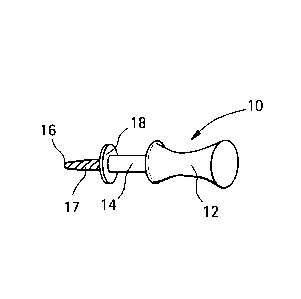
[0041] Referring now to Figs. 9A-9D, there is shown a disposable hand held
perforating device
constructed in accordance with the present invention and a disposable package
that may be
used to contain the hand held perforating device 10. The hand held perforating
device 10 is
comprised of a handle 12 for holding by the user and a shaft 14 extending
therefrom. The shaft
14 has a distal end 16 and a small drill 17. There is a stop 18 displaced a
predetermined distance
inwardly from the distal end 16. The handle 12 of device may be comprised of
plastic while the
small drill 17 may be made of metal (preferably titanium). The perforating
device 10 may be
13
CA 02770642 2012-02-09
WO 2011/019382
PCT/US2010/002202
provided in different lengths or widths. The predetermined length of the small
drill 17, in the
exemplary embodiment, can be 6, 8 or 10 mm while the thickness or diameter may
be between
1.5 or 2mm, however other lengths and diameters can be used. The hand held
perforating device
may be used to make perforations in the alveolar bone, as will be described
with respect to
Figs 11A-11C, of a known, predetermined diameter and depth.
[0042] In Fig. 9B, there is depicted a modified version of a hand perforating
device 20 that has
an abbreviated handle 22 that may be used to make perforations in the
aleveolar bone of a
patient in areas that cannot be accessed by the hand perforating device 10 of
Fig. 9A such as
areas of posterior teeth (second molars), or where the patient cannot fully
open the mouth. This
modified version of a hand held perforating device 20 may also be provided in
different lengths
and widths of the small drill 17.
[0043] The hand held perforating devices 10 and 20 can be provided to the user
in a disposable
package as shown in Fig. 9C. As seen in Fig 9C, there is a sealed container 24
that can contain a
hand held perforation device 10 or 20 in a sterilized format for one time
usage. This package has
a container body 26 and a removable cover 28. In Fig. 9D, it can be seen that
the cover 28 has
been partially peeled back for access to the components contained within the
container body 26.
[0044] In Fig. 10, there is illustrated a slow speed dental handpiece 30 or
other instrument
having a rotating chuck attachable to a perforating device 32 of the present
invention. The
means of attachment may be conventional with such dental handpieces such that
that the ,
perforating device 32 may be rotated at a relatively slow speed. As can be
seen, the perforating
device 32 has a circular shaft 34 and at the end of the shaft 34 is a small
drill 40 with a cutting
end. A stop 36 is located on the shaft 34 at predetermined linear distance
inwardly of a distal
tip 42 of the small drill 40 to stop the small drill 40 from penetrating the
alveolar bone more than
a predetermined depth. The small drill 40 may be provided in different lengths
and widths of
diameters, such as a length D of 4 or 6 mm and a width or diameter of 1.5 mm
or 2 mm.
[0045] Turning now to Fig. 11A-11D, there is shown schematic views
illustrating the use of the
devices of the present invention. In Fig. 11A, a normal alveolar bone 44 is
illustrated. The
alveolar bone 44 is part of the jaw bone that accommodates the teeth and which
is covered by
the gum 46 or gingiva.
14
CA 02770642 2012-02-09
WO 2011/01982
PCT/US2010/002202
[0046] Turning to Fig 11B, there is shown the alveolar bone 44 and gum 46 has
been perforated
directly by hand held perforating device 10 as described with respect to Fig.
9A. Stop 18
determines the depth of penetration of the perforation that the hand held
perforating device 10
will produce inside the bone.
[0047] Fig 11C and 11D demonstrate the steps in using a rotating perforating
device 32 that is
attached to the dental handpiece 30. As before with a hand held device, the
rotating perforating
device 32 has a shaft 33 having a distal end 35 and a small drill 40. As stop
36 is also provided
to function as a limiter to the depth of penetration of the small drill 40.
[0048] While it is not mandatory, it is may be preferable in certain cases
that a soft tissue punch
48 be used before application of a rotating device, see Fig. 11C, especially
in places where gum
tissue is loose. Application of the soft tissue punch 48 with a rotating
perforating device 32 may
prevent damage to gum. The soft tissue punch 48 can create an opening in the
gums of the
patient so that the later use of a rotating perforation device 32 does not
catch up in the gum
tissue with the drill 40 so that the drill 40 enters cleanly into the bone 44.
In areas where the
gum is firmly attached to the bone 44, application of soft tissue punch 48 may
not be necessary.
Following punching of the soft gum tissue, the rotating perforating device 32
can directly access
the bone 44 as illustrated in Fig. 11D.
[0049] The device may be provided in a kit. The kit may also contain one or
more of a
disposable local anesthetic carpule, and topical analgesic swabs, a depth
gauge probe, and an
illustrated detailed instruction manual.
[0050] Fig 12 demonstrates an inventive kit form that can be conveniently used
for performing
the methods of this invention. It is envisioned that the present kit can be
supplied to dentists or
orthodontists so that the doctor will have all of the components necessary to
carry out the
osteoperforation method of the present invention. This kit includes a
container having therein a
local anesthetic 50 (lidocaine HCL 2%), a topical anesthetic 52, a syringe for
application of local
anesthetic 56, short needles 54, soft tissue punch 48 and different length and
widths of hand held
perforating devices in disposable packages 24, short modification of hand
perforating devices
for access to difficult area in disposable packages 20 and different length
and widths of small
CA 02770642 2012-02-09
WO 2011/019382
PCT/US2010/002202
drills for application with dental handpiece 32. As can be seen, one or more
of the previously
listed components may be omitted in a particular kit. The kit may be contained
in a disposable
container similar, but different in dimensions, to that described with respect
to Figs. 9C and 9D.
[0051] As described elsewhere herein, the procedure may include the following
steps: first, a
topical anesthetic is applied in the desired area, followed by local
anesthetic injection using a
syringe and short needle. The anesthetic is used to deaden the tissue where
the perforation is to
be made. In majority of cases, using one standard hand held perforating device
should be
adequate, but if the patient has very dense alveolar bone, a strong device
such as rotatory
perforating device attached to dental handpiece will be helpful. In such
cases, a disposable
punch 48 can be used to facilitate the procedure.
100521 Those skilled in the art will readily recognize numerous adaptations
and modifications
which can be made to the devices used to carry out that method with will
result in improved
devices, yet all of which will fall within the scope and spirit of the present
invention as
defined in the following claims. Accordingly, the invention is to be limited
only by the
following claims and their equivalents.
Summary
100531 It is unclear whether corticotomy facilitates orthodontic tooth
movement by reducing
physical constraints or via a mechanism resembling that in bone response to
injury. Since
inflammation is an underlying mechanism, it is preferable to administer the
minimal injury
capable of eliciting an inflammatory response. Forty-eight rats were fitted
with closing coils
and subjected to either a 50 cN force to the maxillary first molar (0), the
same force after
implementation of a soft tissue flap (OF), force plus flap plus three
perforations of the
cortical plate mesial to the first molar (OFP), or no force (controls: C).
Perforations of
cortical bone resulted in increased inflammatory reaction as shown by RT-PCR
of RNA at 24
h. At 28 days post-treatment, micro-computed tomography, light and fluorescent
microscopy,
and immunohistochemistry revealed increased rates of tooth movement and bone
remodeling.
The increase in rate of bone remodeling extended beyond the first molar region
to the
adjacent alveolar bone. Shallow perforations of cortical bone are sufficient
to stimulate an
inflammatory response capable of accelerating bone remodeling and tooth
movement. The
16
CA 02770642 2012-02-09
WO 2011/019382
PCT/US2010/002202
procedure is easy to perform, minimizes side effects and discomfort, and
shortens recovery
time.
[0054] Corticotomy is sometimes used in difficult adult cases as an
alternative to
conventional orthodontic treatment or orthognathic surgery. (Kole, Oral Surg
Oral Med Oral
Pathol 1959; 12:515-529; Anholm, et al., CDA J 1986; 14:7-11; Gantes, etal. J
Periodontol
1990; 61:234-238; Wilcko, etal., In! J Periodontics Restorative Dent 2001;
21:9-19; Chung,
etal., J Clin Orthod 2001; 35:331-339) The ability to move teeth more rapidly,
it is claimed,
makes it possible to complete treatment in a shorter period of time. The
mechanism of this
action is not clear. Several authors have described rapid tooth movement
observed in
conjunction with corticotomy as movement by "bony block." (Kole, Oral Surg
Oral Med
Oral Pathol 1959; 12:515-529; Anholm, et al., CDA J 1986; 14:7-11) The
practitioner creates
a fissure through the cortical plate surrounding the tooth, in effect making
the tooth a block of
bone connected to surrounding bone only through the medullary bone. The tooth
is thus a
"handle" by which this block of bone can be moved. Others have compared the
effect of
corticotomy-facilitated orthodontics to the repair mechanism that is observed
following injury
of bone. (Wilcko, et al., Int J Periodontics Restorative Dent 2001; 21:9-19)
After bone
injury, accelerated bone turnover and a decrease in regional bone density have
been
described. (Frost, Henry Ford Hosp Med J 1983; 31:3-9; Frost, Clin Orthop
Re/at Res 1989:
294-309; Frost, Clin Orthop Relat Res 1989; 283-293; Yaffe, et al. J
Periodontol 1994;
65:79-83) While the mechanism of this accelerated bone turnover is not
completely
understood, it is reasonable to hypothesize that inflammation plays an
important role.
[0055] Inflammation can alter the physiology and structure of bone by
modifying the normal
pattern of remodeling through stimulation of bone resorption and formation.
The
inflammatory process can affect the recruitment of osteoclast precursors from
the circulation,
including their rate of maturation and their level of activity. Many cytokines
that promote
osteoclast formation and activation, such as IL-1, IL-6, and TNFcc, are
abundantly
synthesized by inflammatory cells. (Seidenberg, et al., Pharmacol Res 2004;
50:151-156;
Glantschnig, et al., Cell Death Differ 2003; 10:1165-1177; Bolander, Proc Soc
Exp Biol Med
1992; 200:165-170; Busti, et al., Pharmacotherapy 2005; 25:1566-1591) These
cytolcines
may thus be central to the biological response in accelerated tooth movement
during
corticotomy.
17
CA 02770642 2012-02-09
WO 2011/01982
PCT/US2010/002202
[0056] Understanding the mechanism by which corticotomy can facilitate
orthodontics tooth
movement is important because the surgical design of corticotomies has been
greatly
influenced by clinicians' mechanistic view of the underlying biological
process. If the
purpose of corticotomy is to weaken the bone around the tooth, then the
surgery should be
designed to create a loose block of bone around the tooth to be moved. If,
however, the goal
of the corticotomy is to accelerate the bone remodeling process by evoking an
inflammatory
response, then the geometry of the surgical cuts is not so crucial, and the
minimal injury that
activates the bone repair system would suffice requiring less traumatic
surgical design. The
current study demonstrates that limited shallow perforations of the buccal
cortical plate of the
maxilla are sufficient to accelerate the bone remodeling process and therefore
tooth
movement.
[00571While there are many case reports of the ability of corticotomy to
accelerate tooth
movement, the biological principle underlying this phenomenon has been
previously unclear.
We used a rat model and created three shallow cortical perforations, mesial to
the first molar,
to elicit an inflammatory response. The rat is considered a good experimental
animal model
for the study of bone biology and physiology. (Frost, Henry Ford Hosp Med Bull
1965;
13:161-172; Tran, J Pharmacol 1982; 13:495-499; Vignery, etal., Anat Rec 1980;
196:191-
200.) The biomechanical system used in this study to apply orthodontic force
to the molar is
also well established. (King, etal., Am J Orthod Dentofacial Orthop 1991;
99:456-465;
Williams, etal., Biomaterials 1984; 5:347-351)
100581The demonstration that inflammation is the key player in controlling
rate of tooth
movement is based in part on the observation that application of
antiinflammatory drugs can
reduce tooth movement. (Arias, etal., Am J Orthod Dentofacial Orthop 2006;
130:364-370;
Chao, et al., Acta Anat (Basel) 1988; 132:304-309) Additionally, studies of
knockout mice
deficient in IL-1 and TNFa receptors showed a slower rate of tooth movement in
response to
orthodontic forces. (Kitaura, et al., J Dent Res 2008; 87:396-400; Jager, et
al., Eur J Orthod
2005; 27:1-11) These observations are also in harmony with studies showing
that application
of orthodontic force, regardless of magnitude, can stimulate an inflammatory
response.
(Arias, etal., Am J Orthod Dentofacial Orthop 2006; 130:364-370; Chao, etal.,
Acta Anat
(Basel) 1988; 132:304-309; Kitaura, etal., J Dent Res 2008; 87:396-400;
Krishnan, etal., Am
18
CA 02770642 2012-02-09
WO 2011/019382
PCT/US2010/002202
J Orthod Dentofacial Orthop 2006; 129:469 e461-432; lino, et al., Am J Orthod
Dentofacial
Orthop 2007; 131:448 e441-448; Garlet, et al. Eur J Oral Sci 2007; 115:355-
362; Kawasaki,
et al., Orthod Craniofac Res 2006; 9:137-142; Ren, et al., J Periodontol 2007;
78:453-458;
Mermut, etal., Angle Orthod 2007; 77:135-141) During early stages of tooth
movement,
there is an initial inflammatory response phase, evidenced by an increase in
vascular
permeability and cellular infiltration of lymphocytes, monocytes, and
macrophages. (Rygh, et
al., Am J Orthod 1986; 89:453-468) High concentrations of inflammatory
cytokines such as
IL-1, IL-2, IL-3, IL-6, IL-8, TNFa, IFNy, and osteoclast differentiation
factor (ODF) have
been found in the gingival crevicular fluid surrounding moving teeth. (Garlet,
et al., Eur J
Oral Sci 2007; 115:355-362; Kawasaki, et al., Orthod Craniofac Res 2006; 9:137-
142; Ren,
et al., J Periodontol 2007; 78:453-458; Mermut, et al. Angle Orthod 2007;
77:135-141,
Alhashimi, etal., J Interferon Cytokine Res 2000; 20:7-12)
[00591The present data demonstrates that limited and shallow perforations of
the cortical
bone can significantly increase the inflammatory response. Increase in
inflammation was
demonstrated not only at the histological level by vascular invasion and
infiltration of
inflammatory cells, but also at the gene level by a significant increase in
the expression of
several cytokines and their receptors. Indeed, markers of lymphocytes (CCL20,
CCR1 (Kao,
etal., J Immunol 2005; 175:6676-6685; Sallusto, etal., J Exp Med 1998; 187:875-
883; Han,
etal., Glia 2000; 30:1-10)), T cells (LFa, IL-3, CCL5, CCR5, CX3CR1, IL-18rb,
IL-Irl
(Schneider, etal., Immunol Rev 2004; 202:49-66; Khapli, etal., J Immunol 2003;
171:142-
151; Xu, etal., Ann Acad Med Singapore 2007; 36:91-95; Ito, etal. J Immunol
1999;
162:4260-4265; Lean, et al. J Cell Biochem 2002; 87:386-393)), monocytes (IL-
1, IL-6, 1111,
IL-18, IL-6ra (Arend, etal., Immunol Rev 2008; 223:20-38; Adachi, etal., Biol
Pharm Bull
1994; 17:1554-1560; de Sa AR, et al., Oral Surg Oral Med Oral Pathol Oral
Radio! Endod
2003; 96:356-360; Dienz, et al., Clin Immunol 2009; 130:27-33; Bai, et al.,
Tissue Antigens
2007; 70:390-397; Bossu, etal. J Neurol Neurosurg Psychiatry 2007; 78:807-811;
Jang, et
al. Clin Exp Rheumatol 2005; 23:S59-63; Lean, et al., J Cell Biochem 2002;
87:386-393;
Knupfer, et al., Immunol Cell Biol 2008; 86:87-91; Yamamoto, et al., J
Periodontal Res
2006; 41:554-559; Leng, et al., Int J Biochem Cell Biol 1997; 29:1059-1062)),
and
macrophages (IL-1, IL-6, IL-11, IL-18, CCL9, CCL12, CCR5, IL-6ra (Arend,
etal., Immunol
Rev 2008; 223:20-38; Adachi, etal. Biol Pharm Bull 1994; 17:1554-1560; de Sa
AR, etal.,
Oral Surg Oral Med Oral Pathol Oral Radio! Endod 2003; 96:356-360; Bai, et
al., Tissue
Antigens 2007; 70:390-397; Yamamoto, etal., J Periodontal Res 2006; 41:554-
559; Leng, et
19
CA 02770642 2012-02-09
WO 2011/019382
PCT/US2010/002202
al., Int J Biochem Cell Biol 1997; 29:1059-1062; Hinton, etal., Am J Orthod
1986; 89:492-
498)) were all found elevated in the OFP group in comparison to the 0 group 24
h after
initiation of the experiment, suggesting substantial differences in the
inflammatory response.
In addition, a significant increase in both CCL2 (monocyte chemoattractant
protein-1
(Piemonti, et al., Diabetes 2002; 51:55-65)) and CCR2 (receptor for CCL2
(Luster, N Engl J
Med 1998; 338:436-445; Shireman, J Vase Surg 2007; 45 Suppl A:A48-
56))¨produced and
expressed in endothelial cells, vascular smooth muscle cells, tubular
epithelial cells,
lymphocytes, and monocyte/macrophages (Piemonti, et al., Diabetes 2002; 51:55-
65)¨ .
confirm the extensive and massive vascular invasion observed in the OFP group.
100601The discovery that an increase in inflammation through minimal bone
perforations
accelerated the rate of bone remodeling is in agreement with previous reports
that an increase
in inflammation during bone injury is accompanied by an accelerated rate of
bone
remodeling. (Frost, Henry Ford Hosp Med J1983; 31:3-9; Frost, Clin Orthop
Re/at Res
1989; 294-309; Frost, Clin Orthop Relat Res 1989; 283-293; Yaffe, etal., J
Periodontol
1994; 65:79-83; Shih, etal., Bone 1985; 6:377-379. The present data
demonstrate that the
increase in bone remodeling rate is not limited to the area of the loaded
tooth, but extends to
the tissues surrounding adjacent teeth. This generalized increase in bone
turnover was
accompanied by osteopenia, as reflected by a decrease in bone density of the
entire
hemimaxilla.
[0061] Higher level of expression of cytokines and their receptors is
important, since it has
been shown that inflammatory cytokines play an important role in recruitment
of osteoclasts
and activation of the bone remodeling machinery (Alhashimi et al., J
Interferon Cytokine Res
(2000) 20(1):7-12; Krishnan, et al., J Dent Res (2009) 88(7):597-608; Ren,
etal., Eur J Oral
Sci (2008) 116(2):89-97). The fact, that the number of osteoclasts and the
bone remodeling
rate was higher in OFP group in comparison with 0 and OF group, supports the
possible role
of inflammatory cytokines in recruiting osteoclasts into the area.
[0062] Similar to previous studies (Verna eta!, Bone (1999) 24(4):371-9), the
present data
demonstrate that the increase in bone remodeling rate is not limited to the
area of the loaded
tooth, but extends to the tissues surrounding adjacent teeth. This generalized
increase in bone
turnover is accompanied by osteoporosity, as reflected by a decrease in bone
density around
CA 02770642 2012-02-09
WO 2011/019382
PCT/US2010/002202
all upper left molars. While a limited number of osteoperforations have a
generalized effect,
the effect is not robust enough to cross to the contra-lateral side.
[0063] Since bone remodeling controls the rate of tooth movement, the increase
in rate of
bone remodeling and osteopenia in response to bone perforations may explain
the increase in
rate and magnitude of tooth movement demonstrated by these data. Our results
further
indicate that the site of the perforations that set this process in motion may
not need to be in
the vicinity of the tooth to be moved.
[0064]The present results were obtained using perforations that were very
small and limited
(only 3). Therefore the majority of the cortical bone remained intact. In
addition, the
perforations were placed far away from the tooth, and could still be observed
at the end of the
study with remaining bone (about 4 mm) between perforations and the moved
tooth. These
results further suggest that the perforations do not need to be in the close
vicinity of the tooth
to be moved in order to accelerate the rate of movement.
[0065] Inflammation can be beneficial by accelerating bone remodeling and
tooth movement,
however, if uncontrolled it may also have a destructive effect on the
periodontium and tooth
structure. Root resorption may be affected by osteoperforation. While
extensive injury to the
cortical plate bone, also referred to as corticotomies, is currently being
used to accelerate
orthodontic tooth movement in private practice, the present data indicate that
this approach
could be simplified to minimize deleterious side effects. Therefore, flapless
minimal cortical
perforations may be used as a means of fine tuning inflammation levels for
enhanced tooth
movement, enabling orthodontists to provide more efficient treatment to their
patients.
[0066] Understanding the biological principles of corticotomy not only
facilitates simplifying
the procedure making it more practical for clinicians to employ, but also
offers other
possibilities. If inducing injury accelerates bone remodeling, then extraction
of teeth should
have a similar effect. Orthodontists may schedule extractions that are part of
the treatment
plan close to the time of major tooth movement. It is also important to
observe that
inflammation is a two-edged sword¨that while it can work to the benefit by
accelerating
bone remodeling and tooth movement, it may also, if uncontrolled, exert a
destructive effect
on the periodontium.
21
CA 02770642 2012-02-09
WO 2011/01982
PCT/US2010/002202
Orthodontic Method
[0067] Orthodontic appliances are installed on the teeth to be moved to exert
force on the
teeth toward the desired positions. Any orthodontic appliances or auxiliaries
either fixed or
removable, installed on teeth may be used in accordance with this invention,
and for any
orthodontic, orthopedic or surgical purpose.
[0068] The basic principles of the orthodontic method of this invention are
applicable in
retraction cases and expansion cases. Retraction cases may also require that
teeth be
expanded, as well, as indicated above. Additionally, since retraction cases
normally require
the extraction of teeth and move teeth in the opposite direction from the
movement of teeth in
expansion cases, retraction cases are handled somewhat differently. The
retraction devices
used in the present methods may be constructed out of components and materials
used by
those skilled in the art to construct orthodontic palatal expansion devices
such as shown in
U.S. Patent No. 4,347,054 Kraus et al., U.S. Patent No. 4,354,832, Wallshein,
U.S. Patent
No. 4,433,956, Witzig, U.S. Patent No. 4,482,318, Forster, U.S. Patent No.
5,281,133,
Farzin-Nia, U.S. Patent No. 5,002,485, Aagesen, U.S. Patent No. 5,439,377,
Milanovich,
U.S. Patent No. 5,472,344, and Binder et al. U.S. Patent No. 4,483,674. The
design and
nature of fixed rapid palatal expanders are discussed by Anthony Viazis
entitled Atlas of
Orthodontics: Principles and Clinical Applications, published by W. B.
Saunders Company,
pp. 205-13, 1993 and by James A. McNamara, etal., entitled Orthodontic and
Orthopedic
Treatment in the Mixed Dentition, published by Needham Press, pp. 131-44,
1993. The
design and nature of removable expanders are described by T. D. Foster
entitled A Textbook
of Orthodontic, published by Blackwell Scientific Publications, 2nd Edition,
pp. 246-61,
1982 and by William R. Proffit, et al., Contemporary Orthodontics, published
by The C. V.
Mosby Company, pp. 272-86.
[0069] Movement processes related to the configuration of palatal expanders
are described by
Handelman, Angle Orthodontic 67(4): 291-305 and a study by Bishara etal., Am.
J. Orthod.
Dentofac, Orthop., 91(1): 3-14, 1987. None of these expanders or physiological
processes
involves the same type of orthopedic movements that we are accomplishing with
the
retraction devices of this invention. Conventional expansion screws start from
a closed
position in a side-to-side position in a patient's jaw. Upon adjustment, two
or more sections of
these screws are spread apart, which in turn widens or spreads apart teeth or
jaws. The design
22
CA 02770642 2012-02-09
WO 2011/019382
PCT/US2010/002202
of these expansion screws is not to pull teeth, sections of teeth, sections of
jaws or jaws
together as is required from the retraction devices of our invention.
EXAMPLE 1
MATERIALS AND METHODS
Animal study
[0070] Forty-eight adult male Sprague-Dawley rats (average body weight of
400g, 120 days
of age) were housed and treated according to a protocol approved by the New
York
University Institutional Animal Care and Use Committee. Animals were divided
into four
groups (12 rats per group): control, which received coil spring without
activation (C),
orthodontic force applied to the spring (0), orthodontic force and soft tissue
flap (OF), and
orthodontic force, soft tissue flap, and shallow perforations of the buccal
cortical plate (OFP).
The health status and body weight of the rats were evaluated daily and no
significant
differences were observed between groups. From each group, 4 animals were used
for gene
expression studies, 4 for microCT and fluorescent studies and 4 for
demineralized
histological studies. Procedures were performed on one side of the maxilla,
which allowed
the contralateral side to be used as an additional control.
Surgical procedure
100711 On day 0, all groups were anesthetized with intraperitoneal injection
of ketamine-
xylazine (0.09 mL/100g) and anesthesia verified by lack of response to toe-
pinch. All groups
were fitted with 50 cN Sentalloy closing coils (GAC International) tied at
both ends to holes
drilled in the maxillary incisors and left maxillary first molar with 0.008
in. ligature wire; the
coil was activated in groups 0, OF, and OFP, but not in C group. In the OF and
OFP groups,
a soft tissue flap was raised around the left first molar. Flaps were sealed
with cyanoacrylate
tissue adhesive (Vetbond, 3M). In the OFP group, the animals received three
shallow
perforations, approximately 0.25 mm in diameter (depth of 0.25 mm), 5 mm
mesial to the left
first molar using a round bur and hand piece. Animals were checked under
general anesthesia
twice weekly, and any springs requiring retying (mostly due to continuous
eruption of the
maxillary incisors) were adjusted. Bone labeling by intraperitoneal injection
of calcein (15
mg/kg) was performed on days 0 and 26 and by demeclocycline (25 mg/kg) on day
14.
Animals were sacrificed by CO2 narcosis on day 28 and hemimaxillae collected,
fixed in
formaldehyde for 48 h before storage in 70% ethanol.
23
CA 02770642 2012-02-09
WO 2011/019,382
PCT/US2010/002202
Micro-CT imaging
100721 Hemimaxillae were scanned using a Scanco MicrocCT ( CT40, Scanco
Medical,
Basserdorf, Switzerland). Results were analyzed utilizing CT V6.0 software on
the HP open
platform (openVMS Alpha Version 1.3-1 session manager). The area extending
from the
coronal to the apical root third was analyzed for bony changes. Maxillae were
analyzed in
fixed coronal and sagittal zones. The ratio of bone volume to total volume
(BV/TV) was
calculated using a threshold of 275.
Histology and immunohistochemistry
100731 Hemimaxillae were collected and fixed in 10% phosphate buffer formalin
and
demineralized in a sodium formate (6.8%) and formic acid (50%) solution for 6-
8 weeks.
Following demineralization, specimens were dehydrated in alcohol series,
embedded in
paraffin, and 5- m-thick sections cut and stained with hematoxylin and eosin
(H&E).
Consecutive specimens were immunostained using antibodies for tartarate-
resistant acid
phosphatase (TRAP; Zymed antibodies, Invitrogen, Carlsbad, CA), a marker of
osteoclasts,
and Vectastain ABC kit (Vector Laboratories, Burlingame, CA) according to the
manufacturer's instructions. As negative control consecutive sections were
exposed to pre-
immune serum. Stained sections were scanned on Scan Scope GL series optical
microscope
(Aperio, Bristol, UK) at 20x magnification. Osteoclasts were defined as TRAP-
positive
multinuclear cells on the bone surface. The area around the mesio-palatal root
of maxillary
first molar was divided into mesial and distal halves and osteoc lasts in the
mesial half were
counted. Data were expressed as the number of TRAP positive cells per 1000 m2
in the area
of PDL and adjacent alveolar bone, excluding the marrow cavities and blood
vessels.
For fluorescent microscopy, after formalin fixation specimens were washed
overnight in
running water, dehydrated in alcohol, cleared in xylene, and embedded in
methyl
methacrylate according to the method of Erben (Erben, J Histochem Cytochem
(1997)
45(2):307-13). The samples were sectioned at 5-7 um thickness on Reichert-Jung
Ultracut E
microtome and viewed under fluorescent microscopy (Nikon Microscopy, NIS-
Elements
software).
24
CA 02770642 2012-02-09
WO 2011/019382
PCT/US2010/002202
RT-PCR ANALYSIS
100741 For RNA extraction, 4 animals from each group were sacrificed by CO2
narcosis at 24
hours and the hemimaxillae dissected and frozen in liquid nitrogen. Isolation
of total RNA
was performed using TRIZOL reagent (Life Technologies, New York, NY), and RNA
cleanup was performed using RNeasy Mini Kit (Qiagen Sciences, Valencia, CA) as
described
before (Serafim et al., 2009). All equipment and tools were cleaned with
RNaseZap (Sigma,
St Louis, MO). Ninety-two inflammatory cytokines and cytokine receptor genes
were
analyzed using primers specific for rat genes (see online appendices for list
of genes), using
QuantiTect SYBR Green RT-PCR kit (both Qiagen, Valencia, CA) on a DNA Engine
Optican 2 System (MJ Research, Waltham, MA). Each mRNA specimen was tested
three
times. Relative levels of mRNA were calculated and normalized to the level of
GAPDH and
acidic ribosomal protein mRNA.
Statistical analysis
[0075] Significant differences between test groups and controls were assessed
by analysis of
variance (ANOVA). Pairwise multiple comparison analysis was performed using
Tukey's
post hoc test. Two-tailed p-values were calculated; p < .05 was set as the
level of statistical
significance.
RESULTS
Osteoperforations increase the rate of tooth movement
[0076] Coil springs were used for mesial movement of the first maxillary molar
crown (Fig.
1A). Three shallow perforations were made in the cortical bone, 5 mm mesial to
the molar as
depicted in figure 1B. At 28 days, the average crown movement (measured in 12
rats per
group) was 0.29 mm in the 0 and OF groups (Fig. 1C), significantly different
from control (p
<.05). The OFP group showed the greatest mean tooth movement, 0.62 mm, which
was
significantly higher (p < .05) than that of C, 0, and OF groups (Fig. IC).
Osteoperforations increase expression of inflammatory cytokines
[0077] Expression of 92 different cytokines/cytokine receptors was studied by
RT-PCR, 24
hours after force application. The expression of 37 cytokines/cytokine
receptors increased
more than 2-fold in the left maxilla of rats in the 0, OF and OFP groups when
compared to
the C group (data not shown). Differences between 0 and OF group were not
statistically
CA 02770642 2012-02-09
WO 2011/019382
PCT/US2010/002202
significant. From these 37 cytokines, expression of 21 cytokines/cytokine
receptors was
statistically higher in the OFP group than in the 0 or OF groups (p<0.05)
(Fig. 2), with 8
cytokines showing a 1.6 to 2.7 fold increase (Fig 2A), 5 chemokines showing a
1.6 to 2.8 fold
increase (Fig 2B), and 8 receptors showing a 1.7 to 2 fold increase in
expression (Fig 2C). All
cytolcines/cytokine receptors expressed in the OFP group were also expressed
in 0 or OF
groups. Expression of cytokines in the contra-lateral side of all groups
showed no statistically
significant differences from group C (data not shown).
Osteoperforations increase osteoclast activity
100781 In both 0 and OF groups, application of the orthodontic force
stimulated an increase
in alveolar bone resorption in the direction of tooth movement and
consequently an increase
in PDL thickness (Fig. 3A, top row). The OFP group showed increased alveolar
bone
resorption in the direction of tooth movement (Fig. 3A, top row).
Immunohistochemical
staining for TRAP positive osteoclasts (Fig. 3B) revealed an increase in
osteoclast number in
the OFP group, compared to the OF, and 0 groups (Fig. 3A, bottom row).
Quantitative
analysis of osteoclasts in the pressure side (mesial) of alveolar bone
adjacent to mesio-palatal
root of maxillary first molar demonstrates a 3 fold increase in number of
osteoclasts in
comparison with 0 and OF group (p<0.05) (Fig 3C). The difference between
number of
osteoclasts in 0 and OF group was not statistically significant.
Osteoperforations increase the rate of bone remodeling and generalized
osteoporosity
100791 Sagittal sections of specimens viewed under fluorescent microscopy
showed more
prominent fluorescence in the OFP group (Fig. 4A), indicative of heightened
bone
remodeling activity. MicroCT quantification was used to evaluate the effect of
osteoperforations on induction of osteoporosity during tooth movement.
Comparison of the
OFP group with the other groups revealed significant findings on all planes of
analysis (Fig.
4B). Bone volume fraction (BV/TV) levels in the OFP group were significantly
lower (p <
.05, ANOVA) than in the C, 0, or OF groups (see online appendices, Table I).
BV/TV
fraction in control group was on average as high as 82% around first maxillary
molar, while
in the OFP group these values decrease to 33%. Both 0 and OF groups also
exhibited
statistically significant changes in BV/TV levels (p < .05, ANOVA) when
compared to the C
group. Interestingly, in comparison with other groups, the BV/TV fraction in
the OFP group
decreased significantly (p<.05) around all left maxillary molars (33% to 35%).
This effect
26
CA 02770642 2012-02-09
WO 2011/019382
PCT/US2010/002202
was limited to the left hemimaxillae and no change in BV/TV fractions was
observed in
contra-lateral hemimaxilla (p>.05).
Osteoperforations induced generalized osteopenia
100801 MicroCT quantification was used to evaluate the effect of
osteoperforations on
induction of osteopenia during tooth movement. Comparison of the OFP group
with the other
groups displayed significant findings on all planes of analysis (Fig. 5). Bone
volume fraction
(BV/TV%) levels in the OFP group were significantly lower (p < .05, ANOVA)
than that in
the C, 0, or OF groups, with an extreme of 31.1%. Both 0 and OF groups also
exhibited
statistically significant changes in BV/TV% levels (p < .05, ANOVA) when
compared to the
C group. Interestingly, bone volume fractions in the OFP group were similar in
all regions of
the rat maxilla, in contrast with the other groups, where a gradient could be
observed from the
mesial to the distal region. Localized osteoperforations resulted in
generalized jaw
osteopenia.
CONCLUSIONS
[0081] The current results help elucidate the relation between bone injury,
inflammation, and
tooth movement, and these results demonstrate that the application of minimal
injury to the
maxilla appears to be sufficient to set in motion an inflammatory cascade that
allows
accelerated movement of teeth during orthodontic treatment.
EXAMPLE 2
Expected result of this study
100821 We expect the flapless shallow perforations that we propose to make to
be safe for
orthodontic patients. We expect that increasing the local inflammatory
response will enhance
the rate of tooth movement with no deleterious side effects. We anticipate the
elimination of
highly invasive surgery as normally required for patients with skeletal
moderate class II
malocclusion.
Study design
[00831 The subjects will be orthodontic patients with class II division I
malocclusion. All
subjects will have the upper 1st premolars extracted and placement of TAD
mesial to upper
27
CA 02770642 2012-02-09
WO 2011/019382
PCT/US2010/002202
2nd premolar. This is a randomized, single blind, single-center, clinical
trial. The
randomization process used in this study is stratified randomization. Group A
control
patients will not receive any osteoperforations and Group B experimental
patients will
receive right side or left side osteoperforations. The subjects will be
assigned in the order
they visit the clinic, for example, using ABABAB. Because of inclusion and
exclusion
criteria, there are no strong confounders.
[0084] This will be a single-blind study, only for the investigator(s).
Subjects and the
resident orthodontist administering the treatment will know the group
assignment due to the
additional procedure being performed on the experimental group. Orthodontists
that are
performing the research procedure are investigators in the study. However,
casts will be
measured to evaluate the rate of tooth movement by investigator who did not
treat subjects.
Therefore, casts will be measured without the information of subjects'
assignment. This
approach will minimize investigator bias.
[0085] The variables in this study will be levels of inflammatory markers and
the rate of tooth
movement. At each visit, an impression to evaluate the rate of tooth movement
will be taken
by measuring casts. Crevicular fluid samples will also be taken from the
patients at each visit
for evaluation of inflammatory markers using a protein array approach. In
addition we will
measure probing depth (PD), PI (Plaque Index) and gingival index (GI) to
assess periodontal
status at each visit. At start of each visit after osteoperforation, patients
will be evaluated for
level of pain or any discomfort. In this regard, patient will be asked to rate
on scale of 1 to 10
the magnitude of pain or other sort of discomfort.
Number of Subjects
[00861A total sample size of 20 patients is being requested for this pilot
study with the
objective of establishing the safety of the procedure and understanding the
levels and
variation of inflammatory markers in this patient population. To calculate the
sample size a
power analysis assuming a type I error frequency of 5% was performed, setting
the power of
the statistical test at 90% (P=0.9, 0=0.1) using results from published data
on tooth
movement (Verna et al., 1999, Bone: 24, 371) as a guide using the following
formula
N=2.(E./8)2. (ta,v +t2(1-p),v)2
28
CA 02770642 2012-02-09
WO 2011/019382
PCT/US2010/002202
Where n=the sample size, e=the population standard deviation, d= the
difference that is
desired to detect (in this case 50% increase in regular rate of tooth movement
¨ 1.5mm),
a=significance level, v= the degrees of freedom, ta,v= the t value
corresponding to a and v,
and P= the desired statistical power. Base on this calculation, a sample size
of 14 is
necessary (7 per group). Considering attrition (for example patients move
away, or do not
continue treatment), a sample size of 10 per group should be attained.
Age of Subjects
[0087] Subjects will be 18 - 40 years of age. This portion of the population
is selected
because a large portion of patients undergoing orthodontic treatment are
within this age
range.
Gender of Subjects
[0088] Male patients are included because of the findings in current
literature, which suggest
that changes in levels of sex hormones can have a significant affect on the
rate and range of
tooth movement and bone remodeling. (Zittermann, et al., J Clin Endocrinol
Metab 2000;
85:95-101) Researchers have suggested that orthodontic tooth movement will
vary
throughout the estrous cycle. (Haruyama, etal., J Dent Res 2002; 81:406-410)
Males are
selected so that confounding variables that are not related to the research
question are
eliminated.
Racial and Ethnic Distribution
[0089] Caucasians will be enrolled in this study. Caucasians that come to the
orthodontic
clinic are selected to eliminate confounding variables that are not related to
the research
question. Baseline levels of cytokines in individuals of different races and
ethnicities can
vary.
Selection criteria
[0090] All subjects will be in good general health, and none will have
received periodontal
therapy or medication during the past 6 months. Participants will have no
history of systemic
diseases, periodontal diseases, gingivitis disease or untreated caries. They
will not be on any
medication that could affect the level of inflammation, such as chronic
antibiotics, phenytoin,
cyclosporin, anti-inflammatory drugs, systemic corticosteroids, or calcium
channel blockers.
29
CA 02770642 2012-02-09
WO 2011/019382
PCT/US2010/002202
[0091] Orthodontists will perform a periodontal examination at the beginning
of each visit,
including probing depth (PD), plaque index (PI) and gingival index (GI)
assessment.
[0092] To avoid the contamination of crevicular fluid samples with blood, GI
and PD will be
measured after these collections. All periodontal disease measurements will be
performed in
four quadrants. PD levels will be measured throughout entire mouth with a
periodontal probe
calibrated in millimeters each month.
Patient oral hygiene control
[0093] If subjects cannot meet inclusion criteria due to poor oral hygiene
they will undergo
cleaning and oral hygiene education program by hygienist. When oral hygiene
improves and
they meet all inclusion criteria (PD is<4mm, GI <1, and PI =1), they can be
enrolled into this
study. During the study patients will receive oral hygiene instructions and
cleanings by
hygienist, at each visit.
Inclusion Criteria
[0094] 1. Caucasian, male subjects ages 18 ¨ 40 years old who have complete
adult dentition,
excluding 3"I molars, in a skeletal class II division I malocclusion
[0095] 2. Subjects do not have any systemic diseases
[0096] 3. PD is<4mm, GI 1, and PI 1
[0097] 4. If any caries present, patient will be referred to dentist for
treatment and
maintenance before beginning treatment
[0098] 5. English speaking
Exclusion Criteria
[0099] 1. Subjects who have taken any antibiotics or periodontal TX in the
previous 6 months
[01001 2. Subjects who have concomitant medical therapy
[0101] 3. Subjects with extreme skeletal class II malocclusion: overjet >10mm,
Pg-
Nper>18mm, ANB>7, SN-GoGn> 38
CA 02770642 2012-02-09
WO 2011/019382
PCT/US2010/002202
[0102] 4. Female subjects
[0103] 5. Individuals who are not Caucasian
Methods and Procedures
[0104] This is a randomized, single blind, single-center, clinical trial. The
subjects will
orthodontic patients with class II division I malocclusion. The subjects will
be randomized
amongst the two study arms. One treatment arm will receive osteoperforations
on the right or
left side. The second treatment arm will not receive any osteoperforations.
All subjects will
have the upper 1st premolar extracted and placement of TAD mesial to upper 2nd
premolar.
At each visit, we will take an impression to evaluate the rate of tooth
movement. We will also
collect crevicular fluid samples from the patients at each visit for
evaluation of inflammatory
markers. The PI, Co-PI, and the student researchers will perform the cytolcine
analysis. We
will also perform an assessment of PD, GI, and PI.
[0105] Additional Procedures and Tests that will be performed exclusively for
research
purposes are:
[0106] For the first 6 months Physical data (weight and height), crevicular
fluid samples, and
assessment of PD, GI, PI will be collected.
[0107] 6 months after extraction we will initiate treatment of topical
anesthesia and
placement of 3 tiny holes in the bone that surrounds upper canine (right or
left). The
following 6 months, at every visit physical data (weight and height),
crevicular fluid samples,
impressions, and assessment of PD, GI, PI will be collected.
Time for recruitment
[0108] A total sample size of 20 subjects is being requested for this pilot
study. A chart
review was conducted at the New York University College of Dentistry
Department of
Orthodontics. 4 patients every week that are seen in the Department of
Orthodontics will
meet the selection criteria for the study. It is estimated that 20% of the
patients (32 patient)
will be willing to participate in the study, therefore we expect it will take
up to 1 year to
recruit 20 patients.
31
CA 02770642 2012-02-09
WO 2011/019382
PCT/US2010/002202
Evaluation for Recruitment
101091 Periodontal evaluation of prospective subjects (Class II, Division 1
patients free of
systemic disease) will be performed by orthodontists in the Clinic of the
Department of
Orthodontics and include (based on American Association of Peridontists'
guidelines)
including a full mouth series, and a full mouth probing depth (PD), plaque
index (PI) and
gingival index (GI) assessment.
Detailed Technical Plan
Phase I:
Physical data
101101 Every month, the weight and height of the subject will be measured and
recorded. The
purpose of recording these measurements is to reduce confounders.
Sampling Gingival Crevicular Fluid (GCF)
101111 GCF samples will be collected from each patient at each visit to
evaluate
inflammation levels. GCF samples in both the experimental and control groups
will be
collected between 10:00 am and 12:00 pm. Before any treatment begins, sample
of crevicular
fluid will be taken from maxillary mesial and distal upper canines that are
affected by the
retraction. Samples will be taken bilateral mesial and distal to upper canine.
Prior to
sampling, we will remove supragingival plaque. Cotton rolls will isolate the
regions where
GCF samples will be taken. The teeth and marginal gingiva will be dried with
air before
sampling. Filter paper strips will be inserted lmm below the gingival margin
into the mesio
labial and mesio labial crevices surrounding each tooth for 30 seconds. About
1.2 lit to 3 L
of GCF will be collected from each side of the tooth on the paper strip. This
will provide
about 1,200,000pg to 3,000,000pg of GCF that will be diluted to obtain the 50
to 100 L of
sample required for analysis using glass slide-based arrays. GCF samples will
be stored in -
70 C refrigerator in a laboratory on 10th floor, room #1038 NYU College of
Dentistry that
will be locked.
Taking impression
101121 Impression will be taken with Alginate. The procedure of taking
impression will be
done before wire placement. After taking impression, immediately Anhydrite
(CaSO4 ) will
32
CA 02770642 2012-02-09
WO 2011/019382
PCT/US2010/002202
be poured over the impression. The casts will be labeled (patient number and
date) and stored
and locked in a laboratory.
X-RAY
101131 Pen-apical x-ray will be taken at enrollment, the day of the TAD
placement and
perforation, 3 and 6 months later evaluate the bone situation, estimate the
placement of TADs
and perforation, and evaluate bone after tiny perforations and tooth movement.
101141 The subjects' x-ray exposure will be carefully monitored and maintained
at safe
levels. This study will require no additional x-ray exposure than what would
be required with
traditional orthodontics treatment. The effective doses that subjects will be
exposed will be
well within the annual effective dose limit of 1 mSv. The effective dose for a
lateral
cephalometric x-ray is 0.002-0.003 mSv. The effective dose for a periapical x-
ray is 0.001-
0.008 mSv. The effective dose for a panoramic x-ray ranges from 0.002-0.03
mSv. (Whaites,
Dental Radiography and Radiology, London: Churchill Livingstone Elsevier,
2007;
Association AD. Oral Health Topics A to Z; 2009) The estimated effective dose
for a
bitewing dental radiograph is 0.038. Throughout the study the patient will
have a panoramic
x-ray (0.03 mSv) at the beginning of treatment. In addition the subject will
have bitewing
dental radiographs (0.038 mSv) taken every 3 months. The total effective dose
exposure will
be much less than the recommended effective dose limit of lmSv.
Initial Treatment
101151 Orthodontic treatment will begin with alignment of teeth into proper
position with
subsequent distal translational movements until proper positioning of teeth
has been
achieved. Orthodontic appliances consisting of Innovation brackets (GAC
International) will
be bonded on upper incisors, lateral incisors, 2nd premolars, 1st molars and
2'd molars. Due to
their occlusion (Class II Division I) we will treat upper arch first.
Occasionally bands will be
cemented on maxillary first molars. The ordinal wire sequence will consist of
first using
0.016NiTi, then 0.016x0.022NiTi archiwires, for initial brackets leveling. And
after that,
0.016x0.022 stainless steel will be used for canine retraction.
Each wire will be used for 2 months.
Phase 2
33
CA 02770642 2012-02-09
WO 2011/019382
PCT/US2010/002202
101161 The PI, Co-PI, or the residents assigned to the study will evaluate
these conditions of
subject's occlusion before initiating phase 2. The wire must be, 0.016x0.022
stainless steel
and no tooth rotations may bepresent.
Anesthesia
101171 Before tiny perforations procedure, orthodontists will deliver the
local anesthetic. The
local anesthetic that will be is used is lidocaine with 1:100,000 epinephrine.
The dentist will
administer an inferior alveolar nerve block (IANB) and a buccal nerve block to
anesthetize
the tooth to be treated. A 27-gauge, long needle will be used and after
multiple negative
aspirations, 1.5 ml of anesthetic will be deposited. More anesthetic will be
delivered and
documented as needed. Patient will be given a complete cleaning at every visit
after all
measurements.
TADs procedure
[0118] The mini screw implants (TADs) (GAC International) will be prepared
(see
appendix). The entire procedure will be carried out under profuse saline
irrigation at room-
temperature. After procedure of anesthesia, we will insert implants between
upper buccal 1st
and 2nd premolar areas.
Perforation
101191 Surgical perforation procedure will be performed following TADs
placement. In the
alveolar bone, three small holes will be made mesial to 2nd premolar,
perpendicular to the
tooth. These 3 tiny holes will form a line (facial to palatal) along the bone.
Appliance
101201 The coil (GAC International) will be connected to an attachment on
canine bracket to
the TAD. The force will be adjusted to 100g at all visits.
Orthodontic Treatment
101211 Before the engagement of appliance, 0.016x0.02255 wire will be in place
for two
months. This wire will be the base wire for the retraction of canine. This
arch wire is of
reasonable for strength.
34
CA 02770642 2012-02-09
WO 2011/019382
PCT/US2010/002202
Lab Procedure
Measurement of casts
101221 Measurements will be taken from the most convex point of canine to the
same point of
15t premolar, in mm. This will be done by investigator/s who did not treat
patients directly, in
order to maintain a single blind study.
Determine volume of GCF sample:
101231 An electronic gingival fluid-measuring device, the Periotron 8000, will
be used to
measure the volume of the GCF samples collected from each patient at every
visit. We will
calibrate the Periotron 8000 with standard volumes of human serum. The
Periotron 8000 will
be calibrated according to the manufacturer's instructions.8 The minimum
concentration of
for each cytokine must be between 5 pg/mL and 45 pg/mL in each sample for
detection using
Glass-slide based array method.
101241 The GCF samples will be placed into microcentrifuge tubes and diluted
to 0.1 ml with
buffer solution provided in the RayBio Human Cytokine Antibody Array kit. The
paper
strips must be incubated for 1 hour in the buffer solution at 4 C. Then, we
will use
centrifugation (14,000 x g for 5 minutes) to collect the fluid from the paper
strip. Paper strips
in sealed microcentrifiige tubes will be labeled and stored at -20 C until
analysis. The
microcentrifuge tubes will be labeled with thedate, patient's number, the
tooth number from
which the sample was collected. Prior to analysis GCF samples, will be thawed
and
recentrifuged. GCF samples will then be analyzed according to the RayBio
Human
Cytokine Antibody Array kit (see appendix II).
Data Analysis and Monitoring
101251 The specific aim is to determine the effect of administering shallow
perforations. The
dependent variables, levels of inflammatory markers will be measured using a
Human
Cytokine Antibody Array kit, and tooth movement; will be measured using in the
casts and
analyzed using the t-test. Measurements of casts and Cytokine Antibody Array
will be
performed by investigators blinded to the group assignment of each patient.
Data will be
plotted using Excel spreadsheet before statistical analysis.
Results
CA 02770642 2012-02-09
WO 2011/019382
PCT/US2010/002202
[0126] The results will demonstrate the role of shallow, small perforations in
the levels of
inflammatory markers and the rate of tooth movement. The patients that receive
osteoperforation, in addition to orthodontics treatment, will finish their
treatment in
significantly shorter period of time. This will decrease the potential side
effects associated
with any lengthy orthodontics treatment such as root resorption, loss of
alveolar bone, white
spots on enamel due to demineralization around brackets and gingivitis.
36
CA 02770642 2012-02-09
WO 2011/019382
PCT/US2010/002202
EXAMPLE 3
[0127] A 12 year old Caucasian male was referred for treatment of moderate
upper and lower
crowding and excessive overjet and overbite. He was near the finishing stage
of his
orthodontic treatment at the age of 14. His Pedodontist recommended extraction
of the lower
right first molar due to severe caries. The extraction of the lower right
first molar produced a
12 mm excessive space between the lower right second premolar and the lower
right second
molar. Therefore, either preserving the space of the lower first molar for a
future implant or
protracting the lower second molar was required. There was a space of almost
12 mm to
replace the lower first molar. Since the patient was near to finishing
orthodontic treatment,
protracting the lower second molar with traditional orthodontic treatment
would lengthen the
treatment for at least 12-16 months. On the other hand, preserving the space
for future
implant replacement required the patient to wear retainers for an additional 4
to 5 years until
growth and development completely stopped around the age of 18-20.
[0128] Therefore after consultation, we decided to protract the lower second
molar and to
shorten the time of treatment by osteoperforation. Three osteoperforations
having a depth of
4-6 mm and a width of 1.5 mm were performed between the lower right second
molar and the
lower right second premolar, using a hand-held device. Then, the orthodontic
force was
applied for protraction of the lower second molar. The total procedure took
less than 5
minutes without any flap or excessive bleeding. No analgesic or extra care
other than mouth
wash was prescribed and a close follow up of the patient during the next few
months did not
reveal any discomfort or side effects. This osteoperforation was repeated
after two months.
After 5 months from the first osteoperforation, the space between the lower
right second
molar and the lower right second premolar was completely closed. This
perforation procedure
decreases the length of treatment from about 12 months to about 5 months.
EXAMPLE 4
[0129] A 24 year old Caucasian male with severe shift of the upper anterior
teeth to the right
(5-6mm) was referred. The lower arch needed minimum orthodontics treatment for
correction of moderate crowding. Correction of the upper midline discrepancy
with such a
degree of severity requires extraction of the upper left first premolar
followed by retraction of
the canine which thereby provides enough space for correction of the midline.
The treatment
37
CA 02770642 2012-02-09
WO 2011/019382
PCT/US2010/002202
time for such a procedure was estimated at about 2 years. We suggested
accelerating the rate
of tooth movement with osteoperforation. Orthodontics treatment was begun, and
the patient
was referred for extraction of the upper left first premolar. After initial
leveling and aligning,
three osteoperforations having a depth of 4-6 mm and a width of 1.5 mm were
performed
between the upper left canine and the upper left second premolar. The total
procedure took
less than 5 minutes without any flap or excessive bleeding. No analgesic or
extra care other
than mouth wash was prescribed, and close follow up of the patient during the
next few
months did not reveal any discomfort or side effects. This procedure was
repeated after 2
months. The canine tooth was retracted with conventional methods. Complete
Canine
retraction was accomplished in 3 months. Another osteoperforation between the
upper left
canine and the upper left lateral was performed for retraction of the other
anterior teeth
accomplished in another 3 months. Finishing and detailing required an
additional 3 months.
The patient's complete treatment required less than one year (11 months). This
osteoperforation treatment reduced the patient's length of treatment from
about 24 months to
11 months.
EXAMPLE 5
[0130IA 38 year old African American female patient was referred due to
congenital missing
of the upper laterals and excessive spacing in the upper and lower. While the
patient had
moderate orthodontic problems in both the upper and lower arches, her main
concern was the
missing upper lateral teeth. For a long time she had been replacing these
teeth with an upper
partial denture. Two options were to either replace the upper laterals with an
implant after
making space for the upper laterals during orthodontics treatment or
protracting the posterior
teeth forward and replacing the upper laterals with natural teeth. The patient
preferred the
second option. The length of treatment for this procedure was estimated at
around two years.
We decided to shorten the treatment time using osteoperforation. After
placement of fixed
appliance (braces) and initial leveling and aligning, osteoperforation was
performed in the
area of the missing laterals by four perforations in each side having 4-6 mm
depth and 1.5
width using a hand instrument. The total procedure took less than 5 minutes
without any flap
or excessive bleeding. No analgesic or extra care other than mouth wash was
prescribed, and
close follow up of patient during the next few months did not reveal any
discomfort or side
effects. Protraction of the posterior teeth was accomplished using
conventional mechanics.
38
CA 02770642 2012-02-09
WO 2011/019382
PCT/US2010/002202
The treatment was accomplished in 13 months. This osteoperforation procedure
reduced the
patient treatment time from about 24 months to 13 months.
EXAMPLE 6
[0131] A 45 year old Hispanic female was referred by another orthodontist due
to the severity
of the case and failure of previous orthodontic treatment. The patient had a
very severe deep
bite that was impinging on the lower gingiva, very dense bone around the upper
anterior teeth
and retroclined upper teeth. The previous orthodontist tried to correct the
deep bite for 3 years
with no success. After evaluation of bone density around the upper anterior
teeth, we decided
to induce temporary osteopenia by osteoperforation and combine that with
intrusion and
retraction forces on the upper anterior teeth. The patient received
ostoeperforation between
the upper anterior teeth, 3 in each space between the anterior teeth, and each
being 4-6 mm
deep and 1.4 mm wide. The total procedure took less than 5 minutes without any
flap or
excessive bleeding. No analgesic or extra care other than mouth wash was
prescribed, and
close follow up of the patient during the next few months did not reveal any
discomfort or
side effects. This procedure was combined with conventional orthodontics to
intrude and
retract the anterior teeth. The overbite was corrected after 4 months, and the
remaining
orthodontic treatment was accomplished in 7 months. Therefore, the total
treatment lasted
for 11 months. While conventional therapy without osteoperforation after 3
years failed,
osteoperforation in combination with orthodontic treatment corrected the
patient's
malocclusion in 11 months.
39