Note : Les descriptions sont présentées dans la langue officielle dans laquelle elles ont été soumises.
CA 02774421 2012-03-16
1
DESCRIPTION
HEPATOCELLULAR CARCINOMA MARKER
TECHNICAL FIELD
[0001]
The present invention relates to a hepatocellular carcinoma marker to be used
for diagnosis of hepatocellular carcinoma in a subject animal, and a method of
differential diagnosis between liver cirrhosis and hepatocellular carcinoma
using the
marker.
BACKGROUND ART
[0002]
In recent years, the fact that many proteins have sugar chains bound thereto
has become widely known, and it is becoming clear that sugar chains bound to
proteins play roles as signals for adhesion or separation between cells, and
that they
also act as cofactors involved in synthesis, transportation, degradation and
the like of
proteins.
It has been reported that even the existence of only a single sugar in a
complicated sugar chain structure may change a function of the protein to
which the
sugar chain is bound. For example, immunoglobulins, which exist in the serum
in
large amounts, have binding regions for N-linked sugar chains in their Fc
region. It
has been reported that not less than 100 times of difference in the antibody-
dependent
cytotoxicity exists between immunoglobulins having, among sugar chain
structures
which are bound to the above binding regions, a sugar chain structure in which
fucose is attached to GIcNAc at the reducing end and immunoglobulins having a
sugar chain structure in which fucose is not attached thereto
However, since purification of a glycoprotein sugar chain requires enormous
time and a systematic analysis method has not been developed yet, simple,
sensitive
CA 02774421 2012-03-16
2
and quantitative analysis of glycoprotein sugar chains has been impossible.
[0003]
Under such circumstances, Ikenaka, one of the present inventors, and
collaborators discovered by a method using an HPLC system that Triantennary
trigalactosylated structure with one outer arm fucosylation (hereinafter
referred to as
"A3G3Fo"), which is a sugar chain, was increased in the sera of patients
suffering
from lung cancer (Non-patent Documents 1 and 2). Further, Patent Document 1
discloses a cancer detection method wherein lung cancer is detected based on
A3G3Fo, and a cancer detection substance to be used in the method.
However, in the above-described Patent Document and Non-patent
Documents, only glycoprotein sugar chains especially specific to lung cancer
are
disclosed, and glycoprotein sugar chains in cancers of other tissues are not
disclosed.
[0004]
It is known that, since the liver synthesizes almost all of the glycoproteins
in
the blood, expression of glycoproteins in the serum fluctuates when
inflammation
occurred in the body or when the liver itself was damaged. a-fetoprotein (AFP)
and
protein induced vitamin K II (PIVKAII) are hepatocellular carcinoma markers
which
have been clinically used so far, and these markers increase not only in the
cases of
hepatocellular carcinoma but also in the cases of inflammatory reaction.
Further,
these markers often do not increase in the cases of early-stage hepatocellular
carcinoma. Thus, these markers were insufficient in terms of accuracy and
specificity of detection of hepatocellular carcinoma. Under such
circumstances, in
terms of AFP, it is known that diagnostic accuracy (specificity) for
hepatocellular
carcinoma can be increased by measuring AFP having a fucosylated sugar chain,
but
no marker exists at present which enables accurate differential diagnosis
between
liver cirrhosis and hepatocellular carcinoma.
[0005]
CA 02774421 2012-03-16
3
Based on a series of recent studies, it has been reported that, in the serum
and
progenitor cells of hepatocytes of patients suffering from hepatocellular
carcinoma,
the activities of glycosyltransferases, which synthesize sugar chains, are
increased,
and sugar chain structures which are not found in normal mature hepatocytes
are
expressed. However, no molecule has been discovered which can be used as an
index for accurate differential diagnosis between liver cirrhosis and
hepatocellular
carcinoma.
Under such a situation, a method based on analysis of abnormality of
glycoprotein sugar chains in the serum of patients suffering from liver
diseases,
which method enables to distinguish between liver cirrhosis and hepatocellular
carcinoma and allows accurate detection of early-stage hepatocellular
carcinoma, has
been demanded.
PRIOR ART DOCUMENTS
Patent Document
[0006]
Patent Document 1: JP 2001-289860 A
Non-patent Documents
[0007]
Non-patent Document 1: J. Biochem. 129, 537-542, 2001
Non-patent Document 2: Anal. Biochem. 267, 336-343, 1996
SUMMARY OF THE INVENTION
[0008]
The present invention aims to provide a biomarker for a method of diagnosis
of hepatocellular carcinoma, especially for differential diagnosis between
liver
cirrhosis and hepatocellular carcinoma, in a subject animal; and to provide a
method
of diagnosis of hepatocellular carcinoma, and the like using the marker.
[0009]
CA 02774421 2012-03-16
4
The present inventors intensively studied to solve the above-described
problems and measured the amounts of biantennary N-linked sugar chains,
especially
neutral sugar chains M5A, A2GO, A2GOB, A2G1(6)FB and A2G2B; and asialo sugar
chains A2G2B and A2G2Fo2; in the sera of subjects, and discovered that the
amounts of those sugar chains contained in the sera of patients suffering from
hepatocellular carcinoma can be clearly distinguished from the amounts of
those
sugar chains contained in the sera of healthy individuals or patients
suffering from
liver cirrhosis. Further, the present inventors discovered that, by using as
indices
the amounts of the neutral sugar chains M5, A2GOB, A2G1(6)FB and A2G2B in the
sera of subjects, patients suffering from early-stage hepatocellular carcinoma
which
is diagnosed as Stage I according to the TNM classification can be detected,
and
differential diagnosis between liver cirrhosis and hepatocellular carcinoma is
possible.
The present inventors also discovered that, by using these hepatocellular
carcinoma
markers, early-stage hepatocellular carcinoma, which cannot be detected with
existing hepatocellular carcinoma markers AFP and PIVKAII, can be detected,
thereby completed the present invention.
[0010]
The present invention provides the followings.
[1] A hepatocellular carcinoma marker comprising a sugar chain represented by
any one of the (1) to (7) shown below:
GaI{11-4GIcNAc1i 1.2Man a1,
6
(1
GIMAc01 3Mano1-4GIcNAcpI 4GIcNAo
GsIP 1.4GIcNAcO 1-2Mana1
Gic.NAc.P1-2Mana1,
8
GIcNAcJi1.4Manfl 1.4G4cNAcj31-4GlcNAc
GcNAc(i1-2Mana1 l (2)
CA 02774421 2012-03-16
GaI01-4GtcNAc(31.2Mana1, Fuca1,
6 6
GIcNAc(31-4Mano l-4GIcNAcXl1-4GIcNAc
GIcNAc131.2Mana t' (3)
GIcNAg11-2Mana1,
6
Man(11-4GIcNAc(i1 alGIcNAc
GICNAc 1.2Mona1' 3 (4)
Mana1,
6
3 Mana1,
Manus' 6Man(1t-4G1cNAcD1-4GIcNAc (5)
3
Mana1-2Mana1'
GaII 1 4GICNACp1.2Mana1,
Sialylated ~FU0ai1' Manpl-4OIcNAcfl1-6GIcNAc
GaI(it 4GIcNAct,1-2Mana1' (6)
Fuca,'
Gal(i14GICNAcp1.2Mann I,
Sialylated C GIcNAc(f13Manp1-4G1cNAcO1-4GIcNAc (7)
Gale 1-4GIcNAcs1-2Mane1'
5
[2] A measurement method for diagnosis of hepatocellular carcinoma in a
subject
animal, comprising measuring the amount of the hepatocellular carcinoma marker
according to [I ] in body fluid collected from said animal, and using the
amount of
the hepatocellular carcinoma marker or a value calculated based on the amount
of the
hepatocellular carcinoma marker as an index.
[3] The measurement method for diagnosis of hepatocellular carcinoma
according to [2], wherein said step of measuring the amount of said
hepatocellular
carcinoma marker according to [1] comprises preliminarily extracting a complex
of a
protein which is bound to the sugar chain as the marker and the sugar chain
from the
body fluid by using a substance which specifically binds to the protein.
[4] A method for evaluating a prophylactic or therapeutic effect against
hepatocellular carcinoma in an animal in need of prophylaxis or treatment of
= CA 02774421 2012-03-16
6
hepatocellular carcinoma, comprising
administering a prophylactic or therapeutic agent for hepatocellular carcinoma
to said animal,
measuring the amount of said hepatocellular carcinoma marker according to
[1] in body fluid collected from said animal, and
using the amount of said hepatocellular carcinoma marker or a value calculated
based on the amount of said hepatocellular carcinoma marker as an index.
[5] The method for evaluating a prophylactic or therapeutic effect against
hepatocellular carcinoma according to [4], wherein said step of measuring the
amount of said hepatocellular carcinoma marker according to [L] comprises
preliminarily extracting a complex of a protein which is bound to the sugar
chain as
the marker and the sugar chain from the body fluid by using a substance which
specifically binds to the protein.
[6] A method for evaluating a prophylactic or therapeutic effect of a
candidate
drug compound against hepatocellular carcinoma, comprising
administering a candidate drug compound to an animal in need of prophylaxis
or treatment of hepatocellular carcinoma,
measuring the amount of said hepatocellular carcinoma marker according to
[1] in body fluid collected from said animal, and
using the amount of said hepatocellular carcinoma marker or a value calculated
based on the amount of said hepatocellular carcinoma marker as an index.
[7] The method for evaluating a prophylactic or therapeutic effect of a
candidate
drug compound according to [6], wherein said step of measuring the amount of
said
hepatocellular carcinoma marker according to [1] comprises preliminarily
extracting
a complex of a protein which is bound to the sugar chain as the marker and the
sugar
chain from the body fluid by using a substance which specifically binds to the
protein.
[8] The method according to any of [2] to [7], wherein said value calculated
CA 02774421 2012-03-16
7
based on the amount of said hepatocellular carcinoma marker is a value
calculated
using the ratio between the amounts of: a sugar chain showing no significant
difference in the content between the samples from healthy animals and the
samples
from animals suffering from hepatocellular carcinoma; and said hepatocellular
carcinoma marker.
[9] A kit for diagnosis of hepatocellular carcinoma, for evaluation of a
therapeutic effect against hepatocellular carcinoma, or for evaluation of a
prophylactic or therapeutic effect of a candidate drug compound against
hepatocellular carcinoma, said kit comprising a reagent with which the amount
of any
of said hepatocellular carcinoma markers according to [1] can be measured.
[0011]
The hepatocellular carcinoma marker provided by the present invention is
effective for clearer diagnosis hepatocellular carcinoma because the abundance
of the
marker in the body fluid is clearly different between patients suffering from
hepatocellular carcinoma and healthy individuals, and also different between
patients
suffering from hepatocellular carcinoma and patient suffering from liver
cirrhosis.
BRIEF DESCRIPTION OF THE DRAWINGS
[0012]
Fig. 1 is a two-dimensional map of standard sugar chains.
Fig. 2-1 is a diagram wherein the abundance values of a hepatocellular
carcinoma marker of the present invention (A2GOB) in the sera of patients
suffering
from various diseases are plotted.
Fig. 2-2 is a diagram wherein the abundance values of a hepatocellular
carcinoma marker of the present invention (A2G0) in the sera of patients
suffering
from various diseases are plotted.
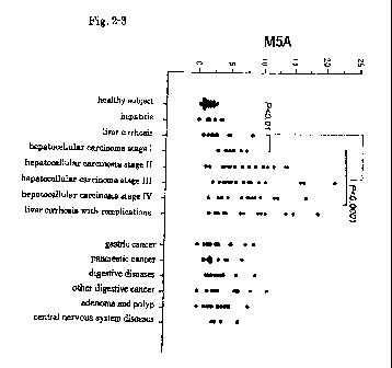
Fig. 2-3 is a diagram wherein the abundance values of a hepatocellular
carcinoma marker of the present invention (M5A) in the sera of patients
suffering
CA 02774421 2012-03-16
8
from various diseases are plotted.
Fig. 2-4 is a diagram wherein the abundance values of a hepatocellular
carcinoma marker of the present invention (A2G1(6)FB) in the sera of patients
suffering from various diseases are plotted.
Fig. 2-5 is a diagram wherein the abundance values of a hepatocellular
carcinoma marker of the present invention (A2G2B) in the sera of patients
suffering
from various diseases are plotted.
Fig. 2-6 is a diagram wherein the abundance values of a hepatocellular
carcinoma marker of the present invention (asialo A2G2Fo2) in the sera of
patients
suffering from various diseases are plotted.
Fig. 2-7 is a diagram wherein the abundance values of a hepatocellular
carcinoma marker of the present invention (asialo A2G2B) in the sera of
patients
suffering from various diseases are plotted.
Fig. 3 is a diagram wherein medical test values for AFP (ordinate) and for
A2G1(6)FB (abscissa) in hepatocellular carcinoma samples are plotted.
Fig. 4 is a diagram wherein medical test values for PIVKAII (ordinate) and
for A2G1(6)FB (abscissa) in hepatocellular carcinoma samples are plotted.
EMBODIMENTS FOR CARRYING OUT THE INVENTION
[0013]
<Hepatocellular Carcinoma Marker>
The hepatocellular carcinoma marker of the present invention at least
comprises a sugar chain represented by any one of the Chemical Structural
Formulae
(1) to (7) described above.
The sugar chain shown in (1) may be hereinafter referred to as "A2G2B".
The sugar chain shown in (2) may be hereinafter referred to as "A2G0B".
The sugar chain shown in (3) may be hereinafter referred to as "A2G1(6)FB".
The sugar chain shown in (4) may be hereinafter referred to as "A2GO".
CA 02774421 2012-03-16
9
The sugar chain shown in (5) may be hereinafter referred to as "M5A".
The sugar chain shown in (6) may be hereinafter referred to as "sialylated
A2G2Fo2". This sugar chain has sialic acid as a side chain in the serum, but
the
asialo type (this may be hereinafter referred to as "asialo A2G2Fo2"), which
is
prepared by deletion of the sialic acid by a known method, is also included
therein.
The sugar chain shown in (7) may be hereinafter referred to as "sialylated
A2G2B". This sugar chain also has sialic acid as a side chain in the serum,
but the
asialo type (this may be hereinafter referred to as "asialo A2G2B"), which is
prepared
by deletion of the sialic acid by a known method, is also included therein.
[0014]
The hepatocellular carcinoma markers of the present invention are sugar chain
moieties of glycoproteins contained in the sera of patients suffering from
hepatocellular carcinoma in large amounts, and, in addition to the above-
described
sugar chains, those to which a peptide is bound are also included in the
hepatocellular
carcinoma markers of the present invention.
[0015]
<Method for Diagnosis of Hepatocellular Carcinoma>
The present invention also include a method for diagnosis of hepatocellular
carcinoma, wherein the amount of the hepatocellular carcinoma marker is
measured,
and the amount of the hepatocellular carcinoma marker or a value calculated
based
on the amount of the hepatocellular carcinoma marker is used as an index.
Examples of the sample to be used in this measurement method include body
fluids
collected from subject animals which may be suffering from hepatocellular
carcinoma. Examples of the body fluid to be used include blood, lymph, spinal
fluid and urine, and processed products thereof. The body fluid to be used is
preferably blood, more preferably serum obtained by subjecting the blood to
separation. In terms of the subject of the method for diagnosis of
hepatocellular
CA 02774421 2012-03-16
1.0
carcinoma, the subject animal is preferably human.
[0016]
In order to measure the content of a sugar chain as the hepatocellular
carcinoma marker of the present invention (this may be hereinafter referred to
as a
"marker sugar chain") in the sample, the marker sugar chain contained in the
sample
needs to be isolated from the protein to which the marker sugar chain is
bound.
Examples of the method for isolating the marker sugar chain from the protein
include
per se known methods which are normally used, more particular examples of the
method include hydrazinolysis and enzyme (N-glycanase) digestion. Among these,
hydrazinolysis is preferred for quantitative cleavage of a sugar chain, and,
for
example, the method described in Y. Otake et al., J Biochem (Tokyo) 129 (2001)
537-42 or the like is preferably employed. Here, in cases where hydrazinolysis
is
employed, reacetylation of the acetyl group eliminated by the hydrazinolysis
is
necessary. More particularly, for example, the method described in K. Tanabe
et al.,
Anal. Biochem. 348 (2006) 324-6 or the like is employed. Further, in the case
of a
sialylated sugar chain, a sialic acid-cleaving enzyme such as neuraminidase
may be
used to cleave sialic acid, followed by purification by ion-exchange
chromatography
or the like.
Before the isolation of the marker sugar chain from the protein, a substance,
more particularly, an antibody or the like, which specifically binds to a
protein bound
to the marker sugar chain may be used to extract a complex comprising the
protein
and the sugar chain from body fluid obtained from the sample, which is
preferred in
view of efficiency of the subsequent structural analysis of the sugar chain,
The
extraction using a substance which specifically binds to the protein may be
carried
out by a per se known method which is normally used. Examples of the protein
which is bound to the marker sugar chain of the present invention include
those
described in Table 1.
CA 02774421 2012-03-16
11
[Table 1]
Gene ID IIC81 f1o Name
5004 AAJ43315 AIAG1 HUMAIJ Alpha-l-acid gi,coprotein 1
5005 hIP 000599 AIAG2 HUMAN Alpha-t-acid 9lycoprotein 2
5265 AA659495 AIAT HUMAN Aloa-1-antitrypsin
1 AAH35719 A180 HUIMIAId Alpha=16=glycoprotein
197 HP 001613 FETUA HUMAN AL1tta-2-HS-glycoptotein
2 EAW88590 A21vG HUMAN Alpha=2=macroglohubn
462 CAA48690 ANT3 HU44AN Antithrombin-III
335 AAD34604 APOA1 HUMAIJ Apolipoprotem Ad
33? IIP 000473 APOA4 HUMAN Apolipoprotem A-IV
338 AAA53373 APO6 HUMAN .Apolipoprotem 6.100
317 A635919 APOD HUMAN Apolipoprotem D
51742 ARI16 HUP,9 AT-itch interact re domain-containing protein 46
350 AAA617G6 APOH HUIa1AII Beta-2-glycoprolein 1
722 NP 000706 C48PA HUMAN C41)-binding protein alpha chain
1356 BAA.08084 CERU HUMAN Cerutoplasmm
1191 AAH10914 CLUB HUMAN Clusterin
710 NP 000055 C03 HUMAN Complement C3
720 AAA51056 CODA HUMAII Complement CJ -A
721 AAA59651 0046 HUMAN Complement C4=6
735 NP 00172"o 09 HUMAN Complement component C9
629 AAH07990 CFAB_NUMAN Complement factor B
3075 CAA 3704 :FAH HUMAN Complement factor H
666 FIP 001747 C8GHUMANJ Cotticosteroid=hinding globulin
60412 AAH26174 E%OC4 HUr1AII Exccyst complex competent 4
233: A.A1437154 FIFIC HUMAN Fibronectin
3959 NP 005558 LG36P HUMAN Galectin-3=binding protein
2934 4AH26033 GELSHUIIAI=I Gelsolei
3240 AAGO1170 HPT HUMAPI Haptoglohin
3250 A4A88081 HPTR HUMAN Haptoglohin=elated ixetem
3263 AAA58678 i=1EMO HUMAf-I Hemopexin
3053 PIP 0001766 HEP2 H MLI I, Heparin colactor 2
32?3 AAH69674 HRG HUMAN Hislidine-rich glycoprotein
68500 AAK00563 fv1LL3 HUMAII Histonadysiee N-ntethyltransieiase MvILL3
3493 A.AA52735 IGHA1 HUMAPI Ig alpha-1 chain C region
3494 AAB59396 -IGHA2 H toAPl Ig alpha-2 chain C region
3500 AAA70227 IGHG1 l1 Atl Ig gamma=1 chain C region
3501 AAI1?6044 1r;HG2 HUMAN Ig gamma-2 chain C region
3502 AAA70228 1GHG3 HUMAN Ig gamma-3 chain C region
3503 AA869394 IGHG4 HUMAN Ig gamma-4 chain C region
3514 AGA58921 1GI<C HUMAN Ig kappa chain C region
3507 AAA52824 IGHPA HUfv1A.1.1 Ig mu chain C region
3512 lIP603247 IGJ HUMAN lmmunoglobulin J char
57323 NP_079357 11305HUFIAlI Integrase catalytic denaimcontairing protein
I<IAA1305
3697 CAA49279 ITIH1 HUMAN Inteoalpha-nypsm ethibitorhoary chain H1
3698 NP 00220? ITIH2 HUFIAII Inleralpha-trypsinmhihdorhear--chainH2
3700 AA138394 MM HUfrlAil Inter=alpha-trvl:sin inhihilor hoary chain H4
3827 AAH26253 I<NGI HUvWI Kininogen-1
4060 AAH35997 LlJI.0 HUMAN Lumican
114770 NP 443122 PGRP2 HUMAN tJ acetylnmramoyl-L-alarine amidase
5176 AAAGO056 PEOF HUNW1 Pigment epithelium.deevnd factor
3818 AA117352 I<LI<B1 HUMAN Plasma kailikrem
710 NP_001027456 IC1 HUh5At9 Plasma protease C1 inhibitor
5340 AAA36451 PLivihl HUMAJI Plasminogen
5473 AAI<29642 CXCL7 HUMFUI Platelet basic protein
11107 NP_061169 PROMS HUI,1APt PR domain zinc finger protein 5
5858 CAA38255 PZP HUI:IAIt Pregnancy zone protein
26091 AAH39600 HERO UMAII Probable E3 uhiquitm=prolem ligase HERC4
259 AAA59196 AMSP HUMAN Protein AMBP
2147 AA106931 THR6 HUIIAlt Prothrembin
15780' AA101378 RLBL2 HUIIWI Retinaldehyde-I:inding prolem 1-like protein 2
7018 NP 00105A TRFE HUMAN Setotransferrut
5444 AAH74719 P0141 HUI,i_J=I Serum paraoxonase'aniesterass I
7276 ABI7i3345 TTHY HUMAN Transthyfetin
2x38 AAA61?01 VTD6 HUMAN Vitamin C-binding protein
7446 AAH05046 6TNC HUlvlAiI Vitronectin
563 HP-00 1176 ZA2G HUMAN Zinc-alpha-2-glycoprolein
CA 02774421 2012-03-16
12
[0017]
The thus isolated marker sugar chain is labeled as required. The method of
labeling is not restricted, and, in cases where a mass spectrograph is used,
TMAPA
(trimethyl(4-aminophenyl)ammonium chloride), which enhances the ionization
efficiency, is especially preferred, and, in cases where a fluorescence
detector is used,
2-aminopyridine is preferred. In terms of the method of derivatization, in the
case
of TMAPA, the method described in M. Okamoto et al., Rapid Commun Mass
Spectrom 9 (1995) 641-3, or the like is used, and, in the case of 2-
aminopyridine, the
method described in Y. Otake et al., J Biochem (Tokyo) 129 (2001) 537-42, or
the
like is used.
[0018]
The method for detecting, or for measuring the content of, a sugar chain as
the
hepatocellular carcinoma marker is not restricted as long as the marker sugar
chain of
the present invention can be detected thereby, and examples of the method
include
normal-phase and reversed-phase high-performance liquid chromatography, mass
spectrometry, nuclear magnetic resonance, and methods using an antibody or
lectin
specific to the marker sugar chain of the present invention. The sugar chain
as a
hepatocellular carcinoma marker of the present invention is an N-linked sugar
chain,
and hence can be detected using a method of simple, highly accurate and
quantitative
detection of a bisecting-type or Lewis X-type sugar chain structure, which is
a N-
linked sugar chain. In the present invention, the bisecting-type sugar chain
structure
means a biantennary N-linked sugar chain structure in which a Man residue of
the
trimannosyl core structure has GIcNAc bound to the 4-position. In the present
invention, the Lewis X-type sugar chain structure means an N-linked sugar
chain
structure in which the G1cNAc residue at the non-reducing end has fucose at
the 3-
position.
[0019]
CA 02774421 2012-03-16
13
Since isolation of the marker sugar chain of the present invention from
similar
sugar chains existing in the serum is very difficult, it is preferred to use a
detection/measurement method by the combination of liquid chromatography and a
mass spectrometer (this may be hereinafter referred to as the "LC-MS method"),
or to
use a detection/measurement method by liquid chromatography. The structure of
the sugar chain may be analyzed by any method, and, more particularly, the
structure
is preferably identified by, for example, a process wherein commercially
available
mannose standard sugar chains are subjected to normal-phase (or reversed-
phase)
liquid chromatography (hereinafter referred to as "normal-phase HPLC") to
calculate
mannose standard sugar chain peaks based on an internal standard, and
commercially
available galactose standard sugar chains are subjected to reversed-phase (or
normal-
phase) liquid chromatography (hereinafter referred to as "reversed-phase
HPLC") to
calculate galactose standard sugar chain peaks based on an internal standard,
to
prepare a two-dimensional map (e.g., the one shown in Fig. 1), which is then
used for
the identification.
[0020]
Examples of the method for measuring the marker sugar chain of the present
invention using the LC-MS method include the method described below in detail.
The specification of the liquid chromatography is not restricted as long as
the liquid
chromatography can stably send a liquid. The ionization method may be either
ESI
or APCI, and ESI is most preferred. The mass spectrometer may be any of the
quadrupole type, TOF type, ion trap type, magnetic field type and Fourier
transform
type, among which the quadrupole type, which is highly quantitative, and the
TOF
type and the ion trap type, which are highly sensitive, are especially
preferred.
[0021]
The column to be used may be any of the normal-phase type, reversed-phase
type and absorption type as long as a plurality of types of sugar chains can
be
CA 02774421 2012-03-16
14
separated from each other with the column. The column is preferably a reversed-
phase column C8, C18 or C30, among which a C30 column is more preferred. The
size of the column is not restricted, and preferably not more than 2.1. mm in
terms of
the inner diameter in order to reduce the flow rate and to thereby increase
the
sensitivity. The size is especially preferably not more than 1.5 mm.
[0022]
The eluent is appropriately selected depending on the properties of the
column, and, in cases where a C30 column is used, for example, 5 mM aqueous
ammonium solution (pH=4) is used as the eluent A, and a mixture of 90% 5 mM
aqueous ammonium acetate solution (pH=4) and 10% acetonitrile is used as the
eluent B. In this case, sufficient equilibration with 0% to 30% B solution (=
100%
to 70% A solution; description of the concentration of the A solution is
hereinafter
omitted) is necessary before injection of a sample. The concentration of the B
solution upon the equilibration is preferably 10 to 25%, especially preferably
1.5 to
20%.
[0023]
About 20 gL of a sample solution is injected to the column, and, immediately
after the injection, the eluent composition is made to linearly change from
about 20%
B solution to about 50% B solution for about 50 minutes. The composition may
be
kept to be 50% B solution from minute 50 to minute 60, but the condition is
not
restricted since the gradient condition is limited by the type of the column.
The
measurement conditions for the mass spectrometry are not restricted as long as
the
conditions are within the ranges in which the marker sugar chain of the
present
invention can be separated, and excellent detection is possible under the
conditions
of, for example, a capillary voltage (ionization voltage) of 4000 V, nebulizer
gas
pressure of 45 psi, dry gas flow rate of 1.0 L/min. and temperature of 350 C.
[0024]
CA 02774421 2012-03-16
1.5
The detection method is most preferably SIM (Selective Ion Monitoring) in
cases where the mass spectrometer is the quadrupole type, and, in such cases,
the ion
to be detected is set to an ion of any of the marker sugar chains of the
present
invention or, when another sugar chain is used as an internal standard, the
ion to be
detected is set to an ion such as its rn/z.
In the cases of the TOF type and ion trap type, the scan mode may be used,
and, in such cases, the mass range is preferably set to 400 to 4000. The
marker
sugar chain is detected in the above-mentioned analysis conditions at the
retention
time and m/z below, but these may vary depending on the column and the eluent
selected, and hence are not restricted. The ion to be detected is not
restricted to the
parent ion, and may be an associated ion such as a fragment ion, loaded ion or
dimeric ion.
In cases where the detection/measurement is carried out using liquid
chromatography, the specification of the liquid chromatography is not
restricted as
long as the liquid chromatography can stably send a liquid. More particularly,
for
example, normal-phase HPLC is carried out using a column such as Asahipak NH2P-
50 4.6 mm I.D. x 250 mm (manufactured by shodex) at a flow rate of 0.6 ml/min.
at a
column temperature of 30 C. In this operation, Solvent A, which is a solution
of
93% acetonitrile and 0.009% acetic acid, whose pH was adjusted to 6.8 with 25%
aqueous ammonia; and Solvent B, which is a solution of 20% acetonitrile and
0.009% acetic acid, whose pH was adjusted to 6.8 with 25% aqueous ammonia; are
used, and the Solvent A:Solvent B ratio is 75:25 at the beginning. After
injection of
a sample, the ratio of Solvent B is linearly increased to 42% for 180 minutes.
HPLC analysis is performed using Prominence (manufactured by Shimadzu
Corporation) and the respective peaks are quantified using LC station
(manufactured
by Shimadzu Corporation).
Further, a method is preferred wherein the elution times of standard sugar
CA 02774421 2012-03-16
16
chains (e.g., PA-sugar chains M2A, M3B, M4B, M5A, M6B, M7A, M8A and M9A,
manufactured by Takara) are used as internal standards (mannose unit, MU) to
calculate MU of the eluted fluorescently labeled sugar chain, and the
respective peaks
sorted based on the mannose unit are subjected to quantitative analysis of the
peak
areas between samples, to thereby determine the sugar chain content.
[0025]
The content of the marker sugar chain detected by the above method or the
value calculated based on the amount of the hepatocellular carcinoma marker is
used
as an index to judge the possibility that the patient who provided the sample
is
suffering from hepatocellular carcinoma. In the method for diagnosis of
hepatocellular carcinoma of the present invention, the content of the marker
sugar
chain does not need to be determined as an absolute amount, and may be
obtained by
digitization of the respective peaks unique to the marker sugar chain detected
by the
above method or the like. Particular examples of the method include a method
wherein the height of each detected peak is digitized and a method wherein the
peak
area is digitized. In liquid chromatography, the method is not restricted to
one of
these since the method has a quantitative nature, whereas in LC-MS, the method
wherein the peak area is digitized is more accurate and preferred. Further, by
using
a lectin or antibody that specifically recognizes the marker sugar chain of
the present
invention, the abundance of the marker sugar chain can be measured by a
conventional method. The lectin to be used for detecting the substance of the
present invention for detection of hepatocellular carcinoma is a substance
having a
specific binding activity to a bisecting-type sugar chain structure among N-
linked
sugar chains. The value calculated by digitizing the thus obtained peak may be
hereinafter referred to as the "peak strength".
[0026]
In the method for diagnosis of hepatocellular carcinoma of the present
CA 02774421 2012-03-16
17
invention, a method using as an index a value calculated based on the amount
of the
above-mentioned marker sugar chain is preferably used, in addition to the
above-
described method using as an index the amount of the marker sugar chain. The
particular method of calculation may be any method as long as the value to he
used
as an index in the measurement method of the present invention is corrected
such that
the value accurately reflects the conditions in the body of the subject, and
examples
of the method include a method wherein the correction is performed using the
amount of the serum to be treated, and a method wherein the correction is
performed
using the weight of the protein to be treated. Further, a method wherein the
amount
of a sugar chain that does not fluctuate, or hardly fluctuates, due to the
disease, that is,
the amount of a sugar chain that does not show a significant difference in the
content
in the body fluid between healthy individuals and patients suffering from
hepatocellular carcinoma (the amount may be hereinafter referred to as the
"internal
standard") is used as a standard to determine its ratio with respect to the
amount of
the marker sugar chain is also effective.
[0027]
As the above-mentioned value calculated based on the amount of the marker
sugar chain, in cases where serum is used as the sample, the amount of the
marker
sugar chain or a value corrected by the internal standard may be used.
Examples of
internal standard herein include sugar chains showing no significant
difference in
their contents in the sample between healthy animals and animals suffering
from
hepatocellular carcinoma.
[0028]
The hepatocellular carcinoma marker of the present invention shows
significant difference in its abundance in the serum between patients
suffering from
liver cirrhosis and patients suffering from hepatocellular carcinoma, and
hence is
preferably used to distinguish between these patients in diagnosis. Patients
CA 02774421 2012-03-16
18
suffering from liver cirrhosis characteristically show extensive tuberculation
in the
liver, extensive fibrosis, necrosis of hepatocytes, reduction of the blood
flow,
decreased secretion of bilirubin, jaundice and/or the like. Thus, in the
diseased state
of liver cirrhosis, complications such as ascites, esophageal varices and/or
hepatic
encephalopathy are observed. In the present invention, liver cirrhosis is a
state
wherein, although extensive tuberculation is observed due to infection with
HCV,
existence of cancer cannot be confirmed by known hepatocellular carcinoma
markers
and diagnostic imaging (Ultra Sound; US, Computer Tomography; CT, Magnetic
Resonance Imaging; MRI), and no complication caused by liver cirrhosis is
observed.
[0029]
The marker sugar chain of the present invention exists in a healthy individual
in a small amount but exists in a patient suffering from hepatocellular
carcinoma in a
remarkably large amount. Therefore, in cases where the content of the
hepatocellular carcinoma marker or a value calculated based on the amount of
the
hepatocellular carcinoma marker is larger than that of a healthy individual,
the
subject can be diagnosed as highly possibly having developed hepatocellular
carcinoma. Further, since the content of the marker sugar chain of the present
invention is higher in a patient suffering from hepatocellular carcinoma than
in a
patient suffering from liver cirrhosis, liver cirrhosis and hepatocellular
carcinoma can
be distinguished from each other, and diagnosis of hepatocellular carcinoma is
thereby possible.
Further, among the sugar chain markers of the present invention, M5A,
A2GOB, A2G1(6)FB and A2G2B showed significant difference also between
patients suffering from early-stage hepatocellular carcinoma (Stage I
according to the
TNM classification) and healthy individuals. Thus, by detecting these marker
sugar
chains, early-stage hepatocellular carcinoma, whose detection has been
impossible
with the hepatocellular carcinoma markers reported so far, can be diagnosed.
CA 02774421 2012-03-16
19
Further, since the content of the sugar chain marker of the present invention
has a tendency to increase as the stage of hepatocellular carcinoma advances
(Fig. 2),
the content of the hepatocellular carcinoma marker of the present invention or
a value
calculated therefrom may also be used for identifying the stage of progression
of
hepatocellular carcinoma.
In the present description, the TNM classification (Stages Ito IV) means a
classification of the stage of progression of hepatocellular carcinoma
according to
TNM Classification, 6th ed., published in 2002 by Union for International
Cancer
Control (UICC) (see Japanese Translated PCT Patent Application Laid-open No.
2008-505143).
[0030]
<Method for Evaluating Therapeutic or Prophylactic Agent for Hepatocellular
Carcinoma>
By administering a prophylactic or therapeutic agent for hepatocellular
carcinoma to an animal requiring prophylaxis or treatment of hepatocellular
carcinoma and measuring the content of the above hepatocellular carcinoma
marker
in body fluid collected from the animal, evaluation of the prophylactic or
therapeutic
effect against hepatocellular carcinoma in the animal can be carried out using
as an
index the content of the hepatocellular carcinoma marker or a value calculated
therefrom.
For example, the content of the hepatocellular carcinoma marker or a value
calculated therefrom observed before administration of the prophylactic or
therapeutic agent for hepatocellular carcinoma is compared with that observed
several days to several months after the administration, and, if the content
of the
hepatocellular carcinoma marker or the value calculated therefrom is lower in
the
latter case, the agent can be judged to have had a prophylactic or therapeutic
effect.
The animal to be evaluated is preferably human.
CA 02774421 2012-03-16
Examples of the therapeutic agent for hepatocellular carcinoma include
tegafur (general name: uracil), epirubicin (general name), mitomycin C
(general
name), fluorouracil (general name), cyclophosphamide (general name) and
mitoxantrone (general name).
5 [00311
<Method for Evaluating Candidate Compound for Therapeutic or Prophylactic
Agent
for Hepatocellular Carcinoma>
Further, by administering a candidate compound for a prophylactic or
therapeutic agent for hepatocellular carcinoma to an animal in need of
prophylaxis or
10 treatment of hepatocellular carcinoma and subsequently measuring the amount
of the
above hepatocellular carcinoma marker in body fluid collected from the animal,
evaluation of the prophylactic or therapeutic effect of the candidate compound
against hepatocellular carcinoma can be carried out using as an index the
amount of
the hepatocellular,carcinoma marker or a value calculated therefrom.
15 For example, the content of the hepatocellular carcinoma marker or a value
calculated therefrom observed before administration of the candidate compound
is
compared with that observed several days to several months after the
administration,
and, if the content of the hepatocellular carcinoma marker or the value
calculated
therefrom is lower in the latter case, the compound can be judged to be a
promising
20 candidate substance for a prophylactic or therapeutic agent for
hepatocellular
carcinoma.
The candidate compound may be either a low molecular weight compound or
a peptide or protein. The animal to be evaluated is preferably human.
[0032]
<Kit for Diagnosis of Hepatocellular Carcinoma, for Evaluation of Therapeutic
Effect against Hepatocellular Carcinoma, or for Evaluation of Prophylactic or
Therapeutic Effect of Candidate drug Compound against Hepatocellular
Carcinoma>
CA 02774421 2012-03-16
21
The kit of the present invention for diagnosis of hepatocellular carcinoma,
for
evaluation of a therapeutic effect against hepatocellular carcinoma, or for
evaluation
of a prophylactic or therapeutic effect of a candidate drug compound against
hepatocellular carcinoma comprises a reagent with which the amount(s) of one
or
more of the marker sugar chains of the present invention can be measured.
Examples of such a reagent include the above-described sugar chains to be used
as
standard substances in normal-phase and reversed-phase high-performance liquid
chromatography, mass spectrometry, nuclear magnetic resonance and the like;
and
antibodies and lectins that specifically recognize the marker sugar chain of
the
present invention. The antibodies that specifically recognize the marker sugar
chain
of the present invention may be any of polyclonal antibodies and monoclonal
antibodies, and fragments thereof, and can be obtained by a known method using
the
marker sugar chain as an antigen. Examples of the lectins that specifically
recognize the marker sugar chain of the present invention include those
described
above.
EXAMPLES
[0033]
The present invention will now be described in more detail by way of
Examples shown below, but the present invention is not limited to thereto as
long as
the gist of the present invention is not impaired.
[0034]
Example I
Determination of Hepatocellular Carcinoma Marker
(1) Measurement of Amount of Maker Sugar Chain in Sample Serum
Sera were obtained from patients clinically diagnosed as having
hepatocellular carcinoma (hereinafter referred to as hepatocellu)ar
carcinoma),
patients clinically diagnosed as having liver cirrhosis accompanied by HCV
infection
CA 02774421 2012-03-16
22
(hereinafter referred to as liver cirrhosis), and healthy individuals with no
sign of
liver diseases (hereinafter referred to as healthy individuals), as subjects.
More
particularly, glycoprotein sugar chain analysis was carried out using sera of
69
healthy individuals, 7 patients suffering from chronic hepatitis, 8 patients
suffering
from liver cirrhosis and 55 patients suffering from hepatocellular carcinoma.
The
55 patients suffering from hepatocellular carcinoma were classified into 4
stages
based on the stage of progression as described in Table 2 (TNM
classification). The
sera of the 69 healthy individuals were collected after obtaining informed
consent in
Soiken. The sera of patients suffering from the liver diseases were collected
after
obtaining informed consent in Faculty of Medicine, Kagawa University. Details
of
the samples are as shown in Table 2.
[0035]
[Table 2]
healthy liver hepatic carcinoma
subject cirrhosis Stage I Stage 11 Stage III Stage IV
number of 69 8 7 17 18 13
subjects
mean age 44.1 69.3 74.6 72.7 71.4 68.7
-age range_ 21-65 57-77 60-85 59-82 63-83 50-80 _
male/female 26/45 3/5 5/2 6/8 15/3 5/8
[0036]
In pretreatment of the samples, 9 volumes of acetone at -20 C was added to
each collected serum and the resulting mixture was mixed well. After leaving
the
mixture to stand for a while, the mixture was centrifuged and the supernatant
was
then removed. The precipitate was freeze-dried, and anhydrous hydrazine was
added to 2 mg dry weight of the sample, followed by decomposition of hydrazine
by
heating at 100 C for 10 hours, to remove sugar chains from proteins.
Subsequently,
hydrazine was evaporated, and reacetylated sugar chains were purified by a
part of
the methods described in JP 2005-308697 A and Anal. Biochem. 384, 324-326,
2006
using a solid-phase-extraction-type graphite carbon column (manufactured by GL
CA 02774421 2012-03-16
23
Sciences Inc.).
[0037]
The purified sugar chains were fluorescently labeled by 2-aminopyridyl
amination (2-aminopyridine; PA, manufactured by Nacarai tesque, Inc.). To
remove unreacted PA, the sample was passed through a solid-phase cellulose
column.
A solution of 66,7% 1-butanol, 16.7% ethanol and 100 mM ammonium acetate was
prepared as a washing solution to be used for the solid-phase cellulose
column, and a
solution of 33% ethanol and 15 mM ammonium bicarbonate was prepared as an
eluent for eluting fluorescently labeled sugar chains. From the obtained pool
of
fluorescently labeled serum sugar chains, neutral sugar chains existing in the
serum
were separated by ion-exchange chromatography (DE52, manufactured by Whatman).
In terms of the solutions to be used in this operation, a solution prepared by
adjusting
pH of pure water to 9.0 with 25% aqueous ammonia was used as Solution A, and a
solution prepared by adjusting pH of 500 mM aqueous ammonium acetate solution
to
9.0 with 25% aqueous ammonia was used as Solution B.
[0038]
At the same time, the same amount of the pool of serum sugar chains was
treated with neuraminidase (Arthrobacter ureafaciens, manufactured by Nacarai
tesque, Inc.) at 37 C for 12 hours, and ion-exchange chromatography was then
performed, to purify only asialo sugar chains produced by removal of sialic
acid from
their side chains.
[00391
The neutral sugar chains and asialo sugar chains that were fluoresccntly
labeled as described above were subjected to normal-phase high-performance
liquid
chromatography (normal-phase HPLC), and the amounts of sugar chains were
measured by a fluorescence detector. For the normal-phase HPLC, a column
Asahipak NH2P-50 4.6 mm I.D. x 250mm (manufactured by shodex) was used at a
CA 02774421 2012-03-16
24
flow rate of 0.6 ml/ nin. at a column temperature of 30 C. In this operation,
Solvent
A, which is a solution of 93% acetonitrile and 0.009% acetic acid, whose pH
was
adjusted to 6.8 with 25% aqueous ammonia; and Solvent B, which is a solution
of
20% acetonitrile and 0.009% acetic acid, whose pH was adjusted to 6.8 with 25%
aqueous ammonia; were used, and the solvent A:solvent B ratio was 75:25 at the
beginning. After injection of each sample, the ratio of Solvent B was linearly
increased to 42% for 180 minutes. HPLC analysis was performed using
Prominence (manufactured by Shimadzu Corporation) and the respective peaks
were
quantified using LC station (manufactured by Shirnadzu Corporation).
[0040]
Further, the elution times of the standard sugar chains (PA-sugar Chains M2A,
M3B, M4B, M5A, M6B, M7A, M8A and M9A, manufactured by Takara) were used
as internal standards (mannose unit, MU) to calculate MU of the eluted
fluorescently
labeled sugar chains. For each peak separated based on the mannose unit,
quantitative analysis of the peak area was carried out among the samples.
[0041]
For identification of the sugar chain structure, commercially available
standard sugar chains were subjected to normal-phase HPLC and reversed-phase
HPLC, and MU calculated based on the internal standard in the normal-phase
HPLC
and GU calculated based on the internal standard (glucose unit, GU3-22,
manufactured by TaKaRa) in the reversed-phase HPLC were used to obtain a two-
dimensional map (Fig. 1).
For the reversed-phase HPLC, a column Develosil C30 (manufactured by
Nomura Kagakus) 4.6 mm I.D. x 150 mm was used at a flow rate of 0.5 ml/min. at
a
column temperature of 30 C. In this operation, Solvent A, which is a solution
adjusted to 5 mM ammonium acetate (pH 4.0); and Solvent B, which is a solution
adjusted to 10% acetonitrile and 5 mM ammonium acetate (pH 4.0); were used,
and
CA 02774421 2012-03-16
the Solvent A:Solvent B ratio was 75:25 at the beginning, After injection of
each
sample, the ratio of Solvent B was linearly increased to 42% for 60 minutes.
[0042]
The identified sugar chain structures and abundances of the thus
5 measured/identified N-linked sugar chains in the healthy individuals (A),
liver
cirrhosis (B) and hepatocellular carcinoma (C) are shown in Table 3. As is
evident
from the table, the sugar chains shown in Table 3 were present in low
abundances in
both the sera of the healthy individuals and the sera of liver cirrhosis,
while these
were present in the sera of hepatocellular carcinoma in very large amounts.
Further,
10 in addition to hepatocellular carcinoma and liver cirrhosis, measurement
was carried
out in the same manner also in patients suffering from hepatitis, liver
cirrhosis
accompanied by complications, gastric cancer, pancreatic cancer, digestive
diseases,
adenoma, polyp and central nervous system diseases, and the measured values
are
plotted in Fig. 2.
15 [0043]
[Table 3]
--~-
marker amount SD
sugar chain healthy subject liver cirrhosis hepatic carcinoma
neutral M5A 6.79 1.88 7.24 3.61 16.93 4.78
sugar A200 1.18 0.55 3.04 2.27 7.20 4.27
chain A2GOB 2.12 0.68 4.34 3.57 12.7 5 6.24
A2G1.(6)FB 3.95 t 1.50 7.05 t 2.21 16.11 6.42
asialo A2G2B 2.04 1.18 3.07 0.67 7.42 3.78
sugar
chain A2G2Fo2 1.91 1.07 2.84 1.31 7.73 5.00
[0044]
(2) Test of Marker Sugar Chains
For the marker sugar chains for detection of hepatocellular carcinoma, which
20 were determined by the above glycoprotein sugar chain analysis, the values
of the
sensitivity, specificity and positive predictive value (PPV) are shown. The
CA 02774421 2012-03-16
26
sensitivity represents the positive rate of each marker in a sample having
hepatocellular carcinoma. The specificity represents the negative rate of each
marker in a sample having no hepatocellular carcinoma. PPV represents the
probability of having hepatocellular carcinoma in cases where each marker is
positive.
The cut-off value employed herein was set to the value 1.2-fold higher than
the mean
abundance of each marker observed for the sera of patients suffering from
liver
cirrhosis.
As shown in Table 4, the neutral sugar chain A2GOB showed a sensitivity of
91%, specificity of 96% and PPV of 96%. Further, the neutral sugar chain
A2G1(6)FB showed a sensitivity of 89%, specificity of 96% and PPV of 94%.
[0045]
[Table 4]
marker sensitivity specificity PPV
neutral M5A 100% 82% 79%
A2GO 84% 98% 96%
sugar A2GOB 91% 96% 94-
chain A2G1 (6) FB 89% 96% 94%
asialo A2G2B 84% 88% 82%
sugar A2G2Fo2 84% 87% 81%
chain
[0046]
On the other hand, as described in J. Gastroenteral. Hepatol. 14. 436-445,
1999 and Cancer Research 53, 5419-5423, 1993, AFP, which is a known
hepatocellular carcinoma marker, is reported to have a sensitivity of 70% and
specificity of 70% (cut-off value, 20 ng/ml), and AFP-L3 is reported to have'a
sensitivity of 70%, specificity of 90% and PPV of 77%. Further, it was
recently
shown that a branch alpha(1.,3)-fueosylated triantennary glycan, which is a
hepatocel.lular carcinoma marker and an N-linked sugar chain, increases in the
serum
of hepatocellular carcinoma patients and hence can be used as a hepatocellular
carcinoma marker (Hepatology 46, 1426-1435, 2008). However, this glycan shows
CA 02774421 2012-03-16
27
a sensitivity of 57%, specificity of 88% and PPV of 81% (limited to HBV-
positive
liver cirrhosis and hepatocellular carcinoma).
[0047]
Table 5 shows the results of analysis of significance of fluctuation of each
sugar chain marker in terms of comparisons between healthy individuals and
liver
cirrhosis, between liver cirrhosis and hepatocellular carcinoma, and between
liver
cirrhosis and hepatocellular carcinoma at various stages of progression, which
analysis was carried out using Student t-test (P value). In the table,
parentheses
indicate control groups, and each sugar chain marker is judged to show a
biologically
significant difference in cases where the P value is not more than 0.05.
[0048]
[Table 5]
hepatic carcinoma vs liver cirrhosis
liver Total
cirrhosis hepatic
marker carcinoma Stagel StageII Stagelll StagelV
(vs
(vs liver
healthy) cirrhosis
M5A 0.7432 <0.0001 0.0055 <0.0001 <0.0001 <0.0001.
neutral A2GO 0.0604 0.0009 0.0758 0.0059 0.0034 0.0034
sugar A2GOB 0.1332 <0.0001 0.0504 0.0001 0.0001 0.0007
chain A2G1 6 FB 0.0060 <0.0001 0.0066 <0.0001 <0.0001 0.0003
A2G2B 0.7079 <0.0001 0.0081 <0.0001 <0,0001 0.0051
asialo A2G2B 0.0048 <0.0001 0.0600 0.0001 <0.0001 0,0037
sugar A2G2Fo2 0.1142 0.01.15 0.1020 0.0033 <0.0001 0.0011
chain
[0049]
As is evident from Table 5, each of the marker sugar chains M5A, A2G0,
A2GOB, A2G1(6)FB and A2G2B, which are neutral sugar chains of the present
invention, and asialo A2G2B and asialo A2G2Fo2 showed a significant difference
between the values in healthy individuals and patients suffering from
hepatocellular
carcinoma. Further, A2G1(6)FB and A2G2B, which are marker sugar chains of the
present invention, showed a significant difference also between liver
cirrhosis and
CA 02774421 2012-03-16
28
healthy individuals. Therefore, for example, these marker sugar chains may be
effectively used for tracing progression of the diseased state from HCV
infection to
liver cirrhosis and then to hcpatocellular carcinoma.
[0050]
In particular, among the sugar chain markers of the present invention, the
neutral sugar chains M5A, A2GOB A2G1(6)FB and A2G2B showed significant
difference between patients suffering from early-stage hepatocellular
carcinoma
(Stage 1) and healthy individuals. By detecting the above sugar chain markers,
early-stage hepatocellular carcinoma, whose detection has been impossible with
the
hepatoeellular carcinoma markers reported so far, can now be diagnosed.
J. Proteome Res. 8, 595-602, 2009 and JP2008-541.060 disclose fucosylated
hemopexin as a hepatocellular carcinoma marker. Fucosylated hemopexin is said
to
have a sensitivity of 92%, specificity of 92% and PPV of 100% as a
hepatocellular
carcinoma marker. However, whether detection of fucosylated hemopexin enables
accurate diagnosis of early-stage hepatocellular carcinoma (Stage I according
to the
TNM classification) has not been revealed. On the other hand, M5, A2GOB,
A2G1(6)FB and A2G2B, which are hepatocellular carcinoma markers of the present
invention, show almost the same or higher significance also in a patient group
(total
hepatocellular carcinoma) equivalent to the one in the above-described
documents.
[0051]
(3) Comparison with Existing Markers
Table 6 shows the rates of detection of hepatocellular carcinoma with marker
sugar chains of the present invention in, among the samples obtained in the
above (1)
from patients diagnosed as having hepatocellular carcinoma, the samples which
are
negative for AFP, which is an existing hepatocellular carcinoma marker (< 30
ng/ml)
(21/55 samples); the samples negative for PIVKAII, which is similarly an
existing
hepatocellular carcinoma marker (< 40 mAU/ml) (18/55 samples); and the samples
CA 02774421 2012-03-16
29
negative for APP and negative for PIVKAII (11/55 samples) (the cut-off value
employed here was set to the value 1.2-fold higher than the mean expression
level of
each sugar chain marker observed for the sera of patients suffering from liver
cirrhosis). The values for AFP and PIVKAII were tested under contract with an
outside examination institute. The EIA method, which is commonly used, was
employed as the examination method.
[0052]
[Table 6]
marker sugar marker sugar chain marker sugar chain
marker chain positive positive positive
/AFP negative (%) JPIVKAII negative /AFP&PIVKAII
% negative (%)
MS 21/21(100) 18/18(100) 11/11(100)
neutral A200 19/21(90) 17/18(94) 11/11 1.00
sugar A2GOB 19/21(90) 17/18(94) 10/1.1 (91)
chain A2G1 6 FB 17/21(81) 16/18(89) 10/11(91)
A2G2B 20/21(95) 18/18 (100) 11/11(100)
asialo A2G2B 18/21 (86) 13/18 (72) 9/11 (82)
sugar
chain A2G2Fo2 19/21 (90) 16/1,8(89) 8/11 (73)
[0053]
As is evident from Table 6, A2G0B, among the sugar chain markers of the
present invention, succeeded in detection of hepatocellular carcinoma in 90%
of the
hepatocellular carcinoma samples negative for AFP (19/21 samples), with values
not
less than the cut-off value. Further, this sugar chain marker also succeeded
in
detection of hepatocellular carcinoma in 94% of the hepatocellular carcinoma
samples negative for PIVKAII (17/1.8 samples), with values not less than the
cut-off
value. Further, this sugar chain marker also succeeded in detection of
hepatocellular carcinoma in 91% (10/1.1 samples) of the hepatocellular
carcinoma
samples negative for both AFP and PIVKAII (11/56 samples), with values not
less
than the cut-off value. There was no correlation between the expression level
of the
sugar chain marker in the serum of hepatocellular carcinoma and the values for
the
existing hepatocellular carcinoma markers AFP and PIVKAII (Figs. 3 and 4).