Sélection de la langue

Search

Sommaire du brevet 2775398 

Énoncé de désistement de responsabilité concernant l'information provenant de tiers

Une partie des informations de ce site Web a été fournie par des sources externes. Le gouvernement du Canada n'assume aucune responsabilité concernant la précision, l'actualité ou la fiabilité des informations fournies par les sources externes. Les utilisateurs qui désirent employer cette information devraient consulter directement la source des informations. Le contenu fourni par les sources externes n'est pas assujetti aux exigences sur les langues officielles, la protection des renseignements personnels et l'accessibilité.

Disponibilité de l'Abrégé et des Revendications

L'apparition de différences dans le texte et l'image des Revendications et de l'Abrégé dépend du moment auquel le document est publié. Les textes des Revendications et de l'Abrégé sont affichés :

  • lorsque la demande peut être examinée par le public;
  • lorsque le brevet est émis (délivrance).
(12) Brevet: (11) CA 2775398
(54) Titre français: PROCEDE DE NORMALISATION DE RELEVES DE DEFORMATION D'IMPLANT POUR EVALUER LA CICATRISATION OSSEUSE
(54) Titre anglais: METHOD OF NORMALIZING IMPLANT STRAIN READINGS TO ASSESS BONE HEALING
Statut: Accordé et délivré
Données bibliographiques
(51) Classification internationale des brevets (CIB):
  • A61B 17/80 (2006.01)
  • A61F 02/28 (2006.01)
(72) Inventeurs :
  • DEIRMENGIAN, CARL (Etats-Unis d'Amérique)
  • MIKHAIL, GEORGE (Etats-Unis d'Amérique)
  • PIERSON, GLEN (Etats-Unis d'Amérique)
(73) Titulaires :
  • DEPUY SYNTHES PRODUCTS, LLC
(71) Demandeurs :
  • DEPUY SYNTHES PRODUCTS, LLC (Etats-Unis d'Amérique)
(74) Agent: BCF LLP
(74) Co-agent:
(45) Délivré: 2019-09-17
(86) Date de dépôt PCT: 2010-10-21
(87) Mise à la disponibilité du public: 2011-04-28
Requête d'examen: 2015-10-08
Licence disponible: S.O.
Cédé au domaine public: S.O.
(25) Langue des documents déposés: Anglais

Traité de coopération en matière de brevets (PCT): Oui
(86) Numéro de la demande PCT: PCT/US2010/053519
(87) Numéro de publication internationale PCT: US2010053519
(85) Entrée nationale: 2012-03-26

(30) Données de priorité de la demande:
Numéro de la demande Pays / territoire Date
61/253,583 (Etats-Unis d'Amérique) 2009-10-21

Abrégés

Abrégé français

L'invention porte sur un dispositif de traitement osseux dans un corps vivant qui comprend (a) un implant conçu pour être fixé à un os; (b) un premier capteur mesurant une déformation sur une première partie de l'implant, cette première partie étant configurée pour être accouplée mécaniquement à une partie affaiblie d'un os lorsque l'implant est accouplé à l'os dans une position cible en combinaison; et (c) un second capteur mesurant une déformation dans une partie non affaiblie de l'os.


Abrégé anglais

A device for treating bone in a living body includes (a) comprises an implant configured for attachment to a bone; (b) a first sensor measuring a strain on a first portion of the implant, the first portion of the implant being configured to be mechanically coupled to a weakened portion of a bone when the implant is coupled to the bone in a target position in combination; and (c) a second sensor measuring strain in a non-weakened portion of the bone.

Revendications

Note : Les revendications sont présentées dans la langue officielle dans laquelle elles ont été soumises.


What is claimed is:
1. A device for treating bone in a living body, comprising:
an implant configured for attachment to an exterior of a bone;
a first sensor mounted along a surface of the implant, the first sensor
measuring a strain on a first portion of the implant, the first portion of the
implant
being configured to be mechanically coupled to a weakened portion of the bone
when the implant is coupled to the bone in a target position; and
a second sensor isolated between two bone fixation element receiving holes
of the implant configured so that, when the implant is positioned as desired,
the
sensor may measure strain in a non-weakened portion of the bone.
2. The device of claim 1, wherein the implant is a plate configured to be
coupled to the
bone overlying a fracture site.
3. The device of claim 1, wherein the second sensor is configured to be
coupled directly
to the non-weakened portion of the bone.
4. The device of claim 1, further comprising:
a third sensor mounted to a portion of a bone fixation element configured for
insertion into the non-weakened portion of the bone in contact with a cortical
wall of
the non-weakened portion of the bone.
5. The device of claim 4, wherein the third sensor extends about a
circumference of a
proximal portion of the bone fixation element.
8

6. The device of claim 1, wherein the implant includes a rigid portion and
a flexible
portion having a first bending stiffness less than a second bending stiffness
of the rigid
portion, the second sensor being mounted on the flexible portion.
7. The device of claim 1, wherein the second sensor is a load sensor
measuring a load
on the bone.
8. The device of claim 2, wherein the second sensor is configured for
placement in a
shoe to measure a load on a leg including the bone simultaneously with a
measurement of
strain across the fracture site from the first sensor.
9. The device of claim 1, wherein the first and second sensors provide data
to an
external data gathering unit wirelessly.
10. The device of claim 9, wherein the first and second sensors are one of
MEMs sensors
and powered chips connected to printed circuit boards.
11. A system for tracking a bone healing progress, comprising:
an implant configured for attachment to an exterior of a bone for fixing a
fracture of the bone, the implant including a first sensor measuring strain
mounted
thereon at a position which, when the implant is mounted on the bone in a
target
position, is mechanically coupled to the fracture of the bone; and
a second sensor isolated between two bone fixation element receiving
holes of the implant to measure strain on a portion of the bone separated from
the fracture such that a ratio of the strain measured by the first sensor to
the
strain measured by the second sensor normalizes an effect of loads on the
bone.
9

12. The system of claim 11, further comprising:
a bone fixation element, a third sensor mounted at a proximal end
thereof so that, when the bone fixation element is inserted into the bone to a
desired position, the third sensor contacts a cortical wall of the bone.
13. The system of claim 11, wherein the implant includes a flexible portion
and a
rigid portion, a first bending stiffness of the rigid portion being greater
than a second
bending stiffness of the flexible portion, the second sensor being mounted on
the
flexible portion while the first sensor is mounted on the rigid portion.
14. The system of claim 13, further comprising:
first and second locking screws configured for insertion through the two bone
fixation element receiving holes on opposing sides of the flexible portion.

Description

Note : Les descriptions sont présentées dans la langue officielle dans laquelle elles ont été soumises.

CA 02775398 2016-12-20
METHOD OF NORMALIZING IMPLANT STRAIN READINGS
TO ASSESS BONE HEALING
Inventors: Carl DEIRMENGIAN, George MIKHAIL and Glen PIERSON
Priority Claim
[0001] The present application claims priority to U.S. Provisional
Application Serial No.
61/253,583 filed on October 21, 2009 and entitled "Method of Normalizing
Implant Strain
Readings to Assess Bone Healing."
Field of the Invention
[0002] The present invention relates to a system and method for tracking
the progress of
bone healing and, in particular, systems and methods that calculate a ratio of
strain at multiple
locations along an implant and/or a bone.
Background
[0003] Strain gages can be placed on orthopedic implants to track the
progress of bone
healing. Upon initial implantation, the implants are expected to experience
higher levels of strain
which decrease during healing as the bone begins to share more of the load
with the implant.
Currently, however, implant strain values need to be assessed with a known
load applied to the
bone in order to evaluate bone healing.
Summary of the Invention
[0004] The present invention is directed to a device for treating bone in a
living body,
comprising an implant configured for attachment to a bone and a first sensor
measuring a strain on
a first portion of the implant, the first portion of the implant being
configured to be mechanically
coupled to a weakened portion of a bone when the implant is coupled to the
bone
1

CA 02775398 2012-03-26
WO 2011/050149
PCT/US2010/053519
in a target position in combination with a second sensor measuring strain in a
non-weakened
portion of the bone.
Brief Description of the Drawings
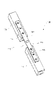
[0005] Fig. 1 shows a perspective view of a system according to a first
exemplary
embodiment of the present invention;
Fig. 2 shows a perspective view of a system according to a second exemplary
embodiment of the present invention;
Fig. 3 shows a perspective view of a system according to a third exemplary
embodiment of the present invention;
Fig. 4 shows a side view of a bone fixation element of the system of Fig. 3;
and
Fig. 5 shows a perspective view of a system according to a fourth exemplary
embodiment of the present invention.
Detailed Description
[0006] The present invention may be further understood with reference to the
following
description and the appended drawings, wherein like elements are referred to
with the same
reference numerals. The exemplary embodiment of the present invention relate
to a system and
method for tracking the progress of bone healing. In particular, the exemplary
embodiments
describe systems and methods that calculate a ratio of strain at multiple
locations along an
implant and/or a bone. An exemplary embodiment of the system may include a
first sensor on a
surface of the implant adapted to be positioned at a location proximate a
weakened portion of the
bone. Strain on the implant at this location will be affected by the strength
or stiffness of the
weakened bone and the load placed on the bone by the patient. A second sensor
may be placed
on the implant at a location in which strain measured by the second sensor is
affected only by the
2

CA 02775398 2012-03-26
WO 2011/050149
PCT/US2010/053519
load placed on the bone such that the measured strain is substantially
unchanged by the bone
healing process. Thus, a ratio between the strains measured by the first and
second sensors
provides information corresponding to bone healing, regardless of the load on
the bone. It will
be understood by those of skill in the art that although the exemplary
embodiment specifically
describe tracking the healing progress of a leg bone, the present invention
may be used to track
the progress of healing of any load bearing bone. It will also be understood
by those of skill in
the art that although the exemplary embodiments specifically show and describe
two sensors, the
present invention may include additional sensors along different areas of the
bone to determine
ratios corresponding to the bone healing progress of the different areas. In
addition, although
exemplary embodiments show a bone plate, the present invention may be used
with any other
fixation element such as, for example, screws, intramedullary devices,
external fixators, spine
fixation implants and prosthetics.
[0007] As shown in Fig. 1, a system 100 according to a first exemplary
embodiment of the
invention comprises an implant 102 (e.g., a bone plate) and first and second
sensors 104, 106,
respectively. The implant 102 is configured for fixation over a target portion
of a bone 108 to,
for example, fix a fracture 110 or to support a weakened portion of the bone
108. The first and
second sensors 104, 106 are mounted along a surface 114 of the implant 102
such that the first
and second sensors 104, 106 may be mechanically coupled to the bone 108.
Although the
surface 114 is shown as facing away from the bone 108 when the implant 102 is
fixed to the
bone 108 in a desired location, it will be understood by those of skill in the
art that the sensors
104, 106 may be mounted along any surface of the implant 102. For example, the
sensors 104,
106 may also be mounted on a surface of the implant 102 facing the bone 108 or
a surface on a
side of the implant 102. The first and second sensors 104, 106, respectively,
are positioned on
the implant 102 so that, when the implant is in a desired position on the bone
108, the first sensor
104 is located over a site of the fracture 110 while the second sensor 106 is
separated from the
fracture 110 over a healthy (i.e., solid) portion 112 of the bone 108 to
measure levels of strain
and/or load on the implant 102, at these positions along the implant 102. The
second sensor 106
should be isolated between two screws locked in a healthy portion 112 of the
bone 108 to
3

CA 02775398 2012-03-26
WO 2011/050149
PCT/US2010/053519
measure a load on the bone 108.
[0008] The sensors 104, 106 in this embodiment may be passively powered MEMs
sensors
that are used to measure strain and include an interface for wireless
connection to a data
collection device as would be understood by those skilled in the art. In
another embodiment, the
sensors 104, 106 may be powered chips that are connected to a printed circuit
board (PCB). This
permits strain on the implant 102 to be measured and transmitted to the data
collection device for
further processing without physically accessing the sensors 104, 106. It will
be understood by
those of skill in the art that the strain measurements detected by the sensors
104, 106 are not
required to represent actual strain values, but may include any signal that
changes based on
changing strains of their substrates. For example, the MEMS sensors 104, 106
may be RF
devices that deform when a strain is placed thereon, resulting in a frequency
shift caused by a
change in capacitance of the sensors 104, 106 such that the frequency shift
corresponds to a
change in strain. As would be understood by those skilled in the art, an
external device may be
employed to wirelessly provide a signal to the sensors 104, 106. Changes in a
returned signal
may then be measured to determine a level of strain to which the sensor is
subject. A ratio of the
strain measured by the first sensor 104 to the strain measured by the second
sensor 106 may then
be deteiiiiined by a physician or other professional to track healing
progress. Alternatively, the
ratio may be determined by a processing device that may also store the strain
measurements and
the determined ratios (e.g., in an internal memory or on an external storage
device) so that
changes in the ratio may be reviewed to more fully understand the progression
of the healing
over time.
[0009] It will be understood by those of skill in the art that when the
bone 108 is initially
broken or fractured, strain on the implant 102 at the location of the fracture
110 will vary based
on changing mechanical properties of the bone 108 during the healing process
and the load
placed on the bone 108 (e.g., the weight that the patient places on the leg)
while the strain
measured in the healthy portion 112 varies based only on the load placed on
the bone 108. Thus,
taking a ratio of the strains measured by the two sensors 104, 106 normalizes
the effects of the
4

CA 02775398 2012-03-26
WO 2011/050149
PCT/US2010/053519
load on the sensors 104, 106 providing data corresponding to the stiffness of
the bone 108 at the
fracture site 110. The ratio of the measurements from the first sensor 104 to
the measurements
from the second sensor 106 during the healing process should trend in a
decreasing pattern over
time, whereas a lack of healing would show no recognizable trend over time.
[0010] As shown in Fig. 2, a system 200 according to a second exemplary
embodiment of the
invention is substantially similar to the system 100, including an implant 202
and at least two
sensors 204, 206. However, rather than both sensors 204, 206 being positioned
on the implant
202, the first sensor 204 is located on a surface 214 of the implant 202 in a
position
corresponding to a fracture of a bone 208, while the second sensor 206 is
placed directly on a
solid portion 212 of the bone 208, outside a perimeter of the implant 202.
Thus, the first sensor
204 measures strain on the implant 202 at a position corresponding to the site
of the fracture 210
while the second sensor 206 measures strain on the solid portion 212 of the
bone 208. Similarly
to the system 100, a ratio between the strains measured by the first and
second sensors 204, 206
is determined and tracked to study the progress of healing in the bone 208. As
indicated above,
the ratio of the strain measurements from the first sensor 204 to the strain
measurements from the
second sensor 206 trend in a decreasing pattern as the bone 208 heals, whereas
a lack of healing
will show no recognizable trend over time.
[0011] As shown in Figs. 3 - 4, a system 300 according to a third exemplary
embodiment of
the invention is substantially similar to the system 200, comprising an
implant 302 and at least
two sensors 304, 306. Similarly to the first sensor 204, the first sensor 304
is placed on a surface
314 of the implant 302 in a location corresponding to a position of a fracture
310 of a bone 308
(when the implant 302 is mounted on the bone 308 in a desired position) to
measure strain on the
implant 302 at the position of the fracture 310 while the second sensor 306 is
placed directly on a
solid portion 312 of the bone 308. However, rather than being placed on an
exterior surface of
the bone 308, the second sensor 306 is placed within the solid portion 312
via, for example, a
bone fixation element 316 (e.g., screw).

CA 02775398 2012-03-26
WO 2011/050149
PCT/US2010/053519
[0012] The second sensor 306 may be attached adjacent to a proximal end 318 of
the bone
fixation element 316 such that when the bone fixation element 316 is inserted
into the solid
portion 312 of the bone, the second sensor 306 contacts a cortical wall of the
bone 308. The
second sensor 306 may be printed or mounted around a portion of the bone
fixation element 316
to measure deformation of the bone 308 which is directly related to strain on
the bone 308. The
ratio of the measurements from the first sensor 304 to those of the second
sensor 306 may then
be determined to track healing progress in the same manner described above.
[0013] As shown in Fig. 5, a system 400 according to a fourth exemplary
embodiment of the
invention is substantially similar to the system 100, comprising an implant
402 and first and
second sensors 404, 406, respectively, both of which are mounted on the
implant 402. Similarly
to the first sensor 104, the first sensor 404 is located on the implant 402 in
a position which,
when the implant 402 is in the desired position, corresponds to the location
of a fracture 410 so
that the first sensor 404 measures strain on the implant 402 at a position
corresponding to the site
of the fracture 410. The second sensor 406 is positioned on a portion 420 of
the implant 402
having greater flexibility than the portion of the implant 402 on which the
first sensor 404 is
mounted. For example, the portion 420 may be made more flexible than other
portions of the
implant 402 by reducing a width (i.e., an extent of the implant 402 across a
bone facing surface
thereof in a direction perpendicular to a longitudinal axis of the implant
402) and/or a thickness
of the portion 420 (i.e., a distance between the bone facing surface and a
surface thereof which
faces away from the bone) as compared to remaining portions of the implant
402. In a preferred
embodiment, the flexible portion 420 is adjacent to an end 422 of the implant
402 so that the
second sensor 406 is separated from the fracture 410 by a distance great
enough to ensure that
the underlying portion 412 of the bone 408 is solid.
[0014] The second sensor 406 on the flexible portion 420 of the implant 402 is
fixed to the
solid portion 412 of the bone 408 via, for example, locking screws inserted in
holes 424 on
opposing sides thereof The second sensor 406 measures strain on a portion of
the implant 402
corresponding to the solid portion 412 of the bone 408 so that measurements
from the second
6

CA 02775398 2012-03-26
WO 2011/050149
PCT/US2010/053519
sensor 406 may be used to normalize measurements from the first sensor.
Similarly to the
placement of a sensor directly in or on a bone, as described in conjunction
with systems 200 and
300, placing the second sensor 406 on a more flexible portion 420 of the
implant 402 between
two locked screws permits a more accurate measurement of the strain on the
underlying solid
portion 412 of the bone 408, as compared to the results from placing the
second sensor 406 on a
stiffer portion of the implant 402. The ratio of the measurements from the
first sensor 404 to the
measurements from the second sensor 406 during the healing process should
trend in a pattern
indicating an increasing stiffness of the bone 408 over time, whereas a lack
of healing should
show no recognizable trend over time.
[0015] It will be understood by those of skill in the art that other
mechanisms may be
employed for normalizing measurements of strain on a portion of an implant
which, when
mounted on a bone in a target location, corresponds to a position of a
fracture or other weakened
portion of that bone. For example, the patient may be provided with load
sensors on which to
push or stand with the affected limb such that a load measurement may be taken
simultaneously
with a strain measurement of the sensor on the implant. Alternatively, the
patient may be
provided with a sensor (e.g., placed in the sole of a shoe) to measure the
load placed on the
affected leg, if the affected bone is the femur or tibia.
[0016] It will be apparent to those skilled in the art that various
modifications and variations
can be made in the structure and the methodology of the present invention,
without departing
from the spirit or the scope of the invention. Thus, it is intended that the
present invention cover
the modifications and variations of this invention provided that they come
within the scope of the
appended claims and their equivalents.
7

Dessin représentatif
Une figure unique qui représente un dessin illustrant l'invention.
États administratifs

2024-08-01 : Dans le cadre de la transition vers les Brevets de nouvelle génération (BNG), la base de données sur les brevets canadiens (BDBC) contient désormais un Historique d'événement plus détaillé, qui reproduit le Journal des événements de notre nouvelle solution interne.

Veuillez noter que les événements débutant par « Inactive : » se réfèrent à des événements qui ne sont plus utilisés dans notre nouvelle solution interne.

Pour une meilleure compréhension de l'état de la demande ou brevet qui figure sur cette page, la rubrique Mise en garde , et les descriptions de Brevet , Historique d'événement , Taxes périodiques et Historique des paiements devraient être consultées.

Historique d'événement

Description Date
Représentant commun nommé 2019-10-30
Accordé par délivrance 2019-09-17
Inactive : Page couverture publiée 2019-09-16
Un avis d'acceptation est envoyé 2019-08-07
Inactive : Q2 réussi 2019-07-23
Inactive : Approuvée aux fins d'acceptation (AFA) 2019-07-23
Modification reçue - modification volontaire 2019-04-03
Inactive : Dem. de l'examinateur par.30(2) Règles 2018-10-11
Inactive : Rapport - CQ échoué - Mineur 2018-09-04
Inactive : CIB attribuée 2018-06-11
Modification reçue - modification volontaire 2018-05-29
Inactive : Dem. de l'examinateur par.30(2) Règles 2017-11-30
Inactive : Rapport - Aucun CQ 2017-11-28
Lettre envoyée 2017-11-09
Requête en rétablissement reçue 2017-11-02
Préoctroi 2017-11-02
Retirer de l'acceptation 2017-11-02
Taxe finale payée et demande rétablie 2017-11-02
Inactive : Taxe finale reçue 2017-11-02
Modification reçue - modification volontaire 2017-11-02
Réputée abandonnée - les conditions pour l'octroi - jugée non conforme 2017-10-19
Un avis d'acceptation est envoyé 2017-04-19
Lettre envoyée 2017-04-19
Inactive : Approuvée aux fins d'acceptation (AFA) 2017-04-06
Inactive : Q2 réussi 2017-04-06
Modification reçue - modification volontaire 2016-12-20
Inactive : Rapport - Aucun CQ 2016-06-21
Inactive : Dem. de l'examinateur par.30(2) Règles 2016-06-21
Lettre envoyée 2015-11-09
Inactive : Transfert individuel 2015-11-02
Modification reçue - modification volontaire 2015-10-30
Lettre envoyée 2015-10-15
Requête d'examen reçue 2015-10-08
Exigences pour une requête d'examen - jugée conforme 2015-10-08
Toutes les exigences pour l'examen - jugée conforme 2015-10-08
Inactive : Page couverture publiée 2012-06-01
Lettre envoyée 2012-05-11
Inactive : Notice - Entrée phase nat. - Pas de RE 2012-05-11
Inactive : CIB en 1re position 2012-05-10
Inactive : CIB attribuée 2012-05-10
Demande reçue - PCT 2012-05-10
Modification reçue - modification volontaire 2012-03-26
Exigences pour l'entrée dans la phase nationale - jugée conforme 2012-03-26
Demande publiée (accessible au public) 2011-04-28

Historique d'abandonnement

Date d'abandonnement Raison Date de rétablissement
2017-11-02
2017-10-19

Taxes périodiques

Le dernier paiement a été reçu le 2018-09-24

Avis : Si le paiement en totalité n'a pas été reçu au plus tard à la date indiquée, une taxe supplémentaire peut être imposée, soit une des taxes suivantes :

  • taxe de rétablissement ;
  • taxe pour paiement en souffrance ; ou
  • taxe additionnelle pour le renversement d'une péremption réputée.

Les taxes sur les brevets sont ajustées au 1er janvier de chaque année. Les montants ci-dessus sont les montants actuels s'ils sont reçus au plus tard le 31 décembre de l'année en cours.
Veuillez vous référer à la page web des taxes sur les brevets de l'OPIC pour voir tous les montants actuels des taxes.

Titulaires au dossier

Les titulaires actuels et antérieures au dossier sont affichés en ordre alphabétique.

Titulaires actuels au dossier
DEPUY SYNTHES PRODUCTS, LLC
Titulaires antérieures au dossier
CARL DEIRMENGIAN
GEORGE MIKHAIL
GLEN PIERSON
Les propriétaires antérieurs qui ne figurent pas dans la liste des « Propriétaires au dossier » apparaîtront dans d'autres documents au dossier.
Documents

Pour visionner les fichiers sélectionnés, entrer le code reCAPTCHA :


Pour visualiser une image, cliquer sur un lien dans la colonne description du document. Pour télécharger l'image (les images), cliquer l'une ou plusieurs cases à cocher dans la première colonne et ensuite cliquer sur le bouton "Télécharger sélection en format PDF (archive Zip)" ou le bouton "Télécharger sélection (en un fichier PDF fusionné)".

Liste des documents de brevet publiés et non publiés sur la BDBC .

Si vous avez des difficultés à accéder au contenu, veuillez communiquer avec le Centre de services à la clientèle au 1-866-997-1936, ou envoyer un courriel au Centre de service à la clientèle de l'OPIC.


Description du
Document 
Date
(aaaa-mm-jj) 
Nombre de pages   Taille de l'image (Ko) 
Description 2012-03-25 7 350
Revendications 2012-03-25 4 114
Abrégé 2012-03-25 2 64
Dessin représentatif 2012-03-25 1 7
Dessins 2012-03-25 4 40
Revendications 2012-03-26 3 90
Description 2016-12-19 7 341
Revendications 2016-12-19 3 71
Revendications 2017-11-01 6 138
Revendications 2018-05-28 3 72
Revendications 2019-04-02 3 80
Dessin représentatif 2019-08-14 1 4
Avis d'entree dans la phase nationale 2012-05-10 1 194
Courtoisie - Certificat d'enregistrement (document(s) connexe(s)) 2012-05-10 1 104
Courtoisie - Lettre d'abandon (AA) 2017-11-08 1 166
Rappel - requête d'examen 2015-06-22 1 124
Accusé de réception de la requête d'examen 2015-10-14 1 174
Courtoisie - Certificat d'enregistrement (document(s) connexe(s)) 2015-11-08 1 102
Avis du commissaire - Demande jugée acceptable 2017-04-18 1 162
Avis de retablissement 2017-11-08 1 170
Demande de l'examinateur 2018-10-10 5 270
PCT 2012-03-25 3 100
Requête d'examen 2015-10-07 2 54
Modification / réponse à un rapport 2015-10-29 2 55
Demande de l'examinateur 2016-06-20 3 210
Modification / réponse à un rapport 2016-12-19 13 367
Rétablissement / Modification / réponse à un rapport 2017-11-01 10 244
Taxe finale 2017-11-01 2 48
Demande de l'examinateur 2017-11-29 5 333
Modification / réponse à un rapport 2018-05-28 12 291
Modification / réponse à un rapport 2019-04-02 13 373