Note : Les descriptions sont présentées dans la langue officielle dans laquelle elles ont été soumises.
WO 2011/057299 PCT/US2010/056084
SYSTEM AND METHOD FOR PROVIDING ACCESS AND CLOSURE TO TISSUE
FIELD OF THE INVENTION
The invention relates generally to devices and methods
for performing surgical procedures, and more specifically to
access and closure technologies pertinent to wound or defect
closure, such as closures required following transapical or
transventrical cardiac diagnostic and interventional
procedures.
BACKGROUND
Minimally invasive diagnostic and interventional
procedure prevalence in US and foreign hospitals continues to
increase, as does the demand for certain procedures which
involve placement of relatively large devices into targeted
locations within tissue structures of criticality. Procedures
such as aortic valve replacement conventionally have been
addressed with open surgical procedures which are highly
invasive. More recently, such procedures have been attempted
using natural lumen (i.e., through large blood vessels after
an initial surgical transcutaneous or percutaneous access to
such vessels) access and delivery systems. Referring to
Figure 1, such systems typically are configured, for example,
to reach the aortic valve (112) location inside of the heart
(102) from an antegrade approach, which generally requires
navigating instrumentation through three of the four chambers
of the beating heart (the right atrium 122, left atrium 108,
and left ventricle 120, by way of the mitral valve 110 and
atrial septum), or from a retrograde approach, which generally
requires navigating instrumentation along the aortic arch,
from the descending aorta (104) to the ascending aorta (106)
and adjacent the aortic valve (112). Each of these approaches
WO 2011/057299 PCT/US2010/056084
presents certain clinical challenges to the surgical team,
some of which may be avoided by using what is referred to as a
transapical approach, whereby the surgeon creates
transcutaneous access to the region around the apex of the
heart (126) with a surgical thoracotomy, followed by direct
access to the left ventricle (120) using a needle or other
device aimed to access the left ventricle (120) around the
left ventricular apex (124), which may be followed by one or
more dilating instruments to create a temporary access port to
the left ventricle. Aspects of a conventional access
procedure are illustrated in Figure 2, wherein a needle device
(134) is puncturing the muscular heart wall (130) to gain
access to the left ventricle (120) around the location of the
left ventricular apex (124). Also shown is a guidewire (136)
which may be advanced (38) toward and through the aortic valve
(112) to assist with diagnostic and interventional aspects of
the procedure. Using these and other instruments such as
dilators, this left ventricular access port may be utilized,
for example, to replace an aortic valve if bleeding and tissue
damage around the access port can be successfully mitigated
during such procedure. Subsequent to such a procedure, the
instrumentation needs to be removed and the access port
closed, usually leaving a prosthetic valve or portion thereof
behind. The successful closure of a transapical wound on a
beating heart of a patient is obviously of high criticality to
such a procedure, as is the minimization of loss of blood.
Conventional transapical closure techniques typically involve
the placement of small sutures to create a purse-string type
effect to close the wound as the instrumentation is withdrawn,
and it may be very difficult to repeatably create acceptable
closures using these techniques without a larger thoracotomy
or improved instrumentation. In other words, one of the key
challenges to transapical intervention remains transapical
2
WO 2011/057299 PCT/US2010/056084
wound closure. Indeed, it is believed that transapical access
may provide enhanced stability and control during procedures
such as aortic valve replacement, due to the fact that the
operator may have a relatively direct mechanical connection
with the pertinent instrumentation, relative to the connection
that he may have using, for example, an antegrade or
retrograde vascular approach with more compliant catheter type
tools. For this reason, it is even more desirable to
successfully address the challenges of transapical access and
closure. Further, it would be desirable to have a wound or
access closure technology that was applicable not only to
transapical access port closure, but also other closure
demands pertinent to other surgical interventions of the human
body wherein wounds or ports are created, such as in
gastrointestinal or gynecological surgery.
BRIEF DESCRIPTION OF THE DRAWINGS
Figure 1 illustrates aspects of the human heart anatomy.
Figure 2 illustrates a conventional transapical access
procedure.
Figures 3A to 3L illustrate various aspects of a closure
configuration wherein a helical tubular member may be utilized
to deploy an elongate prosthesis such as a suture or coil.
Figures 4A-4D depict techniques for implementing various
embodiments of the subject helical closure configurations.
SUMMARY OF THE INVENTION
3
WO 2011/057299 PCT/US2010/056084
One embodiment is directed to a system for closing a
defect in a tissue wall, comprising: a base member having a
proximal end and a distal end, the proximal end being
configured to be manually manipulated by an operator; a
helical tubular member having a proximal end, a distal end, a
longitudinal axis, and a helical length in between the
proximal and distal ends, the helical tubular member defining
a helical lumen therethrough, wherein the proximal end is
coupled to the base member and the distal end defines a distal
outlet of the helical lumen; and an elongate prosthesis
coupled to the helical tubular member along two or more
portions of the helical length and configured to be deployed
into a portion of the tissue wall when decoupled from the
helical tubular member; wherein the helical tubular member is
configured to be helically advanced into an advanced position
in the tissue wall with the defect substantially aligned with
the longitudinal axis of the helical tubular member, and to
decouple from the elongate prosthesis upon helical retraction,
leaving the elongate prosthesis behind in a deployed helical
configuration similar to that previously defined by the
helical advanced position of the helical tubular member, the
deployed configuration being selected to retain coaptation of
the defect. The system may further comprise a plug member
having an elongate central portion, the plug member insertable
into the defect in the tissue wall and configured to span at
least a portion of a depth dimension of the defect in the
tissue wall and place at least a portion of the tissue wall
adjacent the defect into a constrained configuration in
between the plug member and portions of the deployed helical
configuration of the elongate prosthesis. The base member
comprises an elongate shaft. The base member may define a
hole therethrough, the hole configured to facilitate a movable
coupling between the base member and an elongate instrument
4
WO 2011/057299 PCT/US2010/056084
selected from the group consisting of: a catheter, a cannula,
a dilator, a needle, a guidewire, and an elongate probe. The
helical tubular member may have a tubular outer diameter
(i.e., the diameter of the overall helical construct) between
about lmm and about 25mm. The helical tubular member may have
a helical lumen diameter (i.e., the inner diameter of the
helical tubular member material) between about 10 thousandths
of an inch and about 80 thousandths of an inch; the outer
diameter of the helical tubular member material may be between
about 30 thousandths of an inch and about 100 thousandths of
an inch. The helical tubular member may have a helical
winding pitch between about lmm and about 10mm. The helical
tubular member distal end may comprise a cutting tip. The
helical tubular member may comprise a metal or polymer
material. The helical tubular member may comprise a
helically-bent section of metal hypotube. The elongate
prosthesis may comprise an unloaded three dimensional shape
that is different from the shape of the helical lumen. The
unloaded three dimensional shape may substantially approximate
a line. The unloaded three dimensional shape may comprise a
helical shape having a circular section diameter that is less
than that of the helical lumen. The elongate prosthesis may
be highly flexible into a plurality of unloaded three
dimensional shape configurations. The elongate prosthesis may
comprise an element selected from the group consisting of: a
suture, a wire, and a linkage. The elongate prosthesis may
comprise a material selected from the group consisting of: a
metal, a non-bioresorbable polymer, a bioresorbable polymer, a
non-bioresorbable co-polymer, and a bioresorbable co-polymer.
The elongate prosthesis further may comprise a distal anchor.
The distal anchor may comprise an element selected from the
group consisting of: a knot, a barb member, and a catch
geometry. The plug member may comprise a distal portion
5
WO 2011/057299 PCT/US2010/056084
coupled to the distal end of the central portion, and a
proximal portion coupled to the proximal end of the central
portion, each of the proximal and distal portions being
expandable from a collapsed configuration to an expanded
configuration such that the outer diameter of each collapsed
configuration is larger than that of the central portion. The
plug member central portion may comprise a substantially
cylindrical geometry. The plug member may comprise a material
selected from the group consisting of: a metal, a non-
bioresorbable polymer, a bioresorbable polymer, a non-
bioresorbable co-polymer, a bioresorbable co-polymer, and a
fabric.
DETAILED DESCRIPTION
Referring to Figures 3A through 3L, various aspects of
embodiments associated with a transapical access and closure
system are depicted. These same configurations may be applied
to various other cardiovascular and noncardiovascular tissue
wall defect or port closure scenarios. As shown in Figure 3A,
an assembly comprising an elongate member, such as a catheter
defining a working lumen (40) therethrough, a cannula, or
other elongate instrument or probe, is advanced (14) toward a
subject tissue wall (10), in this example a portion of the
myocardium. The elongate member (12) may comprise a
hemostatic valve. The assembly also comprises a base member
(2) movably coupled to the elongate member (12) via a hole or
annulus (3) created through the middle of the base member (2)
to accommodate one or more elongate tools such as the elongate
member (12) shown. The base member preferably is configured
to be manually manipulated in insertion/retraction and also
rotation and may comprise an elongate shape itself (for
example, such as a catheter or hypotube structure) to
6
WO 2011/057299 PCT/US2010/056084
facilitate manual manipulation from an extracorporeal location
by an operator; for illustration a simple embodiment is shown
wherein the base member (2) comprises a relatively thin
cylindrical shape. The depicted assembly also comprises a
helical tubular member (4) or "spring insertion device" with a
proximal end coupled to the base member (2) and a distal end
having a cutting tip configured to readily dive into the
subject tissue structure with helical advancement (i.e., in a
corkscrew type fashion) of the base member (2) and helical
tubular member (4). The cutting tip (16) may comprise a
sharpened end of the helical tubular member (4), or may
comprise a sharpened fitting coupled to the helical tubular
member (4). The helical tubular member (4) may comprise a
metal or polymer material, and in one embodiment comprises a
helically bent or wrapped section of metal hypotube defining a
lumen therethrough which may be utilized for deployment of an
elongate prosthesis member or "helical spring", as described
below.
Referring to Figure 3B, a distal portion of the elongate
member (12) has been advanced across at least a portion of the
subject tissue wall (10), in this example across the entire
thickness of the wall (10) to facilitate a procedure such as
an aortic valve replacement wherein the working lumen (40) of
the depicted elongate member (12) may be utilized as a conduit
for passing various tools and prosthesis portions. Figure 3B
depicts an embodiment wherein the helical tubular member (4)
has not been advanced into or across the tissue wall (10) yet,
and such a configuration may be utilized to facilitate a valve
replacement or other procedure. In another embodiment, as
described below, for example, in reference to Figures 4C and
4D, the helical tubular member (4) indeed may be advanced
across at least a portion of the tissue wall (10) before
utilizing the working lumen (40) for the interventional
7
WO 2011/057299 PCT/US2010/056084
procedure, to ensure that a closure scenario is nearly fully
in place before the intervention begins.
Referring to Figure 3C, after the use of the working
lumen of the elongate member (12) for any interventional
procedure is complete and a closure is desired, a collapsible
and/or pliable sealing device or plug member (8) may be
inserted through the hemostatic valve and working lumen (40)
of the elongate member (12) and allowed to partially expand to
its natural shape, as shown in Figure 3D wherein the distal or
top portion of the plug member (8) has expanded outward.
Also, the assembly of the base member (2) and helical tubular
member (4) may be helically advanced (20) may be advanced into
the tissue wall, as shown in Figure 3E.
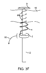
Referring to Figure 3F, with helical retraction (22) of
the the assembly of the base member (2) and helical tubular
member (4), an elongate prosthesis (6), is decoupled and left
behind in the tissue wall (10) a helical or coiled
configuration. In the views illustrated in Figures 3A-3E, the
elongate prosthesis (6) preferably has been located within a
small lumen defined by the helical tubular member (4), and
upon reversing the helical advancement to helical retraction,
the elongate prosthesis (6) is configured to be pulled out of
the helical tubular member (4) and left in place. A distal
end of the elongate prosthesis (6) may comprise an anchor,
such as a knot tied in the material or the elongate prosthesis
(6), a barb coupled to or formed into the elongate prosthesis
(6), or other catch member such as a hook, burr, or toggle
bolt anchor structure. The elongate prosthesis may naturally
have an unloaded three dimensional shape that is different
than that of the helical tubular member (4). For example, in
one embodiment, the unloaded three dimensional shape may
substantially approximate a line - as in a piece of unloaded
straight wire sitting on a tabletop. In the embodiment
8
WO 2011/057299 PCT/US2010/056084
depicted in Figures 3F, and 3J-3L, the elongate prosthesis (6)
has helical unloaded three dimensional shape that has a
helical diameter smaller than that of the helical tubular
member (4), such that when the elongate prosthesis (6) is
released from the lumen of the helical tubular member (4), it
tends to apply a mild constricting load (a mild uniform radial
compression) on tissue trapped between the elongate prosthesis
and the central axis of the tubular member (12), or the
central axis of the elongate prosthesis helix. Thus in the
configuration shown in Figure 3F, the elongate prosthesis (6)
is constricting the trapped tissue toward the outer surface of
the tubular member (12). In Figures 3J-3L, the elongate
prosthesis (6) is constricting the trapped tissue toward the
outer surface of the deployed plug member (8) and/or tubular
member (12) outer surface that remains adjacently positioned.
The elongate prosthesis (6) may comprise a suture, wire, coil,
or linkage made from one or more metals, polymers, co-
polymers, or fabrics. The polymers may be bioresorbable. The
elongate prosthesis (6) may be very thin and/or highly
flexible as an overall construct such that it may be
configured into a variety of unloaded three dimensional shape
configurations.
Referring to Figure 3G, a close-up orthogonal view of one
embodiment of an expandable plug member (8) is shown. A
central portion (26), which may be substantially cylindrical
(28) in shape, may serve as a coupling between a concave
expandable distal portion (32) and an arcuate conical proximal
portion (30). Such a plug member (8) may comprise one or more
metals, polymers, co-polymers, fabrics, or foams. The
polymers may be bioresorbable. Preferably such a plug member
(8) may be folded or collapsed to a substantially small
cylindrical geometry for delivery through a working lumen, as
9
WO 2011/057299 PCT/US2010/056084
shown, for example, in Figure 3C. Other views of an
expandable plug member (8) are shown in Figures 3H and 31.
Referring to Figure 3J, to further complete the
illustrated defect closure process, the elongate member (12)
is further withdrawn (34), as is the base member (2) further
withdrawn (38). With the increased withdrawal (34) of the
tubular member (12), more of the expandable plug member (8) is
allowed to expand out of the distal end of the elongate member
(12) until it is fully expanded, as shown in Figure 3k. The
deployed elongate prosthesis (6) continues to urge captured
tissue against the outer surface of the fully deployed
expandable plug member (8), causing a hemostatic closure of
the defect. Referring to Figure 3L, the assembly of the
elongate member (12), base member (2), and helical tubular
member (4) is withdrawn (36) away from the surgical theater,
leaving behind a hemostatic closure configuration of the
elongate prosthesis (6) and the plug member (8).
In a defect closure scenario wherein the tissue structure
(10) illustrated in Figures 3A-3L is a myocardial wall, the
radial forces applied by the deployed elongate prosthesis
generally apply no axial force to the plug member (8) in the
region of the relatively straight central portion (26) of the
plug member (8), and preferably this central portion (26) is
relatively thin, which may allow the myocardial tissue to
almost completely close. Radial forces from the arcuate
conical portion (30) have an unresolved axial component in the
proximal direction (i.e., tending to pull against the upper
concave portion (32). Such a loading configuration will have
the effect of pulling the inner seal or concave portion (32)
tighter against the inner wall of the heart cavity, and
natural contractile motion of the heart may act in concert
with the elongate prosthesis, or "spring member", forces,
possibly providing a beneficial seal-tightening action with
WO 2011/057299 PCT/US2010/056084
additional heartbeats. Preferably such a deployed
configuration distributes closure stresses more evenly on
local tissues than does a conventional suture-based closure.
While the illustrative embodiments of Figures 3k and 3L
depict the plug member (8) and/or elongate prosthesis (6)
spanning approximately the full thickness of the depicted
tissue wall (10), in other embodiments it is desirable to have
one or both ends of each of these structures (8, 6) ultimately
positioned midsubstance (i.e., not immediately adjacent a
border of the tissue structure 10). Further, while the
embodiments depicted in Figures 3K and 3L show the plug member
(8) and elongate prosthesis (6) substantially aligned
longitudinally (i.e., lengthwise along a longitudinal central
axis of either member) as well as axially (i.e., about a
longitudinal central axis of either member), in other
embodiments, it is desirable to have either longitudinal
misalignment, axial misalignment, or both, to facilitate
stable hemostasis. For example, in one embodiment, it is
desirable to have the proximal portion of the elongate
prosthesis helically extend proximally beyond the
bottom/proximal margin of the arcuate conical portion (30) of
the plug member. In other embodiments, a non-expandable plug
member may be used in place of the structurally
collapsible/expandable plug member (8) shown in Figures 3C-3L.
For example, in one embodiment, a simple cylindrical plug
member sized to be slightly less in diameter than the inner
diameter of the elongate member (12), to facilitate
pushability through the elongate member (12) during
deployment, may be utilized; such a nonexpanding plug member
may comprise a relatively compliant, or relatively
noncompliant, polymer, such as a bioresorbable polymer.
Referring to Figure 4A, one embodiment of deployment
process is illustrated. In this embodiment, an elongate
11
WO 2011/057299 PCT/US2010/056084
member, such as a catheter, cannula, needle, guidewire, or
other probe, is advanced across at least a portion of a
targeted tissue structure (41). This advancement may employ
successive tools, as in an "over-the-wire" procedure wherein a
needle and/or guidewire may serve as pathway predecessors for
a larger instrument such as a catheter or cannula. As shown
in Figure 4A, a working lumen of the installed elongate member
may be utilized to conduct a diagnostic and/or interventional
procedure, such as a heart valve inspection or replacement
(44). In the illustrated embodiment, next a base member
coupled with a helical tubular member may be advanced toward
the tissue structure, the helical tubular member preferably
carrying with it (i.e., threaded through at least a portion of
the helical lumen of the helical tubular member) an elongate
prosthesis (46). The helical tubular member may be helically
advanced into the targeted tissue structure, over the elongate
member (48), then helically retracted (50) to leave the
elongate prosthesis in place, preferably urging at least a
captured portion of the tissue structure toward the outer
surface of the remaining elongate member (i.e., and generally
toward the longitudinal axis of the helically deployed
elongate prosthesis). Finally the wound or defect may be
closed with removal of the elongate member and deployment of a
plug member (52), as shown in Figures 3C-3L.
Referring to Figure 4B, an embodiment similar to that of
Figure 4A is depicted, with exception that a plug member is
not utilized. Rather, after the elongate prosthesis has been
deployed through retraction of the helical tubular member
(50), closure of the defect and hemostasis are achieved merely
through the tissue coaptation provided by the helically
deployed elongate prosthesis, which preferably has a unloaded
three dimensional helical size that is smaller than that of
the helical tubular member, resulting in constriction of the
12
WO 2011/057299 PCT/US2010/056084
elongate prosthesis once deployed and free of the helical
tubular member (54).
Referring to Figure 4C, an embodiment is shown wherein
the elongate prosthesis member is helically deployed around
the installed elongate member (steps 42, 46, 48, and 50)
before any diagnostic and/or interventional procedure is
conducted (56). This allows for an operator or surgeon to
have the confidence that the closure mechanism is safely in
place before conducting the diagnostic and/or interventional
steps. Subsequently the diagnostic and/or interventional
tools are retracted, and retraction of the elongate member and
deployment of a plug member results in closure and hemostasis
(52).
Figure 4D illustrates an embodiment similar to that
described in reference to Figure 4C, with exception that a
plug member is not used, in an analogous scenario to that of
Figure 4B.
Any of the aforementioned deployed structures, including
sutures, anchor members, and ratcheting closure device
assembly components, may comprise resorbable materials in
addition to the aforementioned nonresorbable materials - to
facilitate combinations and permutations which may be
completely resorbed, leaving behind a biologically healed
transapical access wound.
Any of the devices described for carrying out the subject
interventions may be provided in packaged combination for use
in executing such interventions. These supply "kits" further
may include instructions for use and be packaged in sterile
trays or containers as commonly employed for such purposes.
Exemplary embodiments of the invention, together with
details regarding material selection and manufacture have been
set forth above. As for other details of the invention, these
13
WO 2011/057299 PCT/US2010/056084
may be appreciated in connection with the above-referenced
patents and publications. For example, one or more lubricious
coatings (e.g., hydrophilic polymers such as
polyvinylpyrrolidone-based compositions, fluoropolymers such
as tetrafluoroethylene, hydrophilic gel or silicones) may be
used in connection with various portions of the devices, such
as relatively large interfacial surfaces of movably coupled
parts, if desired, for example, to facilitate low friction
manipulation or advancement of such objects relative to other
portions of the instrumentation or nearby tissue structures.
The same may hold true with respect to method-based aspects of
the invention in terms of additional acts as commonly or
logically employed.
Also, it is contemplated that any optional feature of the
inventive variations described may be set forth and claimed
independently, or in combination with any one or more of the
features described herein. Reference to a singular item,
includes the possibility that there are plural of the same
items present. More specifically, as used herein and in claims
associated hereto, the singular forms "a," "an," "said," and
"the" include plural referents unless the specifically stated
otherwise. In other words, use of the articles allow for "at
least one" of the subject item in the description above as
well as claims associated with this disclosure. It is further
noted that such claims may be drafted to exclude any optional
element. As such, this statement is intended to serve as
antecedent basis for use of such exclusive terminology as
"solely," "only" and the like in connection with the
recitation of claim elements, or use of a "negative"
limitation.
Without the use of such exclusive terminology, the term
"comprising" in claims associated with this disclosure shall
allow for the inclusion of any additional element--
14
WO 2011/057299 PCT/US2010/056084
irrespective of whether a given number of elements are
enumerated in such claims, or the addition of a feature could
be regarded as transforming the nature of an element set forth
in such claims. Except as specifically defined herein, all
technical and scientific terms used herein are to be given as
broad a commonly understood meaning as possible while
maintaining claim validity.