Note : Les descriptions sont présentées dans la langue officielle dans laquelle elles ont été soumises.
CA 02785648 2014-06-04
METHOD AND APPARATUS FOR SECURING
PLANAR ORIENTATION OF ANALYSIS CHAMBER
[0001] BACKGROUND OF THE INVENTION
1. Technical Field
[0002] The present invention relates to methods and apparatus for imaging
biologic
specimens disposed within an analysis chamber in general, and to methods and
apparatus for
disposing the chamber in a planar orientation in particular.
2. Background Information
[0003] Historically, biologic fluid samples such as whole blood, urine,
cerebrospinal
fluid, body cavity fluids, etc., have had their particulate or cellular
contents evaluated by
smearing a small undiluted amount of the fluid on a slide and evaluating that
smear under a
manually operated microscope. Different areas of the smear were examined by
manipulating the
slide in an X-Y plane. Focus was accomplished by altering the position of one
or both of the
slide and the microscope objective along the Z-axis. Reasonable results were
attainable using
these techniques, but they relied heavily upon the technician's experience and
technique.
Manually examining the various fields of the sample is also labor intensive
and therefore not
practically feasible for commercial laboratory applications.
[0004] Automated apparatus capable of analyzing quiescent biologic fluid
samples
within a chamber are known. These devices typically maintain the sample in an
X-Y plane.
Image focus is accomplished by moving one or both of the sample or the device
optics relative to
the other along a Z-axis. To efficiently perform such an analysis, it is
necessary to provide such
=
focus at different heights in an accurate, rapid manner.
1
CA 02785648 2012-06-26
WO 2011/082109 PCT/US2010/062046
SUMMARY OF THE DISCLOSURE
[0005] According to an aspect of the present invention, an apparatus for
imaging a
biologic fluid sample quiescently residing within a chamber is provided. The
chamber includes a
first panel and a second panel, between which the biologic fluid sample
quiescently resides. At
least one of the first and second panels is flexible. The chamber has one or
more fields that are
each defined by a cross-sectional area. The apparatus comprises a field
illuminator, a chamber
flattener, a positioner, and an image dissector. The field illuminator has an
objective lens. The
chamber flattener has a platen with a window and a cover plate. The chamber
flattener is
operable to cause the chamber to assume a substantially uniforni Z-axis
position for substantially
all of the fields within the chamber. The positioner is adapted to position
the objective lens and
the chamber relative to one another. The image dissector is adapted to image
the sample residing
within the chamber.
[0006] According to another aspect of the present invention, an apparatus
for orienting a
biologic fluid sample chamber relative to an objective lens is provided. The
apparatus includes a
platen and a cover plate. The platen includes a window and a magnetic source.
The cover plate
has a chamber contact panel with a chamber aperture and at least one
deflectable flap contiguous
with the chamber aperture. The chamber contact panel includes a magnetically
attractive
material. The cover plate is positioned relative to the platen to allow the
chamber to be disposed
there between. The magnetic source and the chamber contact panel are
sufficiently attracted to
one another such that at least one panel of the chamber disposed between the
cover plate and
platen assumes a substantially planar position.
[0007] According to another aspect of the present invention, a method for
imaging a
biologic fluid sample quiescently residing between a first panel and a second
panel of a chamber
is provided. At least one of the first and second panels is flexible, and the
chamber has one or
more fields that are each defined by the cross-sectional area. The method
includes the steps of:
a) positioning the chamber at a Z-axis position relative to an objective lens;
b) flattening the
chamber so that it assumes a substantially uniform Z-axis position for
substantially all of the
fields within the chamber; c) moving one or both of the chamber and the
objective lens relative
to one another to bring the sample in focus; and d) imaging the biologic fluid
sample through the
platen window.
2
CA 02785648 2012-06-26
WO 2011/082109 PCT/US2010/062046
[0008] The present method and advantages associated therewith will become
more
readily apparent in view of the detailed description provided below, including
the accompanying
drawings.
BRIEF DESCRIPTION OF THE DRAWINGS
[0009] FIG. 1 is a diagrammatic illustration of the present analysis
apparatus.
[0010] FIG. 2 is schematic representation of the analysis present
apparatus.
[0011] FIG. 3 is a diagrammatic planar view of an embodiment of a sample
analysis
cartridge, illustrating a fluid module portion of the cartridge in an open
position.
[0012] FIG. 4 is a partially exploded view of the cartridge shown in FIG.
3 with the fluid
module closed in the housing, and an imaging tray portion disposed outside of
the housing.
[0013] FIG. 5 is a diagrammatic sectioned partial view of an analysis
chamber.
[0014] FIG. 6 is a diagrammatic view of an analysis chamber and a chamber
flattener
embodiment.
[0015] FIG. 7 is a diagrammatic view of an analysis chamber and a chamber
flattener
embodiment.
[0016] FIG. 8 is a diagrammatic view of an analysis chamber and a chamber
flattener
embodiment.
[0017] FIG. 9 is a planar view of the chamber side surface of a chamber
flattener platen
embodiment.
[0018] FIG. 10 is an opposite side planar view of the chamber flattener
platen
embodiment shown in FIG. 9.
[0019] FIG. 11 is a cover plate embodiment.
[0020] FIG. 12 is a perspective view of a cover plate embodiment and
collar mounted on
the housing of an objective lens.
[0021] FIG. 13 is a perspective view of a cover plate embodiment mounted
on the
housing of an objective lens.
[0022] FIG. 14 is a planar view of a cover plate embodiment mounted on
the housing of
an objective lens.
3
CA 02785648 2014-06-04
DETAILED DESCRIPTION OF THE INVENTION
[0023] Referring to FIGS. 1-2, the analysis apparatus 17 is configured to
receive a
sample analysis cartridge 18 having an analysis chamber 20 (e.g., see FIGS. 4-
5) adapted to
contain a biologic sample (e.g., anti-coagulated, whole blood) from a subject
for analysis. The
apparatus 17 includes a field illuminator 22, an image dissector 24, a
positioner 26, an analysis
chamber flattener 27, and a programmable analyzer 28. For purposes of this
description, the
terms "analyze" and "analysis" shall be defined as any examination or
evaluation of the fluid
sample, including but not limited to, the examination (e.g., visual,
enumeration, etc.) of
constituents within the biologic fluid sample.
[0024] The analysis apparatus 17 can be used with a variety of different
sample analysis
chambers 20, including those described in co-pending U.S. Patent Application
Ser. Nos.
61/287,955 filed December 18, 2009; 12/061,394; and 10/599,695.
For purposes of this disclosure, unless otherwise
noted, the invention will be described as using the analysis chamber and
cartridge described in
U.S. Patent Application Serial No. 61/287,955. The present invention is not
limited, however, to
use with the aforesaid chamber 20 and cartridge 18.
[0025] Referring to FIGS. 3-5, the sample analysis cartridge 18 includes a
fluid module
30, an image tray 32, and a housing 34. The fluid module 30 includes a sample
port 36
configured to receive a fluid sample from either a syringe or a subject
collection site; e.g., from a
finger or heel stick, or from a sample drawn from an arterial or venous
source. The fluid sample
is subsequently drawn into the cartridge 18 where it can be selectively
transferred to an analysis
chamber 20 located within the image tray 32.
[0026] Referring to FIG. 5, the analysis chamber 20 includes a first panel
38 and a
second panel 40, at least one of which is sufficiently transparent to permit a
biologic fluid sample
disposed between the panels 38, 40 to be imaged for analysis purposes. The
first and second
panels 38, 40 are preferably parallel and aligned with one another, and are
separated from each
other by a distance extending between the opposing surfaces 39, 41 of the two
panels 38, 40.
The alignment between the panels 38, 40 defines an area wherein light can be
transmitted
perpendicular to one panel 38, and it will pass through that panel, the
sample, and the other panel
40 as well, if the other panel is also transparent. The separation distance
between the panel
4
CA 02785648 2012-06-26
WO 2011/082109 PCT/US2010/062046
surfaces (also referred to as the "height" 42 of the chamber 20) is sized such
that a biologic fluid
sample disposed between the two surfaces 39, 41 will be in contact with both
surfaces 39, 41.
[0027] In some embodiments, including the embodiment disclosed in U.S.
Patent
Application Ser. No. 10/599,695, the first and second panels 38, 40 are
separated from one
another by at least three separators 44 (typically spherical beads). Examples
of acceptable
separators include polystyrene spherical beads that are commercially
available, for example,
from Thermo Scientific of Fremont, California, U.S.A., catalogue no. 4204A, in
four micron
(4 m) diameter. At least one of the panels 38, 40 or the separators 44 is
sufficiently flexible to
permit the chamber height 42 to approximate the mean height of the separators
44. The relative
flexibility provides the chamber 20 with a substantially uniform height
despite minor
dimensional tolerance variances in the separators 44. In those embodiments
where one of the
one of the panels (e.g., first panel 38) is formed from a material more
flexible than the separators
44 and the other panel (e.g., second 40), the more flexible panel 38 will
overlay the separators 44
and to the extent that a particular separator 44 is larger than the
surrounding separators 44, the
flexible panel 38 will flex around the larger separator 44 in a tent-like
fashion. In this manner,
although small local areas may deviate from the mean chamber height 42, the
mean height of all
the chamber sub-areas (including the tented areas) will be very close to that
of the mean
separator diameter. Consequently, for purposes of establishing a substantially
uniform Z-axis
chamber position, the deviations of the flexible panel are inconsequential.
[0028] In some embodiments, a bead 51 of adhesive material (e.g., a UV-
curable glue) is
disposed between the panels 38, 40, and is operable to attach the panels 38,
40 to one another.
[0029] Examples of acceptable panel materials include transparent plastic
film, such as
acrylic, polystyrene, polyethylene terphthalate (PET), cyclic olefin copolymer
(COC) or the like.
In some embodiments, one of the panels (e.g., the panel oriented to be the
bottom) is formed
from a strip of material with a thickness of approximately fifty microns (500,
and the other
panel (e.g., the panel oriented to be the top panel) is formed from the same
material but having a
thickness of approximately twenty-three microns (230.
[0030] The chamber 20 is typically sized to quiescently hold about 0.2 to
1.0 ul of
sample, but the chamber 20 is not limited to any particular volume capacity,
and the capacity can
vary to suit the analysis application. The term "quiescent" is used to
describe that the liquid
sample is deposited within the chamber 20 for analysis, and is not
purposefully moved during the
CA 02785648 2012-06-26
WO 2011/082109 PCT/US2010/062046
analysis. To the extent that sample motion is present within the chamber, it
will predominantly
be due to Brownian motion of foffned constituents within the sample, which
motion is not
disabling of the use of this invention.
[0031] Now referring back to FIG. 2, the field illuminator 22 includes a
light source and
objective optics (e.g., objective lens 48, filters, etc.). The light source
produces light throughout
a wavelength range broad enough to be useful for a plurality of analyses
(e.g., approximately 340
nm to 670 nm). The light source can produce light from a single source or from
a plurality
sources that collectively produce the light along the desired wavelengths;
e.g., a zenon arc lamp,
a tungsten halogen lamp, LEDs, or a pulsatile source.
[0032] The path of the light emanating from the illuminator 22 will depend
upon the
whether the sample is being analyzed using fluorescence or transmittance. When
fluorescence is
used, an objective lens 48 focuses light emanating from the light source
within the illuminator 22
into a light beam which, in turn, is directed into the sample quiescently
residing within the
chamber 20. The light beam illuminates at least one field of the sample, which
field is defined
by the cross-sectional area of the sample image which impinges on the image
dissector 24, or a
portion thereof. The light causes material within the sample (e.g.,
fluorescent dye) to fluoresce
and emit light of a particular wavelength. The emitted light passes back
through the objective
lens 48 and is subsequently captured by an image dissector 24. When
transmittance is used, the
field illuminator 22 is configured to direct light through the chamber first
panel 38 and second
panel 40 (which are both sufficiently transparent to allow the light to pass
there through), and the
fluid sample residing there between. The transmitted light passes through the
objective lens 48
and is subsequently captured by an image dissector 24.
[0033] The positioner 26 is adapted to change the relative positions of
the objective lens
48 and the analysis chamber 20. A change in the relative positions of the
objective lens 48 and
the analysis chamber 20 can be accomplished in a variety of different ways;
e.g., by moving one
of the objective lens 48 and analysis chamber 20 relative to the other along
all relevant axes (e.g.,
X, Y, and Z), or by moving the chamber 20 along particular axes (e.g., the X
and Y axes) and the
lens along the other axis (e.g., the Z-axis), etc. For ease of description,
the positioner 26 is
described herein as being adapted to move the cartridge 18 and incorporated
analysis chamber 20
along multiple axes of motion (e.g., X, Y, and Z) relative to a stationary
objective lens 48. The
present invention is not limited to this embodiment, however. The chamber 20
is movable in the
6
CA 02785648 2012-06-26
WO 2011/082109 PCT/US2010/062046
X-Y plane to permit the objective lens 48 to capture all fields of the sample
residing within the
chamber 20, and movable along the Z-axis to change the focal position relative
to the sample
height. Motion of the chamber 20 relative to the objective lens 48 can be
accomplished by a
variety of different devices, including but not limited to, a controllable
stepper motor that can be
operated to selectively produce either continuous motion of the chamber 20
relative to the
objective lens 48 or incremental movement of the chamber 20 relative to the
objective lens 48.
[0034] An acceptable image dissector 24 is a complimentary metal-oxide
semi-conductor
(CMOS) type digital image dissector 24, preferably one that can provide at
least eight (8), and
most preferably twelve (12), bits of resolution per pixel. The image dissector
24 converts an
image of the light into an electronic data format which can be seen and/or
interpreted in real-time
or at a subsequent time, using a data file version of the image.
Alternatively, an image dissector
24 other than a CMOS may be used to convert the image of light into an
electronic data format.
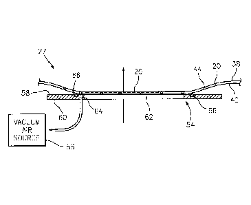
[0035] Referring to FIGS. 6-7, the analysis chamber flattener 27 is
adapted to manipulate
a flexible analysis chamber panel (e.g., one or both of the analysis chamber
panels 38, 40) into a
flat, planar orientation to facilitate the relative positioning of the sample
chamber 20 and the
objective lens 48 (see FIG. 2). A flat, planar chamber orientation positions
substantially all (and
preferably all) of the analysis fields of sample in the same Z-axis position.
As a result, the time
required to find the Z-axis focal plane within the sample is substantially
decreased, and the
ability to image all fields within the sample is greatly enhanced.
[0036] In a first embodiment shown in FIGS. 6 and 7, the chamber flattener
27 includes a
platen 54 and a source of air pressure 56 (e.g., positive air pressure or
suction). The platen 54
includes a chamber side surface 58, an opposite side surface 60, and a
substantially rigid
transparent window 62 extending between the two surfaces 58, 60. The window 62
is sized large
enough to permit light to pass through a substantial area of each chamber
panel 38, 40 when the
chamber 20 is aligned with the window 62; i.e., all of the sample disposed
within the chamber 20
is typically viewable through the window 62. FIG. 6 illustrates a chamber
flattener 27
embodiment wherein the air pressure source 56 is a vacuum. One or more ports
64 open to the
chamber side surface 58 are disposed adjacent the window 62. An air seal 66
disposed outside of
the one or more ports 64 is positioned to contact a chamber panel 40. Once the
chamber panel
40 is placed in contact with the air seal 66, a vacuum drawn by the air
pressure source 56 lowers
the pressure within the pocket formed by the chamber panel 40, the platen 54,
and the air seal 66.
7
CA 02785648 2012-06-26
WO 2011/082109 PCT/US2010/062046
When the pressure drops sufficiently, the chamber panels 38, 40 are drawn
against the flat,
planar platen window 62. As a result, the sample chamber 20 extending in the X-
Y plane is
maintained at a substantially uniform Z-axis position for substantially all of
the fields containing
sample. After the sample is imaged, the vacuum is released.
[0037] In the example shown in FIG. 7, the chamber flattener 27
embodiment includes an
air pressure source 56 adapted to produce positive pressure air that pushes
the sample chamber
20 against the platen window 62. An air seal 68 disposed, for example, on the
side of the
chamber 20 opposite the platen window 62 is configured to create a pocket for
the high pressure
air. Once the chamber 20 is aligned with the platen window 62, positive
pressure air from the air
pressure source 56 is directed into the pocket, consequently pressing the
chamber 20 against the
flat, planar platen window 62. As a result, the sample chamber 20 extending in
the X-Y plane is
maintained at a substantially uniform Z-axis position for substantially all of
the fields containing
sample. After the sample is imaged, the positive air pressure is removed.
[0038] Referring to FIGS. 8-12, a second embodiment of the chamber
flattener 27
includes a platen 70, a cover plate 72, and one or more magnets 73 (see FIG.
10). The platen 70
includes a chamber side surface 74, an second side surface 76, and a
substantially rigid
transparent window 78 extending between the two surfaces 74, 76. The window 78
is sized large
enough to permit light to pass through at least substantially all of the area
of the chamber panels
38, 40 when the chamber 20 is aligned with the window 78. In the embodiment
shown in FIG.
10, the chamber flattener 27 includes a number of magnets 73 (e.g., four)
sufficient to attract the
cover plate 72 as will be described below. A slot 80 is disposed in the second
side surface 76 of
platen 70 for each magnet 73. The slots 80 extend between a first end 82 and a
second end 84.
The first end 82 of each slot 80 is disposed in close proximity to the window
78, where it will be
aligned with the cover plate 72 when the chamber flattener 27 is assembled.
The second end 84
of each slot 80 is disposed away from the window 78, where it will not be
aligned with the cover
plate 72 when the chamber flattener 27 is assembled. The platen 70 is made of
a substantially
rigid non-magnetic material that can be fainted to have a planar chamber side
surface 74 within
acceptable flatness tolerances.
[0039] The cover plate 72 has a chamber contact panel 71, a first side
flange 102, and a
second side flange 104. The chamber contact panel 71 is a relatively thin flat
body that includes
a chamber aperture 86 (see FIG. 11) and typically includes at least one flap
88 contiguous with
8
CA 02785648 2012-06-26
WO 2011/082109 PCT/US2010/062046
the chamber aperture 86. The chamber contact panel 71 embodiment shown in FIG.
11 has four
(4) flaps 88 contiguous with the chamber aperture 86, each separated from the
adjacent flaps 88
by a slot 90. The specific geometry of the chamber contact panel 71, including
the number of
flaps 88, can be altered to accommodate different chamber 20 configurations.
The flaps 88 have
a cantilevered configuration that allows each to deflect as will be described
below. The chamber
contact panel 71 can include (or be made from) a material that is attracted by
magnets (e.g., 420
series martensitic stainless steel shim stock).
[0040] In the embodiment shown in FIGS. 8 and 12, the cover plate 72
comprises a first
side flange 102 and a second side flange 104. The first side flange 102 and
second side flange
104 attach the chamber contact panel 71 to a collar 92 that is slidably
attached to the objective
lens housing 94. The collar 92 (and attached cover plate 72) are moveable in
the Z-axis
direction.
[0041] Referring to FIGS. 8 and 10, in the operation of the second
embodiment of the
chamber flattener 27, the platen 70 is disposed in close proximity to the
chamber 20. Each
magnet 73 is disposed within the second end 84 of a platen slot 80, away from
the platen window
78. The objective lens 48 (and attached collar 92 and cover plate 72) is moved
toward the
chamber 20. After the objective lens 48 moves a distance in the Z-direction,
the chamber contact
panel 71 portion of the cover plate 72 will contact one of the panels 38 of
the chamber 20. If it is
necessary to move the objective lens 48 further toward the chamber 20 for
focusing, the collar 92
will slide up the objective lens housing 94 and will not impede movement of
the objective lens
48 relative to the chamber 20. Either before or after the cover plate 72
contacts the chamber 20,
each magnet 73 disposed within a slot 80 is moved from the second end 84 to
the first end 82 of
the respective slot 80. Movement of the magnet 73 within the slot 80 may be
achieved in a
number of ways, and the present invention is not limited to any particular
embodiment. When
each magnet 73 is positioned at the first end 82 of a slot 80, each flap 88 of
the chamber contact
panel 71 is aligned with one of the first ends 82, and therefore the magnet 73
disposed therein.
Each magnet 73 attracts the aligned flap 88 toward the platen 70. As a result,
each flap 88
presses the chamber 20 toward the flat, planar chamber side surface 74 of the
platen 70, causing
the chamber 20 to assume a substantially uniform Z-axis position for
substantially all of the
fields within the chamber 20. Once the imaging is completed, each magnet 73 is
moved to the
second end 84 of the respective slot 80, out of alignment with the chamber
contact panel 71,
9
CA 02785648 2012-06-26
WO 2011/082109 PCT/US2010/062046
thereby releasing the attractive force with the cover plate 72. In alternative
embodiments of the
chamber flattener, each magnet 73 may be an electromagnet. In these
embodiments, the magnets
73 may be disposed in a fixed location within the platen 70, positioned to
align with the flaps 88
of the chamber contact panel 71. The operation of the electromagnets is
coordinated to provide
attractive force as described above.
[0042] In alternative embodiments of the second chamber flattener 27, each
cartridge 18
can include a chamber contact panel 71 rather than the analysis device having
a chamber contact
panel 71. In addition, the chamber flattener 27 is described above in terms of
an objective lens
that is movable toward the analysis chamber. In alternative embodiments, one
or both of the
objective lens 48 and chamber 20 could be movable toward the other. The
chamber flattener 27
is also described above in teims of a chamber contact panel 71 that is
attracted to magnets, and
magnets disposed within the platen. In alternative embodiments, the chamber
contact panel 71
can be made from a magnetic material and the platen from a material that is
attracted to magnets.
These embodiments illustrate the utility of the present invention, and the
present invention is not
limited thereto.
[0043] Referring to FIGS. 13 and 14, a third embodiment of the chamber
flattener 27
includes a platen 170, a cover plate 172, and one or more magnets (73; see
FIG. 10). The cover
plate 172 comprises a first side flange 174, a second side flange 176, a rear
flange 178, and a
chamber contact panel 171. The flanges 174, 176, 178 each have a cut-out 108.
The cut-outs
108 each have three or more flat sections 110 (shown as 110a-c in FIG. 14) and
three or more
corners 112 (shown as 112a-c in FIG. 14). The corners 112 of the cut-outs 108
each have a
radius r1. Three (3) pins 114 are fixed to the objective lens housing 94 ,
protruding outwardly
from the objective lens housing 94 in a direction orthogonal to the axis of
the objective lens
housing 94 (e.g., the Z-axis). The size of each flange cut-out 108 is such
that a pin 114 may be
inserted therethrough. The pins 114 are oriented about the objective lens
housing 94 such that
one pin 114 is inserted through the cut-out 108 of the first side flange 102,
another pin 114 is
inserted through the cut-out 108 of the second side flange 104, and another
pin 114 is inserted
through the cut-out 108 of the rear flange 106. The pins 114 have a radius r2.
The radius r2 of
the pins 114 is greater than the radius r1 of the corners 112 of the flange
cut-outs 108. In
alternative embodiments, the geometry of the corners 112 may not be uniform,
and may be other
CA 02785648 2012-06-26
WO 2011/082109 PCT/US2010/062046
than a radius. In these embodiments, the cross-sectional geometry of the pins
114 is such that no
pin 114 can be completely received within one of the corners 112.
[0044] In the operation of the third embodiment of the chamber flattener
27, the cartridge
18 and incorporated analysis chamber 20 are initially positioned (i.e., by the
positioner 26 as
shown schematically in FIG. 2) at a Z-axis position such that they are not in
contact with the
cover plate 172. The cover plate 172 is maintained at a Z-axis position by the
pins 114; the
flanges 174, 176, 178 of the cover plate 172 "hang" from the pins 114. In this
initial position,
each pin 114 contacts a corresponding flange cut-out 108 at exactly two
points: e.g., a first flat
section 110a and a second flat section 110b. FIG. 14 shows the cover plate 172
just in contact
with the top panel 38 of the chamber 20, still supported by the pins 114.
Because the radii r2 of
the pins 114 are greater than the radii ri of the corners 112a-c of cut-outs
108, the pins 114 are
unable to contact any of the corners 112a-c of the cut-outs 108. This
configuration is
advantageous because it inhibits rocking of the cover plate 172 as the cover
plate 172 hangs from
the pins 114, and because it aids in alignment and orientation of the cover
plate 172 as the
positioner 26 changes the relative positions of the objective lens 48 (see
FIG. 15) and the
analysis chamber 20.
[0045] As the positioner 26 changes the relative Z-axis positions of the
analysis chamber
20 and the objective lens 48 and the chamber 20 comes in contact with the
cover plate 172, one
or more of the flanges 174, 176, 178 of the cover plate 72 are lifted (i.e.,
they no longer hang)
from the pins 114. Magnets disposed within the platen 170 operate in the
manner described
above to attract the chamber contact panel 171 toward the analysis chamber 20.
As a result,
each flap 88 (see FIG. 11) of the chamber contact panel 171 presses the
chamber 20 toward the
flat, planar chamber side surface 74 (see FIG. 9) of the platen 170, causing
the chamber 20 to
assume a substantially uniform Z-axis position for substantially all of the
fields within the
chamber 20. If it is necessary to move the chamber 20 further toward the
objective lens 48 (i.e.,
in the Z-axis plane) for focusing, the cover plate 172 (including flanges 174,
176, 178) will not
impede movement of the chamber 20 relative to the objective lens 48 to the
extent the pins 114
or the objective lens housing 94 do not come in contact with the flanges 174,
176, 178.
Similarly, if it is necessary to move the chamber 20 in the X-Y plane to
examine different areas
of the chamber 20, the cover plate 172 (including flanges 174, 176, 178) will
not impede
movement of the chamber 20 relative to the objective lens 48 to the extent the
pins 114 or the
11
CA 02785648 2012-06-26
WO 2011/082109 PCT/US2010/062046
objective lens housing 94 do not come in contact with the flanges 174, 176,
178. Once the
imaging is completed, each magnet 73 is moved to the second end 84 of the
respective slot 80,
out of alignment with the cover plate 172, thereby releasing the attractive
force with the cover
plate 172.
[0046] Referring to FIGS. 5 and 8-12, in a fourth embodiment of the
chamber flattener
27, the chamber includes one or both of: 1) a bead 51 of glue that contain
magnetic particles
disposed between the chamber panels 38, 40 (see FIG. 5); and 2) a coating
applied to one or both
panels 38, 40 panels, which coating contains magnetic material. This
embodiment utilizes a
platen 72 similar to that described above in the second embodiment. During
operation, the
chamber 20 is positioned for imaging with the platen 72 disposed in close
proximity to the
chamber 20. Each magnet 73 is moved from the second end 84 of the respective
slot 80 to the
first end 82, adjacent the platen window 78. The magnetic material disposed
within the glue
beads 51 and/or the coating is attracted to the magnets 73, and the chamber 40
is consequently
pressed toward the flat, planar chamber side surface 74 of the platen 70,
causing the chamber 20
to assume a substantially uniform Z-axis position for substantially all of the
fields within the
sample chamber 20. Once the imaging is completed, each magnet 73 is moved to
the second end
84 of the respective slot 80, out of alignment with the chamber 20, thereby
releasing the
attractive force with the chamber 20.
[0047] The programmable analyzer 28 includes a central processing unit
(CPU) that is
adapted (e.g., programmed) to selectively perform the functions necessary to
perfonn the present
method. It should be noted that the functionality of the programmable analyzer
28 may be
implemented using hardware, software, fitinware, or a combination thereof. A
person skilled in
the art would be able to program the processing unit to perform the
functionality described
herein without undue experimentation. The programmable analyzer 28 is in
communication with
and is programmed to coordinate the operation of the field illuminator 22, the
image dissector
24, the positioner 26, and the chamber flattener 27 to image the fluid sample
quiescently residing
within the chamber 20. For example, in those chamber flattener 27 embodiments
that utilize a
source of air pressure 56 (e.g., positive air pressure or suction) to flatten
one or both panels of the
chamber 20, the programmable analyzer can be programmed to operate the air
pressure source at
the appropriate time and in an appropriate manner (e.g., the amount of
pressure, duration, etc.)
12
CA 02785648 2012-06-26
WO 2011/082109
PCT/US2010/062046
In most instances, the analysis apparatus 17 is operated to image the entire
sample within the
chamber 20, which process involves imaging multiple fields (e.g., 50-100) of
the sample.
[0048] While the invention has been described with reference to an
exemplary
embodiment, it will be understood by those skilled in the art that various
changes may be made
and equivalents may be substituted for elements thereof without departing from
the scope of the
invention. In addition, many modifications may be made to adapt a particular
situation or
material to the teachings of the invention without departing from the
essential scope thereof.
Therefore, it is intended that the invention not be limited to the particular
embodiment(s)
disclosed herein as the best mode contemplated for carrying out this
invention.
13