Note : Les descriptions sont présentées dans la langue officielle dans laquelle elles ont été soumises.
Mammography-apparatus
The invention relates to a mammography-apparatus for
detecting malignant cells in a breast comprising an x-ray source
and an x-ray detector that cooperates with the x-ray source for
providing an x-ray image of said breast, and further comprising
a paddle for flattening the breast by pressing it against said
x-ray detector, wherein at least one sensor is applied for meas-
uring a parameter that is used for determining the pressure at
which the paddle presses the breast, and wherein a control sys-
tem is provided which controls the actuation of the paddle de-
pending on the pressure that is applied to the breast. Pressing
the breast with the paddle against the x-ray detector serves the
purpose of the breast's flattening, which is desirable for get-
ting a proper x-ray image of the breast.
Such a mammography-apparatus is known from US-B-
7,656,993. In this known mammography-apparatus the paddle is
shaped according to the curvature of the breast, and the paddle
comprises multiple elements that can be individually moved and
controlled depending on the pressures measured with several
pressure sensors in order to provide the desired pressure to the
breast taking due account of the breast's physical properties.
One of the disadvantages of the known mammography-
apparatus is that in order to improve the quality of x-ray imag-
ing it is suggested to apply an uneven distribution of pressures
to the breast. The general understanding in the art requires
however that a uniform pressure is applied for flattening of the
breast which allows the application of a lesser dose of x-rays
and provides better image quality.
A first objective of the invention is therefore to take
into account specifically those parameters that are considered
relevant for the quality of the x-ray imaging of the breast.
Knowing these parameters and taking them into account is consid-
ered to improve establishing whether or not malignant cells are
present.
A further objective of the invention is to improve the
known mammography-apparatus in that unnecessary pressurization
of the breast during x-ray imaging is avoided.
Still a further objective is to enhance the capability
of the mammography-apparatus to reliably detect malignant cells
CA 2787619 2018-02-02
2
in breast tissue.
Still a further objective is to provide an alternative
for existing methods employing the known method of mammography,
in which pressurization is used to further determine the pres-
ence of possibly malignant cells that are detected with the mam-
mography-apparatus.
These and other objectives of the invention which may
become apparent from the following disclosure, are at least in
part addressed with the features of the mammography-apparatus of
the invention.
In a first aspect of the invention the mammography-
apparatus has a contact area measuring unit for measuring the
contact area between the breast and the paddle. In the mammogra-
phy apparatus of the invention the paddle is preferably made
unitary, that means in one piece, at least without the series of
adjacent paddle elements that are individually movable and con-
trollable as in the prior art according to US-B- 7,656,993.
The contact area measuring unit can be used in a dual
fashion. In an embodiment in which the at least one sensor is
.. used to measure the force that is applied to the breast, this
force together with the contact area provides the average pres-
sure that is applied to the breast. This average pressure can
then be controlled at a pre-established level so as to avoid un-
necessary and avoidable pain during imaging.
Further, the measured contact area between the breast
and the paddle resulting from the breast-compression can be used
together with the pre-established force-level, to calculate and
apply a specific mean compression pressure independent of the
dimensions of the individual breast. Knowing and controlling
this specific mean compression pressure leads to a better stand-
ardization of the mammography operation, with improved accuracy
of screening whilst avoiding unnecessary pain for the persons
being screened. A mean compression pressure in the order of the
diastolic blood pressure avoids excessive discomfort in many
cases, particularly with small breasts.
The aforesaid applies also if the at least one sensor
is used to directly measure the pressure rather than the force
that is applied to the breast, which is possible in an advanta-
geous embodiment which will be discussed hereinafter.
Preferably the contact area measuring unit comprises
optical means. It is then possible to measure the contact area
CA 2787619 2019-06-14
3
between the breast and the paddle without interfering with the
x-ray imaging. Suitably then the optical means comprises a cam-
era, preferably a CCD camera.
One further aspect of the invention relates to such a
mammography-apparatus for detecting malignant cells in a breast
comprising an x-ray source and an x-ray detector that cooperates
with the x-ray source for providing an x-ray image of said
breast, and further comprising a paddle for flattening the
breast by pressing it against said x-ray detector and a contact
area measuring unit comprising optical means, preferably a cam-
era, for measuring the contact area between the breast and the
paddle and wherein the paddle is translucent. In such a mammog-
raphy-apparatus it is beneficial that the paddle or a separate
transparent plate which is assembled to unite with the paddle is
connected to a light source, wherein said paddle or said sepa-
rate transparent plate, and said light source are arranged for
propagating light within the paddle or said plate and for re-
leasing light from the paddle or said plate in the direction of
the camera upon the breast tissue contacting the paddle or said
plate. This provides a very effective and easy way to implement
the contact area measuring unit by making use of the so-called
frustrated total internal reflection effect of the light propa-
gating in the paddle (or the plate assembled to unite with the
paddle) when the paddle (or said plate) is in contact with the
breast. The amount of light as well as the area from which light
escapes the paddle (or from said separate plate) depends on the
(local) pressure applied by the breast on the paddle c.q. said
plate. This phenomenon makes it further possible to measure the
perfusion in the capillaries in the skin of the breast dependent
.. on the (local) pressure e.g. using speckle imaging.
Preferably the paddle and/or the separate plate assem-
bled to unite with the paddle, is made of Lexan polycarbonate
resin. Lexan is a registered trademark of the firm Sabic Innova-
tive Plastics for an amorphous engineering thermoplastic that is
known for its outstanding mechanical, optical, electrical and
thermal properties. The optical properties make the material
very suitable for providing an effective optical path between
the contact area measuring unit and the breast which is under
investigation. The optical properties of the polycarbonate resin
can be further improved by supplying same with traces of titani-
umoxide. It is also possible to cover the paddle and/or the
CA 2787619 2018-02-02
4
seperate plate with graphene, which is known for its strength,
translucency and its excellent behaviour in conducting electric-
ity and heat.
The known mammography-apparatus is provided with a pro-
cessing unit for processing data from the x-ray detector and
converting it into an x-ray image of the breast. Preferably this
processing unit and the contact area measuring unit are connect-
ed to an estimator for determining a ratio between the contact
area between the breast and the paddle, and a breast cross-
section as derived from the x-ray image of the breast. Also this
ratio may provide information that is relevant for establishing
the presence of malignant cells.
Preferably the mammography-apparatus has a thickness
measuring unit for measuring the breast thickness whilst said
breast is compressed and flattened by the paddle. The breast
thickness during compression, which needs not only be steady
state information but may also include information regarding the
breast thickness at several levels of compression, is a parame-
ter that -together with the applied force- provides information
on the mechanical properties of the breast. By measuring this
parameter, nonlinearities in the mechanical properties of the
breast tissue that may be an indication of malignant cell
growth, can be detected.
Suitably the thickness measuring unit is embodied with
means to detect a distance between the paddle and the x-ray de-
tector, in particular a distance between the region of the pad-
dle that contacts the breast and the x-ray detector. This is a
straightforward manner to determine the thickness of the breast
that is being flattened between the paddle and the x-ray detec-
tor.
It is further desirable that the mammography-apparatus
has pressure sensors that are distributed in the paddle and/or
the x-ray detector so as to register local pressures in the
breast. For one thing this provides the possibility that the
pressure sensors are connected to the processing unit, which
then can correlate said local pressures with the x-ray image of
said breast. This local pressure information is indicative for
the breast cross-section during its compression, and the com-
bined information of the local pressures with the x-ray image of
the breast increases the reliability of detecting the presence
of malignant cells.
CA 2787619 2018-02-02
5
It is also possible to directly use these pressure sen-
sors for controlling the actuation of the paddle depending on
the thus measured pressure that is applied to the breast.
The reliability of detecting the presence of malignant
cells may be even further enhanced in a preferred embodiment of
the mammography-apparatus of the invention, wherein the paddle
and/or x-ray detector comprises temperature sensors for regis-
tering a temperature distribution of the breast surface whilst
it is flattened between the paddle and the x-ray detector. Also
the temperature distribution of the breast surface is usable in-
formation which may be used for determining the breast contact
area with the paddle during compression, as well as for detect-
ing the presence of malignant cells per se. The feature that the
temperature sensors are provided in the paddle and/or x-ray de-
tector provides the advantage that the sensors are -due to the
breast's deformation- closer to the heat producing cells and
blood vessels in the breast, and the physical contact of the
breast with the paddle and/or x-ray detector improves heat
transfer to the temperature sensors, reducing the time needed to
.. reach a steady state situation.
It is envisaged that best results in analyzing the in-
formation from the temperature distribution in the breast can be
obtained when the temperature sensors are connected to the pro-
cessing means, and that said processing means are arranged to
correlate the temperature distribution of the breast with the x-
ray image.
Most preferably the pressure sensors and/or temperature
sensors are transparent for x-rays. In this way the quality of
the x-ray image from the breast does not suffer from the appli-
cation of the pressure sensors and/or temperature sensors.
In another embodiment wherein the sensors are not
transparent, it is preferred that the paddle and/or the x-ray
detector and the pressure sensors and/or temperature sensors
provided therein exhibit substantially the same level of absorp-
tion for x-rays. The effect on the x-ray imaging is then limited
to the need to apply a slightly higher energy level of the x-
rays.
In still another embodiment wherein the sensors are not
transparent and wherein a processing unit is applied for provid-
ing the x-ray image, it is preferred that the processing unit is
arranged to remove the image of the pressure sensors and/or tem-
CA 2787619 2018-02-02
6
perature sensors from the x-ray image.
The inventors consider that the pressure sensors and/or
the temperature sensors preferably include thin film sensors
and/or Fiber Bragg grating sensors. As well as being sensitive
to strain, the Bragg wavelength of fiber Bragg grating sensors
is also sensitive to temperature. The measured strain can be
converted to the pressure that is applied to the paddle and/or
the x-ray detector.
An advantage of thin film sensors and Fiber Bragg grat-
ing sensors is that they are optically transparent and to a
large extent translucent for x-rays, and that in as far they do
absorb x-rays, the material of the paddle and/or x-ray detector
can easily be selected with approximately the same absorption
value. If there still remain noticeable images of the fibres in
the x-ray image of the breast, these images of the fibers can
easily be subtracted from the x-ray image of the breast. The op-
tical transparency of the fiber Bragg grating sensors is benefi-
cial for positioning the breast between the paddle and the de-
tector for the x-ray imaging.
Is further preferred that the Fiber Bragg grating sen-
sors are distributed in the paddle and/or the x-ray detector in
a preselected number so as to provide a resolution of approxi-
mately 8 x 8 pixels, or at least 6 x 6 pixels. This provides
sufficient information which can meaningfully be related to the
x-ray image of the breast.
In a broad aspect, moreover, the present invention pro-
vides mammography-apparatus (1) for detecting malignant cells
(8) in a breast (2) comprising an x-ray source (3) and an x-ray
detector (4) that cooperates with the x-ray source (3) for
providing an x-ray image of said breast (2), and further com-
prising a paddle (5) for flattening the breast (2) by pressing
it against said x-ray detector (4), wherein at least one sensor
(7) is applied for measuring a parameter that is used for deter-
mining a pressure at which the paddle (5) compresses the breast
(2), and wherein a control system (10) is provided which con-
trols the actuation of the paddle (5) depending on the pressure
that is applied to the breast (2), characterized in that it has
a contact area measuring unit (6', 6") for measuring the con-
tact area between the breast (2) and the paddle (5).
The invention will hereinafter be further elucidated
with reference to the drawing of a mammography-apparatus in ac-
CA 2787619 2018-02-02
7
cordance with the invention.
In the drawing:
-figure 1 shows schematically the mammography-apparatus
of the invention.
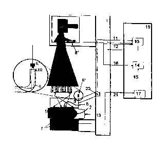
With reference to figure 1 the mammography-apparatus
of the invention is denoted with reference 1. This mammography-
apparatus 1 is used for screening malignant cells 8 in a breast
2. For this purpose the mammography-apparatus 1 comprises an x-
ray source 3 and an x-ray detector 4 that cooperates with the x-
ray source for providing an x-ray image of said breast 2.
The mammography-apparatus 1 further comprises a paddle
5 for flattening the breast 2 by compressing it against said x-
ray detector 4. In the embodiment shown the paddle 5 is provided
with a force- or torque sensor 20 for measuring a force at which
the paddle 5 flattens the breast 2. Further a control system 10,
normally forming part of a computer system 19, is provided that
receives the measurement signals of the force- or torque sensor
via line 11, and controls the actuation of the paddle 5 via a
steering line 12 depending on the force that is measured with
20 the sensor 20 and using the results of the contact area measure-
ment which is discussed in the next paragraph.
In controlling the actuation of the paddle 5, the force
measured with sensor 20 is first converted to a pressure by tak-
ing into account the contact area between the paddle 5 and the
breast 2. For this purpose a contact area measuring unit 6', 6"
is applied for measuring the contact area between the breast 2
and the paddle 5. The contact area measuring unit 6', 6" com-
prises optical means such as a camera 6', preferably a CCD cam-
era. The output of a CCD camera 6'can be directly available in a
digital format for control system 10.
Beneficially the optical means 6', 6" of the contact
area measuring unit comprises further a light source 6", where-
by the paddle 5 is connected to this light source 6", and said
paddle 5 and light source 6" are arranged for propagating light
within the paddle 5 and for releasing light from the paddle 5 in
the direction of the camera 6' upon the breast 2 tissue contact-
ing the paddle 5.
To improve the optical path from the contact area meas-
uring unit 6', 6" to the breast 2, it is advantageous to make
the paddle of Lexan polycarbonate resin. Lexan is a registered
trademark of the firm Sabic Innovative Plastics for an amorphous
CA 2787619 2018-02-02
8
engineering thermoplastic that is known for its outstanding me-
chanical, optical, electrical and thermal properties. The opti-
cal properties of the polycarbonate resin can be further im-
proved by supplying same with traces of titaniumoxide.
Although not shown in the figure, it is also possible
to measure the contact area using a separate plate that is as-
sembled to unite with the paddle on the side of the paddle which
is intended to contact the breast. In this embodiment the plate
is then connected to the light source, and the plate and light
source are then arranged for propagating light within the plate
and for releasing light from the plate in the direction of the
camera upon the breast tissue contacting the plate.
The mammography-apparatus of the invention further has
a processing unit 17 for processing data from the x-ray detector
4 received through line 21 and converting it into an x-ray image
of the breast 2. The processing unit 17 and the contact area
measuring unit 6', 6" are connected via respective lines 15, 16
to an estimator 14 in the computer system 19 for determining a
ratio between the contact area measured between the breast 2 and
the paddle 5, and a breast cross-section as derived from the x-
ray image of the breast 2.
The mammography-apparatus 1 further has a thickness
measuring unit 13 for measuring the breast thickness whilst said
breast 2 is flattened by the paddle 5. The thickness measuring
unit 13 is embodied to detect a distance between the region of
the paddle 5 that contacts the breast 2, and the x-ray detector
4. In the example shown this may be an angle measuring unit con-
nected to the arm that holds the paddle 5.
The paddle 5 is preferably provided with a series of
pressure sensors 7 for measuring local pressures which may be
indicative for the presence of malignant cells. It is desirable
that the pressure sensors 7 are distributed in the paddle 5 and
preferably also in the x-ray detector 4 so as to register local
pressures in the breast 2, and that the processing unit 17 (usu-
ally forming part of the computer system 19) is arranged to cor-
relate said local pressures with the x-ray image of said breast
2. It is also possible that the pressure sensors 7 are directly
employed for controlling the pressure that is applied to the
breast 2. In this way a force sensor for measuring the force at
which the paddle 5 is applied to the breast 2 may be dispensed
with.
CA 2787619 2018-02-02
9
Figure 1 further shows that the paddle 5 and/or x-ray
detector 4 comprises temperature sensors 18 for registering a
temperature distribution of the breast 2 whilst it is pressur-
ized between the paddle 5 and the x-ray detector 4. These tern-
perature sensors 18 for registering a temperature distribution
of the breast 2 are preferably measuring during the whole period
of compression of the breast 2 by the paddle 5. Preferably the
processing unit 17 of the computer system 19 is also arranged to
correlate the temperature distribution of the breast 2 with the
x-ray image.
The above elucidation of the features of the invention
are not intended to limit an understanding of the present inven-
tion to the specific example that is provided herewith. On the
contrary, it is possible that many variations are feasible with-
in the scope of the invention. It is for instance preferred that
the pressure sensors 7 and/or temperature sensors 18 are trans-
parent for x-rays. In another embodiment however it is feasible
that the paddle 5 and/or the x-ray detector 4 and the pressure
sensors 7 and/or temperature sensors 18 provided therein exhibit
substantially the same level of absorption for x-rays. In still
another embodiment it is feasible that the pressure sensors 7
and/or the temperature sensors 18 are not fully transparent for
x-rays, and that the processing unit 17 is arranged to remove
the image of the pressure sensors 7 and/or temperature sensors
18 from the x-ray image.
CA 2787619 2019-06-14