Note : Les descriptions sont présentées dans la langue officielle dans laquelle elles ont été soumises.
CA 02790588 2012-08-21
1
PARTICLE IMAGE VELOCIMETRY SUITABLE FOR X-RAY
PROJECTION IMAGING
FIELD OF INVENTION
The present invention relates to imaging, particularly imaging of
movement.
In one aspect the present invention relates to the field of biomedical
engineering, particularly in vivo or in vitro imaging.
In another aspect, the invention relates to technology for imaging of
function and form in a wide range of research, medical and industrial
applications.
In a yet further aspect the present invention is suitable for use as a method
and device for imaging the movement of living tissue.
It will be convenient to hereinafter describe the invention in relation to in
vivo medical imaging, however it should be appreciated that the present
invention
is not limited to that use only and can also be used for in vitro
applications, and
other medical applications such as diagnosis and treatment as well as research
applications and industrial applications. In particular although the
description will
particularly refer to the pulmonary system and vascular system, the skilled
person
will appreciate that the application of the present invention is not so
limited and
can be extended to other systems that have a mechanically dynamic aspect to
their function.
Furthermore, although it will be convenient to hereinafter describe the
invention in relation to imaging using a source that emits X-rays, such as
those
used for computer tomographic X-ray particle image velocimetry (CTXV), it will
be
appreciated that the present invention extends to any system that provides
imagery using any convenient source.
Many important processes in the human body involve motion. Obvious
examples include the cardiovascular system (motion of heart and blood flow),
the
pulmonary system (motion of the diaphragm and lungs), the renal system (motion
and filtering of blood) and the musculoskeletal system (motion of muscles,
connective tissue, bones and joints). Diseases of the vascular system such as
thrombus formation and pulmonary disease are leading causes of mortality and
morbidity in developed countries. Studying the mechanically dynamic aspects of
CA 02790588 2012-08-21
2
these systems contributes to better understanding of the fundamental operation
of the human body and is a useful aid to the combat of dysfunction and
disease.
The ability to recognise and treat disease or dysfunction in these systems
is dictated by our ability to image them in situ with high resolution. In
particular,
current imaging cannot reveal most forms of lung disease before they become
clinically evident. The earlier these
diseases are detected, the better the
prognosis.
A relatively common feature of many lung diseases such as emphysema
and pulmonary fibrosis is a regional alteration to the distal airway structure
leading to marked regional changes in lung tissue compliance. Thus attempts
have been made to develop imaging techniques that can detect regional
differences in tissue velocities across the lung during the respiratory cycle
and
thus detect lung disease and dysfunction in their early stages.
BACKGROUND ART
It is to be appreciated that any discussion of documents, devices, acts or
knowledge in this specification is included to explain the context of the
present
invention. Further, the discussion throughout this specification comes about
due
to the realisation of the inventor and/or the identification of certain
related art
problems by the inventor. Moreover, any discussion of material such as
documents, devices, acts or knowledge in this specification is included to
explain
the context of the invention in terms of the inventor's knowledge and
experience
and, accordingly, any such discussion should not be taken as an admission that
any of the material forms part of the prior art base or the common general
knowledge in the relevant art in Australia, or elsewhere, on or before the
priority
date of the disclosure and claims herein.
It will also be appreciated that references herein to 'motion' are
interchangeable with 'flow' or 'velocity' (being a function of motion over
time).
The ability to measure three-dimensional (3D) blood flow fields in vivo is an
important capability for studying the effects of blood flow properties on the
development, diagnosis and treatment of cardiovascular diseases, such as
atherosclerosis. To gain useful
information from in vivo blood flow field
measurements, non-invasive measurement through optically opaque tissue at
high resolution is required.
CA 02790588 2012-08-21
3
The development of technologies underpinning in vivo measurements of
form and function of the human body are discussed in various reviews. (See for
example Fouras A, Kitchen MJ, Dubsky S, Lewis RA, Hooper SB and Hourigan K
2009 Journal of Applied Physics Vol.105).
Currently available techniques for flow field measurement in opaque
vessels, such as magnetic resonance imaging based techniques, suffer from poor
spatial and temporal resolution, limiting the application of these techniques
for in
vivo flow analysis. Better results have been achieved with techniques such as
Particle image velocimetry (PIV) in which the displacement of tracer particles
is
determined using statistical cross-correlation of regions within particle
image
pairs. Several variants exist for volumetric flow analysis, including
Tomographic
PIV, volumetric particle tracking and Holographic PIV.
PIV imaging generally
PIV is well known for accurate measurement of instantaneous velocity
fields. PIV techniques using visible light are limited to optically
transparent
sample. However the use of X-rays with PIV has extended the application of
this
method to opaque tissue, making this imaging mode ideal for in vivo blood flow
field measurement.
In PIV, regions of fluid containing multiple tracer particles (typically
illuminated by a visible wavelength laser) are imaged at two points in time,
separated by a known time interval, and processed using correlation software.
Specifically the image pairs are allocated into discrete interrogation
regions.
Cross correlation is performed between image pairs on each interrogation
region
and statistically, the maximum value of the cross correlation is the most
likely
particle displacement within the interrogation region.
In recent years PIV has been combined with X-ray imaging. The
penetrating power of X-rays allows flow to be measured within opaque objects,
with applications for non-invasive, high resolution blood flow field
measurements.
2D Particle Image Velocimetry
Kim and Lee (Kim GB and Lee SJ 2006, Exp. Fluids 41, 195) have
measured flow in tubes with particles and blood cells as tracers using X-ray
PIV.
The methods taught in that study are limited to two components of the velocity
(averaged over the dimension perpendicular to the image plane) within the
CA 02790588 2012-08-21
4
measurement volume. The PIV algorithms used belonged to the prior art relating
to optical/laser based velocimetry. These algorithms assume pulsed
(instantaneous) illumination and zero out-of-plane flow gradients and
therefore fail
to take into account the 3D characteristics of imaging real flows using X-
rays.
This results in a significant under estimation of flow velocity.
3D Particle Image Velocimetry
Recently X-ray PIV analysis has been extended to include 3D flow data.
Fouras et al (Fouras A, Dusting J, Lewis R and Hourigan K et al, 2009 Journal
of
Applied Physics Vol.102:064916) teach that the correlation peak represents a
probability density function (PDF) of the velocity within the measurement
volume.
When combined with certain assumptions about the flow field, it is possible to
convert this volumetric PDF of the velocity to a velocity profile. This
results in the
capability to measure 3D flow data from single projection X-ray images.
CT is a technique used to reconstruct an object in three-dimensional space
from two dimensional projections. Typically, integrated object density in the
projection direction is calculated from the X-ray attenuation, which will be
proportional to the pixel intensity values on a digital projection image. The
object
structure is then reconstructed from projection images taken at different
viewing
angles, using Fourier back-projection or algebraic methods. Variants also
exist for
reconstruction of objects from few projection angles, which use iterative
methods
to reconstruct the sample's structure, often exploiting prior knowledge of the
sample, for example that it is made up of a single material.
CTXV can thus deliver three component velocity measurements for
complex 3D flow fields such as those found in the cardiovascular system.
Single
projection images are insufficient for evaluating three components of
velocity.
Images taken at a single projection angle contain no displacement information
in
the direction parallel to the X-ray beam. This limits single projection X-ray
PIV to
two component velocity measurements. In a method similar to CT, CTXV
overcomes this limitation by using multiple projection angles. Signal-to-noise
ratios can be enhanced using phase contrast imaging and phase retrieval
methods.
Specifically, as in single projection X-ray PIV of the prior art, cross-
correlation functions are calculated for interrogation regions within image
pairs.
CA 02790588 2012-08-21
The velocity field is reconstructed in axial slices, defined by the rows of
interrogation regions for all projection angles. A three component, 2D,
rectangular grid model represents the velocity field for each slice.
Estimated
cross correlation functions are generated for every angle and every
interrogation
5 region within each slice. The estimated cross-correlation functions are
generated
using convolution of the measured autocorrelation function with the velocity
PDF
for the interrogation region within the model. The velocity coefficients in
the
model are iteratively optimized, minimizing the error between measured cross-
correlation function and the estimated cross-correlation functions, across all
projection angles and interrogation regions simultaneously within that slice.
Using this iterative approach, a model is reached which accurately represents
the
three component velocity field within each slice.
A relatively small number of projections are required and this is important
for minimising radiation dosage. It also allows the integration of CTXV with a
CT
reconstruction such as described above, delivering simultaneous measurement of
both form and function.
CT has the advantage of offering the best resolution and penetration of all
medical imaging modalities, but also has the significant disadvantage of
delivering high doses of X-rays. If not for this radiation dose concern high
resolution CT would become a standard screening tool.
But even though they offer the best resolution and penetration of all
medical imaging modalities, the X-ray PIV techniques of the prior art use
particle
images taken at a single viewing angle, which contain no particle displacement
information in the direction parallel to the X-ray beam, and therefore they
suffer
the drawback that they are limited to two component velocity measurements.
Also, no information regarding the velocity profile in the dimension
perpendicular
to the image plane is available, and therefore 3D measurements are not
possible
without prior knowledge of the flow.
There is an ongoing need to expand capabilities for measuring both form
and function in terms of structure, volume and motion and provide a truer 3D
reconstruction of flow fields.
CA 02790588 2012-08-21
6
SUMMARY OF INVENTION
An object of the present invention is to provide improved images that are
truer 3D reconstructions rather than 3D image reconstructions.
A further object of the present invention is to provide an improved method
of converting data sets into truer 3D reconstructions rather than 3D image re-
constructions.
It is an object of the embodiments described herein to overcome or
alleviate at least one drawback of related art systems or to at least provide
a
useful alternative to related art systems.
In essence, embodiments of the present invention stem from the
realisation that imaging can utilise three components (u,v,w) of motion over
2, or
preferably 3 spatial coordinates (x,y,z) plus time (t), which will be referred
to
herein as '3D or '4D' as appropriate, but in practice measures more components
than 3D imaging of the prior art. Furthermore, it has been realised that this
methodology can be applied to measurement of motion of any kind. For example
with reference to physiological measurements the methodology can be applied to
measuring motion fluid, such as blood, air or lymph, and/or measurement of
tissue, such as lung tissue during inspiration and expiration. A further
realisation
is that data relating to characteristics such as compliance and shear can also
be
processed. In practice, the present invention provides the ability to,
(i) make 3D reconstructions of motion that are not possible using 3D
imaging technology of the prior art,
(ii) reconstruct 3D motion (velocity) information without first
reconstructing 3D images ,
(iii) evaluate data such as shear,
compliance and volume flow, in 4D
(x,y,z,t) and present them in image format by reconstructing 3 components of
velocity over 3D or 4D.
The present invention permits the extraction and manipulation of data to
allow presentation of functional information in a format that is easy to
compare
and interpret. In particular it can be used for presentation of regional
functional
information. The term 'region' or 'regional' is used in the sense of
functional
information pertaining to an area or locale (such as, for example, a part of
an
organ such as a lobe of a lung), and may be used in contradistinction to
functional
CA 02790588 2012-08-21
7
information derived from a combination or average of data from multiple
regions
(such as, for example an entire organ, such as a lung). For example a region
can
be global, lobar, functional gas or fluid exchange units or any other desired
segment or locale. Thus the present invention may be used to present
functional
information that is commonly used in scientific and clinical practice (such as
FEV1) but has not hitherto been available regionally.
In a first aspect of embodiments described herein there is provided a
method for imaging of a sample, the method including the steps of:
1. recording images from at least one projection angle and carrying out
image pair cross-correlation analysis encoding velocity data for the
sample in terms of coordinates; and
2. reconstructing a 2D or 3D velocity field directly from the image pair
cross-correlations from the analysis
wherein the reconstruction is performed without first reconstructing 2D or 3D
images and wherein steps 1 and 2 are automated.
It should be noted that although the present invention will be described and
exemplified with reference to Cartesian coordinates, it will be readily
apparent to
the person skilled in the art that other coordinate systems could be used and
data
could be converted from one coordinate system to another. For example,
cylindrical or polar coordinates could be used, or local coordinates that are
oriented to the relevant anatomy.
The reconstruction of step 2 may be carried out by iterative methods, or
alternatively direct methods.
The image of the velocity field can thus convey a large amount of
information visually. However velocity field images, while familiar to
physicists,
are not familiar to other professionals who may need to interpret them such as
medical practitioners or pathologists who are used to seeing physiological
features. In order to provide an image that is more readily recognised and
understood, it may be necessary to associate the velocity field with
corresponding features of a digitised (segmented) image of the sample. The
need to associate image data with recognisable physiological or other features
is not limited to velocity fields but can apply to any appropriate image data
captured by any means.
CA 02790588 2012-08-21
8
There is further provided a method for providing an image of a sample
comprising the steps of:
1. recording images encoding data for the sample in terms of
coordinates;
2. reconstructing a 2D or 30 data field from the information encoded in
the recorded images;
3(a) segmenting an image of the sample, and
3(b) associating each segment with regions of the 2D or 3D data field,
wherein the reconstruction is performed without first reconstructing 2D or 3D
images and wherein steps 1 and 2 are automated.
Preferably the images will be recorded from multiple angles. However, it
will be apparent to the person skilled in the art (as disclosed in Irvine SC,
Paganin
DM, Dubsky S, Lewis RA and Fouras A 2008 Applied Physics Letters 93:153901;
and Fouras, A., Lo Jacono, D., Nguyen, C.V. & Hourigan, K. 2009 Volumetric
correlation PIV: a new technique for 3D velocity vector field measurement.
Experiments in Fluids 47 (4), 569-577) that when a sample has rotational
symmetry in terms of shape or motion, measurement from only one projection
angle will be necessary. Furthermore, only one projection angle will be needed
if
depth data from phase or focus is recorded in addition to velocity data.
Accordingly, in a second aspect of embodiments described herein there is
provided a method for imaging of a sample, the method including the steps of
1. recording images from at least one projection angle and carrying
out
image pair cross-correlation analysis to obtain 2D velocity data for
the sample in terms of three coordinates, and
2. reconstructing a 3D velocity field directly from the image pair cross-
correlations from the analysis
wherein steps 1 and 2 are automated.
Using this method a 2D or 'single projection' image of the sample is thus
obtained. By repeating the steps many times, the 30 data can be expanded to
4D data.
Accordingly, in a third aspect of embodiments described herein there is
provided a method for imaging of a sample, the method including the steps of:
CA 02790588 2012-08-21
9
1. recording images from at least one projection angle and carrying
out
image pair cross-correlation analysis encoding velocity data for the
sample in terms of coordinates; and
2(a) using an iterative method for reconstructing a 3D velocity field
directly from the image pair cross-correlations from the analysis;
2(b) repeating the iterative method to produce a 4D velocity field; and
3. using the 4D velocity field to provide further information
wherein the reconstruction is performed without first reconstructing 2D or 3D
images and wherein steps 1 to 3 are automated.
The further information provided according to the above method may relate
to any useful characteristic such as shear or compliance. These may be
important to analysis of the degree or quality of functionality of the sample.
In
addition to recordal of velocity data, step 1 may include recordal of depth
information from phase (holography) or focus.
Typically the iterative method will be analogous to an iterative CT method.
Any source that provides imagery can be used with the method of this
invention. This includes sources that emit the following types of energy;
= X-rays,
= visible light including visible lasers,
= infrared radiation including infrared lasers,
= ultraviolet radiation including ultraviolet lasers,
= ultrasound,
= electrical impedance, and
= magnetic resonance.
In a preferred embodiment the present invention is a method for CTXV
imaging of a sample.
In a fourth aspect of embodiments described herein there is provided a
method for imaging of a sample, the method including the steps of:
1(a) recording images from multiple projection angles;
1(b) allocating images into rectangular interrogation windows;
1(c) deriving velocity components u, v and w in the x, y and z directions
from the images;
CA 02790588 2012-08-21
1(d) carrying out cross-correlation analysis on image pairs defined by the
interrogation windows; and
2. reconstructing a 3D velocity field directly from the image pair
cross-
correlations derived from the analysis
5 wherein the reconstruction is performed without first reconstructing 20 or
3D
images and wherein steps 1 and 2 are automated.
In a fifth aspect of embodiments described herein there is provided a
method for converting data sets defining a velocity field to a regional
compliance
map the method comprising the steps of:
10 1(a) recording images from at least one projection angle and carrying
out
image pair cross-correlation analysis to measure encoded
parameters of,
(i) velocity (u, v and w) against time opposite Cartesian co-
ordinates (x, y and z),
(ii) a further physical parameter (p) chosen from the group
comprising pressure or volume;
2(a) integrating the measurements to provide a single 3D velocity field;
and
2(b) describing regional compliance in terms of derivatives defined by
(6u/Ox + Ov/6y + i5w/Eiz) I Op.
wherein step 2 is performed without first reconstructing 20 or 3D images and
wherein steps 1 and 2 are automated.
In yet a further aspect of embodiments described herein there is provided a
method for converting data sets defining a velocity field to a regional
compliance
map the method comprising the steps of:
I. recording images from at least one projection angle and carrying
out
image pair cross-correlation analysis to measure encoded
parameters of,
(i) motion (u and v) opposite Cartesian co-ordinates (x and y),
and
(ii) sample thickness (t), pressure (p) and volume (V);
2(a) integrating the measurements to provide a single 30 velocity field;
11
2(b) describing regional compliance in terms of
(6u/6x+ ov/6y).t / 6p; and
2(c) solving for t, by mathematical comparison of total compliance with
the total compliance determined by adding the regional
compliances, according to,
t = V / (6u/6x + 6v/6y),
wherein step 2 is performed without first reconstructing 2D or 3D images and
wherein steps 1 and 2 are automated.
For example, the above method could be used to create a regional
compliance map for inspiration or expiration of a lung, beating of a heart, or
pulsation of blood in an artery. Thus the present invention can be used for
measuring any motion whether fluid or tissue.
With particular reference to the lung, the method of the present invention
can be used to detect lung tissue movement and to measure the velocity fields
that define speed and direction of regional lung motion throughout a breath.
Regional maps of the lung can be generated showing degree and timing of
expansion from the velocity fields, revealing regions of abnormal tissue
properties
caused by experimentally induced non-uniform lung disease. This includes
diseases such as pulmonary fibrosis, cystic fibrosis, cancer and asthma.
Specifically, this would include measuring parameters of air velocity (u, v
and w) against time opposite Cartesian coordinates (x, y and z), segmenting a
3D
image of the airways of the lung, then associating each region of the lung
over
which velocity was measured with a corresponding segment of the 30 image to
depict airflow within the airways over time. This can be summarised in the
flowchart shown in Figure 12.
CA 2790588 2018-03-27
CA 2790588 2017-04-11
12
While the image of the velocity field can thus convey a large amount of
information visually, as mentioned above, velocity field images are familiar
to
physicists, but are not familiar to other professionals who may need to
interpret
them. For example medical practitioners or pathologists are used to seeing
physiological features or textual presentation (such as graphs or tables). In
order
to provide a textual or graphical presentation of functional information that
is more
readily recognised and understood, it may be necessary to associate the
velocity
field with indicia denoting different regions in the sample.
CA 02790588 2012-08-21
13
Accordingly, in a third aspect of embodiments described herein there is
provided a method for presenting information derived from a sample, the method
including the steps of:
1. recording images from at least one projection angle and carrying out
image pair cross-correlation analysis encoding velocity data for the
sample in terms of Cartesian coordinates;
2. reconstructing a 2D or 3D velocity field directly from the image pair
cross-correlations from the analysis wherein the reconstruction is
performed with or without first reconstructing 2D or 3D images; and
3. calculating the expansion of
one or more regions in terms of velocity
(u, v and w) against time opposite Cartesian co-ordinates (x, y and
z) and using derivatives defined by 5u/Ox + ov/oy + 5w/5z for textual
or graphical presentation.
Typically the method also includes the steps of segmenting an image of
the sample and associating each segment with regions of the 2D or 3D data
field.
For example, segmentation can comprise obtaining relevant basic anatomical
detail, such as details of the airways of the lung. The subject can then be
subjected to a physiological manoeuvre such as forced expiration so that the
regional airflow can be associated with the anatomical details. Hence steps 2
and 3 (mentioned above) can be used to calculate airflow throughout the
airways
of the lung. The calculations can be textually represented (or represented in
an
image) to display the volume of air expired in a short period of time (eg 1
second)
in a single region, or multiple regions, or every region of the airway tree,
thus
providing a regional FEV1.
These function-based regional measurements or groups of regional
measurements can be compared to one another.
Required anatomical detail of the lung can be obtained by any convenient
method including:
= high or low resolution CT scan of the subject's lung;
= MRI scan, for example, spin density MRI;
= direct application of anatomical atlas, optionally in conjunction with
subject data such as height, weight, tidal volume;
CA 02790588 2012-08-21
14
= matching data from an anatomical atlas to a plane radiograph or any
other spectroscopic analysis of the subject's lungs;
= combinations of the above methods.
Due to the functional capacity of the method of the present invention,
previously unavailable in X-ray imaging, combined with the expectations of
dramatic reduction in dose, an apparatus or system based on the method could
find great utility, for example as a clinical scanner. Such a scanning system
could
be used to provide early detection and diagnosis of diseases or disorders. The
apparatus could also be applied to industry, for example measuring motion in
minerals processing, or in the laboratory for pre-clinical medicine, minerals
processing, geophysics and fluid mechanics.
In another aspect of embodiments of the invention there is provided an
apparatus when used for the method of the present invention, the apparatus
comprising:
(i) one or more energy source;
(ii) one or more detectors for recording images created by energy from
the one or more energy sources passing through a sample; and
(iii) a sample retainer for locating the sample intermediate the one or
more energy sources and the one or more detectors;
wherein in use, the sample retainer rotates the sample through multiple energy
projection angles and at least one image is recorded at each of the projection
angles.
In another aspect of embodiments of the invention there is provided an
apparatus when used for the method of the present invention, the apparatus
comprising:
(I) one or more energy source; and
(ii) one or more detectors for recording images created by energy from
the one or more energy sources passing through a sample;
wherein in use, a sample is located intermediate the one or more energy
sources
and the one or more detectors, the sources and detectors being rotated
relative to
the sample through multiple energy projection angles and at least one image of
the sample is recorded at each of the projection angles.
=
CA 2790588 2017-04-11
In another aspect of embodiments of the invention there is provided an
apparatus when used for the method of the present invention, the apparatus
comprising:
(i) two or more energy sources having respective projection
angles;
5 (ii) one or more detectors for recording images created by
energy from
the energy sources as it passes through a sample; and
wherein recordings at each of the projection angles are made simultaneously.
Any convenient range of projection angles may be used from 10 to 3600
.
However, typically the range of projection angles does not reach the extremes
of
10 this range. For example, projection angles spaced over as little as 300 or
as
much as 180 are likely to be suitable.
In addition to at least one energy source and detector, the apparatus for
use with the method of the present invention may include a number of other
components including, for example, (i) systems for modulating and aligning the
15 source, the target and/or the detector, (ii) systems for image capture,
processing
and analysis, and (iii) a convenient user interface.
Other aspects and preferred forms are disclosed in the specification.
Although there are a number of prior art systems that can measure 3D
velocity information such as Doppler ultrasound, magnetic resonance imaging
(MRI), holographic PIV, digital in-line holographic PTV, tomographic PIV and
defocusing PIV, these all have drawbacks not included in the present
invention.
For example, Doppler ultrasound has limited spatial and temporal resolution,
the
resolution decreasing with increased depth of measurement. MRI has limited
spatial and temporal resolution, which is particularly evident in temporal
measurement. Holographic PIV, digital in-line holographic PTV and tomographic
PIV are limited to transparent media which virtually eliminates their utility
for in-
vivo imaging. The also cannot provide shape/anatomical information.
Furthermore these techniques are based on reconstructing 4D images to then
measure motion, which means that systems using these techniques are
comparatively complex and have limited spatial resolution compared to the
system of the present invention.
16
Figure 4 illustrates a 3D reconstructed blood velocity field at a single time
point. For clarity only half the sample is plotted, with reduced vector
resolution in
all dimensions so that individual vectors can be seen. The vectors are
depicted in
different colours, with each colour representing a different velocity
magnitude. .
Figure 5 is a velocity field of the type depicted in Figure 4, for a lung and
comprises vectors that show the velocity of lung tissue at a single time point
during inspiration;
Figure 6 is a 3D illustration of lung airway structure image according to the
present invention, wherein the vectors of a velocity field have been matched
to
corresponding physiological features, being airways (trachea, bronchioles,
alveoli);
Figure 7 Illustrates a computer aided design model (Figure 7(a)) used for
the hollow section of the sample of Example 3, and a CT reconstruction (Figure
7(b)) based on particle speckle contrast. The sample geometry is based on the
= 15 union of a cone and a helically swept circle;
Figure 8 illustrates a CTXV reconstruction of flow through the sample
depicted in Figure 7 to show how CTXV can simultaneously measure the 3D
structure and velocity of flow through complex-geometries. A section of the
image
has been rendered transparent to enable the flow and vectors within the sample
to be seen, and for clarity, vector resolution is reduced by 4x in the x, y
and z
directions;
Figure 9(a) shows the 30 nature of X-ray illumination and velocimetric
cross-correlation analysis as described in Example 4 while Figure 9(b)
illustrates
in vivo detection of lung tissue motion according to the present invention;
Figure 10 depicts statistical measures of lung pathology comprising controls
with groups 36 hours after bleomycin exposure (Figures 10 (a) to (d)) and 6
days
after exposure (Figures 10(b) to (h)); and
Figures 11(a) to 11(e) show regional expansion within a lung with comparative
histological imagery.
DETAILED DESCRIPTION
As mentioned above, Fouras et al. have' demonstrated that the cross-
correlation functions calculated from X-ray image pairs represent a velocity
PDF
for the projected measurement volume.
Date Recue/Date Received 2022-01-19
CA 02790588 2012-08-21
17
= during forced expiratory manoeuvers;
= peak inspiratory flow;
= peak expiratory flow;
= regional tidal volume;
= expiratory reserve volume;
= volume flow gradient
= volume flow loops;
= functional vital capacity.
The present invention can also be used to measure other functional
parameters such as:
= regional ventilation heterogeneity;
= functional distribution;
= gravitation distortion;
= gravitation loading;
= regional time constants of any measure, such as expiratory flow;
= regional phase of any measure, such as expiratory flow;
= time of peak of a measure;
= time of minima of a measure;
= determination of organ properties, such as lung properties (for
example, strength of lung recoil or diaphragm action)
Additionally, the present invention could be used on a range of tissue types
including the lung, but also including the heart, vasculature, lymph channels,
brain.
All the above can be viewed globally or regionally and comparisons made
between regions, groups of regions and the global measure.
Further scope of applicability of embodiments of the present invention will
become apparent from the detailed description given hereinafter. However, it
should be understood that the detailed description and specific examples,
while
indicating preferred embodiments of the invention, are given by way of
illustration
only, since various changes and modifications within the spirit and scope of
the
disclosure herein will become apparent to those skilled in the art from this
detailed
description.
CA 02790588 2012-08-21
18
BRIEF DESCRIPTION OF THE DRAWINGS
Further disclosure, objects, advantages and aspects of preferred and other
embodiments of the present application may be better understood by those
skilled in the relevant art by reference to the following description of
embodiments
taken in conjunction with the accompanying drawings, which are given by way of
illustration only, and thus are not limitative of the disclosure herein, and
in which:
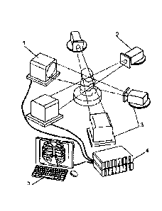
Figure 1 is a schematic diagram outlining the basic design of a CTXV
system according to the present invention. The diagram shows three
polychromatic X-ray beams transmitted through a sample and converted to
visible
light by scintillators. High-speed detector systems then produce a set of
images.
Multiple projection data are gathered simultaneously without rotating the
sample.
Cartesian co-ordinates (x,y,z) are fixed to the sample and rotated at an angle
from the beam axis p;
Figure 2 is as schematic diagram of experimental imaging setup illustrating
Cartesian coordinates x, y, z fixed to the sample, and rotated at an angle 0
from
the beam axis p. The diagram shows a monochromatic X-ray beam transmitted
through a sample and converted to visible light by a scintillator. A high-
speed
detector system then produces an image. Multiple projection data are gathered
by rotating the sample. Cartesian co-ordinates (x,y,z) are fixed to the sample
and
rotated at an angle 0 from the beam axis p;
Figure 3(a) is a schematic of the forward projection model according to the
present invention. Cross-correlation functions are estimated by convolution of
the
velocity PDF, projected from the flow model, with the auto-correlation
function
calculated from the projection images;
Figure 3(b) illustrates 30 CTXV motion reconstruction, the residual between
estimated and measured cross-correlations having been minimized over all
interrogation windows and all projection angles simultaneously to yield a
cross-
sectional flow model which accurately represents the flow field.
Figure 3(c) illustrates one-dimensionalisation of the cross-correlation
functions. Integration across the rows and columns in the 2D cross-correlation
function yields a 1D representation of the velocity PDF in the r and q
directions
respectively;
CA 2790588 2017-04-11
19
Figure 4 illustrates a 3D reconstructed blood velocity field at a single time
point. For clarity only half the sample is plotted, with reduced vector
resolution in
all dimensions so that individual vectors can be seen. The vectors are
depicted in
different colours, with each colour representing a different velocity
magnitude.
Figure 5 is a velocity field of the type depicted in Figure 4, for a lung and
comprises vectors that show the velocity of lung tissue at a single time point
during inspiration;
Figure 6 is a 3D illustration of lung airway structure image according to the
present invention, wherein the vectors of a velocity field have been matched
to
corresponding physiological features, being airways (trachea, bronchioles,
alveoli);
Figure 7 illustrates a computer aided design model (Figure 7(a)) used for
the hollow section of the sample of Example 3, and a CT reconstruction (Figure
7(b)) based on particle speckle contrast. The sample geometry is based on the
union of a cone and a helically swept circle;
Figure 8 illustrates a CTXV reconstruction of flow through the sample
depicted in Figure 7 to show how CTXV can simultaneously measure the 3D
structure and velocity of flow through complex geometries. A section of the
image
has been rendered transparent to enable the flow and vectors within the sample
to be seen, and for clarity, vector resolution is reduced by 4x in the x, y
and z
directions;
Figure 9(a) shows the 3D nature of X-ray illumination and velocimetric
cross-correlation analysis as described in Example 4 while Figure 9(b)
illustrates
in vivo detection of lung tissue motion according to the present invention;
Figure 10 depicts statistical measures of lung pathology comprising controls
with groups 36 hours after bleomycin exposure (Figures 10 (a) to (d)) and 6
days
after exposure (Figures 10(b) to (h)); and
Figure 11 shows regional expansion within a lung with comparative
histological imagery.
Figure 12 shows a flow chart of a method to detect lung tissue movement.
CA 2790588 2017-04-11
19a
DETAILED DESCRIPTION
As mentioned above, Fouras et al. have demonstrated that the cross-
correlation functions calculated from X-ray image pairs represent a velocity
PDF
for the projected measurement volume.
CA 02790588 2012-08-21
The present invention now provides a technique that includes imaging the
flow from multiple projection angles to obtain information regarding the three
components of velocity in three dimensional space. Using this information, the
3D velocity field can be reconstructed directly from image pair cross-
correlations,
5 without the need to reconstruct a volumetric image.
Further uses of data collected
As discussed above the present invention is not limited to imaging the
motion of fluid such as blood, but can also depict the motion of tissues or
entire
organs such as the lungs. Further quantitative processing of the data used for
10 imaging can provide additional useful information such as the airflow
within the
lungs or the amount of shear.
The data collected can also be used to construct images of the
shape/anatomy of a structure such as an organ simultaneously with velocity.
The present invention can also provide useful information relating to the
15 periodic nature of a system such as breathing, heart beating or
peristalsis.
Specifically data collected at different points in time can be combined to
reconstruct one cycle of, for example, a breath, a heart beat or peristaltic
contraction.
The data collected according to the present invention may include depth
20 information from phase (holographic) data or focus data. These types
of data can
be used to improve the quality of data representation for the same number of
projections, or alternatively, reduced the number of projections from which
data is
gathered. At one limiting extreme, data could be collected from as few as one
or
two projections, however some data would be lost if a single projection was
used.
Accordingly, it is preferred that the method includes the steps of;
1. recording images from at least
one projection angle, preferably
multiple projection angles and carrying out image pair cross-
correlation analysis encoding velocity data for the sample in terms of
coordinates, and
2. reconstructing a 2D or 3D
velocity field directly from the image pair
cross-correlations from the analysis
wherein the reconstruction is performed without first reconstructing 2D or 3D
images and wherein steps 1 and 2 are automated.
CA 02790588 2012-08-21
21
Conversion of data sets into compliance maps
The present invention provides a method of converting data sets into
regional compliance maps. For example, for lung ventilation the data set
(defining a velocity field) may comprise measurement of velocity in either 2D
or
3D, the lung pressure range over the course of a breath and possibly the
volume
of air inhaled and exhaled over the course of the patient taking a breath.
When the data set defines a 3D + time (ie 40) velocity field the conversion
method comprises the steps of;
1. recording images from at least one projection angle, preferably
multiple projection angles and carrying out image pair cross-
correlation analysis encoding velocity data for the sample in terms
of Cartesian coordinates, and
2(a) integrating the velocity over any part of either the inspiration or
expiration, to give a single 3D velocity field - this 3D map having
quantities of velocity that can be defined as u, v and w over the
directional co-ordinates x, y and z;
2(b) the regional compliance is then described mathematically as
(6u/6x + i5v/51 + ow/Oz) / Op
(where Op = change in pressure over the same part of inspiration or
expiration in 2a)
wherein the reconstruction of step 2 is performed without first reconstructing
20
or 3D images and wherein steps 1 and 2 are automated.
When the data set defines a 20 + time (ie 3D) velocity field the conversion
method comprises the steps of;
2(a) integrating the velocity over all of either inspiration or expiration, to
give a single 20 velocity field, this 2D map having quantities of
velocity that can be defined as u and v over the directional co-
ordinates x and y.
2(b) describing the regional compliance mathematically as:
(5u/6x+ 6v/6y).t / 5p
(where t = thickness, p = pressure over inspiration or expiration)
2(c) solving for t, by mathematical comparison of total compliance
(commonly measured) with the total compliance determined by
CA 02790588 2012-08-21
22
adding the regional compliances, which in equation form can be
expressed as;
t = V / (6u/ox + Ov/Oy)
(where V = volume inhaled or exhaled)
wherein the reconstruction of this step 2 is performed without first
reconstructing
2D or 3D images and is automated.
Using this method a vector field image of the type in Figure 5 can be
produced. In this image, vectors show the velocity of lung tissue at a single
time
point during inspiration, measured using CTXV. Different shades of grey (or
preferably, different colours) can be used to represent velocity magnitude and
the
vector resolution reduced in all dimensions aids visualisation. In this image
the
spacing between velocity measurements is approximately 0.18 mm. This
illustrates that CTXV is capable of producing high-resolution, accurate
measurements with very few projections.
By relating the vector field to physiological structures of the lung, a more
readily recognisable image can be generated. This can be achieved by the
further steps of:
3(a) segmenting a 30 image of the sample, and
3(b) associating segments of the 3D image with corresponding
derivatives.
Figure 6 is an example of these further steps having been applied to create
a 3D rendering of the lung airway structure. The motion of the lung tissue as
measured using CTXV has been used to calculate the expansion of the lung, and
hence the flow of air into the lung on a regional basis. The major airways are
depicted in shades of grey (or preferably, different colours) to demonstrate
different volume flows in different regions.
Accordingly, lung tissue motion data (Figure 5) is processed using step 3
outlined above to establish measurement of air motion within the lung (Figure
6).
This 'two part' approach has an advantage over direct measurement of fluid
motion in so far as a much lower dose of radiation can be used.
A similar two part approach could be applied to measurement of motion in
other organs such as the heart. Specifically, it would be possible using the
methods of the present invention to directly measure the flow or blood. It
would
CA 02790588 2012-08-21
23
also be possible using the methods of the present invention to measure motion
of
heart tissue (muscle wall, valves and/or vessels) and then derive measurement
of
fluid (blood) flow.
System & Apparatus
The method of the present invention could be implemented in a wide range
of imaging systems. Preferably the method would be implemented using a CTXV
system because this has the advantage of offering the best resolution and
penetration of all medical imaging modalities.
A typical CTXV system incorporating the present invention would consist
primarily of a number of phase contrast X-ray imaging lines - typically at
least
three imaging lines. More than three lines would improve the quality of the
data
collected, but would concomitantly increase system complexity, cost and
potentially the X-ray dosage delivered.
With reference to Figure 1, each imaging line would typically consist of the
following key components:
a. video speed or double shutter X-ray camera (1);
b. cone beam X-ray source (2);
c. source modulation system (3);
d. basic source alignment hardware (4);
e. high-resolution camera alignment hardware (4);
f. image capture and analysis hardware (5); and
g. user interface (6).
In addition to the imaging hardware, there is a requirement for image
capture and analysis hardware and software. The image capture and analysis
hardware and software would typically consist for the following key
components:
h. high speed image capture hardware;
high speed image processing hardware;
j. image processing software; and
k. user interface for alignment, imaging and analysis.
Details of suitable components or component groups are described in the
following paragraphs:
Cone beam X-ray source: As is typical of phase contrast sources there
exists a trade off when considering the size of the source. Larger sources
afford
CA 02790588 2012-08-21
24
less contrast, but more light and hence shorter exposure times. Many current
phase contrast systems employ so called micro or nano sources that are smaller
than 5 micron. Typically, commercial 'off-the-shelf' sources of at least 20-50
micron in size would be suitable for use in the method and system of the
present
invention.
Source modulation system (X-ray shutter): Freeze frame photography is
required for motion measurement. The continuous light sources must be
modulated into short bursts with as close to a temporal square wave as
possible.
Complete control of exposures between 2 and 20 milliseconds are preferred. The
use of a fast shutter also allows minimisation of the delivered dose, as the
sample
will be exposed to X-rays for the minimum time required for image capture.
Basic source alignment hardware: Optimally, the X-ray
source is
positioned so that the brightest region of the source is centred on the sample
region of interest.
Video speed or double shutter X-ray camera: System measurement can
be based on two or more raw images from each imaging line. These images
must be taken in quick succession (at video frame rate or better). A camera
system capable of sustained video frame rates or a 'double-shutter' camera
with
the capacity to acquire two images in quick succession will be required. If
the
optimal pixel size, minimum frame rate, and sensitivity are determined, a
suitable
commercial 'off-the-shelf' camera having the correct specification could be
used.
High-resolution camera alignment hardware: The system preferably
includes automated, robotic alignment of each camera with respect to its
respective source and any other cameras.
High speed image capture hardware: A commercial 'off-the-shelf data
acquisition system can be used to control the cameras and capture the date
from
each imaging line at a speed that will allow analysis to progress almost in
real
time.
High speed image processing hardware: Suitable options include, for
example, GPU, FPGA or DSP processing platforms.
Image processing software: A description of suitable software can be
found for example, in Dubsky S, Jamison RA, Irvine SC, Siu KKW, Hourigan K
and Fouras A (2010) Computed tomographic X-ray velocimetry, Applied Physics
CA 02790588 2012-08-21
Letters 96(2), 023702. The software needs to
be able to carry out a
reconstruction according to the method of the present invention. Using the
embodiment of the invention depicted in Figure 3 as an example, the software
may be able to discretise sample images into rectangular interrogation windows
5 and perform cross-correlation on these windows. The volume may then be
reconstructed in axial cross-sections, defined by the rows of the
interrogation
windows. A rectangular grid model may then be used to define the cross-
sectional velocity profile. Cross-correlation functions can then be estimated
from
each interrogation window measurement region. The 3D velocity field
10 reconstruction then becomes a minimisation of the error between the cross-
correlation functions estimated using the velocity model, and those calculated
from the X-ray image pairs, for all projection angles. The solution can be
implemented using the Levenberg-Marquardt algorithm which performs a
nonlinear least-squares optimisation.
15 User interface for alignment, imaging and analysis: The central
control
system and user interface preferably allows simple activation of technical
functions such as testing, calibration and alignment. The interface preferably
also
allows control of other user related functions such as imaging, image
processing
and visualisation of reconstructed results.
20 Examples
The present invention has been successfully used in 4 distinct animal trials
on the Spring-8 synchrotron. These trials have studied;
a. ventilator induced lung injury,
b. bleomycin induced fibrosis,
25 c. asthma based on the
methacholine challenge and salbutamol
reversal, and
d. cystic fibrosis lung disease.
All four of these trials clearly demonstrated the use of the invention for
measuring lung motion to provide early, accurate and regional detection of
abnormal lung function. In some cases the detection of pathology was possible
with lung motion measurement before it would be clear from histology or
biopsy.
This approach would also be useful for diagnosis of other diseases such as
lung
cancers.
CA 02790588 2012-08-21
26
The present invention will now be further described with reference to the
following non-limiting examples.
Example 1
In this example the method of the present invention has been applied to
the measurement of a strongly 3D flow.
The relevant imaging setup is shown in Figure 1. The monochromatic
beam in this case passes through a particle-seeded fluid (hollow glass spheres
in
glycerine). X-rays are slightly refracted at the interfaces between materials.
The
transmitted and refracted rays are allowed to propagate and interfere before
being co-converted into visible light by the scintillator. This is then imaged
using
a high-speed detector and visible light optics, resulting in a phase contrast
projection image. The image results from the superposition of interference
fringes
generated by the particle-liquid interfaces creating a dynamic speckle pattern
that
faithfully follows the particles.
Unlike visible light based imaging systems, in which images contain focus
or holographic information from which depth can be inferred, the transmission
nature of CTXV results in 2D volumetric projection image in which the entire
volume is in focus, and therefore contains no information of the distribution
of
velocity in planes parallel to the X-ray beam propagation direction.
Furthermore,
from any single viewing angle only two components of displacement can be
determined. This information deficit is overcome by rotating the sample and
imaging from multiple projection angles, allowing tomographic reconstruction
of
the velocity field within the volume. From these multiple
projections,
simultaneous tomographic reconstruction of the object structure is also
possible.
Forward projection
As in traditional PIV, particle image pairs are discretised into interrogation
regions and cross-correlation is performed on these regions (Figure 3(a)(i))
However, due to the large velocity distribution within the projected
interrogation
region, the cross-correlation functions will be highly distorted. The
resulting
projected cross-correlation statistics can be modelled as the velocity
probability
density function (PDF) of the flow projected onto that sub-region of the
image,
convolved with the particle image auto-correlation function (Figure 3(a)(ii)).
Therefore if the flow field and particle image autocorrelation function are
known,
CA 02790588 2012-08-21
27
the cross-correlation functions that would theoretically result from the flow
field
can be estimated. This represents the forward projection model (see Figure
3(a)(iii)). CTXV provides a solution of the inverse problem of reconstruction
the
flow field from the known cross-correlation data.
The effect of finite exposure time on the cross-correlation function of
projection image pairs, must also be taken into account. Due to motion of the
particle during the exposure, the contribution of each velocity to the cross
correlation function will be stretched along the direction of that velocity,
with a
magnitude that is linearly proportional to that velocity. As this effect has
been
well characterised it can be easily accommodated into the forward projection
model to eliminate any errors due to this phenomenon.
Solution to the inverse problem
Figure 3(b) demonstrates the implementation of CTXV. The velocity field
is reconstructed in slices orthogonal to the axis of rotation (Figure
3(b)(i)),
concurrent with the rows of interrogation regions within the projection images
(Figure 3(b)(iii)). A rectangular grid
model represents the flow-field in the
reconstruction domain. The three velocity components are defined at each node
point in the model and bi-linear interpolation is used to define the flow
between
node points. Higher degree interpolation schemes may be used, such as spline
interpolation at the expense of computation time and robustness.
Cross-correlation functions are estimated using the method shown in
Figure 3(a). The convolution is effected through a Fast Fourier Transform
(FFT)
implementation. A Levenberg-Marquardt algorithm is utilised to minimise the
error between the cross-correlation functions estimated from the flow model
and
those measured from the projection image pairs, resulting in a calculated flow
model which accurately represents the flow-field. As the problem is heavily
over-
specified, a Tikhonov-type regularisation scheme is used to ensure convergence
of the reconstruction, where the regularisation function is equal to the sum
of the
difference between each node velocity value and the mean value of its
neighbours.
One-dim ensionalisation of the Cross-Correlation
In order to reduce the number of optimisation parameters and memory
required for the reconstruction, a one-dimensionalisation of the cross-
correlation
CA 02790588 2012-08-21
28
functions is performed, allowing separate reconstruction of the date for vr
and vq.
Projection of the cross-correlation data results in two on-dimensional
representations of the function, for each of the velocity components vr and
vq, as
illustrated in Figure 3(c). By separating the two components they can be
reconstructed individually, greatly reducing the number of optimisation
parameters required per reconstruction. Furthermore, the process significantly
reduces the amount of data that needs to be stored and analysed.
Simultaneous structure reconstruction
To model the forward projection of the velocity PDF correctly, the relative
particle seeding density with in the reconstruction domain must be known.
Assuming homogenous seeding within the working fluid, this corresponds to
knowledge of the flow geometry. According there is provided a CT technique
that
allows the flow geometry to be reconstructed using the date obtained during
the
CTXV scan.
In typical CT reconstruction techniques, integrated object density in the
projection direction is calculated from the X-ray transmission, which will be
proportional to pixel intensity values on a digital projection image. In the
case of
a material of constant density, this integrated object density will be
proportional to
the object thickness. The contrast of the particle speckle (defined as the
ratio of
the standard deviation of the image intensity to the mean intensity) will
increase
with the square root of object thickness and so this statistic may also be
sued for
tomographic reconstruction of the object's structure. This is advantageous, as
in
many cases, including in vivo imaging of blood vessels the absorptions
contrast
alone is insufficient for tomographic reconstruction. Furthermore, the motion
of
the particle between images taken at different projection angles results in
artifacts
in the subsequent reconstructions. In comparison, the particle speckle contras
will be stationary for all viewing angles. The particle speckle
contrast is
calculated for discrete sub-regions in each phase contras image, prior to
phase
retrieval. The flow geometry is reconstructed from the particle speckle
contrast
data using an algebraic reconstruction technique. The use of an algebraic
technique allows for accurate reconstructions with low numbers of projections.
Example 2
CA 02790588 2012-08-21
29
In this example experiments are described which demonstrate the
application of CTXV to the simultaneous measurement of structure and velocity.
The method of the present invention was used with a high resolution medical
imaging beam-line (BL20XU) on a Spring-8 synchrotron at Hyogo, Japan set up
as shown in Figure 2. The source to sample distance of 245 metres provides
highly coherent X-rays for phase contrast imaging. A Si-111 double crystal
monochromator was used to provide a monochromatic beam energy of 25 KeV.
Sample
The sample comprised an optically opaque plastic arterial model, with an
average diameter of 950 pm, manufactured using a 3D-printing technology. The
model was manufactured out of the ObjetTM FullCure acrylic-based
photopolymer material. The high resolution technique, with a layer thickness
of
16 pm, ensured the models were accurate on the small scale being investigated.
The geometry was chosen to mimic a stenosed artery, generating a three-
dimensional flow field similar to that which would occur in vivo. Blood was
pumped through the model at a flow-rate of 4.8 pl/mn, using a syringe pump
(WPI
Inc. UMP2). While PCI has been successful in imaging red blood cells as PIV
tracer particles, to increase signal to noise ratio the blood was seeded with
gas
micro-bubbles. As PCI creates high contrast at a gas-liquid interface,
microbubbles represent an ideal flow tracing media for this imaging modality.
The
ultrasound contrast agent Definityr (Bristol-Myers Squibb Medical Imaging Inc)
was used. When activated, Definityr forms a stable, injectable, homogeneous
suspension of perfluorocarbon-filled microbubbles, with a mean diameter of 2.5
pm.
Data Collection
The imaging setup is shown in Figure 2. From a synchrotron storage ring
(7), an X-ray beam is passed through a monochromator (8) and its emission is
controlled by an X-ray shutter (9). The X-ray beam (10) projects in direction
p
through the sample (12) which flows in the direction z under control of a
syringe
pump (11). The X-ray beam (10) then impinges on a scintillator (14), which
converts X-ray radiation into visible light. The scintillator is imaged using
a high-
speed intensified CMOS detector (20) (IDT Inc. X5i, 4 megapixel) through a
microscope objective lens (16), resulting in a magnification of approximately
15x,
CA 02790588 2012-08-21
or an effective pixel size of 0.52 pm. An optical mirror (18) removes the
detector
from the X-ray beam (10) path. A total scan time of less than 10 seconds was
achieved through the use of a high-speed intensified camera, which allowed
exposure times of 4.5 ms, and a frame rate of 200 Hz. The sample was rotated
5 through 9 projection angles, spaced over 180 degrees, with 195 images taken
at
each angle to provide a particle projection image (22). The sample to detector
distance was optimized experimentally for phase contrast of the blood-Definity
mixture, and an optimum of 900 mm was found to provide the best signal.
Image Pre-Processing
10 X-ray phase contrast particle images require pre-processing prior to
cross-
correlation analysis. A spatial high-pass filter was applied to remove the
effects
of inhomogeneous illumination. Stationary structures such as the vessel walls,
monochromators effects, and dust on the detector or associated optics, are
removed by average image subtraction. A single-image phase-retrieval algorithm
15 as described by Paganin et al (Paganin D, Mayo SC, Gureyev TE, Miller PR
and
Wilkins SW 2002 Journal of Microscopy 206(1):33-40) is then implemented to
remove phase contrast fringes and improve the images for cross-correlation
analysis, as described by Irvine et al (Irvine SC, Paganin DM, Dubsky S, Lewis
RA and Fouras A 2008 Applied Physics Letters 93:153901).
20 Velocity Reconstruction
Figure 3 outlines the reconstruction method used. The particle images are
allocated into rectangular interrogation windows and cross-correlation is
performed on these windows between projection image pairs (Figure 3a). The
volume is reconstructed in axial cross-sections, defined by the rows of the
25 interrogation windows. A rectangular grid model is used to define the cross-
sectional flow profile (Figure 3c). Velocity components in the x, y and z
directions, vx, vy, and v2, are defined at each node in the model. Bi-linear
interpolation is used between node points to define the velocity profile in
the
model space. A point on the model P(x,y) will be projected onto the image
plane
30 as P(q), where,
q = ycos(8) - xsin(8) for a given cross-section in z. Similarly, velocity
components
are transformed onto the image plane as
vg = vysin(8) - vxcos(8)
CA 02790588 2012-08-21
31
vr = vz
where vq and vr are the velocity components in the q and r directions.
Cross-correlation functions are estimated for each interrogation window
measurement region by projecting the PDF from the flow model onto the image
plane. This projected PDF is convolved with the image auto-correlation
function to
yield the estimated cross-correlation functions. The 3D velocity field
reconstruction then becomes a minimization of the error between the cross-
correlation functions estimated using the flow model, and those calculated
from
the X-ray image pairs, for all projection angles (Figure 3b). The solution is
implemented using a non-linear least-squares solver.
Figure 4 shows the 3D velocity field of blood flow inside the optically
opaque vessel model. Maximum velocity reduces as the vessel geometry
expands, as predicted by the conservation of mass. Independently reconstructed
cross-sections are self-consistent with respect to volume flow rate to within
2%,
and are consistent with the syringe pump setting. The result demonstrates the
ability of the present invention to measure all three components of velocity
within
a volume, with no optical access required.
Example 3
In this example a further experiment is described which demonstrate the
application of CTXV to the simultaneous measurement of structure and velocity.
The method of the present invention is again used with a high resolution
medical
imaging beam-line (BL20XU) of the Spring-8 synchrotron at Hyogo, Japan.
Sample
The sample used was an opaque plastic model with a complex three-
dimensional geometry (Figure 7(a)), manufactured using Object rapid
prototyping
technology. The test section consisted of a solid cylinder of 14nrim diameter,
with
a hollow section allowing internal flow of the working fluid. The geometry of
the
hollow section was constructed as the union of a cone and a helically swept
circle, resulting in corkscrew geometry with a decreasing cross-sectional
area.
The geometry was chosen to exhibit a strongly three-dimensional flow. The
working fluid, glycerine seeded with 35um (nominal) solid glass spheres was
pumped through the model at 0.1m1/min using a syringe pump. The propagation
distance, defined as the distance form the front face of the object to the
scintillator
CA 02790588 2012-08-21
32
was optimised for maximum signal to noise ratio of the glycerine/glass mixture
at
6m.
Data Collection
The imaging setup was that shown in Figure 2 and describe with reference
to Example 2. Specifically the BL20B2 beamline used a bending magnet
insertion device. An X-ray energy of 25keV was selected using an Si-111
monochromator. A fast X-ray shutter was used to minimise sample dose and also
to protect the P43 scintillator from the high flux X-ray beam. An EM-CCD
detector
(Hamamatsu C9100-02) was sued for its sensitivity and low noise
characteristics.
The optics used resulted in an effective pixel size of 9.5 x 9.5 um2, allowing
a field
of view of 9.5 mm x 9.5 mm. Images were acquired at 19 angles, evenly spaced
over 180 (inclusive). The 1800 projection was included to allow the
calculation of
the centre of rotation of the sample; however this may be excluded in place of
simple calibration/alignment process. The detector acquired images at 28.5
frames per second with an exposure time of 30 ms.
Velocity Reconstruction
The flow geometry was reconstructed using the method described in
Example 1. A substantially higher signal was achieved using the particle
speckle
contrast data (as compared with using the raw phase contrast image. The
speckle contrast map was generated using 16 x 16 px2 sub-regions with 50%
overlap. Figure 7 shows the computer aided design (CAD) model (Figure 7(a))
used for the manufacture of the hollow section of the flow model and the
tomographically reconstructed geometry, segmented using at gradient-based
edge detection method (Figure 7(b)).
For the velocity reconstruction, cross-correlation function ere calculated
using 64 x 64 px2 interrogation windows with 75% overlap. Correlation
averaging
was used with an ensemble of 99 image pairs taken at each projection angle to
produce the averaged correlation data. The 69 axial slices were individually
reconstructed on a rectangular grid of approximately 300 node points,
depending
on the size of the object within each slice, interpolated onto a 124 x 124 px2
sub-
grid. The resulting structure and velocity fields are shown in Figure 8, As
expected, the flow follows the helical geometry, increasing in speed as the
vessel
CA 02790588 2012-08-21
33
constricts through the cone section. The results illustrate the ability for
the
technique to measure complex 4D flows, even with few projections.
Example 4
This example describes the coupling of PIV with phase contrast X-ray
imaging (PCXI) for detection of lung tissue movement and for measurement of
the
velocity fields that define speed and direction of regional lung motion
through a
breath. Regional maps of the lung are generated to show the degree and timing
of expansion from the velocity fields, revealing regions of abnormal tissue
properties cause by experimentally induced non-uniform lung disease.
Methods
Animal studies: Adolescent Balb/c nude male mice were exposed to
bleomycin (20mg/kg body weight in 20 ul saline. Sigma, n=8) or saline (20 ul;
n=6) by intranasal instillation under isoflurane anaesthesia. During imaging
mice
were anesthetized (Somnopentyl; 15 mg/kg i.p.) and muscle relaxed (Mioblock 1
gh/kg i.m.), then surgically intubated and placed in a pre-warmed (37 C) water
column for ventilation and imaging. Mice were then
humanely killed
(Somnopentyl; 100 mg/kg i.p.); the lungs were excised and pressure fixed at 20
cm H20 in 10% formalin. Paraffin embedded lung sections (5 um) stained with
Massons Trichrome were used to determine the Ashcroft score; five fields of
view
from at least three randomly selected lung sections/mouse. Unpaired one-tailed
T-tests were used to compare mean tidal volume and parameters for the Ashcroft
score. Two-way repeated measures ANOVA was used to determine differences
in frequency distributions of lung expansion and time flung expansion. Results
were considered statistically significant at a probability level of 5%. Values
are
reported as mean +1- SEM (unless stated otherwise).
Mice were examined during two separate experiments with a total for four
groups (n=14). Each experiment consisted of two groups: controls (n=3) and a
group treated with bleomycin (n=4), with measurements performed at 36 hours
and 6 days after exposure. Mouse exposure to bleomycin causes progressive
lung injury. Inhaled bleomycin is a well characterised and commonly used
experimental model of pulmonary fibrosis that begins with the initiation of an
inflammatory cascade. Since Balb/c nude mice (an immune-deficient strain) were
utilised in the study, it is not surprising that the pulmonary fibrotic
response was
CA 02790588 2012-08-21
34
reduced in these mice compared with reports in other strains because
inflammatory responses are reduced in these mice.
X-ray imaging: X-ray imaging was conducted using the high resolution
medical imaging beam-line (BL20XU) on a Spring-8 synchrotron at Hyogo, Japan
set up as shown in Figure 2. Propagation based phase contrast imaging was
conducted at 25keV with a sample-to-detector distance of 2 m. X-ray photons
were converted to visible light using a Hamamatsu Beam Monitor (BM5) and
acquired by a Hamamatsu EMCCD(09100-02) camera. Images were acquired
with an exposure time of 20 ms, an interframe time of 34.5 ms and an effective
pixel size of 19 um. Image acquisition was synchronised with ventilation with
70
frames acquired during inspiration and 30 frames during expiration.
Velocimetry analysis: Velocimetry analysis use custom software. Bulk
animal motion was calculated and removed from image sequences by PIV
analysis of upper vertebrae, followed by interpolation of images onto a static
reference frame. Lungs were isolated from images by band-pass filtering based
on appropriate frequency cut-offs. Regions of image occupied by lungs were
identified and masked with velocimetric analysis of lung tissue motion
conducted
over masked regions for 5 consecutive inspirations. These data were then
phase-averaged, resulting in a data set of 70 frames of velocimetry
representing
the inspiration phase of respiration for each animal. At every time-point, the
local
rate of expansion can be expressed as the divergence of the vector field where
the spatial derivates are accurately and easily evaluated from the velocity
vector
field. The total expansion over inspiration is the sum of the expansion
between
each pair of subsequent time points. As the data was integrated over the
entire
inspiration, total expansion was represented in a single map. If the vector
field
could be measured in 3 dimensions (3D) over time, then the 3D quantification
of
expansion would be directly related to regional compliance, and the average
time
of compliance directly related to airway resistance. To preserve the temporal
quality of the expansion map, while still reducing the volume of data, a map
of the
time at which the average expansion occurred was developed. The average time
was evaluated as the sum of the product of expansion and time, normalised by
the sum of the expansion.
Velocimetry application
CA 02790588 2012-08-21
Projection of the X-ray beam through multiple overlying airways produced
high contrast speckle intensity patterns whose movement over subsequent X-ray
images takes the place of the introduced tracker particles used in
conventional
PIV. By adapting the analysis methods of PIV to phase contrast X-ray images; a
5 comprehensive map of regional lung velocity over the breathing cycle was
generated.
Figure 9(a) shows the 3D nature of X-ray illumination and velocimetric
cross-correlation analysis. Each 2D sampling region in the projection images
represents a 3D volume for which a distribution of velocities may be present.
The
10 present analysis selects the modal velocity which may differ
significantly from the
mean. Figure 9(b) depicts
this in vivo detection of lung tissue motion.
Specifically, in this example, lung images were divided into 2641 overlapping
segments with a velocity vector associated with each segment. The spacing
between each velocity measurement was 155 urn, which is of similar dimensions
15 to a single alveolus. One hundred images were acquired per breath over 5
breaths and the complete data set was used to generate movies that demonstrate
the temporal pattern of the change in speed and direction of lung motion
throughout a breath. The velocity was measured relative to the upper
vertebrae,
the motion of which was measured and then removed form the image sequences
20 prior to analysis.
Lung tissue motion is a complex function of the local characteristics of
regional compliance, the compliance and motion of nearby tissue, as well as
the
proximity to structures such as the diaphragm, heart and chest wall. For
example, lung tissue near the diaphragm displayed significantly more motion
than
25 tissue near the apex of the lung, irrespective of the local compliance. To
accommodate differential degrees of motion across the lung, two measures of
compliance were developed and evaluated: the local rate of expansion and the
average time of expansion ¨ both normalised using the average for all controls
within their experiment.
30 Figure 10 depicts statistical measures for lung pathology comprising
controls
with groups 36 hours after bleomycin exposure (Figures 10 (a) to (d)) and 6
days
after exposure (Figures 10(b) to (h)). Frequency distribution of the magnitude
of
expansion (top, measured via velocimetry) is compared for treated groups (n=4)
CA 02790588 2012-08-21
36
with controls (n=3). Datum were normalised by average of controls. At 36 hours
post treatment, treated mice have 24% greater expansion on average and 14% of
treated lungs expand at over 2x the control average compared with less than 5%
for control lungs. At 6 days post treatment treated mice have 76% greater
expansion on average and 47% of treated lungs expand at over 2x the control
average compared with less than 4% for control lungs. Asterisks indicate
significant difference between control and treated animals (p<0.001).
Frequency
distribution of the average time of expansion (middle) in control and treated
mice
are shown. Asterisks indicate significant difference between control and
treated
animals (p<0.001). Bottom: comparisons of the compliance between control and
treated mice (statistically insignificant) and Tidal volumes (VT)(tidal
volumes in
controls are significantly lower than treated groups) but are non specific and
global in nature.
Figure 11 shows regional expansion within the lung and comparative
histological imagery. Maps of regional expansion (normally in colour) have
been
determined using PIV for typical control mice (Figure 11(a)) and bleomycin-
treated mice 6 days after exposure (Figure 11(b)). Data are normalised by the
average regional expansion across the control group and maps generated using
a colour scale. The mice treated with bleomycin (Figure 11(b)) had dramatic
regional alterations in the pattern of lung expansion. Histological image
Figure
11(c) from the lung imaged in Figure 11(a) is typical of the control group.
Histological images Figures 11(d) and 11(e) from the lung imaged in Figure
11(b)
are typical of the diseased group 6 days after bleomycin treatment. Scale bars
in
Figures 11(a) and 11(b) show relative size of images and inset boxes for
Figures
11(c), 11(d) and 11(e) are shown to scale in Figures 11(a) and 11(b).
While this invention has been described in connection with specific
embodiments thereof, it will be understood that it is capable of further
modification(s). This application is intended to cover any variations uses or
adaptations of the invention following in general, the principles of the
invention
and including such departures from the present disclosure as come within known
or customary practice within the art to which the invention pertains and as
may be
applied to the essential features hereinbefore set forth.
CA 02790588 2012-08-21
37
As the present invention may be embodied in several forms without
departing from the spirit of the essential characteristics of the invention,
it should
be understood that the above described embodiments are not to limit the
present
invention unless otherwise specified, but rather should be construed broadly
within the spirit and scope of the invention as defined in the appended
claims.
The described embodiments are to be considered in all respects as illustrative
only and not restrictive.
Various modifications and equivalent arrangements are intended to be
included within the spirit and scope of the invention and appended claims.
Therefore, the specific embodiments are to be understood to be illustrative of
the
many ways in which the principles of the present invention may be practiced.
In
the following claims, means-plus-function clauses are intended to cover
structures
as performing the defined function and not only structural equivalents, but
also
equivalent structures.
It should be noted that where the terms "server", "secure server" or similar
terms are used herein, a communication device is described that may be used in
a communication system, unless the context otherwise requires, and should not
be construed to limit the present invention to any particular communication
device
type. Thus, a communication device may include, without limitation, a bridge,
router, bridge-router (router), switch, node, or other communication device,
which
may or may not be secure.
It should also be noted that where a flowchart is used herein to
demonstrate various aspects of the invention, it should not be construed to
limit
the present invention to any particular logic flow or logic implementation.
The
described logic may be partitioned into different logic blocks (e.g.,
programs,
modules, functions, or subroutines) without changing the overall results or
otherwise departing from the true scope of the invention. Often, logic
elements
may be added, modified, omitted, performed in a different order, or
implemented
using different logic constructs (e.g., logic gates, looping primitives,
conditional
logic, and other logic constructs) without changing the overall results or
otherwise
departing from the true scope of the invention.
Various embodiments of the invention may be embodied in many different
forms, including computer program logic for use with a processor (e.g., a
CA 02790588 2012-08-21
38
microprocessor, microcontroller, digital signal processor, or general purpose
computer), programmable logic for use with a programmable logic device (e.g.,
a
Field Programmable Gate Array (FPGA) or other PLD), discrete components,
integrated circuitry (e.g., an Application Specific Integrated Circuit
(ASIC)), or any
other means including any combination thereof. In an exemplary embodiment of
the present invention, predominantly all of the communication between users
and
the server is implemented as a set of computer program instructions that is
converted into a computer executable form, stored as such in a computer
readable medium, and executed by a microprocessor under the control of an
operating system.
Computer program logic implementing all or part of the functionality where
described herein may be embodied in various forms, including a source code
form, a computer executable form, and various intermediate forms (e.g., forms
generated by an assembler, compiler, linker, or locator). Source code may
include a series of computer program instructions implemented in any of
various
programming languages (e.g., an object code, an assembly language, or a high-
level language such as Fortran, C, C++, JAVA, or HTML) for use with various
operating systems or operating environments. The source code may define and
use various data structures and communication messages. The source code may
be in a computer executable form (e.g., via an interpreter), or the source
code
may be converted (e.g., via a translator, assembler, or compiler) into a
computer
executable form.
The computer program may be fixed in any form (e.g., source code form,
computer executable form, or an intermediate form) either permanently or
transitorily in a tangible storage medium, such as a semiconductor memory
device (e.g., a RAM, ROM, PROM, EEPROM, or Flash-Programmable RAM), a
magnetic memory device (e.g., a diskette or fixed disk), an optical memory
device
(e.g., a CD-ROM or DVD-ROM), a PC card (e.g., PCMCIA card), or other memory
device. The computer program may be fixed in any form in a signal that is
transmittable to a computer using any of various communication technologies,
including, but in no way limited to, analog technologies, digital
technologies,
optical technologies, wireless technologies (e.g., Bluetooth), networking
technologies, and inter-networking technologies. The computer program may be
CA 02790588 2012-08-21
39
distributed in any form as a removable storage medium with accompanying
printed or electronic documentation (e.g., shrink wrapped software), preloaded
with a computer system (e.g., on system ROM or fixed disk), or distributed
from a
server or electronic bulletin board over the communication system (e.g., the
Internet or World Wide Web).
Hardware logic (including programmable logic for use with a programmable
logic device) implementing all or part of the functionality where described
herein
may be designed using traditional manual methods, or may be designed,
captured, simulated, or documented electronically using various tools, such as
Computer Aided Design (CAD), a hardware description language (e.g., VHDL or
AHDL), or a PLD programming language (e.g., PALASM, ABEL, or CUPL).
Programmable logic may be fixed either permanently or transitorily in a
tangible storage medium, such as a semiconductor memory device (e.g., a RAM,
ROM, PROM, EEPROM, or Flash-Programmable RAM), a magnetic memory
device (e.g., a diskette or fixed disk), an optical memory device (e.g., a CD-
ROM
or DVD-ROM), or other memory device. The programmable logic may be fixed in
a signal that is transmittable to a computer using any of various
communication
technologies, including, but in no way limited to, analog technologies,
digital
technologies, optical technologies, wireless technologies (e.g., Bluetooth),
networking technologies, and internetworking technologies. The programmable
logic may be distributed as a removable storage medium with accompanying
printed or electronic documentation (e.g., shrink wrapped software), preloaded
with a computer system (e.g., on system ROM or fixed disk), or distributed
from a
server or electronic bulletin board over the communication system (e.g., the
Internet or World Wide Web).
"Comprises/comprising" and "includes/including" when used in this
specification is taken to specify the presence of stated features, integers,
steps or
components but does not preclude the presence or addition of one or more other
features, integers, steps, components or groups thereof. Thus, unless the
context clearly requires otherwise, throughout the description and the claims,
the
words 'comprise', 'comprising', 'includes', 'including' and the like are to be
construed in an inclusive sense as opposed to an exclusive or exhaustive
sense;
that is to say, in the sense of "including, but not limited to".