Note : Les descriptions sont présentées dans la langue officielle dans laquelle elles ont été soumises.
CA 02790593 2012-08-21
1
Cannula device and method and device for providing a cannula device with an
implant
The invention relates to a cannula device and to a loading device for loading
a cannula holder
with an implant. The invention relates further to a method for loading the
cannula device with an
implant.
Implantation devices are used, for example, in the treatment of tumour
patients and serve to
administer a medicament subcutaneously to tumour patients. To that end, small
rods of a
biodegradable material, such as, for example, a plastics material, are
produced as implants.
The implant releases in the patient's body an active ingredient which, for
example, causes a
reduction in testosterone and accordingly inhibition of tumour growth. The
implant is broken
down, for example, by hydrolysis due to the natural water balance in the human
body. The
period for which the medicament is released in the patient's body typically
extends over about
four weeks.
The implantation device is used to insert the implant into the human body. The
implantation
device conventionally consists of three functional parts: an injection
cannula, which contains the
implant, a cannula holder, which can be permanently connected to the cannula
or can comprise
the cannula, and an applicator, which applies the implant from the injection
cannula into the
patient's tissue.
According to the prior art, an implant in the form of a small rod is inserted
into the proximal
opening of the cannula (the cannula tip) using tweezers and by means of a
piston rod. In order
to prevent the implant rod, which is thus loose in the cannula, from
accidentally falling out, a
solution of plastics material is then applied to the implant in the cannula by
way of the proximal
opening of the cannula, in order thus to fix the implant rod. The solution of
plastics material can
be introduced into the cannula in a predefined amount in the form of a drop by
means of a
dosing pipette. The drop then sits proximally in front of the implant rod and
adheres both to the
inside wall of the cannula and to the implant rod and cures at room
temperature. It thus
prevents the implant rod from falling out of the cannula.
If such an implant rod is to be applied, it is pressed against the cured
plastics drop by means of
a piston rod acting at the distal end of the implant rod, the plastics drop
thereby becoming loose
and releasing the implant rod.
CA 02790593 2012-08-21
2
That prior art has the disadvantage that several working steps are required in
order to make a
cannula with an implant rod ready for use. In addition, it is difficult to
ensure a reproducible
position of the plastics drop in the cannula. Finally, the surface of the
implant rod, via which the
active ingredient is to be released, is sealed by the plastics solution to
differing degrees, so that
parts of the surface are at times not available for the release of the active
ingredient from the
implant rod, which can result in a disadvantageous manner in uneven release of
the active
ingredient. Also, when the implant is applied, the plastics plug is injected
at the same time,
which can place additional stress on a patient.
It is an object of the present invention to find a solution in which the
implant is fixed securely
and reproducibly in the cannula and at the same time it is ensured that no
additional material
must be injected and that the properties of the implant are not adversely
affected by additional
material.
The object is achieved by the invention by the provision of an improved
cannula device having
an implant or for an implant, a loading device for loading a cannula device
with an implant, and
a method for loading a cannula device with an implant. According to the
invention there are
provided a cannula device according to claims 1 and 3, a loading device
according to claim 10
and a method according to claim 12. Advantageous further developments of the
invention are
defined in the dependent claims.
A cannula device according to the invention with a proximal end and a distal
end, which is
provided for connection to an applicator, comprises an implant channel, which
extends from the
distal end to the proximal end of the cannula device, and an implant in the
implant channel,
wherein the implant is formed by solidification in the implant channel of a
material introduced in
a flowable state into the implant channel. An advantage of this cannula device
is that the
production of the implant and the loading of the cannula device with the
implant take place in a
single process and accordingly with an especially small number of process
steps. As a result,
the risk of error and the production costs are reduced. The cannula device
preferably comprises
a hose- or tube-like lining of at least a portion of the implant channel.
According to the invention, the implant is formed by solidification of a
flowable material. The
flowable material preferably comprises all the constituents of the implant
that is to be produced.
The flowable material is preferably a polymer melt which comprises all the
constituents of the
CA 02790593 2012-08-21
3
implant that is to be produced. The implant, and accordingly the flowable
material, preferably
comprises one or more active ingredients as well as a matrix, which is formed
of biodegradable
materials. There are preferably used as the matrix-forming material polymers,
especially
polylactide coglycerides and/or polylactides and/or lipids. Particular
preference is given to poly-
(lactide-co-glycol ides) having an inherent viscosity of from 0.1 to 0.7 dl/g.
Particular preference
is given to polylactides having an inherent viscosity of from 0.1 to 0.7 dl/g.
Particular preference
is given to triglycerides, mono- and di-glycerides (of glycerol and fatty
acids), wherein fatty
acids are long-chained acids having 8 or more carbon atoms, preferably having
from 8 to 30
carbon atoms. The fatty acids are preferably monocarboxylic acids having from
8 to 30 carbon
atoms, which are saturated or mono- or poly-unsaturated and can optionally be
branched. The
fatty acids can also be partially replaced by acetic acids (acylated
glycerides). Modified
glycerides, such as, for example, PEGylated glycerides, can further be used.
Phospholipids
(e.g. lecithin) are also suitable. The active-ingredient-containing matrix
melts above the
pressure-dependent glass transition temperature. The mass preferably becomes
flowable at
temperatures of from 50 to 120 C, preferably from 70 to 90 C, under pressures
of up to 20 kN,
preferably from 0.5 to 2 kN. Melting of the mass and the glass transition
temperature thereof
are preferably measured by means of DSC (differential scanning calorimetry):
In the case of
phase changes such as melting or evaporation, temperature changes delta-T in
comparison
with a blank sample TRef occur. If the sample is heated in the measuring
device, heat flows
through the sample and the reference. If a sample changes during the
measurement, for
example by melting, a difference in the heat flow of the sample and the
reference occurs, which
is recorded by integration of the delta-T/TRef curve. The DSC method and
suitable measuring
devices are known in the specialist field.
The flowable material is preferably introduced into the implant channel under
those conditions,
for example by extrusion. Particularly preferably, the flowable material is
extruded into the hose
matrix at a temperature in the range of from 50 to 120 C at a pressure of up
to 20 kN.
According to the invention there can be used as active ingredients, for
example, LHRH
analogues, especially goserelin and/or leuprorelin.
In another embodiment, a cannula device with a proximal end and a distal end,
which is
provided for connection to an applicator, comprises an implant channel, which
extends from the
distal end to the proximal end of the cannula device, and a tube-like lining
of at least a portion
CA 02790593 2012-08-21
4
of the implant channel. Lining with a material which can be different from the
material otherwise
used for the cannula device allows the material properties to be adapted to
the implant material
and other boundary conditions. Optionally, the cannula device further
comprises a piston rod,
which is arranged in the implant channel and which preferably has a sealing
piston which closes
the implant channel at the location of the sealing piston. The piston rod
permits particularly
simple and at the same time accurate dimensioning of the implant, as is
described below.
The lining of the implant channel is preferably formed by a tube or a hose,
which is inserted into
a bore in the cannula device. The implant channel preferably has an enlarged
inside diameter in
the region of the bore. The tube or hose preferably comprises a plastics
polymer or copolymer.
Preferred polymers are inert towards a reaction with the extrusion mass.
Preferred plastics
polymers are polyfluoropolymers, such as, for example, PFA, PTFA or FEP,
especially
polytetrafluoroethylene (e.g. Teflon). They can be used either on their own or
together with
other polymers, also in copolymerised form.
The material of the lining should be flexible in order to allow the implant to
be removed after
solidification, because the mass that is introduced can expand slightly after
the extrusion
pressure is removed. It is to be ensured that the injection unit has
sufficient stability so that it
can be handled easily by the user (doctor). The balance between the required
resilient
properties of the tube lining and the mechanical properties of the cannula
device can be
achieved by suitably choosing the materials for the device and the lining.
Particular preference is given to the combination of a cannula device of
polycarbonate with an
implant channel lining of Teflon. It has been found that the combination of
polycarbonate and
Teflon is especially stable to exposure to gamma radiation and heat, as act
upon the cannula
device during sterilisation, for example. The capacity for exposure to gamma
radiation and heat
was determined empirically. The combination of polycarbonate with an inserted
Teflon hose
was found to be particularly stable.
The piston rod (plunger) must be able to withstand the expected extrusion
pressure of up to 20
kN and must ensure that the implant can readily be detached from the plunger
after
solidification of the extrusion mass. The piston rod (plunger) is preferably
made of stainless
steel, the adhesion properties of which can preferably be reduced by a
suitable surface
treatment, for example with silicone.
CA 02790593 2012-08-21
For insertion of the lining, the bore has an inside diameter and the tube has
an outside diameter
which are larger than the inside diameter of the implant channel. The tube can
be rigid or
flexible, that is to say hose-like. The bore and the tube can each have a
smaller length than the
implant channel, so that only a portion of the implant channel is lined with
the tube, especially
the portion that is filled by the implant or into which the implant is
introduced. That portion is
advantageously adjacent to the distal end of the cannula device. The inside
diameter of a
portion of the implant channel adjoining the bore is preferably smaller than
the inside diameter
of the bore and the outside diameter of the tube. The shoulder located in
between prevents the
tube from slipping during implantation.
The inside diameter of the hose- or tube-like lining is dependent upon the
desired implant
diameter, which is conventionally from 0.5 to 5 mm and preferably from 1 to 2
mm. The inside
diameter of the lining preferably corresponds to the desired implant diameter.
The diameter is
preferably from 1 to 2 mm, more preferably from 1.2 to 1.8 mm and particularly
preferably
approximately 1.5 mm. The lower limit is determined by the mechanical
stability of the implant.
Implants having a diameter of less than 0.5 mm are generally not sufficiently
stable. The upper
limit is governed by the acceptability of the injection needle, the inside
diameter of which must
be larger than the diameter of the implant. The inside diameter of the
injection needle is
preferably from 0.1 to 0.5 mm larger than the diameter of the implant.
Injection needles having
an inside diameter of more than 5.5 mm cause large wounds and are not
acceptable. The bore
diameter is particularly preferably from 2 to 3 mm with, at the same time, a
diameter of the
implant channel of from 1 to 2 mm.
The length of the lining is preferably equal to or longer than the desired
implant length.
Preferred implant lengths are from 10 to 25 mm. The implant length will be
established in
dependence upon factors such as, for example, the desired use or dosage, the
composition of
the implant, the active ingredient concentration, etc. Accordingly, the lining
length is preferably
from 10 to 30 mm, more preferably from 20 to 30 mm and particularly preferably
from 25 to 30
mm. The cannula device according to the invention can thus advantageously be
used
universally with different products.
The described cannula devices can be varied in many ways and optimised for
different uses
and boundary conditions. For example, the length of the implant can be smaller
than the length
of the implant channel. Furthermore, the implant channel can have a smaller
inside diameter in
CA 02790593 2012-08-21
6
the region of the implant than in other places. Those different inside
diameters allow the implant
to be held securely in its intended location during storage and transport and
at the same time
allow it to be moved easily on implantation.
The cannula device is preferably a cannula holder and can comprise a cannula
which is
connected to the cannula holder by a friction-based, interlocking or material-
bonded connection.
Alternatively, the cannula device is configured for connection to a cannula,
for example by way
of a luer or luer-lock connection. The implant channel preferably has a
smaller inside diameter
than the cannula at least in the region of the implant, or of an implant
chamber provided to
receive an implant. This has the advantages already mentioned. However, the
implant
chamber, in which the implant is formed or arranged, can also be located in
the cannula, as a
result of which an especially simple form of construction is achieved, with
low production costs.
There can be used as the cannula a conventional cannula with the desired
dimensions.
The cannula device preferably comprises a transparent material and/or a
viewing window of
transparent material. The viewing window is preferably arranged close to the
proximal end of
the cannula device, or close to the cannula. It preferably allows the implant
channel to be
observed in a viewing direction perpendicular to the implant channel. As a
result, it is possible
visually to check before implantation whether the cannula device is loaded
with an implant
and/or to observe the movement of the implant during implantation. The window
can also be a
cut-out portion in the cannula or be formed by the transparency of the cannula
fastening in the
region between the tube- or hose-like lining of the implant channel. This
arrangement offers the
advantage that, after the implant has solidified in the tube or hose, it can
be displaced slightly
so that it becomes visible in the window.
Both the material of the cannula device and the transparent material of the
viewing window and
the material of the sealing piston are each preferably pharmaceutically inert.
Preferred materials
are plastics, especially silicone, polycarbonate, as sold, for example, by
Bayer under the trade
name Makrolon, polypropylene, polyethylene and polytetrafluoroethylene. If a
viewing window in
the region of the needle holder is desired, polycarbonate is the preferred
material therefor. For
a viewing window in the region of the injection cannula, a cut-out portion is
formed in the
cannula. Non-transparent regions can, if desired, be obtained by the
introduction of suitable
non-transparent fillers. For the sealing piston required during production, a
coating of silicone or
CA 02790593 2012-08-21
7
polytetrafluoroethylene (Teflon) is preferably applied to a piston of
optionally hardened stainless
steel. The materials for the cannula device and insert must be heat-resistant
up to the
temperature at which the implant is extruded (from 50 to 120 C). Furthermore,
they must not
become cloudy when exposed to gamma rays for sterilisation. It has been found
that these
requirements are met by commercially available materials which, as regards
their monomer
and/or additive composition, comply with the legal requirements for allowable
plastics, such as,
for example, EU directive 2002/72/EC relating to "Plastic materials and
articles intended to
come into contact with foodstuffs", the German "commodities act", the American
FDA-modified
ISO 10993-1 "Biological Evaluation of Medical Devices", the European
Pharmacopoeia (4th
supplement 2002, 3.2.2), the US Pharmacopeia XXII Class VI (biocompatibility)
or the like. A
preferred material is a polycarbonate which is obtainable under the name
"Makrolon 2858" from
Bayer, e.g. colourless (Makrolon 2858 550115) and meets the legal requirements
according to
material specification and safety certificate.
The implant preferably comprises a plastics material which contains or is
mixed with a
pharmaceutical active ingredient. Particularly preferably, the implant
comprises a biodegradable
plastics material. Particular preference is given to biodegradable plastics
materials which are
broken down by hydrolysis due to the natural water balance in the human body,
after the
implant has been injected, preferably subcutaneously, into a patient.
Preferred examples of
biodegradable plastics materials include polymers and copolymers of lactic
acid and/or glycolic
acid, especially polylactides and poly-lactide-co-glycoIides. Also preferred
are polyesters and
lipids. Particularly preferred implants comprise a plastics material/active
ingredient combination
of poly-lactide-co-glycolide and goserelin.
The proximal end of the cannula device preferably has a cannula coupling for
connecting the
proximal end of the cannula device to the cannula by a friction-based,
material-bonded or
interlocking connection. The distal end of the cannula device preferably has a
cannula coupling
for connecting the distal end of the cannula device to an applicator by a
friction-based, material-
bonded or interlocking connection.
At the distal end of the cannula device there is particularly preferably used
a modification of a
conventional luer-lock coupling, in which the counter-piece of the luer-lock
attached to the
applicator, unlike a conventional luer-lock, does not have an inner cone.
Penetration of the cone
into the distal end of the cannula holder is not possible in the device of the
present invention
CA 02790593 2012-08-21
8
because the extruder die must be able to form a tight seal with the cannula
holder during the
extrusion.
A loading device for loading an implant channel of a cannula device with an
implant comprises
an extrusion die for extruding a flowable implant material and a sealing
surface on the extrusion
die. A holding device holds a cannula device at the extrusion die in such a
manner that the
cannula device is in contact with the sealing surface. A material feed device
is provided for
feeding the flowable implant material through the extrusion die into the
implant channel, the
implant material preferably being fed free of air bubbles. The loading device
preferably further
comprises a stop for a piston rod, which is arranged in an implant channel of
a cannula device
held at the extrusion die by the holding device and is movable along the
implant channel. The
length and accordingly the mass of the implant are determined in an especially
simple and
reliable manner by the stop.
The feed and positioning device preferably consists of optionally hardened
stainless steel, the
surfaces that come into contact with the extrusion mass optionally being
coated with an inert
coating of silicone or polytetrafluoroethylene (Teflon) in order to prevent
adhesion to the
extrusion mass and/or undesirable interaction with any aggressive or corrosive
constituents
thereof. There can be used as the piston rod a sealing piston as described
above. It is
preferably made of optionally hardened stainless steel, likewise with an
optional coating of
silicone or polytetrafluoroethylene (Teflon).
Any conventional extruder can be used for the extrusion, such as, for example,
a 1-screw
extruder, a 2-screw extruder, a piston extruder or the like.
A method for loading a cannula device comprises the following steps: holding
the cannula
device at an extrusion die so that the cannula device is in contact with a
sealing surface of the
extrusion die; feeding a flowable implant material through the extrusion die
into an implant
channel of the cannula device; solidifying the implant material in the implant
channel.
Solidification is effected, especially in the case of a thermoplastic implant
material, preferably by
cooling or by polymerisation. The cannula device can be separated from the
extrusion die
before or after the solidification. This method combines the two steps of
producing the implant
and loading the cannula device with the implant. As a result, the method is
particularly simple,
quick and reliable. In particular, it is not necessary to prepare and clean a
mould for the implant,
CA 02790593 2012-08-21
9
to handle the implant after its production and to insert the implant into the
cannula device. The
risk of loss, damage or contamination of the implant is minimised as a result.
The parameters of the individual production steps will be determined in
dependence upon the
extrusion mass. Conventional process steps in the production of implants
comprise cold pre-
compression at from 1000 to 5000 N for from 10 to 30 minutes followed by warm
pre-
compression at from 70 to 120 C and from 200 to 1000 N for from 10 to 60
minutes and
extrusion at from 70 to 120 C and from 100 to 2000 N. Further conditions of
the individual
process steps can be found, for example, in W02007/107328 Al (p. 10-11).
Before the flowable implant material is fed in, a piston rod is preferably
introduced into the
implant channel. The piston rod is displaced out of the implant channel when
the implant
material is fed in. The piston rod preferably has a sealing piston which
closes the implant
channel at its location. The implant material is preferably fed until the
piston rod abuts a stop.
The piston rod allows the length and accordingly the mass of the implant to be
determined in an
especially simple and reliable manner.
Furthermore, before the flowable implant material is fed in, and especially
also before the
cannula device is held at or connected to the extrusion die, a tube is
preferably inserted into a
bore in the cannula device. In order to prevent the implant from adhering to
the cannula device
or the hose, the implant is preferably moved after the implant material has
solidified or partially
solidified, without thereby leaving the cannula device.
Preferred exemplary embodiments of the present invention are explained in
detail below by
means of the accompanying figures.
Figure 1 shows in schematic views A) and B) cross-sections through an
implantation device
according to a first embodiment of the invention;
Figure 2 shows in schematic views A) and B) cross-sections through an
implantation device
according to a second embodiment of the invention;
Figure 3 shows in schematic views A), B) and C) the loading of a cannula
holder with an
implant;
CA 02790593 2012-08-21
Figure 4 shows in schematic views A), B) and C) the loading of an implantation
device with an
implant;
Figure 5 shows in schematic views A), B) and C) an implantation device on
application of the
implant;
Figure 6 shows a schematic diagram of a novel applicator;
Figure 7 shows a partial perspective schematic diagram of a loading device.
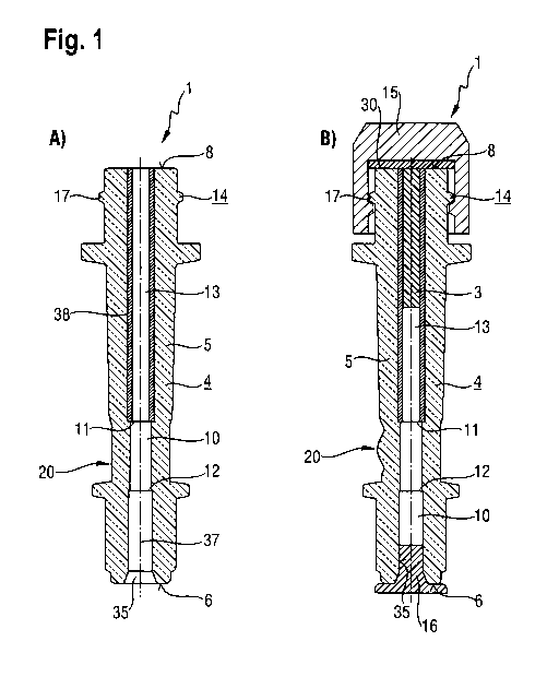
Figure 1 shows in schematic views A) and B) cross-sections through an
implantation device 1
according to a first embodiment of the invention. Figure 1 A) shows a cannula
device 4,
especially a cannula holder, which has an implantation device 1 together with
a housing 5 with a
proximal end 6 and a distal end 8. Arranged along a cannula holder axis 37 is
a central channel,
which forms an implant channel 10 and has a first shoulder 11 and a second
shoulder 12, the
channel cross-section being reduced between the shoulders 11 and 12. The
shoulder 12 serves
as a stop for a cannula in the form of an injection needle, which can be
inserted as far as the
shoulder 12 by way of a proximal opening 35 of the cannula holder 4 and can
there be
connected to the cannula device 4 preferably by a material-bonded, friction-
based or
interlocking connection.
The shoulder 11 delimits the region of an implant chamber 13, which in this
embodiment of the
invention is formed by a Teflon tube or hose section 38 and in turn forms a
portion of the
implant channel. The Teflon tube or pipe section is arranged in a bore of the
cannula device 4.
An implant material, preferably an active ingredient/polymer mixture, can be
extruded directly
into the implant chamber 13. The weight of such an implant in the implant
chamber 13, and
accordingly also the active ingredient content in the case of a fixed active
ingredient
concentration in the mixture, is determined by the inside diameter of the hose
section 38 and by
the length of the hose section 38 that is filled with an implant material.
Depending upon the
embodiment of the implantation device, the hose diameter for the implant
chamber 13 can be
varied.
CA 02790593 2012-08-21
11
The length of the implant is fixed by a piston rod having a sealing piston.
The piston rod is
inserted from one end, preferably from the proximal opening 35, with the
sealing piston first,
into the implant channel until the sealing piston is flush with the other end
of the implant
channel, preferably the distal opening. When the implant chamber is filled
with the implant
material, the sealing piston, and accordingly the piston rod as a whole, is
displaced from or
pushed out of the implant channel until the piston rod abuts a stop. The
length of the implant is
accordingly fixed by the arrangement of the stop.
The hose section 38, and accordingly the implant chamber, extend from the
distal end 8 of the
cannula holder 4 to the first shoulder 11, at which the cross-section of the
implant channel 10 is
narrowed. In that transition region from the implant chamber 13 to the cannula
arranged in the
proximal opening 35 there is provided a viewing window 20, which allows a view
into the implant
chamber in a viewing direction perpendicular thereto. In the embodiment of
Figure 1 A), the
viewing window 20 is simply a smooth or polished outer surface of the housing
5, which in this
embodiment is made of a transparent plastics material.
Instead of inserting a cannula in the form of an injection needle into the
proximal opening 35, it
is also possible to provide on the outer periphery of the cannula holder 4 an
additional coupling
device for releasably connecting a cannula to the proximal end of the cannula
holder 4.
On the outer surface of the cannula holder 4 in the region of the distal end 8
there is provided a
coupling device 14 in the form of a luer-lock external thread 17, which is
used on the one hand
for attaching a cap in a media-tight manner after the implant chamber 13 has
been filled with an
implant and on the other hand for attaching an applicator to the distal end 8
of the cannula
holder for application of the implant.
Figure 1 B) shows a schematic view of a cross-section of the cannula holder 4
shown in Figure
1 A) with an implant 3 introduced into the implant chamber 13, a cap 15 with a
luer-lock internal
thread having been screwed onto the distal end 8 of the cannula holder 4 so
that a gasket 39 in
the cap 15 closes the distal end 8 in a media-tight manner. The proximal end 6
of the cannula
holder 4 can be closed in a media-tight manner by means of a suitable stopper
16. In the region
of the viewing window 20, a concave contour is ground into the transparent
housing 5 in order
to permit a lens effect for observation during application of the implant.
CA 02790593 2012-08-21
12
Figure 2 shows in schematic views A) and B) cross-sections through an
implantation device 2
according to a second embodiment of the invention. Components of Figure 2
having the same
functions as in Figure 1 are labelled with the same reference numerals and are
not discussed
further. The difference with respect to the first embodiment of the invention
is that the housing 5
of the cannula holder 4 according to Figure 2A) is made of an optically non-
transparent
material. In this case, an opening has been formed in the optically non-
transparent material in
the region of the viewing window 20, which opening is filled with a
transparent plastics material.
It is thus ensured that, here too, it is possible to observe the movement of
the implant during
application in the transition region between the two shoulders 11 and 12.
Figure 2 B) shows this second embodiment of the invention with an implant 3,
which is arranged
in the implant chamber 13, the cannula holder 4 being closed in a media-tight
manner during
storage and transport by means of a cap 15, as is already known from Figure
113), and a
stopper 16.
Figure 3 shows in schematic views A), B) and C) the loading of a cannula
holder 4 with an
implant 3. To that end, as is shown in Figure 3 A), a piston rod 21 is
inserted into the implant
channel 10 of the cannula holder from the proximal opening 35 of the cannula
holder 4. To that
end, the piston rod 21 has at its distal end 40 a sealing piston 22, which can
be coated with a
medicinally inert material. The coating 23 ensures that the implant material
that is to be
introduced is not contaminated. The material of the coating 23 is preferably
likewise Teflon. The
piston rod 21 is pushed through the implant channel 10 and especially through
the implant
chamber 13 until the distal end 40 of the piston rod 21, and accordingly the
sealing piston 22, is
flush with the distal end 8 of the cannula holder 4. The distal end 8 of the
cannula holder has a
sealing surface which is preferably flat, so that the distal end 8 can be
positioned at an
extrusion die in a media-tight manner. There is additionally provided a stop
36 which can be set
at a distance a from the proximal end 49 of the piston rod 21 in order to
specify the length I of
an implant 3 in the implant chamber 13.
In Figure 3 B), the cannula holder 4 with the piston rod 21 is located at an
extrusion die (not
shown), which is positioned in a media-tight manner at the distal end 8 of the
cannula holder 4.
A flowable implant material, which preferably comprises a mixture of an active
ingredient and a
polymer, is extruded directly into the implant chamber 13 in the direction
indicated by arrow D.
The cross-section of the implant chamber 13 is smaller than the inside cross-
section of the
CA 02790593 2012-08-21
13
transition region between the shoulders 11 and 12 and also smaller than the
inside cross-
section of the cannula that is to be positioned. When the active
ingredient/polymer mixture is
brought into a loading position of an automatic loading device, the piston rod
21 is displaced
from or pushed out of the proximal opening 35 of the piston holder 4 until the
stop 36 has
reached the loading position through the proximal end 49 of the piston rod 21.
Accordingly, the
length I of the implant in the implant chamber is given by the distance a.
Figure 3 C shows the cannula holder 4 with an implant 3 in the region of the
distal end 8 of the
implant chamber 13. The cannula holder 4 can then be sterilised by means of
irradiation,
preferably by gamma rays, and packaged and supplied in sterile form. To that
end, as is shown
in Figure 1 B) and Figure 2 B), a cap can be placed in a media-tight manner on
the distal end 8
and a stopper can be placed on the proximal end 6 of the cannula holder 4.
Figure 4 shows in schematic views A), B) and C) the loading of an implantation
device 2 with an
implant. Unlike in Figure 3, in the case of this loading the cannulas 7 are
already arranged with
their distal end 18 in the proximal opening 35 of the cannula holder 4, the
distal end 18 abutting
the shoulder 12 of the cannula holder 4. In order to define the length of the
implant material that
is to be introduced there is provided a piston rod 21 which is longer than the
piston rod shown in
Figure 3 by the length of the cannula 7. Components in Figures 4 A), B) and C)
which have the
same functions as in Figure 3 are labelled with the same reference numerals
and are not
discussed further. Accordingly, Figure 4 merely demonstrates that it is also
possible to load the
implant chamber 13 with a predetermined amount of active ingredient/polymer
mixture with the
cannula 7 already inserted. Sterilisation can then again be carried out, and a
cannula holder 4
loaded with an implant 3 and with an attached cannula 7 can be supplied.
Alternatively to loading the cannula holder from its distal end, the cannula
holder can also be
loaded with an implant from its proximal end, especially when the cannula
holder is not provided
with the cannula until after loading.
Figure 5 shows in schematic views A), B) and C) an implantation device 2 on
application of the
implant 3. To that end, as is shown in Figure 5 A), an applicator 9 is
attached to the distal end 8
by means of the coupling device 14, the mouthpiece of the applicator having a
disk-like seal 41
which connects the distal end 8 of the cannula holder 4 to the applicator 9 in
a media-tight
manner. The mouthpiece of the applicator 9 preferably has a luer-lock internal
thread, which
CA 02790593 2012-08-21
14
engages in a luer-lock external thread 17 of the cannula holder 4. A central
applicator rod 42 is
brought into a coaxial position relative to the cannula holder axis 37 and is
in contact with the
distal end of the implant 3 with a sealing piston 43, which has a medicinally
inert coating 44.
In Figure 5 B), a pressure is exerted on the implant 3 in the direction
indicated by arrow E, so
that the implant 3 is pushed out of the implant chamber 13 and guided past the
viewing window
20, whereby monitoring by the personnel carrying out the application is
possible.
In Figure 5 C), the implant 3 has been introduced into the injection needle 19
in the direction
indicated by arrow E by means of the applicator rod 42 and, if the injection
needle 19 is already
arranged subcutaneously, can finally be pushed out of the injection needle 19
subcutaneously
and positioned. To that end, a simultaneous movement of the injection needle
19 in the distal
direction is advantageous in order to position the implant 3 subcutaneously.
Figure 6 shows a schematic diagram of a novel applicator 9, which has a gear
wheel 45. This
couples two toothed rods 46 and 47 together and has the effect that, when the
implant 3 is
pushed in direction E, a withdrawal of the injection needle in direction F
takes place
synchronously. However, before that synchronous movement takes place, the
implant 3 is
brought beyond the position shown in Figure 5 C) to the proximal end of the
cannula 7.
Figure 7 shows a partially perspective schematic diagram of a loading device
24 for loading
cannula holders 4 with an implant 3. To that end, the loading device 24 has a
feed device 25,
which in this embodiment of the invention has two conveyor belts 32 and 33 as
well as a rotary
plate 34 and an extruder 27 having a mixing device 28. The entire loading
device 24 is
controlled by a control and monitoring device 31. A polymer granulate and the
active ingredient
are introduced into the opening 48 of the extruder 27 in the direction
indicated by arrow G and
are brought into a mixing device 28 by way of a screw drive by rotation in the
direction indicated
by arrow H, a pressure at the same time building up, which pushes the mixture
of active
ingredient and polymer granulate to an extrusion die 29. A heater 30 heats the
extrusion die 29
to a softening temperature of the polymer granulate, so that it becomes liquid
or at least
viscous, that is to say flowable under pressure.
The cannula holders 4 with empty, unfilled implant chambers are fed by way of
a feed conveyor
belt 32 to the rotary plate 34, which transports them cyclically into a
loading position 26 beneath
CA 02790593 2012-08-21
the extrusion die 29. By opening and closing of the extrusion die 29, an
amount of implant
material set beforehand by means of a piston rod is introduced into implant
chambers of the
cannula holders 4 in the loading position 26.
The cannula holders 4 loaded or filled with implant material are provided at
their distal ends 8
with caps shown in Figures 1B) and 2B) and are sealed at their proximal ends 6
with
corresponding stoppers and fed to a take-off conveyor belt 33. The conveyor
belts 32 and 33
can be in the form of endless belts and are loaded and emptied by an automatic
unit (not shown
here). Sterilisation positions can be provided on the removal conveyor belt
33, before the filled
or loaded cannula holders 4 are removed from the take-off conveyor belt 33 and
dispatched
packaged in a sterile manner.
CA 02790593 2012-08-21
16
List of reference numerals
1 Implantation device (first embodiment)
2 Implantation device (second embodiment)
3 Implant
4 Cannula holder
Housing of the cannula holder
6 Proximal end of the cannula holder
7 Cannula
8 Distal end of the cannula holder
9 Applicator
Implant channel
11 Shoulder in the channel
12 Shoulder in the channel
13 Implant chamber
14 Coupling device
Cap
16 Stopper
17 Luer-lock external thread
18 Distal end of the cannula
19 Injection needle
Viewing window
21 Piston rod
22 Sealing piston
23 Coating of the sealing piston
24 Loading device
Feed device
26 Loading position
27 Extruder
28 Mixing device
29 Extrusion die
Heater
31 Control and monitoring device
32 Conveyor belt
CA 02790593 2012-08-21
17
33 Conveyor belt
34 Rotary plate
35 Proximal opening of the cannula holder
36 Stop
37 Cannula holder axis
38 Tube
39 Gasket
40 Distal end of the piston rod
41 Seal
42 Applicator rod
43 Sealing piston
44 Coating
45 Gear wheel
46 Toothed rod
47 Toothed rod
48 Opening
49 Proximal end of the piston rod
a Distance
D Direction of arrow
E Direction of arrow
F Direction of arrow
G Direction of arrow
H Direction of arrow
I Length of the implant rod