Sélection de la langue

Search

Sommaire du brevet 2790792 

Énoncé de désistement de responsabilité concernant l'information provenant de tiers

Une partie des informations de ce site Web a été fournie par des sources externes. Le gouvernement du Canada n'assume aucune responsabilité concernant la précision, l'actualité ou la fiabilité des informations fournies par les sources externes. Les utilisateurs qui désirent employer cette information devraient consulter directement la source des informations. Le contenu fourni par les sources externes n'est pas assujetti aux exigences sur les langues officielles, la protection des renseignements personnels et l'accessibilité.

Disponibilité de l'Abrégé et des Revendications

L'apparition de différences dans le texte et l'image des Revendications et de l'Abrégé dépend du moment auquel le document est publié. Les textes des Revendications et de l'Abrégé sont affichés :

  • lorsque la demande peut être examinée par le public;
  • lorsque le brevet est émis (délivrance).
(12) Demande de brevet: (11) CA 2790792
(54) Titre français: APPAREIL MEDICAL A RAYONS X ET PROCEDE DE FONCTIONNEMENT
(54) Titre anglais: MEDICAL DEVICE OPERATING WITH X-RAYS AND METHOD FOR OPERATING SAME
Statut: Réputée abandonnée et au-delà du délai pour le rétablissement - en attente de la réponse à l’avis de communication rejetée
Données bibliographiques
(51) Classification internationale des brevets (CIB):
  • A61N 5/10 (2006.01)
  • A61N 5/01 (2006.01)
(72) Inventeurs :
  • HEID, OLIVER (Allemagne)
(73) Titulaires :
  • SIEMENS AKTIENGESELLSCHAFT
(71) Demandeurs :
  • SIEMENS AKTIENGESELLSCHAFT (Allemagne)
(74) Agent: SMART & BIGGAR LP
(74) Co-agent:
(45) Délivré:
(86) Date de dépôt PCT: 2011-02-02
(87) Mise à la disponibilité du public: 2011-09-01
Licence disponible: S.O.
Cédé au domaine public: S.O.
(25) Langue des documents déposés: Anglais

Traité de coopération en matière de brevets (PCT): Oui
(86) Numéro de la demande PCT: PCT/EP2011/051459
(87) Numéro de publication internationale PCT: WO 2011104075
(85) Entrée nationale: 2012-08-22

(30) Données de priorité de la demande:
Numéro de la demande Pays / territoire Date
10 2010 009 019.0 (Allemagne) 2010-02-24

Abrégés

Abrégé français

L'invention concerne un appareil médical à rayons X, comprenant : - une source de rayons X apte à émettre un faisceau de rayons x présentant un maximum d'intensité le long d'un faisceau central présente, - un dispositif de rotation apte à faire tourner la source de faisceau rayons X autour d'un isocentre, l'axe central du faisceau de rayons X étant excentré par rapport à l'isocentre de telle façon que, notamment lors d'une rotation autour de l'isocentre, les faisceaux centraux émis depuis différentes directions spatiales sont tangentiels à un cercle fictif ayant pour centre l'isocentre. L'invention concerne également un procédé de fonctionnement d'un appareil médical, comprenant les étapes suivantes consistant à : prendre une source de faisceaux de rayons X apte à émettre un faisceau de rayons X qui présente une intensité maximale le long d'un faisceau central; à faire tourner la source de faisceau de rayons X autour d'un isocentre, ledit axe central du faisceau de rayons X étant excentrique par rapport à l'isocentre de telle façon que, notamment lors d'une rotation autour de l'isocentre, les faisceaux centraux émis depuis différentes directions spatiales sont tangentiels à un cercle fictif ayant pour centre l'isocentre.


Abrégé anglais

The invention relates to a medical device operating with X-rays, comprising: an X-ray source, from which an X-ray beam that has an intensity maximum along a central ray can be emitted, a rotation unit, with which the X-ray source can be rotated about an isocentre, wherein the central axis of the X-ray beam is oriented eccentrically to the isocentre such that, in particular upon rotation about the isocentre, the central rays emitted from different spatial directions are tangential to an imaginary circle around the isocentre. Furthermore, the invention relates to a method for operating a medical device, comprising the following steps: providing an X-ray source, from which an X-ray beam that has an intensity maximum along a central ray is emitted, rotating the X-ray source about an isocentre, wherein the central axis of the X-ray beam is oriented eccentrically to the isocentre such that, in particular upon rotation about the isocentre, the central rays emitted from different spatial directions are tangential to an imaginary circle around the isocentre.

Revendications

Note : Les revendications sont présentées dans la langue officielle dans laquelle elles ont été soumises.


Claims
1. A medical device operating with x-rays, comprising:
- an x-ray radiation source, with which an x-ray radiation
cone can be emitted, which has a maximum intensity along a
central beam,
- a rotation apparatus, with which the x-ray radiation
source can be rotated about an isocenter,
wherein the central axis of the x-ray radiation cone is aligned
paraxially past the isocenter.
2. The medical device operating with x-rays according to
claim 1, wherein
the central axis of the x-ray radiation cone is aligned
paraxially past the isocenter such that upon rotation around the
isocenter, the central beams emitted from different spatial
directions are tangential to an imaginary circle around the
isocenter.
3. The medical device operating with x-rays as claimed in
one of claims 1 or 2, wherein the device is embodied as a
radiation therapy device.
4. The medical device operating with x-rays as claimed in
one of claims 1 to 3, wherein a collimator exists in the
radiation path of the x-ray radiation cone for lateral
delimitation of the beam profile of the x-ray beam.
5. The medical device operating with x-rays as claimed in
one of claims 1 to 4, wherein no beam flattening filter which is

11
used for spatial dose homogenization is arranged in the radiation
path of the x-ray radiation cone.
6. A method for operating a medical device, comprising the
following steps:
- providing an x-ray radiation source, with which an x-ray
radiation cone is emitted, which has a maximum intensity along a
central beam,
- rotating the x-ray radiation source about an isocenter,
wherein the central axis of the x-ray radiation cone is directed
paraxially past the isocenter.
7. The method as claimed in claim 6, wherein
the central axis of the x-ray radiation cone is directed
paraxially past the isocenter such that upon rotatation around
the isocenter, the central beams emitted from different spatial
directions are tangential to an imaginary circle around the
isocenter.
8. The method as claimed in claim 6 or 7, wherein no beam
flattening filter used for spatial dose homogenization is
arranged in the radiation path of the x-ray radiation cone.
9. The method as claimed in one of claims 6 to 8, wherein
the x-ray radiation cone is collimated.
10. A use of an x-ray radiation cone, which has a maximum
intensity along a central beam, which, with a medical device
operating with x-rays, is emitted by an x-ray source from
different spatial directions about an isocenter, for dose
homogenization in an area around the isocenter, by the central

12
beam of the x-ray radiation cone being aligned laterally past the
isocenter in each spatial direction.

Description

Note : Les descriptions sont présentées dans la langue officielle dans laquelle elles ont été soumises.

CA 02790792 2012-08-22
PCT/EP2011/051459 / 2010P00420W0
1
Description
Medical device operating with x-rays and method for operating
same
The invention relates to a medical device operating with x-rays,
in particular a radiation therapy device, as well as method for
operating the same.
The irradiation of a finite target volume by means of x-rays from
different spatial directions, such as is implemented for instance
for the purposes of imaging in diagnostic methods or dose
synthesis in therapeutic methods, usually requires as homogenous
a gross illumination of the target volume as possible.
Since the x-ray sources usually used have a preferred beam
direction, along which the maximum dose is located, and the beam
direction is usually aligned toward the center of the target
volume, the illumination of the boundary areas of the target
volume is usually reduced compared with the center of the target
volume.
The problem with radiation therapy devices and with imaging
devices was previously solved by a beam flattening filter, which
flattens the central portions of the radiation lobe and thus
homogenizes the profile of the x-ray beam.
It is the object of the invention to provide a medical device
operating with x-rays, which enables a rapid and efficient
illumination of a target volume. Furthermore, it is the object of

CA 02790792 2012-08-22
PCT/EP2011/051459 / 2010P00420W0
2
the invention to provide a method with which it is possible to
illuminate a target volume rapidly and efficiently.
The object of the invention is achieved by the features of the
independent claims. Advantageous developments of the invention
are found in the features of the dependent claims,
The inventive medical device operating with x-rays includes:
an x-ray radiation source, with which an x-ray can be
emitted, which has a maximum dose along a central beam,
a rotation apparatus, with which the x-ray radiation
source can be rotated about an isocenter,
wherein the central axis of the x-ray beam is aligned
eccentrically to the isocenter, so that in particular upon
rotation around the isocenter, the central beams emitted from
different spatial directions are tangential to an imaginary
circle around the isocenter.
The invention is based on the knowledge that the radiation lobe
of an x-ray source, in other words the x-ray cone emitted by the
x-ray source, usually has a maximum along the central beam. The
radiation lobe of the x-ray source is now no longer directed at
the center, but instead more at the boundary area. The
irradiation therefore takes place paraxially and/or tangentially
from different spatial directions.
By rotating the source around the target volume, a significantly
more homogenous illumination of the entire volume is achieved on
the whole, even if the radiation lobe itself indicates an
inhomogeneous dose profile. The more homogenous illumination is
enabled by the combination of inhomogeneous dose profile of the

CA 02790792 2012-08-22
PCT/EP2011/051459 / 2010P00420W0
3
radiation lobe, which flattens around'a maximum dose along the
central beam, an eccentric alignment of the radiation lobe and of
the rotation of the radiation lobe.
The geometric arrangement and alignment of the x-ray source
together with the rotation of the x-ray source therefore
contribute to the homogenization of the illumination.
Compared with previous solutions, in which the homogenization is
largely effected by an absorber, a significantly improved
efficiency of the x-ray source can be achieved.
The absorber can namely be embodied such that it absorbs less
radiation power, since it no longer has to cater for the
homogenization of the dose profile on its own. It can even be
completely omitted, so that the complete dose output of the
source strikes the target volume. The latter case is free of beam
flattening filters used for homogenization in the x-ray radiation
path. Overall, it is therefore possible to dispense with a lossy,
radiation-blasted absorber.
The dose rate with which a target volume can be illuminated
increases as a result. The irradiation duration depends on the
fraction (desired dose)/ (local dose rate) formed across the
tumor volume. The site to be irradiated at which the minimal dose
rate occurs, in other words the least irradiated region,
.therefore defines the irradiation duration. Time can therefore be
reduced using the inventive medical device.
With an inhomogeneous dose distribution, it also applies that
locations irradiated at the same time with higher dose rates are

CA 02790792 2012-08-22
PCT/EP2011/051459 / 2010P00420WO
4
protected from the radiation for a part of the irradiation
duration, since otherwise an excessively high dose would be
applied there. A more homogenous dose distribution also solves
this problem. Locations with an excessively high"dose rate need
not be"masked out after a short period of time, e.g. with a
collimator, so that the desired dose can be applied with a lower
dose rate at another location.
In particular, the device can be embodied as a radiation therapy
device, for instance with a therapeutic x-ray radiation source,
which can be rotated about an isocenter.
A collimator can be present in the radiation path of the x-ray,
with which the beam profile of the x-ray can then be laterally
delimited and can thus be adjusted to a target volume to be
irradiated for instance. Only certain partial volumes can
therefore be irradiated with an object to be irradiated.
The inventive method for operating a medical device, comprising
the following steps:
providing an x-ray radiation source, with which an x-ray beam is
emitted, which has a maximum intensity along a central beam,
rotating the x-ray radiation source about an isocenter,
wherein during rotation the central axis of the x-ray beam is
aligned eccentrically to the isocenter such that in particular
,upon rotation about the isocenter, the central beams emitted from
different directions are tangential to an imaginary circle around
the isocenter.

CA 02790792 2012-08-22
PCT/EP2011/051459 / 2010P00420W0
No beam flattening filter is preferably arranged in the radiation
path of the x-ray. The radiation path of the x-ray beam can be
collimated in order to form the lateral profile.
In accordance with the invention, the x-ray beam, which has a
maximum intensity in the.central beam and which, with a medical
device operating with x-rays is emitted from an x-ray source from
different spatial directions about an isocenter, is used to
homogenize the dose in an area around the isocenter, by the
central beam of the x-ray beam being aligned laterally past the
isocenter in each spatial direction.
The preceding and subsequent description of the individual
features and the advantages and effects thereof relate both to
the apparatus category and also to the method category, without
this being explicitly mentioned in detail in each case; the
individual features disclosed here can also be essential to the
invention in other combinations than shown.
Embodiments of the invention with developments according to the
features of the dependent claims are explained in more detail
with the aid of the following drawing, without however being
restricted thereto, in which:
Fig. 1 shows the principle of dose generation in a radiation
therapy device according to the prior art,
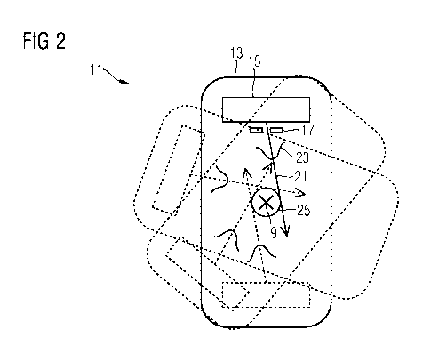
Fig. 2 shows the principle of dose generation in an embodiment
of the inventive radiation therapy device.

CA 02790792 2012-08-22
PCT/EP2011/051459 / 2010P00420W0
6
With the aid of Fig. 1, the principle'of dose generation about an
isocenter in a radiation therapy device is explained according to
the prior art.
The radiation therapy device 11 includes a rotatable gantry 13,
with which an x-ray radiation source 15 can be rotated around the
object to be irradiated. A conical x-ray beam is directed at the
isocenter 19 from the x-ray radiation source. The x-ray beam is
laterally delimited by a collimator 17. The irradiation usually
takes place from different directions by rotating the x-ray
source 15 around the isocenter 19, indicated by the positions of
the gantry shown with dashed lines.
The central beam 21 of the conical x-ray beam strikes the
isocenter 19 in the process. The x-ray beam in this way has a
dose distribution 23, indicated by the Gaussian-type curve, which
has a maximum dose along the central beam 21, which tapers toward
the edge. If a target volume is irradiated with the particle
beam, the dose rate in the isocenter 19 is therefore at its
highest and attenuates toward the edge.
This principle is made clearly apparent by the applied dose
generated by the two irradiation directions shown opposite one
another. The dose maxima illuminate the target volume at the same
point, so that the dose applied by the two irradiation directions
in the target volume has the same inhomogeneous distribution as
the inhomogeneous dose. distribution 23 in the x-ray beam.
A beam flattening filter (not shown here) is therefore generally
used in a radiation therapy device 11 according to the prior art,
said beam flattening filter being arranged in the radiation path

CA 02790792 2012-08-22
PCT/EP2011/051459 / 2010P00420W0
7
of the x-ray beam and rendering the inhomogeneous dose profile
more homogenous. This however takes place at the cost of the
radiation dose which is destroyed in the beam flattening filter.
Fig. 2 shows the principle of dose generation in an inventive
radiation therapy device 11.
The central beam 21 of the x-ray source 15 is now no longer
aligned toward the isocenter 19, but aims paraxially past the
isocenter. Upon rotation of the x-ray source 15 about the
isocenter 19, this results in the central beams 21 emitted being
tangential to a circle 25 around the isocenter.
The dose which is set up around the isocenter 19 in geometric
configurations of this type, is clearly more homogenous compared
with a dose which was applied when aligning the central beam 23
to the isocenter 19.
This is clearly apparent with the aid of the two irradiation
directions shown opposite one another. The maximum dose which is
applied by the respective irradiation directions is now no longer
in the isocenter 19, but instead respectively at an opposite
point of the isocenter 19. Dose portions which clearly lie below
the maximum dose are added together in the isocenter 10 itself by
the opposite irradiation. By adding the dose portions, a dose is
applied in the isocenter 19, which is significantly closer to the
maximum dose than the individual dose portions themselves. A
significantly more homogenous dose can be applied overall as a
result.

CA 02790792 2012-08-22
PCT/EP2011/051459 / 2010P00420W0
8
Ideally it is therefore even possible'to completely dispense with
a beam flattening filter. No dose output is then destroyed in the
beam flattening filter, as a result of which the target volume
can be illuminated with a higher dose rate, which significantly
reduces irradiation time.
Provision can also be made here for a collimator 17, which
adjusts and restricts the applied dose to a target volume.
Even if the principle was explained with the aid of a radiation
therapy device 11, the same principle of achieving a dose
homogenization of the dose distribution set up about an isocenter
13 can be transferred to imaging devices operating with x-rays,
such as for instance a computed tomography system (CT) or a cone
beam CT.

CA 02790792 2012-08-22
PCT/EP2011/051459 / 2010P00420W0
9
List of reference characters
11 Radiation therapy device
13 Gantry
15 x-ray radiation source
17 Collimator
19 Isocenter
21 Central beam
23 Dose distribution
25 Circle

Dessin représentatif
Une figure unique qui représente un dessin illustrant l'invention.
États administratifs

2024-08-01 : Dans le cadre de la transition vers les Brevets de nouvelle génération (BNG), la base de données sur les brevets canadiens (BDBC) contient désormais un Historique d'événement plus détaillé, qui reproduit le Journal des événements de notre nouvelle solution interne.

Veuillez noter que les événements débutant par « Inactive : » se réfèrent à des événements qui ne sont plus utilisés dans notre nouvelle solution interne.

Pour une meilleure compréhension de l'état de la demande ou brevet qui figure sur cette page, la rubrique Mise en garde , et les descriptions de Brevet , Historique d'événement , Taxes périodiques et Historique des paiements devraient être consultées.

Historique d'événement

Description Date
Le délai pour l'annulation est expiré 2014-02-04
Demande non rétablie avant l'échéance 2014-02-04
Réputée abandonnée - omission de répondre à un avis sur les taxes pour le maintien en état 2013-02-04
Inactive : Page couverture publiée 2012-10-29
Inactive : Notice - Entrée phase nat. - Pas de RE 2012-10-10
Inactive : CIB attribuée 2012-10-10
Demande reçue - PCT 2012-10-10
Inactive : CIB en 1re position 2012-10-10
Inactive : CIB attribuée 2012-10-10
Exigences pour l'entrée dans la phase nationale - jugée conforme 2012-08-22
Demande publiée (accessible au public) 2011-09-01

Historique d'abandonnement

Date d'abandonnement Raison Date de rétablissement
2013-02-04

Historique des taxes

Type de taxes Anniversaire Échéance Date payée
Taxe nationale de base - générale 2012-08-22
Titulaires au dossier

Les titulaires actuels et antérieures au dossier sont affichés en ordre alphabétique.

Titulaires actuels au dossier
SIEMENS AKTIENGESELLSCHAFT
Titulaires antérieures au dossier
OLIVER HEID
Les propriétaires antérieurs qui ne figurent pas dans la liste des « Propriétaires au dossier » apparaîtront dans d'autres documents au dossier.
Documents

Pour visionner les fichiers sélectionnés, entrer le code reCAPTCHA :


Pour visualiser une image, cliquer sur un lien dans la colonne description du document. Pour télécharger l'image (les images), cliquer l'une ou plusieurs cases à cocher dans la première colonne et ensuite cliquer sur le bouton "Télécharger sélection en format PDF (archive Zip)" ou le bouton "Télécharger sélection (en un fichier PDF fusionné)".

Liste des documents de brevet publiés et non publiés sur la BDBC .

Si vous avez des difficultés à accéder au contenu, veuillez communiquer avec le Centre de services à la clientèle au 1-866-997-1936, ou envoyer un courriel au Centre de service à la clientèle de l'OPIC.


Description du
Document 
Date
(aaaa-mm-jj) 
Nombre de pages   Taille de l'image (Ko) 
Dessins 2012-08-22 1 17
Abrégé 2012-08-22 1 25
Revendications 2012-08-22 3 71
Description 2012-08-22 9 290
Dessin représentatif 2012-08-22 1 8
Page couverture 2012-10-29 2 49
Rappel de taxe de maintien due 2012-10-10 1 111
Avis d'entree dans la phase nationale 2012-10-10 1 193
Courtoisie - Lettre d'abandon (taxe de maintien en état) 2013-04-02 1 173
PCT 2012-08-22 15 489