Note : Les descriptions sont présentées dans la langue officielle dans laquelle elles ont été soumises.
CA 02793483 2012-09-17
WO 2011/123239 PCT/US2011/028437
H-RM-01572 WO
MULTI-WAVELENGTH PHOTON DENSITY WAVE SYSTEM
USING AN OPTICAL SWITCH
BACKGROUND
The present disclosure relates generally to non-invasive measurement of
physiological parameters and, more particularly, to multi-wavelength photon
density
wave measurements of physiological parameters.
This section is intended to introduce the reader to various aspects of art
that
may be related to various aspects of the present disclosure, which are
described and/or
claimed below. This discussion is believed to be helpfiul in providing the
reader with
background information to facilitate a better understanding of the various
aspects of
the present disclosure. Accordingly, it should be understood that these
statements are
to be read in this light, and not as admissions of prior art.
Pulse oximetry may be defined as a non-invasive technique that facilitates
monitoring of a patient's blood flow characteristics. For example, pulse
oximetry may
be used to measure blood oxygen saturation of hemoglobin in a patient's
arterial blood
and/or the patient's heart rate. Specifically, these blood flow characteristic
measurements may be acquired using a non-invasive sensor that passes light
through a
portion of a patient's tissue and photo-electrically senses the absorption and
scattering of
the light through the tissue. Typical pulse oximetiy technology may employ two
light
emitting diodes (LEDs) and a single optical detector to measure pulse and
oxygen
saturation of a given tissue bed.
A typical signal resulting from the sensed light may be referred to as a
plethysmograph waveform. The plethysmograph waveform is largely based on
absorption of emitted light by specific types of blood constituents and may be
used
with various algorithms to estimate a relative amount of blood constituent in
the
tissue. For example, the plethysmograph waveform may provide a ratio of
oxygenated
hemoglobin to total hemoglobin in the volume being monitored. The amount of
arterial blood in the tissue is generally time-varying during a cardiac cycle,
which is
reflected in the plethysinographic waveform.
1
CA 02793483 2012-09-17
WO 2011/123239 PCT/US2011/028437
H-RM-01572 WO
The accuracy of blood flow characteristic estimation via pulse oximetry may
depend on a number of factors. For example, variations in light absorption
characteristics can affect accuracy, and such variations may depend on where
the
sensor is located and/or the physiology of the patient being monitored,
Additionally,
various types of noise and interference can create inaccuracies. For example,
electrical noise, physiological noise, and other interference can contribute
to
inaccurate blood flow characteristic estimates.
BRIEF DESCRIPTION OF THE DRAWINGS
Advantages of the presently disclosed subject matter may become apparent
upon reading the following detailed description and upon reference to the
drawings in
which:
FIG. 1 is a perspective view of a pulse oximeter system in accordance with an
embodiment;
FIG. 2 is a block diagram of the pulse oximeter system of FIG. 1, in
accordance with
an embodiment;
FIG, 3 is a plot of a first single-wavelength photon density wave signal for
use in the
system of FIG. 1, in accordance with an embodiment;
FIG. 4 is a plot of a second single-wavelength photon density wave signal for
use in
the system of FIG. 1, in accordance with an embodiment;
FIG. 5 is a plot of a multi-wavelength photon density wave signal combining
the first
and second single-wavelength photon density wave signals of FIGS. 3 and 4, in
accordance with an embodiment;
PIG. 6 is a plot representing a comparison between a portion of the multi-
wavelength
photon density wave signal of FIG. 5 and a received single-wavelength photon
density wave signal after the signal of FIG. 5 has been passed through a
patient, in
accordance with an embodiment;
2
CA 02793483 2012-09-17
WO 2011/123239 PCT/US2011/028437
H-RM-01572 WO
FIG. 7 is a flowchart representing an embodiment of a method for obtaining
physiological measurements using the system of FIG. 1, in accordance with an
embodiment; and
FIG. 8 is a flowchart representing an embodiment of an algorithm for use by
the
system of FIG. 1 for determining scattering and absorption properties of
patient
tissue.
DETAILED DESCRIPTION
One or more specific embodiments of the present disclosure will be described
below. In an effort to provide a concise description of these embodiments, not
all
features of an actual implementation are described in the specification. It
should be
appreciated that in the development of any such actual implementation, as in
any
engineering or design project, numerous implementation-specific decisions must
be
made to achieve the developers' specific goals, such as compliance with system-
related and business-related constraints, which may vary from one
implementation to
another. Moreover, it should be appreciated that such a development effort
might be
complex and time consuming, but would nevertheless be a routine undertaking of
design, fabrication, and manufacture for those of ordinary skill having the
benefit of
this disclosure.
Present embodiments relate to non-invasively measuring physiological
parameters corresponding to blood flow in a patient. Specifically, light may
be
emitted into a patient and photoelectrically detected after having propagated
through
(e.g., transmitted through, scattered by, and/or reflected by) pulsatile
tissue of a
patient. The propagation of light through pulsatile tissue may by affected by
the
composition of the tissue, which may vary as blood enters and exits the
pulsatile
tissue. Rather than emitting a light signal modulated at a rate that is
effectively DC
through the pulsatile patient tissue, present embodiments involve emitting a
light
modulated at frequencies sufficient to produce waves of photons known as
photon
density waves. A photon density wave may refer to light that is modulated at
frequencies of approximately 50 MHz - 3 GHz. Photon density waves may be
3
CA 02793483 2012-09-17
WO 2011/123239 PCT/US2011/028437
H-RM-01572 WO
resolvable in pulsatile tissue because the photon density waves may have a
wavelength that is shorter than a mean absorption distance in pulsatile
tissue.
A photon density wave ("PDW") signal that has propagated through the
pulsatile tissue of the patient may be detected and analyzed to obtain
absorption
and/or scattering properties of the pulsatile patient tissue. Certain changes
between
the PDW signal emitted into the tissue (i.e., the input signal) and the PDW
signal
detected after propagating through the tissue (i.e., an output signal) may be
indicative
of tissue conditions. In particular, a change in amplitude between the output
signal
and the input signal may correspond to absorptive components in the pulsatile
tissue.
A change in phase between the output signal and the input signal may
correspond to
scattering components in the pulsatile tissue.
Changes in amplitude of the PDW signals may correspond to the amount of
absorptive components in the pulsatile patient tissue, as certain components
of the
tissue may absorb different wavelengths of light, such as red or infrared
light, in
different amounts. By analyzing decreases in amplitudes between the output
signal
and the input signal, a ratio of different types of particles in the pulsatile
patient tissue,
such as oxygenated and deoxygenated hemoglobin, may be estimated.
Changes in phase of the PDW signals may correspond to the total number of
scattering particles in the area of measurement of the patient tissue. More
specifically,
the phase change between the output signal and the input signal may correspond
to the
total number of particles (e.g., total hemoglobin) which scatter the PDW
signal, and
not merely a ratio of particles (e.g., oxygenated and total hemoglobin).
Further,
variations in the phase change may also be measured to provide information
associated with variations of total particles in the patient tissue. With
measurements
of absorption and scattering by components of the patient tissue,
physiological
parameters such as SpO2, regional oxygen saturation, total hemoglobin,
perfusion, and
many others may be monitored.
Thus, PDW signals may be used in the present embodiments to provide
physiological information based on the amplitude and phase change of the
detected
output signal. It is now recognized that using multiple PDW signals having
different
4
CA 02793483 2012-09-17
WO 2011/123239 PCT/US2011/028437
H-RM-01572 WO
wavelengths may provide even more physiological information, as patient tissue
may
have different components of interest (e.g., various types of cells or
structures), which
may each have different absorption and scattering coefficients. To emit
multiple
wavelengths of PDW signals to the patient tissue, multiple optical fibers may
be used
to transmit the generated PDW signals from the monitor to the sensor and into
the
patient tissue. Likewise, multiple detectors may be used to detect the PDW
signals
that have propagated through the patient tissue. However, such a configuration
may
increase system complexity and cost.
In accordance with the present techniques, multiple PDW signals of various
wavelengths of light may be time-division multiplexed using an optical switch,
such
that one wavelength of light is transmitted through a single emission optical
cable at
any one instant. Thus, the single emission optical cable may transmit to a
sensor a
multi-wavelength PDW signal which includes multiple single-wavelength PDW
signal components transmitted in series over time. Such a multi-wavelength PDW
signal (i.e., the input signal), emitted from the sensor into pulsatile
patient tissue, may
be received by a detector in the sensor after propagating (e.g., reflecting,
scattering,
and/or passing) through the tissue. Thereafter, a single optical cable may
carry the
received signal (i.e., the output signal) to the patient monitor. Since the
multi-
wavelength PDW input signal is time-division multiplexed, only one wavelength
of
the output signal is received at one time. Thus, a single detector may
photoelectrically
detect and digitize the output signal. The detected and digitized output
signal may be
demultiplexed into its component single-wavelength PDW signals and processed
to
determine various physiological parameters based on comparisons with the multi-
wavelength PDW input signal.
With the foregoing in mind, FIG. 1 illustrates a perspective view of a PDW
pulse oximetry system 10, which may include a patient monitor 12 and a pulse
oximeter sensor 14. A sensor cable 16 may connect the patient monitor 12 to
the
sensor 14, and may include two fiber optic cables. One of the fiber optic
cables
within the sensor cable 16 may transmit a multi-wavelength PDW input signal
from
the patient monitor 12 to be emitted into the patient tissue 26 by an emitter
22 on the
sensor 14. The multi-wavelength PDW input signal may propagate through the
5
CA 02793483 2012-09-17
WO 2011/123239 PCT/US2011/028437
H-RM-01572 WO
patient tissue 26 and be received as an output signal by a detector 24 on the
sensor 14.
Another of the fiber optic cables may transmit the output signal from the
sensor 14 to
the patient monitor 12. The cable 16 may couple to the monitor 12 via an
optical
connection 18. Based on signals received from the sensor 14, the patient
monitor 12
may determine certain physiological parameters that may appear on a display
20.
Such parameters may include, for example, a plethysmograin or numerical
representations of patient blood flow (e.g., partial oxygen saturation or a
measurement
of total hemoglobin).
FIG. 2 represents a block diagram of the system 10 of FIG. 1. Specifically,
FIG. 2 more clearly illustrates that the patient monitor 12 may generate
several single-
wavelength PDW signals which may be transmitted via the sensor cable 16 to the
sensor 14 at alternating periods of time (e.g., approximately I - 10 ms, and
4.5 ins in
one embodiment) using a driving circuit 28. The driving circuit 28 may include
multiple light sources 30 (e.g., laser diodes) which may each emit a
wavelength of
light. Such wavelengths may include red wavelengths of between approximately
600-
700 nm (e.g., 660 nm), infrared wavelengths of between approximately 800-1000
nm
(e.g., 900 nm), and/or any other wavelength which may be emitted into and
detected
from the patient tissue 26 to provide physiological information. For example,
in one
embodiment, the light sources 30 of the driving circuit 28 may also emit a far
red
wavelength of between approximately 690-770 nm (e.g., 730 nm). Other
wavelengths
that may be emitted by the multiple light sources 30 of the driving circuit 28
may
include, for example, wavelengths of between approximately 500-600 urn and/or
1000-1100 nm.
The driving circuit 28 may modulate light emitted from the light sources 30 at
a modulation frequency between approximately 50 MHz to 3 GHz, which may
produce resolvable photon density waves in pulsatile tissue. In some
embodiments,
the driving circuit 28 may sweep the modulation frequency of one or more of
the light
sources in a range from 50 MHz to 2.4 GHz. Some embodiments of the patient
monitor 12 may be configured to modulate light between 100 MHz and 1 GHz or to
sweep a range from 100 MHz to 1 GHz. The driving circuit 28 may, in certain
embodiments, modulate the light sources primarily at a frequency of
approximately
6
CA 02793483 2012-09-17
WO 2011/123239 PCT/US2011/028437
H-RM-01572 WO
500 MHz. Examples of PDW signals that may be generated by the driving circuit
28
of the patient monitor 12 may be illustrated below with reference to FIGS. 3
and 4.
The modulation frequency used to modulate each wavelength of light may vary,
and
light emitted from one of the light sources 30 may be modulated at a higher or
lower
modulation frequency than light emitted from another of the light sources 30.
The
driving circuit 28 may represent one or more components of commonly available
drive circuits (e.g., DVD R/W driver circuits) for modulation. Examples of
such
devices may include the LMH6525 device available from National Semiconductor
Inc. In FIG. 2, the driving circuit 28 is illustrated as being configured to
generate two
single-wavelength photon density wave signals of different wavelengths
respectively
through optical cables 32 and 34. However, it should be appreciated that the
driving
circuit 28 may be designed to generate any suitable number of single-
wavelength
PDW signals.
An optical switch 36 may switch between the two optical cables 32 and 34,
selecting one of the two modulated single-wavelength PDW signals, such that a
multi-
wavelength PDW signal is produced with multiple single-wavelength PDW signal
portions in series. The switching of the optical switch 36 may time-division
multiplex
the single-wavelength PDW signals from the optical cables 32 and 34 to form
the
multi-wavelength PDW signal. Each of the single-wavelength PDW signals is
alternatingly the sole wavelength active in the multi-wavelength PDW signal
for brief
periods of time (e.g., on the order of several ms). In other words, the multi-
wavelength PDW signal includes several time-multiplexed components of single-
wavelength PDW signals. Generally, periods of time that each single wavelength
PDW signal is active may be brief enough to enable each of the single-
wavelength
PDW signals to pass through the pulsatile tissue with substantially no
perceptible
change in the pulsatile tissue of the patient occurring between the start of
the first
single-wavelength PDW signal and the end of the last single-wavelength PDW
signal
in the multi-wavelength PDW signal. Furthermore, the periods may be brief
enough
such that a pulse through the pulsatile tissue may be adequately sampled by
each of
the single-wavelength PDW signals. An example of a multi-wavelength PDW signal
composed oftinie-multiplexed single-wavelength PDW signals is illustrated
below
with reference to FIG. 5.
7
CA 02793483 2012-09-17
WO 2011/123239 PCT/US2011/028437
H-RM-01572 WO
The optical switch 36 may be any circuit element capable of selecting one
single-wavelength PDW signal generated by the driving circuit 28 at one time.
In
some embodiments, the optical switch 36 may switch between the two optical
cables
32 and 34 mechanically. For example, the optical switch 36 may alternate
between
providing an optical route for each of the two optical cables 32 and 34 to the
emitter
output 22. Further, the optical switch 36 may be capable of steering light by
switching
or altering the wavelengths of a signal provided by either of the two optical
cables 32
and 34. In some embodiments, the optical switch 36 may enable the constant
operation of multiple light sources 30 in the driving circuit 28, which may
decrease
the complexity of the driving circuit 28 and improve the stability of the
light sources
30 while still producing a multi-wavelength PDW signal.
The multi-wavelength PDW signal resulting from the switching of the optical
switch 36 may be transmitted through a single optical cable 38 to be emitted
into the
patient tissue 26 via the emitter output 22 of the sensor 14. An input signal
23 (i.e.,
multi-wavelength PDW signal emitted by the emitter output 22 into the
pulsatile
patient tissue 26) may then propagate through the pulsatile tissue 26. The
detector
input 24 may receive a resulting output signal 25 (i.e., portions of the input
signal 23
that have propagated through the patient tissue 26) and transmit the output
signal 25 to
the patient monitor 12 over an optical cable 40. In one or more embodiments,
the
optical cable 40 may be a second of only two optical cables of the sensor
cable 16.
Because the multi-wavelength PDW signal represents a time-division
multiplexed combination of the several single-wavelength PDW signals, only one
of
the single-wavelength PDW signals generally may pass through the patient
tissue 26
at any given time. As such, the output signal 25 may generally be
photoelectrically
detected in a single photodetector 42, which may receive and convert the
optical
output signal 25 to an electrical signal referred to as a digitized output
signal 27,
The digitized output signal 27 from the detector 42 may enter phase detection
circuitry 44, and the output of the phase detection circuitry 44 may be
entered into a
processor, such as a digital signal processor (DSP) 46, to be analyzed for
phase and
amplitude changes. The driving circuit 28 may provide the phase detection
circuitry
44 and the DSP 46 with information regarding the input signal 23 generated by
the
8
CA 02793483 2012-09-17
WO 2011/123239 PCT/US2011/028437
H-RM-01572 WO
driving circuit 28. The information may include reference signals and time-
division
information relating to the input signal 23, The reference signals may be
digital
representations of the input signal 23, and may enable the phase detection
circuitry 44
and the DSP 46 to analyze amplitude and phase changes between the output
signal 25
and corresponding portions of the input signal 23. The time-division
information may
indicate which single-wavelength PDW signal is currently being received and
may
enable the phase detection circuitry 44 and the DSP 46 to distinguish between
the
multiple single-wavelength PDW signals of the multi-wavelength PDW signal.
Thus,
the DSP 46 may use the time-division information to demultiplex the multi-
wavelength PDW signal into its component single-wavelength PDW signals,
By analyzing changes in amplitude and phase between the output signal 25 and
the input signal 23, absorption and scattering properties of the patient
tissue 26 for
that wavelength of light may be determined. To determine amplitude and phase
changes corresponding to absorption and scattering in the patient tissue 26,
the phase
detection circuitry 44 may obtain the output signal 25 (which may be digitized
by the
detector 42), time-division information, clock signals, and/or reference
signals relating
to the corresponding original input signal 23. By comparing amplitude changes
between the digitized output signal 25 and the input signal 23 (or a digital
reference
signal corresponding to the input signal 23), absorption properties of the
patient tissue
26 for each wavelength of light may be determined. Further, the phase
detection
circuitry 44 may detect phase changes between the output signal 25 and the
input
signal 23 to determine scattering properties in the patient tissue 26. In
certain
embodiments, the phase detection circuitry 44 and the driving circuit 28 may
be
individual components of a single semiconductor device, such as a DVD R/W
driver
circuit.
The DSP 46 may receive the output from the phase detection circuitry 44 and
time-division information and/or reference signal information from the driver
circuit
28. Using the absorption and scattering information associated with the
amplitude
changes and phase changes between the input signal 23 and the output signal
25, the
DSP 46 may determine a variety of properties based on algorithms stored in
memory
on the DSP 46 or received from external sources, such as a microprocessor 48
or other
9
CA 02793483 2012-09-17
WO 2011/123239 PCT/US2011/028437
H-RM-01572 WO
devices via a bus 49. One example of such an algorithm may be described below
with
reference to FIG. 8.
In general, the DSP 46 may ascertain certain properties of the patient tissue
26
based on the relationships described below. For a modulation frequency where
the
product of the frequency and the mean time between absorption events is much
larger
than 1, the change in phase Arp between two points located a distance r from
each
other on a tissue bed may be given by the following relation:
00
where c is the speed of light, w is the angular frequency of modulation, and
,us is the
reduced scattering coefficient. The reduced scattering coefficient for a
tissue bed
accounts for both blood and surrounding tissue components. This can be written
as:
Its tafal v Vblaod gs bland + y l ssxePs tissue (2).
The time varying component of equation (1) at a single wavelength will
generally be
only the portion due to arterial blood. The time varying component of equation
(1) at
a second wavelength will allow for the deconvolution of the scattering
coefficient.
The scattering coefficient for blood is related to the hematocrit (HCT)
through the
following relation:
ftS ,rand =o (1-g)(HCTIV,.)(1--HCT)(1.4-HCT) (3),
where g is the anisotropy factor, a is the scattering cross section of an
erythrocyte, Vi
is the volume of an erythrocyte and HCT is the hematocrit.
As indicated above, the phase of the PDW signals may be sensitive to changes
in the scattering coefficient, while the amplitude of the photon density waves
may be
sensitive to the concentration of absorbers in the medium. Specifically, with
regard to
amplitude measurements, the AC amplitude and DC amplitude may yield
information
about absorption in the volume. Thus, detection of amplitude changes in the
photon
density waves may be utilized to calculate absorber concentration values in
the
CA 02793483 2012-09-17
WO 2011/123239 PCT/US2011/028437
H-RM-01572 WO
observed medium, such as blood oxygen saturation values. Such calculations may
be
made using a standard ratio of ratios (e.g., ratrat) technique for the
constant and
modulated values of the photon density wave amplitudes at two wavelengths.
Once
the ratio of ratios values is obtained, it may be mapped to the saturation
from clinical
calibration curves. In general, the amplitude of the resulting photon density
waves
after passing through the patient tissue 26 may be described as follows:
A=- AD exp -7d/[y `co2]2 +p c
(4),
47rDr;,r 2D
where A0 is the initial amplitude, D is the diffusion coefficient given as
D = C 6la is the absorption coefficient, and r5d is the distance between the
3(ps +pR)
emitter and the detector.
With regard to phase shift measurements, when the wavelength of the photon
density waves is less than a mean absorption distance of the pulsatile tissue
26, the
phase becomes almost exclusively a function of the scattering coefficient.
While
dependent upon the tissue bed being probed, this is generally believed to
occur at a
modulation frequency in the range of approximately 500 MHz. Thus, the phase
shift
measurement may yield information about the number of erythrocytes or red
blood
cells in the local probed volume. The HCT discussed above is proportional to
the
number of erythrocytes. Accordingly, by sweeping frequencies, a multi-
parameter
output may be obtained that relates to standard pulse oximetry measurements as
well
as the puddle hematocrit. In general, the change in phase of the resulting
photon
density waves after passing through the patient tissue 26 may be described as
follows:
1
1 ~~RC)2 +0)212 -fRC
= rs~ D o a(D +C (5),
where (Do is a constant.
11
CA 02793483 2012-09-17
WO 2011/123239 PCT/US2011/028437
H-RM-01572 WO
The amplitude and phase at a given frequency may be proportional to the
scattering and absorption coefficient at a given wavelength until the product
of the
frequency and the mean time between absorption events is much larger than 1.
When
the product of the frequency and the mean time between absorption events is
much
larger than 1, the amplitude is a function of the absorption and phase is only
a function
of the scattering. Thus, in some embodiments, the driving circuit 28 may
perform a
frequency sweep over time (e.g., from 100 MHz to I GHz) to reduce the error in
the
determination of a single value of reduced scattering coefficient for the
blood and a
single value of absorption coefficient.
hi some embodiments, by modulating the light sources at a sufficient
frequency, and, thus, facilitating a detectable phase shift that corresponds
to scattering
particles, present embodiments may provide an extra degree of certainty for
blood
flow parameter measurements. Indeed, the detected amplitude for the photon
density
waves may be utilized to calculate traditional pulse oximetry information and
the
phase may be utilized to confirm that such values are correct (e.g., within a
certain
range of error). For example, the amplitude information may be utilized to
calculate a
blood oxygen saturation (Sp02) value and empirical data may indicate that a
particular
SpO2 value should correspond to a particular phase variation at a given
frequency. In
other words, there may be a certain phase change that should accompany a given
increase in absorber that may be observed as a change in amplitude. Various
known
techniques (e.g., learning based algorithms such as support vector machines,
cluster
analysis, neural networks, and PCA) based on the measured phase shift and
amplitude
change may be compared to determine if the amplitude shift and phase shift
correlate
to a known Sp02. If both the measured amplitude shift and phase shift
correlate to a
known Sp02, the measured Sp02 value may be deemed appropriate and displayed or
utilized as a correct Sp02 value. Alternatively, if the measured amplitude
shift and
phase shift do not agree, the calculated Sp02 value may be identified as being
corrupt
or including too much noise and, thus, may be discarded.
As shown in FIG. 2, the patient monitor 12 may include the general- or
special-purpose microprocessor 48 on the bus 49, which may govern other
general
operations of the patient monitor 12, such as how data from the DSP 46 is
employed
12
CA 02793483 2012-09-17
WO 2011/123239 PCT/US2011/028437
H-RM-01572 WO
by other components on the bus 49. A network interface card (NIC) 50 may
enable
the patient monitor 12 to communicate with external devices on a network. A
read
only memory (ROM) 52 may store certain algorithms, such as those used by the
DSP
46 to determine absorption and scattering properties of the patient tissue 26,
and
nonvolatile storage 54 may store longer long-term data. Additionally or
alternatively
the nonvolatile storage 54 may also store the algorithms for determining
tissue
properties.
Other components of the patient monitor 12 may include random access
memory (RAM) 56, a display interface 58, and control inputs 60. The RAM 56 may
provide temporary storage of variables and other data employed while carrying
out
certain techniques described herein. The display interface 58 may allow
physiological
parameters obtained by the patient monitor 12 to appear on the display 20. The
control inputs 60 may enable a physician or other medical practitioner to vary
the
operation of the patient monitor 12. By way of example, a practitioner may
select
whether the patient is an adult or neonate, and/or whether the patient tissue
26 is high
perfusion or low perfusion tissue. Such a selection with the control inputs 60
may
vary the modulation frequency of one or more of the single-wavelength PDW
signals,
may disable one or more of the single-wavelength PDW signals, or may cause a
preprogrammed sequence of operation, such as a sweep of modulation frequencies
for
one or more of the single-wavelength PDW signals, to begin.
As noted above, the driving circuit 28 may emit several single-wavelength
PDW signals that may be selected by the optical switch 36 to generate one
multi-
wavelength PDW signal which is sent to the sensor 14. FIG. 3 includes a plot
62
representative of one single-wavelength PDW signal of a multi-wavelength PDW
signal. As the multi-wavelength PDW signal includes more than one single-
wavelength PDW signal alternating in time, the single-wavelength PDW signal
illustrated in the plot 62 may be referred to as the first PDW signal 68 in
order to
distinguish it from subsequent single-wavelength PDW signals which may
alternate
with the first PDW signal 68 in a multi-wavelength PDW signal. In the plot 62,
the
amplitude 64 of the first PDW signal 68 (e.g., a 660 nm PDW signal) is plotted
with
respect to time 66. The first PDW signal 68 may be generated by the driving
circuit
13
CA 02793483 2012-09-17
WO 2011/123239 PCT/US2011/028437
H-RM-01572 WO
28 by modulation of one of the light sources 30. It should be understood that
in other
embodiments, the first single-wavelength component 68 may have a different
amplitude, modulation frequency, and/or phase.
The first PDW signal 68 may be active at regular intervals for a given period
of time (e.g., approximately several milliseconds) in the time-multiplexed
multi-
wavelength PDW signal. An active interval 70 of the first PDW signal 68 may
correspond with a time period during which the optical switch 36 selects a
first single-
wavelength PDW signal (e.g., via a first optical cable 32) from one of the
light
sources 30 in the driving circuit 28, such that the first PDW signal 68 is
transmitted to
the emitter output 22 in the sensor 14 to be emitted into the patient tissue
26. An
inactive interval 72 of the first single-wavelength PDW signal 68 may
correspond
with a time period where the optical switch 36 does not select the first
single-
wavelength PDW signal, and instead selects a single-wavelength PDW signal
modulated from another one of the light sources 30 (e.g., via a second optical
cable
34).
As noted below with reference to FIGS. 4 and 5, the first PDW signal 68 may
be combined with other single-wavelength PDW signals into a multi-wavelength
PDW signal. The optical switch 36 may thus select the first PDW signal 68 at
intervals and for periods of time related to the number of other emitted
single-
wavelength PDW signals to be combined into the multi-wavelength PDW signal.
For
example, if the first PDW signal 68 is one of two single-wavelength PDW
signals
emitted by the driving circuit 28, the first PDW signal 68 may be selected by
the
optical switch 36 for approximately half of the time, as shown in the plot 62.
In some
embodiments, optical switch 36 may also select different single-wavelength PDW
signals to be combined into the multi-wavelength PDW signal at
disproportionate
amounts of time. For example, disproportionate emissions may be random, or may
be
affected by factors such as efficiency of the driving circuit 28, the detector
42, and/or
the optical switch 36 in emitting and receiving a signal of a certain
wavelength, or by
the scattering or absorption coefficients of certain patient tissue 26
components.
FIG. 4 is a plot 74 that represents another single-wavelength PDW signal from
a multi-wavelength PDW signal in accordance with present embodiments, This
14
CA 02793483 2012-09-17
WO 2011/123239 PCT/US2011/028437
H-RM-01572 WO
single-wavelength PDW signal may be referred to as the second PDW signal 76,
as it
may be the second single-wavelength PDW signal to become active in a multi-
wavelength PDW signal. In the plot 74, the amplitude 64 of the second PDW
signal
76 (e.g., an 808 Hz PDW signal), is plotted with respect to time 66. The
second PDW
signal 76 may be generated by the driving circuit 28 by modulation of one of
the light
sources 30. The second single-wavelength PDW signal 76 may be active at
regular
intervals for a given period of time (e.g., approximately several
milliseconds) for
time-division multiplexing with one or more other PDW signals, such as the
first
PDW signal 68. This active period 80 may correspond an inactive period of the
first
PDW signal 68 of the plot 62, due to the selective switching of the optical
switch 36
(e.g., between optical cables 32 and 34). Similarly, the inactive period 78 of
the
second PDW signal 76 may correspond to an active period of the first PDW
signal 68.
It should be understood that the second PDW signal 76 may alternatively have a
different amplitude, modulation frequency, and/or phase.
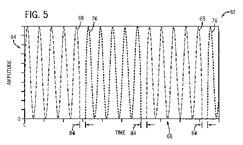
The first and second PDW signals 68 and 76 emitted by the driving circuitry
28 may be selected by the optical switch 36 and combined as a single multi-
wavelength PDW signal transmitted through a single emitting optical cable 38
to the
sensor 14. FIG. 5 is a plot 82 illustrating such a multi-wavelength PDW signal
having single-wavelength PDW signals 68 and 76 time-multiplexed by the optical
switch 36 such that the first and second PDW signals 68 and 76 are transmitted
in
series. In the plot 82, the amplitude 64 of the multi-wavelength PDW signal is
plotted
with respect to time 66. Because the optical switch 36 may select one single-
wavelength PDW signal at one time (e.g., optical cable 32 or optical cable
34), the
first and second PDW signals 68 and 76 may occur at different, non-
overlapping,
times in the multi-wavelength PDW signal of the plot 82. Time division
information
and/or clock signals related to the plot 82 may be sent to the phase detection
circuitry
44 and the DSP 46 such that the two single-wavelength PDW signals 68 and 76
may
be later separated into distinct single-wavelength PDW signals. Although not
necessary, in some embodiments, a brief dark period 84 (e.g., 3 ns) may
separate the
first and second PDW signals 68 and 76.
CA 02793483 2012-09-17
WO 2011/123239 PCT/US2011/028437
H-RM-01572 WO
When the multi-wavelength PDW signal of the plot 82 has passed through the
pulsatile patient tissue 26, single-wavelength components of the output signal
(i.e.,
portions of the signals 68 and 76 which have propagated through the patient
tissue 26)
may be isolated (i.e., demultiplexed) in the phase detection circuitry 44 and
the DSP
46 using time-division information from the driving circuit 28. Comparing one
of the
de-multiplexed output signals with the corresponding first or second PDW
signals 68
or 76 of the plot 82 may indicate various properties of the patient tissue 26.
For example, FIG. 6 illustrates the superimposition of an emitted single-
wavelength PDW signal (referred to here as the input signal 68) which may be
part of
a multi-wavelength PDW signal, with the corresponding detected single-
wavelength
PDW signal (referred to as the output signal 92). Like the plots 62, 74, and
82 of
FIGS. 3-5, plot 86 of FIG. 6 plots a relative amplitude 88 of the input signal
68 and
output signal 92 with respect to time 90 in units of nanoseconds (ns). The
input signal
68 and the corresponding output signal 92 may have a DC amplitude difference
of
ADC,, an AC amplitude difference of AAC,, and a phase difference of Arp, . The
amplitude measurements ADC, and AAC, may correspond essentially only to
absorption in the patient tissue 26, while the phase difference AV, may
correspond
essentially only to scattering in the patient tissue 26, as generally
described with
reference to FIG. 8 below.
Since another single-wavelength PDW signal, such as the second PDW signal
76, may occur very shortly thereafter, performing a similar comparison with
the
following single-wavelength PDW signal may yield additional measurements for
absorption and scattering properties of the patient tissue 26 for the second
wavelength,
at substantially the same time. Thus, the patient monitor 12 may determine at
least
four measurements associated with properties of the patient tissue 26 for
substantially
the same time for medical purposes associated with pulse oximetry, including
two
absorption and two scattering properties. In other words, because
substantially no
perceptible change in the pulsatile tissue of the patient may occur between
the start of
the first PDW signal 68 and the start of the second PDW signal 76, for
purposes of
pulse oximetry, the four measurements may be understood to occur at
substantially the
same time. Furthermore, the emissions of the first PDW signal 68 and the
second
16
CA 02793483 2012-09-17
WO 2011/123239 PCT/US2011/028437
H-RM-01572 WO
PDW signal 76 may be sufficiently frequent to adequately sample pulse from the
patient tissue 26 using, for example, the patient monitor 12 (from FIGS. 1 and
2).
FIG. 7 illustrates a flowchart 94, which represents an embodiment of a method
for performing a multi-wavelength PDW measurement using an optical switch. In
a
first step 96, the driving circuit 28 may modulate a light source (one of the
light
sources 30) emitting a first wavelength (e.g., a 660 mn wavelength) at a
modulation
frequency sufficient to produce resolvable photon density waves within the
patient
tissue 26 to produce a first single-wavelength PDW signal 98. The modulation
frequency may result in a PDW wavelength shorter than a mean absorption
distance of
the pulsatile tissue 26. In other words, such modulation frequency may exceed
the
product of the mean absorption coefficient multiplied by the speed of light.
Thus,
depending on the patient tissue 26, the modulation frequency may be between 50
MHz
to 3 GHz. In some embodiments, the light source may be modulated at a
frequency of
approximately 500 MHz. Similarly, in step 100, the driving circuit 28 may also
modulate another light source (another of the light sources 30) emitting a
second
wavelength in substantially the same manner as in step 96 to produce a second
single-
wavelength PDW signal 102. While only two single-wavelength PDW signals (e.g.,
98 and 102) are depicted, in accordance with the present techniques, a
plurality of
single-wavelength PDW signals may be produced and time-multiplexed to form a
multi-wavelength PDW signal.
In step 104, the optical switch 36 may select the first PDW signal 98 or the
second PDW signal 102 for transmission via the single optical cable 38 to be
emitted
into the patient tissue 26. The selected single-wavelength PDW signal,
referred to as
the input signal 106, may pass through the patient tissue 26 in step 108
(e.g., emitted
from the emitter output 22 of the sensor 14). Once the input signal 106 has
propagated through the patient tissue 26, portions of the input signal 106
(including
light that has been scattered by, reflected by, or transmitted through the
tissue) may be
received at a detector input 24 of the sensor 14 and passed through a
receiving optical
cable 40 to the detector 42, as represented by step 110. In step 114, such
portions of
the detected signal, also referred to as the output signal 112, may be used by
the DSP
46 and/or the microprocessor 48 to determine properties of the patient tissue
26 based
17
CA 02793483 2012-09-17
WO 2011/123239 PCT/US2011/028437
H-RM-01572 WO
on phase and amplitude changes between the output signal 112 and the input
signal
106 which correspond to scattering and absorption properties in the tissue.
Portions of the process 94, including steps 104, 108, 110, and 114, may repeat
indefinitely to generate and emit a multi-wavelength PDW signal into the
patient
tissue 26 tissue. More specifically, in each iteration of steps 104, 108, 110,
and 114,
the selected input signal 106 may represent a single-wavelength PDW signal of
a
multi-wavelength PDW signal, and by alternately selecting a different input
signal 106
(e.g., alternating between the first signal 98 and the second signal 102), a
multi-
wavelength PDW signal may be emitted and detected. In some embodiments, the
repetition may be between steps 104, 108, and 110, and the determination
and/or
analyses of phase and amplitude changes may occur after multiple repetitions
of steps
104, 108, and 110, and after a certain length of a multi-wavelength PDW signal
has
been emitted and detected from the patient tissue 26. Furthermore, the line
115 drawn
from the first and second signals 98 and 102 to step 114 may represent that
information, such as time-division information, clock signals, and/or
reference signals
relating to the corresponding original emitted single-wavelength PDW signals
98 and
102 may be used in step 114.
FIG. 8 is a flowchart 114, which represents an algorithm that may be used by a
processor, such as the DSP 46 of the patient monitor 12, to determine
physiological
properties of the patient tissue 26 using values obtained by passing a multi-
wavelength PDW signal through the patient tissue 26. It should be understood
that
the flowchart 114 is essentially a more detailed illustration of step 114 of
FIG. 7. In a
first step 116 of the flowchart 114, phase change L\rp, and/or amplitude
change ADCI
and/or AAC1 values of an output signal 112 may be received into or determined
by a
processor, such as the DSP 46. Such phase or amplitude changes may be
determined
based on a comparison between the output signal 112 and a reference signal of
a
corresponding input signal 106. In step 118, the DSP 46 may determine a
scattering
property of the patient tissue 26 for the moment in time at which the single-
wavelength PDW component of the multi-wavelength PDW signal has passed through
the pulsatile tissue 26. Generally, the scattering property may be represented
by a
18
CA 02793483 2012-09-17
WO 2011/123239 PCT/US2011/028437
H-RM-01572 WO
scattering coefficient, and may be determined based on the phase change Arp,
value
obtained in step 116 by using Equation (I).
In step 120, the DSP 46 may determine an absorption property of the patient
tissue 26 for the moment in time at which the single-wavelength component of
the
multi-wavelength PDW signal has passed through the pulsatile tissue 26.
Generally,
the absorption property may be represented by an absorption coefficient, and
may be
determined based on the amplitude change ADC, and/or SAC, values obtained in
step 116 by using Equations (1) and (4).
While the embodiments set forth in the present disclosure may be susceptible
to various modifications and alternative forms, specific embodiments have been
shown byway of example in the drawings and have been described in detail
herein.
However, it should be understood that the disclosure is not intended to be
limited to
the particular forms disclosed. The disclosure is to cover all modifications,
equivalents, and alternatives falling within the spirit and scope of the
disclosure as
defined by the following appended claims.
19