Note : Les descriptions sont présentées dans la langue officielle dans laquelle elles ont été soumises.
WO 2011/124922 PCT/GB2011/050696
-1-
Ultrasound Simulation Training System
The present invention relates generally to the field of medical training
systems, and in
particular to ultrasound training systems using ultrasound simulation.
Medical sonography is an ultrasound-based diagnostic medical technique wherein
high
frequency sound waves are transmitted through soft tissue and fluid in the
body. As the
waves are reflected differently by different densities of matter, their
'echoes' can be built up
to produce a reflection signature. This allows an image to be created of the
inside of the
human body (such as internal organs) such that medical data can be obtained,
thus
facilitating a diagnosis of any potential medical condition.
In clinical practice, ultrasound scans are performed by highly trained
practitioners who
manipulate a transducer around, on or in a patient's body at various angles.
In the case of
trans-vaginal ultrasound, an internal probe is rotated or otherwise
manipulated.
Medical and other health practitioners undergo extensive training programmes
when
learning how to use ultrasound machines appropriately and correctly. These
programmes
consist of in-classroom sessions, plus clinical training sessions during which
the student
observes an expert in the performance of an ultrasound scan. The student, by
watching and
copying, is taught how to identify and measure anatomical entities, and
capture the data
required for further medical examination or analysis.
In order to acquire the necessary skills, the ultrasonography student must
develop a
complex mix of cognitive skills and eye-hand movement coordination. Thus, the
more
practice a student gets at performing ultrasound operations, and the more
anatomies (i.e.
different patients) he/she experiences during the training the process, the
better the
student's skills are likely to be.
However, this is a lengthy and time consuming process, as well as being
resource
intensive. The present shortage of ultrasound-trained radiographers and the
additional
introduction of ultrasound techniques in many specialities such as obstetrics
and
WO 2011/124922 PCT/GB2011/050696
-2-
gynaecology, cardiology, urology and emergency medicine have placed
considerable
pressure on the limited number of qualified trainers. The constant demand to
meet health
service delivery targets adds to the pressure. The essential challenge of
ultrasound training
therefore lies in resolving the conflict by expediting the acquisition of
skills and increasing
trainees' competency prior to hands-on patient contact. Thus, there is a need
for an
ultrasound training solution which provides an effective and reproducible
training
programme without the use of clinical equipment and/or expert supervision and
leads to
the reduction of time required to competency. In addition, this solution
should be cost
effective whilst reducing current pressures on resources and time. Ideally,
such a solution
would be capable of incorporating anatomies and pathologies not often seen in
the learning
environment, thus improving the quality and breadth of ultrasound training
prior to
students' exposure to live patients.
Thus, in accordance with a first aspect of the present invention, there is
provided a
simulator training system for simulation training in ultrasound examination or
ultrasound-
guided procedures, the training system comprising:
a simulator input device to be operated by the user, the input device being
movable;
means for displaying an ultrasound scan view image, being an image or
facsimile
image of an ultrasound scan, the scan view image being variable and related to
the
position and/or orientation of the simulator input device;
wherein:
a) the system further includes means for displaying a second image, the second
image being an anatomical graphical representation of the body structure
associated with the ultrasound scan view, wherein the ultrasound scan view
image and the second image are linked to vary in a coordinated manner as the
position and/or orientation of the simulator input device changes; and/or,
b) the system further includes means for electronically recording aspects of
the
users interaction with the system enabling an assessment or measure of the
users performance to be made; and/or
WO 2011/124922 PCT/GB2011/050696
-3-
c) the ultrasound scan view image is a composite image composed of scan view
image data obtained from different sources and merged; and/or,
d) the ultrasound scan view image is generated from a scan volume, the scan
volume being a 3-Dimensional (3-D) scan volume created by converting 2-
Dimensional ultrasound scans or images to form the 3-Dimensional scan
volume.
In a preferred realisation of the invention the system will include two or
more of features a)
b) c) and d).
The user (i.e. student or trainee or a trained professional undertaking a
continued
professional activity) may manipulate, re-orientate or otherwise move the
simulator input
device. Preferably, the simulator input device is configured to provide force
feedback via
the device to the user relating to the position and/or orientation and/or
degree of force
applied to the device by the user. It is preferred that data pertaining to the
force applied to
the control device is fed back to the student to enhance the realism of the
student's
experience. This feedback may be provided via the control device itself. The
simulator
input device may be a "replica intelligent" probe simulating that of a
conventional
ultrasound machine. The probe may be an intelligent probe such as a haptic
device.
However, other types of control device may be used.
The simulator may be called a `virtual ultrasound machine'. Preferably, the
simulator is
configured to present a visualisation which resembles at least partially the
features and
visualisation which would be presented by a clinical ultrasound machine. This
is the
ultrasound scan view image. The scan view image may be a mosaic produced using
data
obtained from a variety of sources such as patient scans. The patient scans
may be 2-
dimensional images obtained by scanning a patient's body using a clinical
ultrasound
device.
Preferably, the ultrasound simulation includes a scanned image of part of a
patient's body,
the view of the image being changeable in response to movement or manipulation
of the
simulator input device. Thus, the simulator coordinates and controls the
perspective of the
WO 2011/124922 PCT/GB2011/050696
-4-
scanned anatomy as viewed by the user. In addition, the simulator system may
provide a
representation of at least one other ultrasound machine feature. For example,
it may
provide brightness and contrast controls.
It is preferred that the simulator input device corresponds or is mirrored by
a `virtual'
ultrasound device which simulates the movement, orientation and/or position of
the
simulator input device.
Thus movement of the physical simulator input device causes a corresponding
movement
of the virtual ultrasound device. By manipulating the physical input control
device, a user
is able to alter the view or perspective of an image of an anatomy displayed
via the system.
This enables a user undergoing an assessment or practice session to perform
virtual (i.e.
simulated) scan-related tasks by manipulating the physical simulator input
device. As the
user moves the simulator input device, he/she is able to observe the virtual
change effected
by that movement. It is preferred that data pertaining to the movement of the
control
device is recorded or noted during the user's interaction with the system.
This data may
relate to the position, orientation, applied force and/or movement of the
control device.
It is preferred that the movement or scan plane of the virtual device and
anatomy are
presented to the student for viewing of the scan view image in real time,
preferably on a
computer screen or, for example, as a holographic display. Preferably, this
presentation
resembles or mimics the scan view image which would be presented to the user
of a `real'
ultrasound machine, thus providing a simulated yet realistic experience for
the student.
In one preferred embodiment, a corresponding graphical representation of the
scanned
anatomy is provided in addition to the ultrasound scan view image. This
second, graphical
anatomical image is linked to the scan view image in a coordinated manner. The
graphical anatomical representation of the anatomy may show the virtual
control device or
the scan plane and a `slice through' of the anatomy based on the position of
the simulator
input device. As the user moves the physical simulator input device, the
virtual control
device shown in the representation mirrors that movement and the plane of the
slice
through the anatomy, is adjusted accordingly.
WO 2011/124922 PCT/GB2011/050696
-5-
In those embodiments wherein both the ultrasound scan view image and graphical
representation are both displayed, it is preferred that they are displayed
adjacent to or near
one another, for example in different windows on the same computer screen.
Preferably,
the graphical representation and the scanned images are two different
renderings of the
same anatomy. Thus, movement of the control device causes a corresponding
movement
in both versions of the viewed anatomy.
It is preferred that the training system further comprises an assessment
component. This
can be realised by the system including means for electronically recording
aspects of the
users interaction with the system enabling an assessment or measure of the
users
performance to be made. This may be referred to as a `learning management
system'
(LMS). Preferably, the LMS is configured to provide an assessment of the
student's
performance of tasks based on the manipulation of the control device.
Preferably the LMS
comprises a plurality of further components, such as a user interface. The LMS
may
comprise a security and/or access control component. For example, the student
may be
required to log into the LMS or undergo some type of authentication process.
It is preferred that the LMS provides training related content to the user
before during
and/or after use of the training system. This training content may include
instructions
regarding the type or nature of task to be accomplished, and/or how to
accomplish it. The
content may be provided in a variety of formats. For example, it may be
presented as text
or in an audible form.
In an alternative embodiment, the LMS may `remember' data relating to the
user's
previous interactions with the system and may present these to the user for
feedback,
teaching and/or motivational purposes.
In accordance with a second aspect of the present invention, there is provided
at least one
pre-determined metric or performance-related criterion. Preferably, a
plurality of metrics
is provided wherein each criterion serves as a benchmark or gauge against
which an aspect
of the student's performance may be measured. The comparison of the student's
WO 2011/124922 PCT/GB2011/050696
-6-
performance against the metrics may be performed by a metric analysis
component of the
system.
It is preferred that the metrics are stored in a simulator definition file.
Preferably, a
simulator definition file (and set of metrics contained therein) is provided
for each
assignment or pedagogical objective that the student may undertake. Thus, the
metrics are
task-oriented and enable the student's performance to be assessed in
comparison with the
performance expected of a competent or expert user, or with standards set down
by a
professional body. In addition to the results themselves, it is preferred that
the simulator
definition file contains text relating to each metric. This text may provide a
recommendation as to whether the student has succeeded or failed in achieving
the
particular learning objective. In an alternative embodiment, multiple metrics
may be
assessed in combination to provide enhanced analysis based on the assessment
of multiple
criteria.
It is preferred that throughout a given training session, data pertaining to
the student's use
of the control device is noted. Preferably, this data is recorded within an
audit trail.
Preferably, the position, orientation and applied force of the probe are
recorded at spaced
or timed intervals. Preferably, the student's performance data are analysed in
view of the
metrics at the end of the simulation session. Thus, the results which have
been accrued in
the audit trail file during the training session are received as input by the
metrics analyser.
However, the skilled addressee will understand that the metrics comparison may
also be
performed at any time during the learning session.
The metric criteria may be determined in a number of ways. For example, it may
be
determined empirically, or by assessing the performance of at least one expert
using the
invention, or from known medical knowledge
In accordance with one aspect of the present invention the ultrasound scan
view image is a
composite image generated from merging data obtained from different sources.
The
sources may be 2 dimensional scans obtained by scanning a volunteer subject's
body using
a conventional ultrasound machine. Effectively a 3-D ultrasound volume is
provided for
WO 2011/124922 PCT/GB2011/050696
-7-
use with an ultrasound training system, the 3-D ultrasound volume comprising a
composite
volume in which one portion has been imported into the 3-D volume from at
least one
other volume, or separate volumes combined. This is achieved by merging the
electronic
data of the scan view and/or the graphical anatomy representation from a
number of
different sources, volunteers or subjects.
The 3-D volume may be created as a composite of real volunteer subjects'
anatomies. One
or more selected portions of a scan of a real volunteer subject's anatomy may
be copied
and superimposed (or `pasted') onto the corresponding area of the virtual
volume. The
selected portion may be an area corresponding to, for example, a the subjects
ovaries or
other internal organ. Thus, a new, virtual volume may be built up as a mosaic
of scanned
data originally derived from more than one volunteer subject. For example, it
may be
decided that, for pedagogical reasons, a particular volume would be preferred
with larger
ovaries than those possessed by the actual subject. Thus, the present
invention provides
such a tailored virtual volume.
The 3-D volume is created by converting 2-Dimensional ultrasound scans or
images into a
3-Dimensional volume by creating a 3-D grid of voxels from a stream of 2-D
grids of
pixels. Thus, a 3D anatomical volume may be created from a `sweep' of a 2-D
ultrasound
image. As a single sweep may not cover the full area required for the image
(because the
beam width may not be wide enough), multiple `sweeps' may be performed wherein
each
`sweep' may record a video of consecutive 2-D images with respect to time.
Multiple
sweeps may then be merged to build up a larger dataset pertaining to the 2-D
ultrasound
scanned image. This may be needed because one sweep cannot cover the full area
of
interest required for the simulator due to 2-D ultrasound beam limitations.
It is preferred that, having compiled a collection of `sweeps' from the
scanned 2-D data,
the sweeps are alpha blended together. This is preferably performed using a
mask, the
mask defining which pixels in the sweeps are to be ignored and/or which are to
be used as
input into the resulting 3-D volume.
WO 2011/124922 PCT/GB2011/050696
-8-
In a preferred embodiment, the resulting alpha blend may then be edited to
import data
from one or more alternative datasets, such that desired portions of that
other data set are
incorporated into the alpha blend to create a 3-D volume having the desired
anatomical
attributes. Thus, the resulting virtual volume is a representation of a
portion of a virtual
patient's body designed in accordance with pedagogical motivations.
This provides the advantage that additional virtual volumes can be created
quickly and
easily. In addition, this provides the advantage that students can be exposed
to a greater
variety of anatomies and structures in less time than would be possible if
he/she were
training by clinical practice alone.
Alternatively, the 3-D volume may comprise an artificially generated dataset
designed to
represent a specific subject anatomy.
Furthermore, the dataset maybe processed in such a way or to vary with time or
force
applied via the control input device in order to mimic movement of the subject
such as
fetal heartbeat, baby in womb movement, or spatial relationship changes
induced by the
force applied by the input control device.
Thus, the present invention eliminates or alleviates at least some of the
drawbacks of the
current ultrasound training environment whilst providing the advantages
outlined above.
These and other aspects of the present invention will be apparent from, and
elucidated with
reference to an exemplary embodiment of the invention as described herein.
An embodiment of the present invention will now be described by way of example
only
and with reference to the accompanying drawings, in which:
Figure 1 shows the components and events of an embodiment of the present
invention.
Figure 2 shows a typical view of a simulation based ultrasound training
session presented
to a student in accordance with an embodiment of the present invention.
WO 2011/124922 PCT/GB2011/050696
-9-
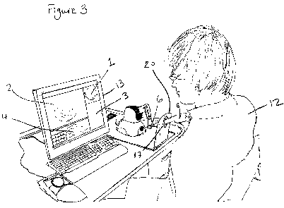
Figure 3 shows a user interacting with a system in accordance with the present
invention.
The following exemplary embodiment describes the invention's use in relation
to
transvaginal scanning. However, this application is for illustrative purposes
only and the
invention is not intended to be limited in this regard. Other embodiments may
be applied
to other types of medical use;
Turning to Figure 1, a medical ultrasound training simulator is provided and
comprises the
following components:
= Learning Management System (LMS) 5 which oversees or manages the learning
experience presented to the user;
= User assessment component 7. This enables a judgement or analysis of the
user's
performance to be formed.
= Ultrasound simulation component 2 configured to replicate the key features
of a
conventional ultrasound machine. This may be referred to as the `virtual
ultrasound machine'.
= Replica `intelligent' ultrasound probe 6 as an input device to be
manipulated by the
user and provide electronic input into the system. The input device 6 may be,
for
example a haptic device in communication with the simulator component of the
system.
= Computer and other associated hardware for running the software components
of
the invention
= High resolution screen 13 for displaying and presenting information to the
user 12.
This may be a touch screen.
With reference additionally to Figures 2 and 3, in use a user 12 logs into the
LMS 5 of the
ultrasound training system to begin a training session. This may require
authentication via
a variety of known methods (e.g. by providing a user ID and password). The
interaction
WO 2011/124922 PCT/GB2011/050696
-10-
between the user and the system components is handled via a user interface,
which may be
written in any appropriate programming language.
After logging into the system, the LMS 5 provides the user with an overview of
the course
content 3. This overview presents the student with information regarding the
objectives
and learning outcomes of the modules. Each module is divided into a number of
tutorials
and assignments. A tutorial relates to themes of a particular technique such
as orientation
conventions or introduction of the transvaginal probe, whilst an assignment is
a group of
tasks within a module which constitute a key learning point (such as the
orientation in
sagittal and coronal planes or direction and positioning and pressure for the
latter).
The user then selects which training modules (s)he wishes to undertake (e.g.
examination
of the normal female pelvis, normal early pregnancy or assessment of fetal
well being).
When the user indicates that (s)he wishes to undertake an assignment, (i.e.
run the
simulator), the LMS 5 provides initial instructions to the student. The
instructions may be
provided orally or visually. The LMS also passes a simulator definition 10 to
the
simulation component so that the assignment can be performed.
The simulator definition 10 is a package of information and data pertaining to
a particular
assignment for testing and training a student with regard to a particular
objective or task.
For example, the simulator definition 10 may include a full description of the
relevant
assignment, including text to be displayed, parameters relating to the
ultrasound volume to
be used, which volume is to be used, which force feedback files should be used
and a full
description of the metrics to be tested. Associated pass/fail criteria may
also be included.
The training content 11 is stored within XML files, thus enabling the training
content 11 to
be configured, updated and altered.
The user may be offered the option of using the simulator in `practice mode'
without
feedback or an `interactive mode' whereby the user follows instructions to
under-take
specific tasks which will then be measured against a set of `gold standard'
metrics. These
instructions may be provided in textual form e.g. on screen or in audible form
e.g. via a
speaker.
WO 2011/124922 PCT/GB2011/050696
-11-
Thus, when the user selects an assignment via the LMS interface, the
appropriate simulator
definition 10 is loaded in the simulator 7 and the training session begins.
During the
training session, the user completes the selected assignment or task by
manipulating the
haptic input device 6 (i.e. `intelligent probe'). The user operates the
physical input device
6 to navigate a virtual ultrasound probe 14 around a virtual patient's
anatomy. This may
appear on the screen 1 as a recreated ultrasound scan view image 2 and/or as a
simulated
ultrasound beam corresponding to the plane and movement of the virtual probe
14. As the
intelligent replica probe 6 is moved, the display 1 shows the progress of the
beam in the
simulation of the patient's anatomy.
Thus, by using the haptic input device 6, the training system allows the user
12 to perform
ultrasound operations in a virtual world which mimics how the operation would
be
performed in a clinical session on a living patient. For example, the user is
able to perform
operations such as examining and measuring the virtual patient's internal
organs.
During the session, the system shows the ultrasound volume and the virtual
anatomy in
two side-by-side views which are shown in separate windows on the user's
screen, as
shown in Figure 2:
1. a recreated ultrasound scan view image generated during real-time scanning
2.
Thus, the virtual ultrasound machine 2 enables presentation of a simulated
ultrasound machine showing a scan view image based on the probe input
device's current position. This is shown in screen 2 of Figure 2. As the user
moves the haptic input device, the perspective of the scan view image 2 is
changed accordingly, as would occur if the user was operating a `real'
ultrasound machine.
2. a view of the progress of the simulated scanning beam 21 in the anatomy of
the
virtual patient 1. Screen 1 of Figure 2 shows such a graphical representation
of
the anatomy as created by a graphic artist (this process is discussed in more
detail below). The graphical representation of the anatomy is shown from the
perspective of the virtual probe 14. The virtual probe and its orientation are
shown, along with the scan plane 21 resulting from the position of the virtual
WO 2011/124922 PCT/GB2011/050696
-12-
probe 14. A `slice through' of the anatomy is shown based on the plane 21 of
the virtual probe 14. As the user moves the haptic device, the virtual probe
14
mirrors the movement and is seen to move on the screen 2. Accordingly, the
viewed perspective of the anatomy is altered (e.g. rotated) so as to reflect
the
change in the simulated scan plane 21.
The two images (i.e. the simulated scan view image in screen 2 and the
graphical
representation in screen 1) both track the movement of the haptic input 6
device so that as
the user performs the required learning tasks, (s)he is able to see the
results of her/his
actions in two forms or representations. This provides an enhanced
understanding of the
results of manual actions.
While both of the views described above may be presented to the user at the
same time, the
skilled addressee will appreciate that in some embodiments only one of the
above images
may be displayed. In other words, the system may display only the ultrasound
volume or
the graphical representation of the virtual anatomy.
A third window 3 may also be presented to the user during the training
session, containing
instructions and/or information regarding the selected training module.
Alternatively, these
instructions and/or information may be provided in an audible form rather than
via the
screen. Thus, the screen may provide the user with one or both of the
anatomical views
described above, with or without an additional third screen for presentation
of training-
related material.
The interaction between the user and the simulator 2 is managed by an
interface 9 which
enables data to be obtained from the haptic input device 6 (e.g. position
within the virtual
anatomy) and fed back to the haptic input device (i.e. force feedback). Thus,
the haptic
device 6 provides feedback to the user regarding the force (s)he is applying
via the probe
and the resistance which the tissue or other matter is providing.
In some embodiments, a hardware constraint such as an aperture 17of defined
perimeter in
a support frame 20 may be used to limit the movement of the haptic input probe
6 thus
WO 2011/124922 PCT/GB2011/050696
-13-
replicating the range of movement of a real probe, which would be inhibited by
the
patient's body. The system may also artificially constrain the exit point of
the probe from
the virtual body opening e.g. mouth, vagina or anus or an operative entry
point e.g.
laparoscopic port such that it is at the correct point in the virtual anatomy.
This avoids an
incorrect visualisation in the event of a mismatch in the measurement of the
probe position
or angle. For example, in such an event the probe may otherwise exit
incorrectly through
the virtual anatomy's leg or other body part. However, other embodiments of
the system
may not require the use of a hardware constraint.
Thus, a sophisticated level of interaction is provided with the system which
mimics the
experience obtained in a clinical training session. The user is provided with
a realistic
sensation of a scanning operation, both through pressure when pushing against
organs and
by preventing the probe from moving to anatomically impossible positions.
During the simulation, the known techniques are used to deform the virtual
anatomy to
simulate the effect of the probe e.g. within a cavity such as the vaginal
canal or on the
external surface of the body. Other techniques are also used to simulate some
of the key
functionality of an ultrasound machine, thus enhancing the realism of the
student's
experience. These may be presented and controlled by the student during the
training
session via an area of the screen 4. These features may include including:
= Brightness, contrast and Time Gain Compensation (TGC) controls
= Image annotation (labelling and text annotation)
= Changing image orientation
= Freeze and split screen functionality
= Magnify and zoom image
= Take pictures or make video recordings
= Take measurements of a distance or an area or calculate a volume from a
series
of measurements
WO 2011/124922 PCT/GB2011/050696
-14-
Via the LMS 5, the student is also able to view saved screenshots and/or video
recordings
of his performance.
Throughout the training session, user interaction and session data are stored
or recorded by
the system within an audit trail 8. Additionally, the haptic position and/or
orientation, and
applied force, are recorded at spaced or timed intervals (e.g. every 100ms).
At the end of
the simulation, this information is analysed to determine the user's
performance in respect
of the relevant metrics.
The user's performance is assessed by use of the metric analysis component 7.
Whilst the
analysis may be performed at any time during the session, it will more
typically take place
as a batch operation at the end of the simulation run (i.e. the assignment)
using the results
stored in the audit trail file 8.
The metric analyser 7 compares the data obtained during the simulation
regarding the
student's performance against a set of pre-determined criteria stored in the
simulator
definition file 10 for the selected assignment (i.e. the `metrics'). Metrics
are associated
with each task within an assignment and enable assessment of the student's
performance of
that task against key performance criteria. For example, if the task is to
fully examine and
measure the size of the patient's right ovary, the metrics may check the
maximum force
applied by the simulated probe, the time taken to complete the examination,
the probe
movement profile, the measurements taken e.g. length, width and height of the
ovary and
the measurements position.
Comparison is made against a number of different metrics, each of which
measures a
single aspect of the student's performance. The following metrics may be
included in the
system although the following list is not intended to be finite or absolute:
Time Time taken to perform the task
WO 2011/124922 PCT/GB2011/050696
-15-
FlightPath How closely the student followed the `expert'
probe path.
The algorithm used is as follows:
For each expert probe (haptic) position recorded
find the closest student point by absolute distance
(C)
Metrics are min (C), max (C), mean (C)
LocatePlane Checks position of a frozen ultrasound view
compared to that recorded by the expert.
AngularDeviation Checks the deviation from a specific orientation
vector made by the student during a scan
MultipleChoice Multiple choice questions
Force Maximum force applied
Contrast Checks screen contrast against limits
Brightness Checks screen brightness against limits
TGC (Time Gain Compensation) Checks TGC against limits
UltraSound Orientation Checks ultrasound orientation (ie orientation of
ultrasound image which can be flipped or rotated
on the user interface)
Label Checks the position of an annotation label
1 dMeasurement Checks value and position of a 1 d measurement
in the ultrasound view
2dMeasurement Checks value, position and perpendicularity of
two 1 d measurements in the ultrasound view
WO 2011/124922 PCT/GB2011/050696
-16-
3dMeasurement Checks value, position and perpendicularity of
three 1 d measurements in the ultrasound view
VerifyArrow Checks the orientation of an arrow drawn on the
screen against the expert's arrow
It should be noted that the above examples of metrics are provided by way of
an example
only. The skilled addressee will understand that the system may be adapted so
as to be
used for other types of ultrasound applications and, therefore, a different
set of metrics may
be drawn up which relate more closely to that particular type of operation.
The metric criteria may be determined in a number of ways:
= Empirically (e.g. it may determined that a student must take less than 30s
for a
particular task)
= By assessing the performance of a number of experts using the simulator
(e.g.
by using the simulator itself to find the average probe path followed by an
expert).
= From medical knowledge (e.g. doctors and practitioners may supply a
specified
maximum force limit because this is the level which, in their experience,
causes
patient discomfort).
In addition to the results themselves, the simulator definition file 10 also
contains specific
text for each metric giving a recommendation with regard to whether the user
has passed or
failed that particular aspect of the assignment. Alternatively, multiple
metrics may be
assessed as a combination to provide improved guidance based on multiple
criteria.
When the user has completed the assignment, (s)he returns to the LMS interface
5 so that
her/his results may be reviewed and assessed. The user may then re-take the
assignment if
the feedback indicates that the performance was not satisfactory in comparison
to what is
expected by the metrics, or may progress to the next assignment.
WO 2011/124922 PCT/GB2011/050696
-17-
Additionally, for users who are enrolled in a specific training programme, the
user's
supervisor may have access rights to the user's reports on the LMS 5, thus
enabling the
supervisor to monitor progress and performance on an ongoing basis.
Prior to use, at least one (but typically more than one) 3-D ultrasound volume
of an
anatomy is created for use with the training system.
In order to create the required volume, a 2D ultrasound scan view image is
captured using
a `conventional' ultrasound machine. The captured 2D ultrasound may be stored
inside the
ultrasound machine itself or on a DVD for subsequent use and replay.
As a 3-D ultrasound volume is used with the present invention, the 2D
ultrasound image
must be converted or transformed into the requisite 3-D format. Thus, tracked
sensor data
relating to position and orientation must be combined with the 2-D ultrasound
scan. This
process requires spatial and temporal calibration of the tracking apparatus.
An example of such calibration techniques will now be discussed as performed
during
construction of an exemplary embodiment of the present invention.
1. Spatial calibration
Two tracked magnetic sensors were used to achieve the spatial calibration. One
sensor
was attached to the ultrasound probe, the other being left "loose". The probe
was
suspended in a container of water (to transport the ultrasound), whilst the
other probe was
intersected into the ultrasound beam.
The positions of both sensors were recorded, along with the orientation of the
ultrasound
probe sensor. The "loose" sensor was positioned such that the tracked centre
of the sensor
was in the ultrasound beam, thus producing a sparkle or discernable entity
within the
ultrasound image. The image was recorded, and the position noted. This was
carried out
many times to provide a good sample range (e.g. > 20).
WO 2011/124922 PCT/GB2011/050696
-18-
The 3D position of the "loose" sensor was then mapped to the sensor connected
to the
ultrasound probe. This enabled the calculation of where ultrasound pixels in
the image
were actually located in space, because the position of the target (i.e.
tracked sensor) was
known.
2. Temporal calibration
During the temporal calibration, two tracked sensors were used. One sensor was
strapped
to the ultrasound probe, and the other attached to a nearby wooden pole (to
hold it steady).
The operator tapped the wooden pole with the ultrasound probe. As a result,
the wooden
pole becomes instantly visible in the ultrasound image whilst the second
sensor registered
the sudden movement. This was carried out at the start and end of a scan, to
calibrate and
demark the start and stop of the scan in both movement and ultrasound imagery.
The
movement in the 2 d sensor was more pronounced than the movement in the 1St
sensor, and
the 2nd sensor was usually stationary (until it was tapped) making it easier
to find in the
stream of position and orientation data.
3. Volume generation
Given the spatial and temporal calibration, the 2D ultrasound image could be
accurately
"Swept" in 3D. Thus, it was possible to `paint' using a 2D ultrasound video as
a
paintbrush.
A volume conversion utility was used to paint the 2D ultrasound images into a
3D volume,
the volume being a 3D grid of voxels created from a stream of 2D grids of
pixels. This
enabled a single "sweep" to create a 3D volume of ultrasound.
Multiple "sweeps" were then merged to build up a larger dataset. These were
then alpha
blended by creating a "mask" which defined which pixels were to be ignored and
which
pixels were to be used in the input ultrasound image, enabling blends to be
achieved
between images. The correct blend was then calculated manually to minutely
adjust the
WO 2011/124922 PCT/GB2011/050696
-19-
2n1(or subsequent) sweep(s) to align them correctly, or at least minimise
(visible) overlap
error.
The alpha blends were then used to merge in data from an alternative dataset,
enabling the
creation of a new 3-D ultrasound volume by merging volunteer subject data. For
example,
small ovaries in a dataset can be replaced with larger ovaries from a
different volunteer
subject. Although the result was the product of two different bodies being
merged, the
result appears sufficiently accurate to the eye. Thus, multiple virtual
patients may be
created from a base collection of virtual volunteer subjects.
In addition, a 3-dimensional anatomical graphical representation of a volume
was created
by segmenting out the organs of interest (e.g. the ovaries) from `real'
ultrasound volumes.
These were sent to a graphic artist for transformation into an anatomical
graphical
representation. The anatomical graphical representation may then be
manipulated on the
screen during the training session as described above. Screen 1 of Figure 2
shows an
example of such a graphical representation in accordance with an embodiment of
the
invention, and shows the simulated probe and associated scanning plane, and
the virtual
anatomy from the perspective of the scanning plane. The ultrasound scan view
image and
the anatomical graphical image are linked to vary in a matched relationship as
the input
device 6 is manipulated.
The invention has been primarily described in an embodiment in which scan data
is
obtained from ultrasound scans conducted on `real' subjects. It should be
appreciated that,
alternatively, virtual datasets may be created artificially through forward
simulation or by
other methods. Such artificial data maybe merged with real data, in certain
embodiments,
where preferred.
Furthermore, the data may be processed or manipulated to provide variations in
time or in
response to a force applied by the input device. Such manipulation may, for
example,
enable the scan view image to vary to represent fetal heartbeat, baby in womb
movement,
or changes to the shape of physical area under investigation as a result of
the application of
force to the baby via the input device.
WO 2011/124922 PCT/GB2011/050696
-20-
Thus, the present invention provides the advantage of teaching key skills to
the student
whilst providing real-time feedback on performance and charting a path for the
student to
achieve full competence. Other advantages arise from the present invention as
follows:
= Provision of non-clinical learning environment, thus solving the current
resource conflict between provision of clinical service and need to train,
releasing expensive ultrasound equipment for clinical use;
= Assist in overcoming the current shortages of suitably qualified trainers as
well
as learning capacity in hospitals and training centres;
= Improvement of the quality and breadth of ultrasound learning prior to the
trainee's exposure to patients;
= Provides the trainee with accurate feedback `active learning', monitoring
performance and providing structure to the training process;
= Eliminates the need for an expert's direct supervision, thus providing a
highly
cost-effective solution;
= Enables the student to experience a wider variety of anatomies in a more
condensed period of time than would be possible during clinically-based
training;
= The learning modules and/or metrics can be developed in accordance with
industry curriculum so as to meet the learning objectives set out by
professional
bodies, thus meeting professional gold standards;
= Provides an effective and reproducible training programme.