Note : Les descriptions sont présentées dans la langue officielle dans laquelle elles ont été soumises.
CA 02796187 2012-10-11
WO 2011/140006 PCT/US2011/034882
TITLE OF THE INVENTION
Therapeutic Agent Delivery System and Method for Localized Application of
Therapeutic Substances to a Biological Lumen.
INVENTORS
Matthew D. Cambronne, a citizen of the United States, resident in Stillwater,
MN
CROSS-REFERENCE TO RELATED APPLICATIONS
The present application claims priority under 35 U.S.C 119(e) to provisional
application No. 61/330617, filed on May 3, 2010 entitled DEGRADEABLE DRUG
DELIVERY DEVICE.
BACKGROUND OF THE INVENTION
[001] Field of the Invention
[002] The invention relates to systems, devices and methods for treating walls
of
biological lumens, e.g., animal lumens, with localized delivery of therapeutic
agents.
[003] Description of the Related Art
[004] A variety of techniques and instruments have been developed for use in
the
removal or repair of tissue in biological conduits, e.g., without limitation,
blood
vessels and similar body passageways. A frequent objective of such techniques
and
instruments is the removal of atherosclerotic plaques in a patient's arteries.
Atherosclerosis is characterized by the buildup of fatty deposits (atheromas)
in the
intimal layer (under the endothelium) of a patient's blood vessels. Very often
over
time, what initially is deposited as relatively soft, cholesterol-rich
atheromatous
material hardens into a calcified atherosclerotic plaque. Such atheromas
restrict the
flow of blood, and therefore often are referred to as stenotic lesions or
stenoses, the
blocking material being referred to as stenotic material. If left untreated,
such
CA 02796187 2012-10-11
WO 2011/140006 PCT/US2011/034882
stenoses can cause angina, hypertension, myocardial infarction, strokes, leg
pain
and the like.
[005] Rotational atherectomy procedures have become a common technique for
removing such stenotic material. Such procedures are used most frequently to
initiate the opening of calcified lesions in coronary arteries. Most often the
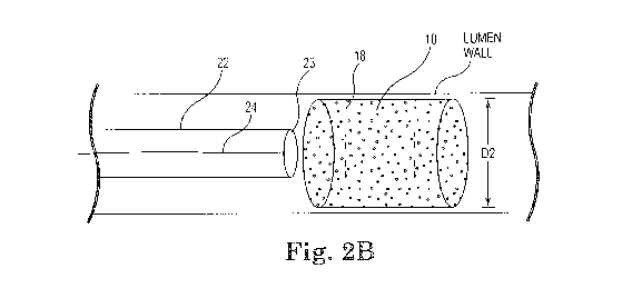
rotational
atherectomy procedure is not used alone, but is followed by a balloon
angioplasty
procedure, which, in turn, is very frequently followed by placement of a stent
to assist
in maintaining patency of the opened artery. For non-calcified lesions,
balloon
angioplasty most often is used alone to open the artery, and stents often are
placed
to maintain patency of the opened artery. Studies have shown, however, that a
significant percentage of patients who have undergone balloon angioplasty and
had
a stent placed in an artery experience stent restenosis--i.e., blockage of the
stent
which most frequently develops over a period of time as a result of excessive
growth
of scar tissue within the stent. In such situations an atherectomy procedure
is the
preferred procedure to remove the excessive scar tissue from the stent
(balloon
angioplasty being not very effective within the stent), thereby restoring the
patency of
the artery.
[006] Several kinds of rotational atherectomy devices have been developed for
attempting to remove stenotic material. In one type of device, such as that
shown in
U.S. Pat. No. 4,990,134 (Auth), a burr covered with an abrasive abrading
material
such as diamond particles is carried at the distal end of a flexible drive
shaft. The
burr is rotated at high speeds (typically, e.g., in the range of about 150,000-
190,000
rpm) while it is advanced across the stenosis. As the burr is removing
stenotic tissue,
however, it blocks blood flow. Once the burr has been advanced across the
stenosis,
the artery will have been opened to a diameter equal to or only slightly
larger than
the maximum outer diameter of the burr. Frequently more than one size burr
must be
utilized to open an artery to the desired diameter.
[007] U.S. Pat. No. 5,314,438 (Shturman) discloses another atherectomy device
having a drive shaft with a section of the drive shaft having an enlarged
diameter, at
least a segment of this enlarged surface being covered with an abrasive
material to
define an abrasive segment of the drive shaft. When rotated at high speeds,
the
2
CA 02796187 2012-10-11
WO 2011/140006 PCT/US2011/034882
abrasive segment is capable of removing stenotic tissue from an artery. Though
this
atherectomy device possesses certain advantages over the Auth device due to
its
flexibility, it also is capable only of opening an artery to a diameter about
equal to the
diameter of the enlarged abrading surface of the drive shaft since the device
is not
eccentric in nature.
[008] U.S. Pat. No. 6,494,890 (Shturman) discloses an atherectomy device
having
a drive shaft with an enlarged eccentric section, wherein at least a segment
of this
enlarged section is covered with an abrasive material. When rotated at high
speeds,
the abrasive segment is capable of removing stenotic tissue from an artery.
The
device is capable of opening an artery to a diameter that is larger than the
resting
diameter of the enlarged eccentric section due, in part, to the orbital
rotational motion
during high speed operation. Since the enlarged eccentric section comprises
drive
shaft wires that are not bound together, the enlarged eccentric section of the
drive
shaft may flex during placement within the stenosis or during high speed
operation.
This flexion allows for a larger diameter opening during high speed operation,
but
may also provide less control than desired over the diameter of the artery
actually
abraded. In addition, some stenotic tissue may block the passageway so
completely
that the Shturman device cannot be placed therethrough. Since Shturman
requires
that the enlarged eccentric section of the drive shaft be placed within the
stenotic
tissue to achieve abrasion, it will be less effective in cases where the
enlarged
eccentric section is prevented from moving into the stenosis. The disclosure
of U.S.
Pat. No. 6,494,890 is hereby incorporated by reference in its entirety.
[009] U.S. Pat No. 5,681,336 (Clement) provides an eccentric tissue removing
burr
with a coating of abrasive particles secured to a portion of its outer surface
by a
suitable binding material. This construction is limited, however because, as
Clement
explains at Col. 3, lines 53-55, that the asymmetrical burr is rotated at
"lower speeds
than are used with high speed ablation devices, to compensate for heat or
imbalance." That is, given both the size and mass of the solid burr, it is
infeasible to
rotate the burr at the high speeds used during atherectomy procedures, i.e.,
20,000-
200,000 rpm. Essentially, the center of mass offset from the rotational axis
of the
drive shaft would result in development of significant centrifugal force,
exerting too
3
CA 02796187 2012-10-11
WO 2011/140006 PCT/US2011/034882
much pressure on the wall of the artery and creating too much heat and
excessively
large particles.
[010] Another method of treatment of occluded vessels may include the use of
stents. Stents may be placed at the site of a stenosis and expanded to widen
the
vessel, remaining in position as a vessel implant.
[011] No matter the technique used to open an occluded conduit, e.g., blood
vessel,
and restore normal fluid flow therethrough, one problem remains: restenosis. A
certain percentage of the treated conduits and vessels will reocclude
(restenose)
after a period of time; occurring in as many as 30-40% of the cases. When
restenosis does occur, the original procedure may be repeated or an
alternative
method may be used to reestablish fluid, e.g., blood, flow.
[012] The relevant commonality shared by each of the above treatment methods
is
that each one may result in some trauma to the conduit wall. Restenosis occurs
for
a variety of reasons; each involving trauma. Small clots may form on the
arterial
wall. Small tears in the wall expose the blood to foreign material and
proteins which
are highly thrombogenic. Resulting clots may grow gradually and may even
contain
growth hormones released by platelets within the clot. Moreover, growth
hormones
released by other cells, e.g., macrophages, may cause smooth muscle cells and
fibroblasts in the affected region to multiply in an abnormal fashion. There
may be
an injury in the conduit wall due to the above methods that results in
inflammation
which may result in the growth of new tissue.
[013] It is known that certain therapeutic substances may have a positive
effect on
prevention and/or inhibition of restenosis. Several difficulties present
themselves in
the application of these substances to the affected region in a therapeutic
dose. For
example, the region in need of treatment is very small and localized. Fluid,
e.g.,
blood, flow in the conduit is continuous, resulting in a flow boundary along
the wall
which must be disrupted so that the therapeutic substances may reach the
localized
region of interest within a dose range considered therapeutic. The art fails
to
adequately provide a mechanism for breaking through this flow boundary to
target
the region of interest; electing instead generally to place the therapeutic
substance
4
CA 02796187 2012-10-11
WO 2011/140006 PCT/US2011/034882
into the general flow of the conduit, either by intravenous means or intra-
lumen
infusion, at a dose that is much higher than therapeutic since the majority of
the
therapeutic substance will simply flow downstream and either be absorbed
systemically or eliminated as waste. For example, intravenous medications are
delivered systemically by vein, or regionally, e.g., through intra-lumen
infusion
without targeting the subject region. Such unnecessary systemic exposure
results
with unknown and unnecessary adverse results in regions, tissue, and/or organs
that
are distant from the region of interest. Clearly, systemic delivery and
exposure is not
well suited to treatment of diseases or conditions having a single intra-lumen
region
of interest.
[014] The potential utility of localized application of a therapeutic dose of
therapeutic substances is not limited to treatment of coronary arteries.
Beyond
coronary artery delivery, other sites of atherosclerosis, e.g., renal, iliac,
femoral,
distal leg and carotid arteries, as well as saphenous vein grafts, synthetic
grafts and
arterio-venous shunts used for hemodialysis would be appropriate biological
conduits for a localized therapeutic substance delivery method and mechanism.
Nor
is the potential utility limited to blood vessels; any biological conduit
having a region
of interest amenable to treatment may benefit from such a treatment method and
mechanism.
[015] The present invention overcomes these deficiencies.
[016] BRIEF SUMMARY OF THE INVENTION
[017] The invention provides a system and method for localized application of
therapeutic substances within a biological lumen and to the wall of the lumen.
In
various embodiments, a biodegradable tubular prosthesis comprising a plurality
of
pores is deployed within a biological lumen. Subsequent to, or in conjunction
with,
the deployment of the prosthesis, a drug-eluting balloon comprising at least
one
therapeutic agent is expanded within the lumen of the tubular prosthesis,
thereby
releasing the agent(s) from the balloon and delivering them to the prosthesis
pores.
The at least one therapeutic agent is then allowed to diffuse through the
pores to the
lumen wall.
CA 02796187 2012-10-11
WO 2011/140006 PCT/US2011/034882
[018] The Figures and the detailed description which follow more particularly
exemplify these and other embodiments of the invention.
[019] BRIEF DESCRIPTION OF THE DRAWINGS
[020] The invention may be more completely understood in consideration of the
following detailed description of various embodiments of the invention in
connection
with the accompanying drawings, which are as follows.
[021] FIG. 1A is a side partial cutaway view of one embodiment of the present
invention.
[022] FIG. 1 B is an end view one embodiment of the present invention.
[023] FIG 2A is a side partial cutaway view of one embodiment of the present
invention.
[024] FIG 2B is a side partial cutaway view of one embodiment of the present
invention.
[025] FIG 3A is a side partial cutaway view of one embodiment of the present
invention.
[026] FIG 3B is a side partial cutaway view of one embodiment of the present
invention.
[027] DETAILED DESCRIPTION OF THE INVENTION, INCLUDING THE BEST
MODE
[028] While the invention is amenable to various modifications and alternative
forms, specifics thereof are shown by way of example in the drawings and
described
in detail herein. It should be understood, however, that the intention is not
to limit the
invention to the particular embodiments described. On the contrary, the
intention is
to cover all modifications, equivalents, and alternatives falling within the
spirit and
scope of the invention.
6
CA 02796187 2012-10-11
WO 2011/140006 PCT/US2011/034882
[029] For the purposes of the present invention, the following terms and
definitions
apply:
[030] "Bodily disorder" refers to any condition that adversely affects the
function of
the body.
[031] The term "treatment" includes prevention, reduction, delay,
stabilization,
and/or elimination of a bodily disorder, e.g., a vascular disorder. In certain
embodiments, treatment comprises repairing damage cause by the bodily, e.g.,
vascular, disorder and/or intervention of same, including but not limited to
mechanical intervention.
[032] A "therapeutic agent" comprises any substance capable of exerting an
effect
including, but not limited to therapeutic, prophylactic or diagnostic. Thus,
therapeutic
agents may comprise anti-inflammatories, anti-infectives, analgesics, anti-
proliferatives, and the like including but not limited to antirestenosis
drugs.
Therapeutic agent further comprises mammalian stem cells. Therapeutic agent as
used herein further includes other drugs, genetic materials and biological
materials.
The genetic materials mean DNA or RNA, including, without limitation, of
DNA/RNA
encoding a useful protein, intended to be inserted into a human body including
viral
vectors and non-viral vectors. Viral vectors include adenoviruses, gutted
adenoviruses, adeno-associated virus, retroviruses, alpha virus, lentiviruses,
herpes
simplex virus, ex vivo modified cells (e.g., stem cells, fibroblasts,
myoblasts, satellite
cells, pericytes, cardiomyocytes, skeletal myocytes, macrophage), replication
competent viruses, and hybrid vectors. Non-viral vectors include artificial
chromosomes and mini-chromosomes, plasmid DNA vectors, cationic polymers,
graft copolymers, neutral polymers PVP, SP1017, lipids or lipoplexes,
nanoparticles
and microparticles with and without targeting sequences such as the protein
transduction domain (PTD). The biological materials include cells, yeasts,
bacteria,
proteins, peptides, cytokines and hormones. Examples for peptides and proteins
include growth factors (FGF, FGF-1, FGF-2, VEGF, Endotherial Mitogenic Growth
Factors, and epidermal growth factors, transforming growth factor.alpha. and
.beta.,
platelet derived endothelial growth factor, platelet derived growth factor,
tumor
necrosis factor .alpha., hepatocyte growth factor and insulin like growth
factor),
7
CA 02796187 2012-10-11
WO 2011/140006 PCT/US2011/034882
transcription factors, proteinkinases, CD inhibitors, thymidine kinase, and
bone
morphogenic proteins. These dimeric proteins can be provided as homodimers,
heterodimers, or combinations thereof, alone or together with other molecules.
[033] Therapeutic agents further includes cells that can be of human origin
(autologous or allogeneic) or from an animal source (xenogeneic), genetically
engineered, if desired, to deliver proteins of interest at the transplant
site. Cells
within the definition of therapeutic agents herein further include whole bone
marrow,
bone marrow derived mono-nuclear cells, progenitor cells (e.g., endothelial
progentitor cells) stem cells (e.g., mesenchymal, hematopoietic, neuronal),
pluripotent stem cells, fibroblasts, macrophage, and satellite cells.
[034] Therapeutic agent also includes non-genetic substances, such as: anti-
thrombogenic agents such as heparin, heparin derivatives, and urokinase; anti-
proliferative agents such as enoxaprin, angiopeptin, or monoclonal antibodies
capable of blocking smooth muscle cell proliferation, hirudin, and
acetylsalicylic acid,
amlodipine and doxazosin; anti-inflammatory agents such as glucocorticoids,
betamethasone, dexamethasone, prednisolone, corticosterone, budesonide,
estrogen, sulfasalazine, and mesalamine; antineoplastic/antiproliferative/anti-
miotic
agents such as paclitaxel, 5-fluorouracil, cisplatin, vinblastine,
vincristine,
epothilones, methotrexate, azathioprine, adriamycin and mutamycin; endostatin,
angiostatin and thymidine kinase inhibitors, taxol and its analogs or
derivatives;
anesthetic agents such as lidocaine, bupivacaine, and ropivacaine; anti-
coagulants
such as heparin, antithrombin compounds, platelet receptor antagonists, anti-
thrombin anticodies, anti-platelet receptor antibodies, aspirin, dipyridamole,
protamine, hirudin, prostaglandin inhibitors, platelet inhibitors and tick
antiplatelet
peptides; vascular cell growth promotors such as growth factors, Vascular
Endothelial Growth Factors, growth factor receptors, transcriptional
activators, and
translational promotors; vascular cell growth inhibitors such as
antiproliferative
agents, growth factor inhibitors, growth factor receptor antagonists,
transcriptional
repressors, translational repressors, replication inhibitors, inhibitory
antibodies,
antibodies directed against growth factors, bifunctional molecules consisting
of a
growth factor and a cytotoxin, bifunctional molecules consisting of an
antibody and a
cytotoxin; cholesterol-lowering agents; vasodilating agents; and agents which
8
CA 02796187 2012-10-11
WO 2011/140006 PCT/US2011/034882
interfere with endogenous vasoactive mechanisms; anti-oxidants, such as
probucol;
antibiotic agents, such as penicillin, cefoxitin, oxacillin, tobranycin
angiogenic
substances, such as acidic and basic fibrobrast growth factors, estrogen
including
estradiol (E2), estriol (E3) and 17-Beta Estradiol; and drugs for heart
failure, such as
digoxin, beta-blockers, angiotensin-converting enzyme, inhibitors including
captopril
and enalopril. The biologically active material can be used with (a)
biologically non-
active material(s) including a solvent, a carrier or an excipient, such as
sucrose
acetate isobutyrate, ethanol, n-methyl pymolidone, dimethyl sulfoxide, benzyl
benxoate and benzyl acetate.
[035] Further, "therapeutic agent" includes, in particular in a preferred
therapeutic
method of the present invention comprising the administration of at least one
therapeutic agent to a procedurally traumatized, e.g., by an angioplasty or
atherectomy procedure, mammalian vessel to inhibit restenosis. Preferably, the
therapeutic agent is a cytoskeletal inhibitor or a smooth muscle inhibitor,
including,
for example, taxol and functional analogs, equivalents or derivatives thereof
such as
taxotere, paclitaxel, abraxane TM, coroxane TM or a cytochalasin, such as
cytochalasin B, cytochalasin C, cytochalasin A, cytochalasin D, or analogs or
derivatives thereof.
[036] Additional specific examples of "therapeutic agents" that may be applied
to a
bodily lumen using various embodiments of the present invention comprise,
without
limitation:
[037] L-Arginine;
Adipose Cells;
Genetically altered cells, e.g., seeding of autologous endothelial cells
transfected
with the beta-galactosidase gene upon an injured arterial surface;
Erythromycin;
Penicillin:
Heparin;
Aspirin;
Hydrocortisone;
Dexamethasone;
9
CA 02796187 2012-10-11
WO 2011/140006 PCT/US2011/034882
Forskolin;
GP Ilb-Ills inhibitors;
Cyclohexane;
Rho Kinsase Inhibitors;
Rapamycin;
Histamine;
Nitroglycerin;
Vitamin E;
Vitamin C;
Stem Cells;
Growth Hormones;
Hirudin;
Hirulog;
Argatroban;
Vapirprost;
Prostacyclin;
Dextran;
Erythropoietin;
Endothelial Growth Factor;
Epidermal Growth Factor;
Core Binding Factor A;
Vascular Endothelial Growth Factor;
Fibroblast Growth Factors;
Thrombin;
Thrombin inhibitor; and
Glucosamine, among many other therapeutic substances.
[038] The therapeutic agent delivery system of the present invention can be
used to
apply the therapeutic agent to any wall surface of a biological lumen where a
catheter can be inserted. Such biological lumen includes, inter alia, blood
vessels,
urinary tract, coronary vasculature, esophagus, trachea, colon, and biliary
tract.
[039] A therapeutically effective, or therapeutic, or effective, dose refers
to that
amount of therapeutic agent, which mitigates and/or provides therapy for the
CA 02796187 2012-10-11
WO 2011/140006 PCT/US2011/034882
symptoms or condition. As the skilled artisan will readily recognize,
therapeutic
efficacy and toxicity may be determined by standard pharmaceutical procedures
in
cell cultures or with experimental animals, such as by calculating the ED50
(the dose
therapeutically effective in 50% of the population) or LD50 (the dose lethal
to 50% of
the population) statistics. Pharmaceutical formulations which exhibit large
therapeutic indices are preferred. The data obtained from cell culture assays
and
animal studies are used to formulate a range of dosage for human use. The
dosage
contained in such formulations is preferably within a range of circulating
concentrations that includes the ED50 with little or no toxicity. The dosage
varies
within this range depending upon the dosage form employed, the sensitivity of
the
patient, and the route of administration.
[040] The exact dosage will be determined by the practitioner, in light of
factors
related to the subject requiring treatment. Dosage and administration are
adjusted to
provide sufficient levels of the active moiety or to maintain the desired
effect. Factors
which may be taken into account include the severity of the disease state, the
general health of the subject, the age, weight, and gender of the subject,
time and
frequency of administration, drug combination(s), reaction sensitivities, and
response
to therapy. Long-acting pharmaceutical formulations may be administered every
3 to
4 days, every week, or biweekly depending on the half-life and clearance rate
of the
particular formulation. Normal dosage amounts may vary from about 0.1 pg to
100,000 pg, up to a total dose of about 1 g, or more in certain embodiments.
[041] Moreover, the diffusive dose rate of the at least one therapeutic agent
delivered and applied to the lumen wall may vary depending on the application
and
the size of the patient. An acceptable dose rate of the at least one
therapeutic agent
is within the range of about 0.01 mg/day to about 100 mg/day, more preferably
about
0.2 mg/day to about 20 mg/day, still more preferably between 1 mg/day to about
5
mg/day.
[042] In some embodiments, the formulation contains at least 1% by weight of
the
drug. For example, the formulation can contain at least 1 %, at least 2%, at
least 5%,
at least 7%, at least 10%, at least 15%, at least 17%, at least 20%, at least
30%, at
least 40%, at least 45% at least 50%, at least 60%, or at least 70%, e.g. 1-
20%, 5-
11
CA 02796187 2012-10-11
WO 2011/140006 PCT/US2011/034882
30%, 10-30%, 10-50%, 20-30% or 20-50% by weight of the drug. In other
embodiments, the formulation can contain less than 1 % of the drug.
[043] Turning now to Figures 1 A and 1 B, the various embodiments of the
present
invention comprise a tubular therapeutic agent delivery prosthesis 10
comprising a
cylindrical profile, a lumen 12 to allow biological fluid, e.g., blood, to
flow
therethrough, a cylindrical inner lumen surface 14, a cylindrical outer
surface 16, a
thin wall 20 defined by the cylindrical inner lumen surface 14 and cylindrical
outer
surface 16, and an open pore structure wherein a plurality of pores 18 allow
fluid
communication between the inner lumen surface 14 and the outer surface 16.
[044] The tubular prosthesis may be comprised of at least one biodegradable
material. Such material is known in the art. For example and without
limitation, poly-
L,D-lactic acid, poly-L-lactic acid, poly-D-lactic acid, polyglycolic acid,
polylactic acid,
polycaprolactone, polydioxanone, poly(lactic acid-ethylene oxide) copolymers,
or
combinations thereof may be suitable for the present invention. Further,
Vainionp at
al., Prog Polym. Sci., vol. 14, pp. 697-716 (1989); U.S. Pat. No. 4,700,704,
U.S. Pat.
No. 4,653,497, U.S. Pat. No. 4,649,921, U.S. Pat. No. 4,599,945, U.S. Pat. No.
4,532,928, U.S. Pat. No. 4,605,730, U.S. Pat. No. 4,441,496, and U.S. Pat. No.
4,435,590, all of which are incorporated herein by reference, disclose various
compounds from which bioabsorbable stents can be fabricated. Materials may
further include aliphatic polyesters, e.g., PLGA, PLAA, PLA, PDLLA, PDLA, PCL,
PGA and PHB, polyanhydrides, aliphatic polycarbonates, POE, PDXO and the
biodegradable polymer family known as polyketals. The material may, in
addition to
being biodegradable, also be bioabsorbable as is known in the art. Further,
preferred time ranges for the degradation of the tubular prosthesis 10 when
inserted
in the biological lumen include a preferred range of about 1 week to about 6
months,
a more preferred range of about 2 weeks to about 6 months, a most preferred
range
of about 2 weeks to about 4 months.
[045] Pore 18 size is one of the factors to consider when controlling the
release rate
of the at least one therapeutic agent from the inserted prosthesis 10. A
preferred
pore size is within the range of 0.02 micron to 100 micron, a more preferred
pore
12
CA 02796187 2012-10-11
WO 2011/140006 PCT/US2011/034882
size is within the range of 5 micron to 100 micron. Larger pore sizes may be
needed
for larger molecules or stem cells.
[046] Moreover, the pores 18 may comprise a gradient of diameter moving from
the
inner surface 14 to the outer surface 16. Depending upon the therapeutic
agent(s)
being used, the time frames involved and various other factors known to the
skilled
artisan, the pore gradient may comprise a smaller pore size at the inner
surface 14
and a larger pore size at the outer surface 16, with a smooth gradual pore
size
increase moving from inner 14 to outer 16 surface. This arrangement will cause
the
therapeutic agent(s) to diffuse into the lumen wall more quickly.
Alternatively, the
pore gradient may comprise a larger pore size at the inner surface 14 and a
smaller
pore size at the outer surface 16, with a smooth gradual pore size decrease
moving
from inner 14 to outer 16 surface. This latter pore gradient configuration
will slow the
diffusion of the therapeutic agent(s) out of the pore 18 and into the lumen
wall. The
manufacturing process can, as the skilled artisan will readily recognize, be
modified
to accommodate the particular therapeutic agent(s) being delivered by the
present
invention.
[047] As illustrated in Figs 2A and 2B, the tubular prosthesis 10 of the
present
invention is in certain embodiments self-expanding. Thus, the material in
these
embodiments may allow deformation to a deformed configuration with a first
diameter D1 and an expanded configuration with a second diameter D2, wherein
the
first diameter D1 is smaller than the second diameter D2. This allows delivery
of the
tubular prosthesis 10 through a delivery sheath or catheter 22 to the region
of
deployment within the patient's lumen L. Translating the tubular prostheses 10
in the
deformed configuration through the delivery sheath or catheter 22 out of the
distal
end 23 of the sheath or catheter thus allows the tubular prosthesis 10 to
realize the
expanded configuration with the larger second diameter D2 as illustrated in
Fig 2B.
Deployment of the tubular prosthesis 10 is complete when the self-expanding
tubular
structure 10, specifically the cylindrical outer surface 16 of the prosthesis
10, presses
against the lumen wall.
[048] In other embodiments, illustrated in Figs 3A and 3B, the tubular
prosthesis 10
of the present invention may be releasably adhered to the outer surface of an
13
CA 02796187 2012-10-11
WO 2011/140006 PCT/US2011/034882
inflatable balloon 24 by which it is expanded for deployment within the lumen
L and
pressed against the lumen wall. Axially translating the balloon 24 and tubular
prosthesis distally through, and ultimately out of the distal end 23 of the
delivery
sheath or catheter 22 allows the balloon 24 to be inflated by means well known
in the
art. In this manner, the outer surface 16 of the tubular prosthesis 10 is
expanded to
press against the wall of the lumen, thereby deploying the prosthesis 10.
Deflation
of the balloon 24 breaks the releasable adhesion of the tubular prosthesis 10
to the
outer surface of the balloon 24, allowing the balloon 24 to be removed.
[049] The present invention comprises deploying the tubular prosthesis within
the
lumen without preloading of any therapeutic agent in the pores 18. Nor does
the
tubular prosthesis material comprise any therapeutic agent therein whereby, as
is
known in the art, the agent is slowly released as the prosthetic material
degrades.
The present invention comprises introducing therapeutic agent(s) is introduced
into
the open cells, i.e., the pores 18, at the inner surface 14 of the tubular
prosthesis 10
only after deployment in the lumen is complete, whereby the agent(s) slowly
diffuse
into the lumen wall through the pores 18 at the tubular prosthetic outer
surface 16.
[050] Introduction of the at least one therapeutic agent into the deployed
tubular
prosthesis may be achieved by a drug eluting balloon as is well known in the
art.
Thus, in certain embodiments the inflatable balloon 24 may serve two
functions:
expanding the tubular prosthesis 10 and deploying the prosthesis 10 within the
lumen, and delivering therapeutic agent(s) from the drug eluting balloon 24
through
pores 18 or the like as is well known in the art to the pores 18 of the
tubular
prosthesis 24. Delivery of the agent(s) from balloon 24 to the pores 18 of
prosthesis
may be achieved in ways well known to the artisan skilled in drug eluting
balloons, e.g., inflation of the balloon 24 may drive the agent(s) out of the
balloon's
reservoir. Alternative methods of delivering agent(s) to the balloon 24 for
subsequent emission or elution therefrom and into the pores 18 of the
prosthesis are
disclosed in co-pending and commonly owned application 13/026,567 filed
February
14, 2011 and entitled "Devices and Methods for Low Shearing Local Delivery of
Therapeutic Agents to the Wall of a Body Lumen", the entire contents of which
are
hereby incorporated by reference.
14
CA 02796187 2012-10-11
WO 2011/140006 PCT/US2011/034882
[051] In the case where the tubular prosthesis 10 is self-expanding, i.e.,
moving
from a first deformed configuration to a second expanded and deployed
configuration, the inflatable balloon 24 may then be moved into the lumen 12
of the
tubular prosthesis 10 and expanded, thereby releasing the therapeutic agent(s)
contained in the drug eluting balloon 24 and delivering the agent(s) to the
pores 18
of the tubular prosthesis 10.
[052] The preferred material for the tubular prosthesis is, in certain
embodiments, a
biogradable open-celled foam. Various manufacturing methods for such material
are
known. For example, a composite of the biodegradable polymer and gelatin
microspheres may be created. A thin walled tubular structure, i.e., the
tubular
prosthesis, may then be compression molded at a temperature greater than the
glass transition point of the polymer. The gelatin may then be leached from
the
composite using DD water, thereby leaving an open-cell foam material with a
pore
size and morphology defined by the size of the gelatin spheres that were
leached out
of the composite. See U.S. Patent 5,866,155 to Thompson, the entire contents
of
which are hereby incorporated by reference. Additional manufacturing methods
for
an open-celled material are disclosed in the following references, each of
which is
incorporated herein by reference: U.S. Patent 5,699,175 to Mikos; 5,626,861 to
Laurencin; 6,281,256 to Harris.
[053] The present invention should not be considered limited to the particular
examples described above, but rather should be understood to cover all aspects
of
the invention. Various modifications, equivalent processes, as well as
numerous
structures to which the present invention may be applicable will be readily
apparent
to those of skill in the art to which the present invention is directed upon
review of the
present specification.