Note : Les descriptions sont présentées dans la langue officielle dans laquelle elles ont été soumises.
CA 2798123
METHOD OF PROCESSING DRIED SAMPLES USING DIGITAL
MICROFLUIDIC DEVICE
CROSS-REFERENCE TO RELATED APPLICATION
This application claims priority to U.S. Application No. 61/331,679, titled
"METHOD OF DIGITAL MICROFLUIDIC SAMPLE PREPARATION FOR MASS
ANALYSIS OF METABOLIC DISORDERS" and filed on May 5, 2010.
BACKGROUND
This invention relates to methods of processing of dried samples for
subsequent
analysis. Blood samples stored as dried blood spots have emerged as a useful
sampling and
storage vehicle for clinical and pharmaceutical analysis in a wide range of
applications. For
example, the Newborn Screening Ontario facility at the Children's Hospital of
Eastern Ontario
evaluates dried blood spot samples from approximately 140,000 babies each year
for 28
inherited diseases. In each screening test, a dried blood spot sample is
collected and then
mailed to facility for analysis by tandem mass spectrometry (MS/MS).
Unfortunately, this
technique is slowed by an extensive sample preparation regimen (including
excision/punching,
extraction, evaporation, resolubilization, and derivatization), and in
addition, high-throughput
screening typically requires robotic sample handling.
The success of dried blood spot sampling and MS/MS for newborn screening has
led to
a surge in popularity for similar techniques for a wide spectrum of
applications in
1
CA 2798123 2019-06-21
CA 02798123 2012-11-02
WO 2011/137533 PCT/CA2011/050205
clinical labs and the pharmaceutical industry. Dried blood spot sampling
methods allow
for the collection of small amounts of sample and are convenient for long-term
storage
and cataloguing. MS/MS methods allow for the unambiguous identification and
quantification of many different analytes in a single shot.
Unfortunately, the throughput and turn-around-time associated with this
technique are
problematic as a result of time-consuming sample preparation. In particular,
the off-line
sample preparation of blood spots on filter paper necessitates the labor
intensive and time
consuming steps of extraction via centrifugation, in which the analyte is
obtained in a
supernatant. Furthermore, the maintenance of instruments (sample preparation
robots and
mass spectrometers) and plumbing (capillary tubes and associated connections)
requires
many hours of laboratory-time, which reduces the throughput of such analyses.
In
addition, the costs are magnified by the scale of operation (for example,
nearly 150,000
samples are processed a year in Ontario alone).
SUMMARY
Methods are provided for the preparation of a sample using a digital
microfluidic
platform and the optional subsequent mass analysis of an extracted analyte. A
sample is
dried, optionally on a solid phase support, and contacted with digital
microfluidic array.
An analyte present within the dried sample is extracted into an extraction
solvent by
electrically addressing the digital microfluidic array to transport a droplet
of extraction
solvent to the dried sample spot. The extracted sample may be dried and
subsequently
processed on the digital microfluidic array for derivatization. The digital
microfluidic
device may further include an integrated microtluidic channel having an output
aperture,
2
CA 2798123
and the method may further include contacting a droplet containing extracted
analyte
with the microfluidic channel and applying a suitable electric field for
generating nano-
electrospray, thereby enabling the device to be directly interfaced with a
mass analysis
device.
Accordingly, in one aspect, there is provided a method of sample preparation
comprising the steps of: providing a solid phase support comprising a dried
sample;
providing the solid phase support at a first location between an upper plate
and a lower
plate of a two-plate digital microfluidic device, wherein the first location
is dropwise
addressable under actuation of the digital microfluidic device; providing an
extraction
solvent at an additional location that is dropwise addressable under actuation
of the
digital microfluidic device; actuating the digital microfluidic device to
transport a droplet
of the extraction solvent to the first location; and incubating the droplet of
the extraction
solvent and extracting an analyte in the dried sample.
A further understanding of the functional and advantageous aspects of the
invention can be realized by reference to the following detailed description
and drawings.
The claimed invention relates to method of sample preparation in a two-plate
digital
microfluidic device comprising an array of digital microfluidic elements that
can be electrically
actuated, the method comprising the steps of: placing a solid phase support at
a first location so that
said solid phase support is sandwiched between an upper plate and a lower
plate of the two-plate
.. digital microfluidic device, wherein said solid phase support comprises a
dried sample, wherein
said first location is dropwise addressable under actuation of said two-plate
digital microfluidic
device, further wherein a lateral extent of said solid phase support is
limited to a portion of said
array of digital microfluidic elements to permit a transfer of droplets by
said array of digital
3
CA 2798123 2019-06-21
CA 2798123
microfluidic elements; actuating said two-plate digital microfluidic device to
transport a droplet
of an extraction solvent from a second location that is dropwise addressable
under actuation of
said two-plate digital microfluidic device to said first location; and
incubating said droplet of
said extraction solvent to extract an analyte from said dried sample into said
droplet of said
extraction solvent, wherein said dried sample is selected from the group
consisting of: dried
whole blood, dried serum, dried plasma, dried urine, dried sputum, and dried
cerebral spinal
fluid.
The claimed invention also relates to a method of sample preparation in a two-
plate
digital microfluidic device comprising an array of digital microfluidic
elements that can be
electrically actuated, the method comprising the steps of: placing a solid
phase support at a first
location so that the solid phase support is sandwiched between an upper plate
and a lower plate
of said two-plate digital microfluidic device, wherein said solid phase
support comprises a
dried sample, wherein said first location is dropwise addressable under
actuation of said two-
plate digital microfluidic device, further wherein a lateral extent of said
solid phase support is
limited to a portion of said array of digital microfluidic elements to permit
a transfer of droplets
by said array of digital microfluidic elements, wherein said solid phase
support comprises filter
paper; punching said filter paper to a pre-selected size prior to placing said
solid phase support
at said first location; actuating said two-plate digital microfluidic device
to transport a droplet
of an extraction solvent from a second location that is dropwise addressable
under actuation of
said two-plate digital microfluidic device to said first location; and
incubating said droplet of
said extraction solvent to extract an analyte from said dried sample into said
droplet of said
extraction solvent.
3a
CA 2798123 2019-06-21
CA2798123
BRIEF DESCRIPTION OF THE DRAWINGS
Embodiments will now be described, by way of example only, with reference to
the drawings, in which:
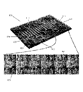
Figure 1 illustrates steps in sample processing by digital microfluidics. The
top
image is a schematic of a digital microfluidic device which allows for the
processing of 3
blood samples simultaneously. The bottom image is a sequence of frames from a
movie
(left-to-right) depicting several stages in sample processing including: (1) a
dried blood
3B
CA 2798123 2018-07-26
CA 02798123 2012-11-02
WO 2011/137533
PCT/CA2011/050205
sample; (2) mixing and incubating an extracted droplet with the sample; (3) a
droplet
containing sample extractate after translation away from the dried sample; (4)
a dried
extract; (5) mixing and incubating a derivatization reagent droplet with the
dried extract;
and (6) the dried, derivatized product.
Figure 2 illustrates three digital microfluidic methods for processing
different
sample formats, where (a) shows a droplet of blood spotted directly onto the
device
surface and allowed to dry, (b) In method 2 shows a punch from filter paper
bearing dried
blood that is positioned on the device surface.
Figure 3 illustrates the steps in processing blood samples for quantification
of
1 0 amino acid by tandem mass spectrometry. (a) Reaction scheme involving
derivatization
of the extracted amino acid, followed by derivatization with n-butanol,
followed by the
formation of a daughter ion by collision induced dissociation in the mass
spectrometer.
(b) Mass spectrum generated from primary analysis of derivatized phenyalanine
(Phe).
(c) Mass spectrum generated from the secondary analysis of derivatized Phe
showing the
1 5 loss of 102 amu as a result of collision induced dissociation.
Figure 4 provides schematics showing the hybrid digital microfluidic device
used
for in-line mass spectrometry analysis. Figure 3(a) shows the individual
layers forming
the device, and Figure 3(b) shows the integrated device.
Figure 5 provides calibration curves generated by digital microfluidic sample
20 preparation for quantification of three amino acids in blood.
Figure 6 provides a comparison of Met, Phe, and Tyr concentrations in normal
(green) and spiked (red) blood samples as biomarkers for homocystinuria,
phenylketonuria, and tyrosinemia, respectively. The dashed lines indicate the
upper levels
4
CA 02798123 2012-11-02
WO 2011/137533
PCT/CA2011/050205
for normal concentrations in newborn blood samples. Each data point represents
at least
four replicate measurements, and error bars represent 1 S.D.
Figures 7 (a)-(c) shows a series of frames from a movie (top-to-bottom)
demonstrating derivatization and extraction of amino acids, resolubilization
in solvent,
and analyte solution on a hybrid microfluidic device.
Figure 8 is an image of sample spraying from the fabricated emitter into a
mass
spectrometer inlet.
Figure 9 plots the secondary analysis spectra of (a) Phe and (b) d5-Phe
generated
from blood samples.
Figure 10 illustrates the analysis of amino acids in dried blood spots by
digital
microfluidic methods, where (a) and (b) show frames from a movie depicting
sample
processing of 3.2 mm diameter punch of a dried blood spot on filter paper by
digital
microfluidics, and (c) provides a graph of Phe concentrations measured by the
digital
microfluidic method involving punches from three patients.
DETAILED DESCRIPTION
As required, embodiments of the present invention are disclosed herein.
However,
the disclosed embodiments are merely exemplary, and it should be understood
that the
invention may be embodied in many various and alternative forms. The Figures
are not to
scale and some features may be exaggerated or minimized to show details of
particular
elements while related elements may have been eliminated to prevent obscuring
novel
aspects. Therefore, specific structural and functional details disclosed
herein are not to be
interpreted as limiting but merely as a basis for the claims and as a
representative basis
5
CA 02798123 2012-11-02
WO 2011/137533 PCT/CA2011/050205
for teaching one skilled in the art to variously employ the present invention.
For purposes
of teaching and not limitation, the illustrated embodiments are directed to
methods of
processing dried samples using a digital microfluidic device. .
As used herein, the terms, "comprises" and "comprising" are to be construed as
being inclusive and open ended, and not exclusive. Specifically, when used in
this
specification including claims, the terms, "comprises" and "comprising" and
variations
thereof mean the specified features, steps or components are included. These
terms are
not to be interpreted to exclude the presence of other features, steps or
components.
As used herein, the terms "about" and "approximately, when used in conjunction
1 0 with ranges of dimensions of particles, compositions of mixtures or
other physical
properties or characteristics, is meant to cover slight variations that may
exist in the upper
and lower limits of the ranges of dimensions so as to not exclude embodiments
where on
average most of the dimensions are satisfied but where statistically
dimensions may exist
outside this region. It is not the intention to exclude embodiments such as
these from the
1 5 present invention.
As used herein, the term "exemplary" means "serving as an example, instance,
or
illustration," and should not be construed as preferred or advantageous over
other
configurations disclosed herein.
In a first embodiment, a method of sample preparation is provided in which a
20 digital microfluidic array is employed to extract and prepare an analyte
for subsequent
analysis. The sample preparation method is especially suitable for use with a
subsequent
mass analysis step such as a tandem mass spectrometry.
Figure 1 illustrates a digital microfluidic device 100 for performing a method
6
CA 02798123 2012-11-02
WO 2011/137533 PCT/CA2011/050205
according to a first embodiment. The device 100 includes an insulating
substrate 105
having formed thereon an array 110 of digital microfluidic elements that can
be
electrically actuated to transport droplets between array elements. The array
elements
each include a conductive surface that is coated with a hydrophobic insulator.
For
.. example, the device may be formed on a glass substrate, onto which
patterned chromium
electrodes are formed by photolithography and etching, and coated with
Parylene-C and
Teflon-AF.
Each element is electrically connected to a contact pad (not shown) for
electrically addressing the array. The array may further include reagent
reservoirs 120
1 0 that are suitable for containing a reagent volume. An array element 125
adjacent to a
reservoir may be actuated to form reagent droplets and transport the reagent
droplets to
samples 130 within the array. As shown in Figure 1, the digital microfluidic
array 110
may include multiple regions in which various samples may be processed in
parallel.
Additionally, side walls and a top plate may also be provided for forming a
dual-layer
1 5 device, in which array elements may be actuated based on a voltage
difference between
the array and an electrode on the top plate electrode. For example, the top
plate may be
transparent and may comprise an unpattemed indium tin oxide (ITO) coated glass
substrate coated with a hydrophobic material such as Teflon-AErm.
The bottom portion of Figure 1 provides a series of images from a movie
20 illustrating an example of the present embodiment, in which an amino
acid analyte is
extracted from a dried blood spot and derivatized for a subsequent mass
analysis assay.
As shown in the Figure, the images correspond to a single region 140 of the
illustrated
digital microfluidic device 100.
7
CA 02798123 2012-11-02
WO 2011/137533 PCT/CA2011/050205
In image 1 in the bottom portion of Figure 1, a droplet of blood is provided
to the
digital microfluidic array and dried to form a dried blood spot 145. It is to
be appreciated
that the sample may be dispensed directly onto the array, or may be dispensed
onto a
sample reservoir and the array may be electrically addressed to extract a
droplet from the
sample reservoir and transport the droplet to an element of the array.
An extraction solvent is then provided to the array, and may be provided by
dispensing the extraction solvent to an extraction solvent reservoir 150. As
shown in
image 2, the array is then electrically addressed to transport one or more
droplets 155 of
extraction solvent to the dried blood spot. The extraction solvent is
incubated ( it has been
found that approximately 5 to 10 min is sufficient to incubate the extraction
solvent) over
the dried blood spot and analyte present in the dried blood spot is extracted
into the
extraction solvent. It will be understood that a suitable incubation time to
extract the
analyte may depend on the properties of the analyte and extraction solvent. In
image 3,
the array is again electrically actuated to transport the extraction solvent
to a second array
1 5 element 160, where it is subsequently dried, as shown in image 4.
In image 5, the array is electrically addressed to transport one or more
droplets
170 of a derivatization reagent from reagent reservoir 165 to the second array
element
160. The derivatization reagent is incubated (for example, for approximately 5
to 10 min)
to solubilize and derivatize the analyte previously dried onto second element
160. It will
be understood that a suitable incubation time to solubilize and derivatize the
analyte may
depend on the properties of the analyte and the derivatization solvent. In
image 6, the
derivatization reagent droplet is evaporated to provide a dried derivatized
analyte spot
175.
8
CA 2798123
Although embodiments disclosed herein illustrate various methods using blood
as the
sample matrix, it is to be understood that the sample need not be blood, and
may be or may
include any suitable sample matrix, such as, but not limited to, whole blood,
serum, plasma,
urine, sputum, and cerebral spinal fluid.
For example, as shown in Figure 2(a), a sample 180 could be spotted directly
and dried
onto a digital microfluidic device 100 at the point of collection, after which
the device could be
directly processed or transported to a remote testing location for analysis.
If the sample is
spotted directly onto the device, it is necessary for the device to be
provided at the point of
collection. This may be achieved by providing only a component of the device,
such as the
bottom plate of a two-plate digital microfluidic device. After having received
the spotted plate
at the laboratory, the full device could be assembled and electrically
connected to a device
controller. It is to be understood that embodiments involving sample spots
dried directly onto
the surface of a digital microfluidic array device may be performed using a
single plate digital
microfluidic device, or a two-plate microfluidic device.
Alternatively, the sample may be spotted onto a solid phase support, such as
an
exchangeable carrier or exchangeable sheet, as disclosed in International
Patent Application
No. PCT/EP2009/062657. titled "EXCHANGEABLE CARRIERS PRELOADED WITH
REAGENT DEPOTS FOR DIGITAL MICROFLUIDICS", and filed on September 301h, 2009.
The exchangeable carrier is an electrically insulating sheet having a
hydrophobic surface,
where the insulating sheet may be contacted with a digital microfluidic array
device to form an
external surface of the device. After contacting the exchangeable carrier with
the
9
CA 2798123 2019-06-21
CA 02798123 2012-11-02
WO 2011/137533
PCT/CA2011/050205
digital microfluidic array, the digital microfluidic array may be actuated to
transport
droplets that are provided on or in contact with the exchangeable carrier.
This allows for
repeated use of the device without requiring device disposal or
decontamination of the
device surface.
Although PCT/EP2009/062657 disclosed the use of a exchangeable carrier for
providing a exchangeable device component including dried reagents, the
exchangeable
sheets may additionally or alternatively employed to remotely obtaining a
dried sample
and providing the dried sample to another location (such as a laboratory) for
testing. The
sample is dried and spotted onto the exchangeable carrier prior to device
assembly. The
1 0 sample is dried at a location such that the dried sample spot is
accessible to droplets
actuated over the front hydrophobic surface of the exchangeable sheet after
attachment to
the digital microfluidic device. The dispensing of the sample onto an
appropriate location
of the exchangeable carrier may be achieved using alignment marks or features,
or, for
example, using an external masking device that places an aperture at the
appropriate
1 5 location to guide the dispensing process. The exchangeable carrier may
also include dried
reagents at one or more pre-selected positions, where the positions are
selected to be
accessible to droplets actuated over the front hydrophobic surface of the
exchangeable
carrier after attachment to the digital microfluidic device. The exchangeable
carrier with
a dried spot on a surface thereof may be applied to the surface of a single
plate digital
20 microfluidic device, or to the channel-facing surface of one or both of
the top and bottom
plates of a two-plate digital microfluidic device.
In an alternative embodiment, the sample may be first dried onto a solid phase
support, after which at least a portion of the solid phase support is locally
contacted with
CA 02798123 2012-11-02
WO 2011/137533 PCT/CA2011/050205
a location on the digital microfluidic for analyte extraction and optional
further sample
processing steps. This allows for the remote collection of dried sample in a
simple and
convenient format for subsequent processing with a digital microfluidic
device.
An example of this embodiment is shown in Figure 2(b), where a filter paper
punch 185 bearing dried blood is contacted with a surface of a microfluidic
device at a
location that is dropwise accessible under actuation of the digital
microfluidic device. In
Figure 2(b), the filter paper punch is positioned over a digital microfluidic
element and
contacted with the surface of the first plate as shown. Prior to processing
the sample, the
top plate of the digital microfluidic device is installed to complete the
assembly of the
two-plate device (as described above, a gap or channel is formed by a spacer
layer
provided between the upper and lower plates of the digital microfluidic
device). This
method allows for the convenient placement of the solid phase support at any
internal
addressable location of the digital microfluidic array. Alternatively, a two-
plate digital
microfluidic device may be pre-assembled, and the filter paper punch may be
inserted
.. into the gap between the two plates of the digital microfluidic device and
positioned at a
pre-determined location that is dropwise addressable by the digital
microfluidic array.
Any of the surfaces of the digital microfluidic array may be initially
contacted with a
exchangeable carrier, and the solid phase support may contact the digital
microfluidic
device indirectly through contact with the exchangeable carrier.
After having provided the solid phase support at a suitable location between
the
upper and lower plates of the digital microfluidic device, where the suitable
location is in
accessible to droplets actuated by the digital microfluidic device, the
digital microfluidic
array may be actuated to transport one or more extraction solvent droplets to
the location
11
CA 02798123 2012-11-02
WO 2011/137533 PCT/CA2011/050205
of the solid phase support for incubation and extraction of the dried analyte.
The
extraction and other sample processing steps, such as derivatization of the
extracted
analyte, may further be performed as described above.
The solid phase support has a thickness that is selected to be compatible with
droplet actuation in a digital microfluidic device. For example, a suitable
thickness range
for a two-plate digital microfluidic device is approximately 90 to 450
microns. As noted
above, a suitable solid phase is filter paper, which has a thickness that is
compatible with
digital microfluidic devices. Other suitable solid phase supports include
other porous
materials, such as, but not limited to paper, cellulose filters,
nitrocellulose, polymer based
1 0 monoliths such as porous polymer monoliths, and hydrogel forming
materials. It has been
shown (see below) that when a porous solid phase support such as filter paper
is
sandwiched between two plates of a digital microfluidic device, the support
need not be
adhered or otherwise fixed to the device, and is held in place after being
contacted with a
droplet by inherent forces such as capillary forces. In an alternative
embodiment, a non-
porous solid phase may be employed, for example, as described above in the
context of
removable carriers. In another embodiment, a punched non-porous solid phase
may be
adhered or secured to a surface of the digital microfluidic array using one of
many
suitable techniques, including bonding methods such as thermal bonding and/or
gluing.
The solid phase support may be selected to have a hydrophobic surface in order
to
support the transport of a droplet from a location where the droplet is
contacting the solid
phase support to a location elsewhere on the digital microfluidic array.
The lateral extent (e.g. surface area and/or diameter) of the solid phase
support is
sufficiently small to support the actuation of droplets to and from the solid
phase support,
12
CA 02798123 2012-11-02
WO 2011/137533 PCT/CA2011/050205
In some cases, it may be beneficial to limit the lateral extent of the solid
phase support to
less than one array element. For example, this can allow the transport of
droplets along
neighbouring channels without contacting the solid phase support. However, it
is to be
understood that the lateral extent of the solid phase support need not be
smaller than an
array element, and the lateral extent of the solid phase support may overlap
with
neighbouring array elements, as demonstrated in Figure 10.
For example, a larger lateral extent of the solid phase support will provide a
larger
radial extent of dried solvent, and a correspondingly higher amount of analyte
for
extraction. In some applications, it may be desirable to provide a greater
number of
analyte molecules, particularly in applications where the analyte
concentration range of
interest is near to the limit of detection of an analytical method employed
for subsequent
detection.
It may also be useful to provide, on a single digital microfluidic array,
multiple
solid phase supports, where each solid phase support contains a common dried
sample.
The multiple solid phase supports may be subjected to extraction steps as
described
above, where the extraction steps are performed in parallel or in serial
format. The
extracted analyte from the multiple solid phases may be dried onto a common
array
element in order to concentrate the extracted analyte.
It another embodiment, multiple solid phase supports may he provided on a
single
digital microfluidic array, where the solid phase supports contain analyte or
analytes from
different samples for parallel processing on a single digital microfluidic
array. This
allows for multiplexed extraction from multiple samples, and is beneficial in
further
reducing labor costs and turn around time. For example, such an embodiment may
he
13
CA 02798123 2012-11-02
WO 2011/137533 PCT/CA2011/050205
useful for the multiplexed extraction and processing of analyte in
applications involving
high-throughput screening.
After utilizing the digital microfluidic array for the extraction and
derivatization
of the analyte as described above (either using direct sample deposition and
drying or
indirect dried sample processing), an assay for the analyte may be
subsequently
performed using a mass analyzer. To prepare the derivatized analyte for mass
analysis,
the derivatized analyte is resuspended in a solvent compatible with the
subsequent mass
analysis step.
The extraction solvent may additionally contain an internal standard for use
with
1 0 the subsequent mass analysis step. The standard may include a
concentration of
isotopically labeled analyte. The mass analysis may involve analysis by
collision-
induced dissociation for tandem mass analysis. In another embodiment, the
resuspended
analyte may be first provided to a chromatographic separation system (such as
a high
performance liquid chromatography system) prior to subsequent analysis of the
1 5 separation system eluent with a mass analysis device.
In one embodiment, the analyte is one or more amino acids, fatty acids
(acylcarnitines), organic acids, and a combination thereof. The extraction
solvent in this
case is preferably methanol. A suitable derivatization step is illustrated in
Figure 3(a) for
the non-limiting case of amino acid (amino acid) analyte, in which a
derivatization
20 reagent comprising HCl-butanol transforms each amino acid to its
corresponding butyl
ester (derivatized amino acid), and subsequent formation of daughter ions via
collision
induced dissociation makes each analyte convenient to quantify by multiple
reaction
monitoring. The capacity to carry out similar processes in parallel for many
different
14
CA 02798123 2012-11-02
WO 2011/137533 PCT/CA2011/050205
amino acids simultaneously with minimal steps makes the present digital
microfluidic
and MS/MS method a useful tool for a wide range of applications, including the
screening of metabolic disorders on a large scale.
Figures 3(b) and 3(c) provide illustrative primary (MS1) and secondary (MS2)
mass spectra for the amino acid, phenylalanine, with peaks at m/z 222 and 120.
This
technique, using collision induced dissociation and MS/MS, is useful for
selectively
analysing such species because they exhibit a characteristic loss of
butylformate
(HCO0C4H9, 102 D), making them easy to identify.
In one embodiment, more than one extraction step may be performed using the
1 0 digital microfluidic array for the purpose of extracting multiple
analytes that are
beneficially processed using different extraction and/or derivatization
solvents. In a one
embodiment, the multiple extraction methods are performed on a common digital
microfluidic platform. The multiple extraction methods may be performed
serially using
a common dried sample spot or in parallel using two separate dried sample
spots, where
the dried sample spots may be dried directly onto the digital microfluidic
device, or dried
onto an intermediate matrix as described above. The final droplets containing
the
extracted analyte, in which the droplets are provided in a fluid that is
compatible with a
subsequent mass analysis step, may be combined and provided to the mass
analyzer in a
single aliquot for multiplexed mass analysis. In a non-limiting example, the
marker
succinulacetone may be extracted in order to perform an assay for tyrosinemia
type I. The
extraction method is similar to that discussed above, in which an acidic
extraction solvent
containing hydrazine is provided to the digital microfluidic array.
Preferably, this
extraction solvent comprises an acetonitrile/water/formic acid solution having
relative
CA 02798123 2012-11-02
WO 2011/137533 PCT/CA2011/050205
concentrations of 80:20:0.1% by volume, respectively, and further containing
0.1%
hydrazine monohydrate and isotopically labeled succinylacetone (13C5-
succinylacetone),
as discussed in Turgeon et al. (C. Turegeon et al., Clin. Chem. 54, 657,
2008). The array
is then electrically actuated to transfer a droplet of the extraction solvent
to a dried
sample (e.g. a dried blood spot), which may have already been processed
according to the
above protocol using a different solvent (e.g. methanol). The extraction
solvent droplet
containing the extract succinulacetone is then transported to a different
element of the
digital microfluidic array under electrical actuation of the array, where it
may be dried
and resolubilized in another buffer prior to analysis.
1 0 In one embodiment, the digital microfluidic device is interfaced a the
mass
spectrometer to support a method that may be performed without the need for
intermediate manual or robotic liquid handling steps. This overcomes many of
the
difficulties associated with convention nanoflow electrospray ionization
(nESI), which is
known to be a complex technique requiring operator expertise and vigilance to
achieve
reproducible results. For example, this limitation is part of the reason why
newborn blood
samples are often mailed to a single remote screening facility for processing.
Accordingly, in one embodiment, a nESI device is integrated into the
microfluidic
device to provide a hybrid digital microfluidic and nESI device that may be
formed by
standard batch-processing. Sample processing is performed as described above,
where an
analyte is extracted and processed from a dried sample (directly dried onto
the digital
microfluidic array, or indirectly dried onto a suitable matrix which is
contacted with the
array), and mass analysis is realized by positioning the hybrid device in
front of the mass
16
CA 02798123 2012-11-02
WO 2011/137533 PCT/CA2011/050205
spectrometer inlet and applying an electrical potential to achieve nESI. This
process
requires only a few minutes to accomplish and can be implemented by non-
experts.
Such an integrated system is illustrated in Figure 4, which shows the various
layers that form the device in Figure 4(a) and the overall integrated device
in Figure 4(b).
As shown in Figure 4(a), the device comprises a top plate 200, a digital
microfluidic layer
205, and a microfluidic channel layer 210. Top plate 205 comprises a non-
conductive and
substrate, having formed on its lower surface an electrode that is further
coated with a
hydrophobic material. Preferably, top plate is transparent and comprises an
unpatterned
indium tin oxide (ITO) coated glass substrate coated with a hydrophobic
material such as
1 0 .. Teflon-AFThl.
Digital microfluidic layer 205 is similar to the digital microfluidic device
described above, and comprises an insulating substrate having formed thereon
an array
215 of digital microfluidic elements that can be electrically actuated to
transport droplets
between array elements. Preferably, the array elements each comprise a
conductive
1 5 surface that is coated with a hydrophobic insulator. For example, the
device may be
formed on a glass substrate, onto which patterned chromium electrodes are
formed by
photolithography and etching, and coated with Parylene-C and Teflon-AF. As
described
above, each element is electrically connected to a contact pad (not shown) for
electrically
addressing the array. The array preferably further comprises reagent
reservoirs 220 that
20 are suitable for containing a reagent volume. Vertical spacing elements
225 are provided
between top plate 200 and digital microfluidic layer 205 for forming a planar
channel
within which fluidic droplets may be transported by electrically addressing
the digital
microfluidic array.
17
CA 02798123 2012-11-02
WO 2011/137533 PCT/CA2011/050205
Digital microfluidic layer 205 further includes a vertical hole 230 located
adjacent
to array element 235, enabling droplets residing on the digital microfluidic
array to be
transported to, and contacted with, the aperture of vertical hole 230, whereby
hole 230
may be filled under capillary action.
Microfluidic channel layer 210 comprises a microfluidic channel 240 formed in
an upper surface of a substrate, and extending to the edge of the substrate.
By positioning
microfluidic channel layer 210 in contact with the underside of digital
microfluidic layer
205 so that an end portion of microchannel 240 is contacted with vertical hole
230. an
inlet is formed in microchannel 240 that may be filled by fluid captured by
vertical hole
230 under further capillary forces. Accordingly, microfluidic channel 240 may
be filled
with liquid from a droplet by electrically addressing the digital microfluidic
array to
transport the droplet to contact vertical hole 230, which leads to the
subsequent filling of
channel 240 under via capillary forces. The contacting of microfluidic channel
layer 210
with digital microfluidic layer 205 also forms an external outlet 245 of
microfluidic
1 5 channel 240. In a preferred embodiment, microfluidic channel outlet 245
is located in the
corner of the device. Figure 4(b) illustrates an integrated device 250
containing a sample
droplet 260 and a reagent 270 loaded onto a reagent reservoir.
In another embodiment, the digital microfluidic array is employed to perform a
sample preparation method as disclosed above, and the derivatized analyte is
solubilized
in a fluidic droplet that is compatible with a subsequent mass analysis step.
The droplet is
transported to contact and fill the microfluidic channel, and the device is
positioned in
close proximity to the inlet of a mass analysis device. An electrical
conductor is then
made to contact the liquid in the microchannel, for example, by removing the
top plate
18
CA 2798123
and placing a wire into the vertical hole 230. Alternatively, a conductor may
be made to contact
the contents of the microfluidic channel by forming a secondary access hole
and inserting the
electrical conductor into the secondary hole in such a way as to provide a
suitable fluidic seal,
thereby preventing leakage from the microchannel. Electrospray may be
subsequently
generated in a cone emerging from outlet 245 by applying a voltage between the
conductor and
the inlet of the mass analysis device. In yet another embodiment, the
electrical contact point
may be made at the top plate (ITO slide) with a suitable contact means such as
a soldered wire
or an alligator clip. This contact is connected to the MS power supply for
applying a voltage
between the microfluidic channel outlet and the inlet to the mass
spectrometer.
While the present embodiment illustrates the interfacing of a digital
microfluidic array
with a microfluidic channel located beneath the digital microfluidic array, it
is to be understood
that the digital microfluidic array may be interfaced with the microfluidic
channel in a variety
of different geometries. For example, the digital microfluidic array may be
interfaced with a
microfluidic channel having an inlet that is laterally adjacent to an element
of the digital
microfluidic array, as disclosed in PCT Application No. PCl/CA2009/001439,
filed Oct. 13,
2009 and titled "Hybrid Digital and Channel Microfluidic Devices and Methods
of Use
Thereof'.
The methods provided herein are generally automated and are thus typically
faster and
less prone to operator error when compared to the conventional techniques in
terms of sample
preparation and reagent use. Specifically, the methods disclosed herein avoid
the need to
manually process samples dried on filter paper, namely centrifuging the filter
19
CA 2798123 2019-06-21
CA 02798123 2012-11-02
WO 2011/137533 PCT/CA2011/050205
paper in an extraction solvent, which involves laborious and time consuming
steps.
The aforementioned embodiments may be applied for a wide range of sample
types,
analytes, and processing applications. Although embodiments disclosed above
have
focused on three specific metabolic diseases (homocystinuria, phenylketonuria,
and
.. tyrosinemia), it is to be understood that the scope of the various
embodiments includes,
but is not limited to a wide range of analytes that are compatible with
digital microfluidic
array based extraction and processing.
Example assays include, but are not limited to, amino acid assays, fatty acid
disorders (acylcamitines), organic acid disorders, and markers for metabolic
disorders.
1 0 Tables 1, 2 and 3 below provide a non-limiting list of various known
amino acid
disorders, fatty acid disorders, and organic acid disorders, respectively, and
their markers.
The analytes below merely provide an illustrative list and are not intended to
limit the
scope of the embodiments provided in the present disclosure.
Table 1: List of Amino Acid Metabolic Disorders and their Markers
Amino Acid Disorders Marker(s)
Argininemia or Arginase deficiency Arginine
Citrullinemia-I or Argininosuccinate Citrulline
synthase deficiency
Argininosuccinic aciduria or Citrulline,
Argininosuccinate lyase deficiency argininosuccinic acid
Omithine transcarbamylase deficiency Alanine, Citrulline
Carbamoylphosphate synthetase deficiency Alanine, Citrulline
Citrullinemia-II or Citrin-mitochondrial Citrulline
aspartate-glutamate transporterdeficiency
Hyperammonemia-Hyperornithine mia- Ornithine
Homocitrullinuria Syndrome
Phenylketonuria or Phenylalanine Phenylalanine
hydroxyl ase deficiency
Maple Syrup Urine Disease or Branched Leucine, Valine
chain ketoacid
dehydrogenase deficiency
Homocystinuria or Cystathioninebeta- Methionine
CA 02798123 2012-11-02
WO 2011/137533
PCT/CA2011/050205
synthase deficiency
Non ketotic hyperglycinemia Glycine
Tyrosinemia I or Fumarylacetoacetase Tyrosine
deficiency
Tyrosinemia II or Tyrosine Tyrosine
aminotransferase deficiency
Tyrosinemia III or 4-hydroxyphenylpyruvic Tyrosine
acid dioxygenase deficiency
5-0xoprolinuria or Glutathione synthetase 5-oxoproline
deficiency
Biopterin defects Phenylalanine
Table 2: List of Fatty Acid Metabolic Disorders and their Markers
Fatty Acid Oxidation Disorders Marker(s)
Very long-chain acyl-CoA Tetradecenenoylcarnitine,
dehydrogenase deficiency Tetradecanoylcarni tine
Long-chain hydroxyacyl-CoA Hydroxyhexadecanoylcarnitine,
dehydrogenase deficiency Octadecenenoylcamitine,
Hydroxyoctadecenenoylcamitine
Medium-chain acyl-CoA Octanoylcamitine, Hexanoylcarnitine,
dehydrogenase deficiency Decanoylcarnitine
Short-chain acyl-CoA Butyrylcamitine & Isobutyrylcamitine
dehydrogenase deficiency
Tr-functional protein deficiency Hydroxyhexadecanoylcamitine,
Hydroxyoctadecenenoylcamitine,
Hydroxyoctadecanoylcarnitine
Glutaric aciduria-II or Multiple acyl- Butyrylcarnitine & Isobutyrylcarnitine,
CoA dehydrogenase deficiency Isovalerylcamitine & 2-
methylbutyrylcamitine,
Glutarylcamitine, Octanoylcarnitine,
Tetradecanoylcarnitine
Carnitine palmitoyl transferase Free camitine
deficiency-I
Carnitine palmitoyl transferase Octadecenenoylcarnitine,
deficiency-II Hexadecanoylcamitine
Carnitine/acylcarnitine translocase Octadecenenoylcamitine,
deficiency Hexadecanoylcamitine
Carnitine uptake defect or 2,4- Free camitine, Decadienoylcarnitine
Dicnoyl-CoA reductase deficiency
Hydroxyacyl-CoA dehydrogenase Hydroxybutyrylcamitine
deficiency or Short/Medium-chain
hydroxyacyl-CoA dehydrogenase
deficiency
21
CA 02798123 2012-11-02
WO 2011/137533 PCT/CA2011/050205
Propionic acidemia or Propionyl- Propionylcarnitine
CoA
carboxylase deficiency
Table 3: List of Organic Acid Metabolic Disorders and their Markers
Organic Acid Disorders Marker(s)
Methylmalonic aciduria or Propionylcarnitine
Methylmalonyl-CoA mutase
deficiency
Cobalamin defects (A, B) Propionylcarnitine
Cobalamin defects (C, D) Propionylcarnitine. Methionine
Multiple carboxylase deficiency Propionylcarnitine,
Hydroxyisovalerylcamitine
3-Hydroxyisobutyric aciduria Hydroxybutyrylcarnitine
Isovaleric acidemia or Isovaleryl- Isovalerylcamitine & 2-
CoA dehydrogenase deficiency methylbutyrylcarnitine
2-Methylbutyrylglycinuria or 2- Isovalerylcamitine & 2-
Methylbutryl-CoA dehydrogenase methylbutyrylcarnitine
deficiency
3-Methylcrotonyl-CoA carboxylase Hydroxyisovalerylcamitine
deficiency
3-Hydroxy-3-methylglutaryl-CoA Hydroxyisovalerylcamitine,
lyase deficiency
Mitochondrial acetoacetyl-CoA Tiglylcamitine plus 3-
thiolase deficiency or Beta- methylcrotonylcamitine
ketothiolase deficiency
Methylglutaconic aciduria or 3- Tiglylcarnitine plus 3-
Methylglutaconyl-CoA hydratase methylcrotonylcamitine
deficiency
2-Methyl-3-hydroxybutyryl-CoA Hydroxyisovalerylcamitine
dehydrogenase deficiency
MaIonic aciduria or Malonyl-CoA Malonylcarnitine
decarboxylase deficiency
Glutaric aciduria-I or Glutaryl-CoA Glutarylcamitine
dehydrogenase deficiency
It is to be understood that while the preceding embodiments have included a
derivatization step for processing the extracted analyte using the digital
microfluidic
array, this step may not be required for extraction protocols that are
suitable for other
analytes. As such, the derivatization step, or any further on-chip processing
steps, may be
22
CA 02798123 2012-11-02
WO 2011/137533 PCT/CA2011/050205
optionally performed, as appropriate for a given analyte or application.
Example applications include, but are not limited to, neonatal screening of
metabolic
disorders (i.e. amino acids and organic acids), other disorders (for example,
congenital
adrenal hyperplasia, congenital hypothyroidism, biotinidase deficiency and
galactosemia), and genetic disorders (e.g. cystic fibrosis and sickle cell
diseases).
For example, in each newborn blood spot analysis, a sample is obtained by
pricking
the subject's heel (or by venipuncture) and allowing a spot of blood to dry on
filter paper.
The dried blood spot is typically couriered to a lab, where 3.2 mm diameter
circular discs
are punched, and the analytes are extracted, mixed with isotope-labeled
internal
standards, derivatized, and then reconstituted for analysis by tandem mass
spectrometry
(MS/MS). As shown in Figure 3(a), the derivatization step transforms each
amino acid to
its corresponding butyl ester (derivatized amino acid) that allows for a
characteristic
fragmentation pattern (neutral loss of 102) via collision induced dissociation
. Figures
3(b-c) contains representative primary (MS1) and secondary (MS2) mass spectra
for the
amino acid, phenylalanine, with peaks at m/z 222 and 120. In addition to amino
acids, the
same derivatization step butylates acylcarnitines (AC), which serve as markers
of inborn
errors of fatty acid and organic acid metabolism.
An additional and clinically relevant advantage of the present digital
microfluidic
methods is the reduction in sample size that may be achieved relative to
conventional
processing methods. In some cases, the reduction in volume may be
approximately 15-
20x. This reduction has the potential to be beneficial for applications in
which it is
desirable to employ small sample volumes, such as in the testing of newborn
patients,
23
CA 02798123 2012-11-02
WO 2011/137533 PCT/CA2011/050205
from which five spots of blood are typically collected for analysis, and in
high throughput
screening applications.
In the examples provided herein, the small volume required (5 x 5 4 = 25 4)
for certain clinical in-vitro diagnostic assays can be collected as capillary
blood with a
single needle-prick, but in conventional sample processing methods, the volume
(5 x 75-
100 4 = 375-500 4) often requires multiple pricks and tissue-squeezing, which
can
contaminate the sample with interstitial fluids, invalidating it for testing.
Other sample
size-related advantages of the present embodiments are a reduction in reagent
use (fpr
example, 20 4 vs. 170-450 4), and a reduction in analysis time (-1 h vs. >3.5
h). This
reduction in reagents and analysis time, combined with the potential
elimination of
sample preparation robotic liquid handling systems, makes the present methods
an
attractive option for diagnostic testing in a time of increasing costs for
health care.
The following examples are presented to enable those skilled in the art to
understand and to practice the present invention. They should not be
considered as a
limitation on the scope of the invention, but merely as being illustrative and
representative thereof.
EXAMPLE 1
Device Fabrication and On-Chip Processing of Amino Acids
Digital microfluidic devices were fabricated in the University of Toronto
Emerging Communications Technology Institute (ECTI) cleanroom facility, using
a
transparent photomask printed at Norwood Graphics (Toronto, ON). Glass devices
bearing patterned chromium electrodes were formed by photolithography and
etching and
were coated with 2.5 jam of Parylene-C and 50 nun of Teflon-AF. Parylene-C was
applied
24
CA 02798123 2012-11-02
WO 2011/137533 PCT/CA2011/050205
using a vapor deposition instrument (Specialty Coating Systems), and Teflon-AF
was
spin-coated (1% wt/wt in Fluorinert FC-40, 2000 rpm, 60 s) followed by post-
baking on a
hot-plate (160 'V, 10 min). The polymer coatings were removed from contact
pads by
gentle scraping with a scalpel to facilitate electrical contact for droplet
actuation.
A prototype similar to the device shown in Figure 1 was fabricated to analyze
5-
f.t,L blood samples. As shown in the top image, an array of 88 driving
electrodes connects
a series of 10 reservoirs dedicated to microliter volumes of sample and
reagents. As
depicted in the bottom image, blood samples are spotted onto the device and
dried, after
which the sample is extracted into methanol and the solvent is allowed to
evaporate. The
extract is then derivatized, and the product is isolated by allowing the
solvent to
evaporate. The entire process requires 50 min to complete (compared with >3.5
h for
clinical laboratories, not including mailing time).
The device featured an array of eighty-eight actuation electrodes (2.2 x 2.2
mm
ea.) connected to ten reservoir electrodes (5 x 5 mm ea.), with inter-
electrode gaps of 40
pm. Devices were assembled with an unpattemed ITO¨glass top plate and a
patterned
bottom plate separated by a spacer funned from four pieces of double-sided
tape (total
spacer thickness 360 pm). Unit droplets (covering a single driving electrode)
were ¨1.8
pL. To actuate droplets, driving potentials (70-100 VRms) were generated by
amplifying
the output of a function generator (Agilent Technologies, Santa Clara, CA)
operating at
18 kHz. As described elsewhere, droplets were sandwiched between the two
plates and
actuated by applying driving potentials between the top electrode (ground) and
sequential
electrodes on the bottom plate via the exposed contact pads. Droplet actuation
was
monitored and recorded by a CCD camera mounted on a lens.
CA 02798123 2012-11-02
WO 2011/137533 PCT/CA2011/050205
Blood samples were collected from a healthy adult male volunteer after a 10 h
fasting period and were kept at -20 C until analysis. Immediately prior to
use, samples
were thawed and evaluated as described.
Working solutions of all amino acids (amino acids) (25, 50, 100 and 500 M
ea.)
were prepared in DI water. For derivatization of extracted amino acids, a 3 N
HC1-
butanol solution was prepared from a mixture of 12 N HO/neat butanol (1:3
v/v). For
analysis of amino acids in blood samples, the extracting solvent (Me0H)
contained 25
pM of the appropriate deuterated amino acid (d3-Met, d5-Phe or d4-Tyr). For
quantitative
analysis of amino acid recovery from blood and for experiments mimicking
.. diseased/healthy infant blood, samples were spiked with 200 pM of the
appropriate
amino acid (Met, Phe or Tyr). In all experiments, organic solvents were HPLC
grade and
deionized (DI) water had a resistivity of 18 MQ-cm at 25 C.
5-1iL droplets containing the sample (i.e., amino acid standards, whole blood
or
spiked whole blood) were pipetted onto the bottom plate of a device and dried.
The top
plate was then affixed and two solvents were loaded into the appropriate
reservoirs,
including Me0H containing 25 M of deuterated amino acid (extraction solvent),
and 3
N HCl-butanol (derivatization solvent). A reservoir volume (10 pL) of
extraction solvent
was dispensed and driven by digital microfluidic to the dried sample and
allowed to
incubate (5 min). The extraction solvent was then actuated away from the
sample and
dried (-15 min, room temperature) at a second site, after which a reservoir
volume (10
L) of derivatization solvent was dispensed to the dried extract and incubated
for 15 min at
75 C. Following the reaction, the top plate was removed and the solvent was
allowed to
evaporate (-15 min, room temperature).
26
CA 02798123 2012-11-02
WO 2011/137533 PCT/CA2011/050205
Calibration curves were generated by digital microfluidic sample preparation
for
quantification of (a) methionine (Met), (b) phenylalanine (Phe), and (c)
tyrosine (Tyr) in
blood. As shown in Figure 5, data was generated by plotting the intensity
ratios of the
daughter ions of each amino acid relative to their deuterated internal
standard (i.e., d3-
Met, d5-Phe, d4-Tyr, respectively) as a function of amino acid concentration.
Each data
point represents at least four replicate measurements, and error bars
represent 1 S.D.
Regression lines were linear with R2 > 0.996 for each analyte.
For comparison, amino acids were also extracted and derivatized on the
macroscale using known methods. Amino acid samples (20 tiL) were pipetted and
dried
in a microcentrifuge tube and extracted in methanol (500 pL, 30 min)
containing isotope-
labeled internal standards. The solution was centrifuged (13,000 rpm, 15 min),
and the
supernatant transferred to a second tube and evaporated to dryness using
nitrogen. The
extractate was then resuspended in 3 N HC1-butanol solution (250 pL) to
derivatize the
amino acids at 65 C for 20 min, followed by evaporation of the derivatized
mixture.
Most samples were processed by digital microfluidic and then were analyzed
offline by nanoelectrospray tandem mass spectrometry (nESI-MS/MS). Such
samples
(stored dry on device or in centrifuge tube until analysis) were reconstituted
in 70 pl of
acetonitrile/water (4:1 v/v); samples originating from blood were, in
addition, passed
through PVDF membrane centrifuge-filters with 0.1 pm pore diameter (Millipore,
ON).
Samples were injected into an LTQ Mass Spectrometer (Thermo Scientific) via a
fused
silica capillary transfer line (100 pm i.d.) mated to a New Objective Inc.
(Woburn, MA)
nanoelectrospray emitter (100 pm i.d. tapering to 50 pm i.d.) at a flow rate
of 0.81aL min,
with an applied voltage of 1.7-1.9 kV and capillary temperature of 200 C.
Tandem
27
CA 02798123 2012-11-02
WO 2011/137533 PCT/CA2011/050205
MS/MS analysis was carried out by introducing 30% collision energy to the
parent ions
and then the fragments over the m/z range of 100-300 were scanned. For amino
acid
samples, the daughter ions detected in the second mass selection, which
exhibit a loss of
butylfomiate (HCO0C4H9, 102 m/z), were observed and used for quantification.
Spectra
were collected as an average of 50 acquisitions, and replicate spectra were
obtained for
digital microfluidic-derivatized samples of both control and blood.
Some samples were analyzed by nES1-MS/MS in-line on hybrid digital
microfluidic-microchannel devices bearing an integrated nESI emitter. In these
experiments, hybrid devices were mounted on a 3-axis micromanipulator (Edmund
Optics, NJ) positioned near the inlet of the LTQ MS. After sample processing,
a spray
was generated by applying 2.5-3.0 kV to a platinum wire inserted in the access
hole (see
Figure 6).
Tandem mass spectrometry was used to (i) quantify amino acids in blood samples
and (ii) evaluate the recovery efficiency of the digital microfluidic method.
For the
former (quantification of amino acid in blood samples), calibration plots were
generated
by plotting the intensity ratio of daughter ions from the extracted amino
acids relative to
the those of the internal standards (i.e., Met m/z 104:107, Phe m/z 120:125,
and Tyr m/z
136:140) as a function of amino acid concentration in standard solutions (25-
500 1jM in
DI water). Data points included in the calibration plots represent an average
of at least 4
replicate measurements, and the data in each plot were fit with a linear
regression. Blood
samples were then evaluated (with on-chip derivatization and extraction, and
measurement by MS/MS relative to internal standards, as above), and the values
were
compared to the calibration plots to determine the amino acid concentrations.
For the
28
CA 02798123 2012-11-02
WO 2011/137533 PCT/CA2011/050205
latter (evaluation of % recovery), blood samples of known amino acid
concentrations
were spiked with 200 /aM of amino acid standards and extracted (as above).
Knowing the
total concentration of amino acids in blood spots (e.g. native methionine
concentration
plus spiked methionine), % recovery was obtained by comparing the
concentration values
(obtained from calibration curves) vs. the known values.
The % recovery of amino acids was evaluated quantitatively using a
fluorescence-
based assay. Control samples (Met, Phe or Tyr; 50 iuM of each) were processed
by digital
microfluidic (as above), excluding the derivatization step. The dried extracts
were diluted
into 95 1_, aliquots of borate buffer (20 mM, pH 8.5) in wells in a 96-well
microplate.
Upon addition of 5 ,tiL of fluorescamine (5 mg/mL in acetone) the microplate
was
inserted into a fluorescence microplate reader (Pherastar, BMG Labtech,
Durham, NC)
equipped with a module for 390 nm excitation and 510 nm emission. The plate
was
shaken (5 s) and the fluorescence was measured. As a control, identical
samples that had
not been extracted were evaluated using the same fluorescent assay. To ensure
that
controls were processed in identical manner relative to extracted samples,
each control
sample was spotted on a device, dried and then reconstituted in buffer for
analyses. Four
replicate measurements were made for each sample and control.
Nanoelectrospray ionization tandem mass spectrometry (nESI-MS/MS) is used to
quantify amino acids in samples of blood processed by the digital microfluidic
method.
Calibration curves with R2 greater than 0.996 (Figure 4) were generated by
analyzing
standards processed by digital microfluidic at known concentrations from the
abundance
ratio of each amino acid to its detterated standard peak in the secondary
(MS2) spectra.
The calibration curves facilitated measurement of amino acid concentrations in
blood
29
CA 02798123 2012-11-02
WO 2011/137533 PCT/CA2011/050205
samples from a healthy male volunteer. As listed in Table 4, the values
obtained were in
the expected physiological range and the precision in the method was high with
coefficients of variation (CVs) ranging from 5 to 11%.
Measured Blood
Normal Blood
Amino Acid Concentration (tM) in an
Concentration (iuM)
Adult Male Volunteer
Methionine 25 2 16-33
Phenylalanine 38 2 41-68
Tyrosine 46 5 45-74
Table 4: Measured ( 1 S.D.) and normal adult concentrations of amino acids in
blood.
Fluorescence and MS/MS were used as an orthogonal test to evaluate the
extraction efficiency of the new digital microfluidic technique. The former
method relies
on fluorescamine, a fluorogenic reagent that exhibits no fluorescence until it
reacts with
primary amines. Recovery was determined by comparing the fluorescence
intensity of
multiple samples before and after extraction. In the latter (MS/MS) method,
blood
samples were spiked with amino acid and recovery was determined by comparing
the
amino acid concentration (native amino acid plus spiked amino acid) vs. known
concentration. As listed in Table 5, the two orthogonal methods (fluorescence
and
MS/MS) agree and reveal the new technique to be very efficient and recovery
was >80%
for each standard and blood sample evaluated. As above, the precision of these
measurements was high, with CVs ranging from 1 to 10%.
% Recovery by % Recovery by MS/MS
Amino Acid
Fluorescence in Standards in Blood
Methionine 98 10 100 1
CA 02798123 2012-11-02
WO 2011/137533 PCT/CA2011/050205
Phenylalanine 86 9 85 5
Tyrosine 82 10 84 7
Table 5: /G Recovery of the digital microfluidic method measured by
fluorescence and
MS/MS ( 1 S.D.)
To validate the new digital microfluidic method as a platform for analyzing
amino
acid disorders in blood, spiked blood samples (mimicking diseased states) and
non-spiked
blood samples (mimicking healthy state) were analyzed by mass spectrometry.
Figure 6
shows a comparison of measured concentration of amino acids in normal and
spiked
blood samples. The dashed line indicates the threshold value for diagnosis of
homocysteinuri a (67 laM Met), phenylketonuri a (120 laM Phe), and tyrosinemia
(150 laM
Tyr). As shown, the method is useful for distinguishing between these states.
As shown in Figure 4, a hybrid digital microfluidic system was fabricated
(using
process similar to those disclosed above) in which an nESI device was directly
integrated
with the microfluidic device. The hybrid digital microfluidic-microchannel
devices were
fabricated in four steps.
First, a DMF bottom substrate (layer 205 in Figure 4(a)) bearing an array of
DMF
driving electrodes) was fabricated as described above, but without a Teflon-AF
coating). The design was similar to that of the DMF-only devices described
above, but
with fewer electrodes ¨ 2 rows of 9 actuation electrodes 215 (2.2 xx 2.2 mm)
and 3
reservoir electrodes 220 (5 xx 5 mm). Moreover, the substrates were first
modified by
drilling an access hole 230 (-2 mm diameter) through the substrate using a
micro drill-
press before the photolithographic processes, where the hole was drilled
adjacent to the
planned location of an electrode 235. After patterning the electrodes, the
opposite side
31
CA 02798123 2012-11-02
WO 2011/137533 PCT/CA2011/050205
was first coated with 7 m of Parylene-C for bonding with substrate 210.
Secondly, a glass substrate 210 bearing a microchannel 245 nanoelectrospray
tip
240 formed in Parylene was fabricated. 37 grams of Parylene-C were deposited
on
piranha cleaned, silanized glass slide (25 x 55 mm) via vapor deposition.
After Cr
deposition, a microchannel (25 um wide x 5 mm long) was photolithographically
patterned on the substrate by UV radiation (365nm, 35mW/cm2, 50s) using a Karl-
Suss
MA6 mask aligner (Garching, Germany).
Third, the channel side of substrate 210 was mated to the non-electrode side
of
substrate 205, placed under pressure in a precision vise (-20 MPa), and
thermally bonded
in a vacuum oven (200 C, 24 h). After cooling, the top of substrate 205 was
first coated
with 2 pm of parylene followed by spin-coating 50 nm of Teflon-AF with a small
piece
of dicing tape covering the drilled hole. The tape was removed before post-
baking on a
hot plate (160 C, 10min).
Fourth, the top plate 200 was assembled with spacers formed from four pieces
of
double-sided tape as described above for droplet actuation.
Using this hybrid device, droplets are manipulated on the top surface, and are
subsequently transferred to microchannels on the bottom of the device through
the hole.
The principle of operating the hybrid device for on-chip sample analysis is
shown in
Figure 7, which shows several frames from a movie demonstrating derivatization
and
extraction of amino acids, resolubilization in solvent, and analyte solution
on a hybrid
microfluidic device. A blood sample 310 was first spotted on the device 300
and the
amino acids were extracted and derivatized as described above. The dried,
derivatized
sample 320 was then resuspended in acetonitrile and the droplet 330 was
actuated
32
CA 02798123 2012-11-02
WO 2011/137533 PCT/CA2011/050205
towards the access hole 340 such that it filled the underlying channel 350.
The filled
channel can be seen at 360 in Figure 7(c).
As shown in Figure 8, the fluid in the channel was delivered to a corner 400
of the
device and nanoelectropsray 410 was generated by applying a high voltage to
the counter
electrode 420. Representative mass spectra generated from samples processed
and
analyzed on-chip are shown in Figure 9. The entire process requires ¨1 h from
sampling
to analysis, and requires only the hybrid digital microfluidic device and a
mass
spectrometer (i.e., no complex nanoflow pumps, robots, samplers, or control
software).
EXAMPLE 2
Direct Digital Microfluidic Processing of Sample Dried on Solid Phase
In the present example, a digital microfluidic method is demonstrated using
the direct
processing, on a digital microfluidic array, of samples dried onto filter
paper. A digital
microfluidic array device was fabricated as described in Example 1, except
that in the
present case, the two plates were separated by six pieces of double-sided tape
(total
spacer thickness 540 pm). This increased spacing was provided to accommodate
the
thickness of the filter paper. A blood sample was collected and dried onto
filter paper,
and a 3.2 mm diameter filter paper disc was punched for subsequent analysis.
The punch
was placed onto an element of a digital microfluidic array.
A portion of an experiment is depicted in Figure 10(a). As shown, a droplet of
extraction solvent 510 was dispensed on the digital microfluidic array 500 and
driven to
the filter paper punch 520, and the extract was then moved away and dried 530
for further
processing (i.e., derivatization and solvent exchange, similar to Figure 1).
As can be seen
in Figure 10(b), after wetting, the filter paper punch 520 remains adhered to
the surface
33
CA 02798123 2012-11-02
WO 2011/137533 PCT/CA2011/050205
through capillary forces. This process requires ¨ 50 min to complete.
To evaluate this digital microfluidic method relative to gold standard
practices, a
series of punches from blood samples containing various concentrations of Phe
were
processed by this digital microfluidic method, and punches from the same
samples were
evaluated using the conventional newborn screening technique. As listed in
Table 6
below, a paired 1-test revealed no significant difference between the two data
sets at a
95% confidence level. To validate the new technique for application to
clinical samples,
dried blood spot punches from three newborn patients of NSO were evaluated by
the
digital microfluidic method. As shown in Figure 10(b), the new technique
correctly
identified patients 1 and 3 as suffering from phenylketonuria, and patient 2
as being
unaffected.
Measured Phe Concentration (iuM) Measured
Phe Concentration (JIM)
Sample
Using DMF Method 2 Using NSO Technique
1 70 70
2 550 548
3 93 88
4 93 92
5 368 302
6 534 539
7 735 871
Table 6: Measured phenylalanine (Phe) concentration in 3.2 mm dia. punches
from filter
paper bearing dried blood using digital microfluidic method 2 (left) and
standard
techniques at NSO (right). A paired t-test (P = 0.05, t = 0.69) revealed no
significant
difference between the two data sets.
The foregoing description of the embodiments of the invention has been
presented to
illustrate the principles of the invention and not to limit the invention to
the particular
34
CA 02798123 2012-11-02
WO 2011/137533 PCT/CA2011/050205
embodiment illustrated. It is intended that the scope of the invention be
defined by all of
the embodiments encompassed within the following claims and their equivalents.