Note : Les descriptions sont présentées dans la langue officielle dans laquelle elles ont été soumises.
CA 02799736 2012-12-27
WO 2012/000073 PCT/CA2010/001005
1
IMAGING DEVICE INFORMATION SYSTEM AND METHOD
FIELD OF THE INVENTION
[0001] The present invention relates to systems and methods for managing the
use of imaging devices.
BACKGROUND OF THE INVENTION
[0002] A radiology information system (RIS) used in a hospital typically
employs
the Health Level 7 (HL7) international standard for messaging that defines a
series of healthcare events, records and messages to support administrative,
logistical, financial as well as clinical processes. HL7 is not designed to
support
the exchange of image data. HL7 messages are broadcast on internal and inter-
hospital networks to indicate events such as the admission, discharge or
transfer
of a patient, or, for example, to record the fact that the results of a
Diagnostic
Imaging (DI) test have been recorded in the RIS. The HL7 data stored in the
RIS
may include fields such as patient ID, patient name, gender and address, date
and time of admission, name of referring physician, name of radiologist, as
well as
test-specific information. For example, if a magnetic resonance (MR) study has
been requested for a patient, then the HL7 data for that patient may indicate
the
procedure code for such a request, the requesting physician, imaging status
and
report status.
[0003] Diagnostic image data is typically handled by a picture archiving and
communication system (PACS). PACS systems generally store and transmit data
in accordance with the Digital Imaging and Communications in Medicine (DICOM)
international standard. Imaging modalities, such as MR and other diagnostic
imaging devices, generally communicate directly with the PACS over a network
using DICOM. The function of the PACS is to maintain a database of diagnostic
images taken on connected devices along with related information for image
display and patient demographics.
[0004] In addition to the image data, the DICOM records include accession
number, the start and end times of tests, demographic information such as
patient
CA 02799736 2012-12-27
WO 2012/000073 PCT/CA2010/001005
2
identification information, and may include identification of the attending
staff
member(s).
[0005] Information must be transmitted between such RIS and PACS systems
and DI modality devices, for example, in order to indicate which tests have
been
ordered on which patients, and report when image studies have been completed.
For example, after an MR test has been completed, the interpreting radiologist
will
render a medical opinion report on the images, which is stored the RIS and
PACS
in one of several possible different formats.
[0006] The lack of interoperability between HL7 and DICOM has been a
longstanding problem as is the variability in the use and interpretation of
these
standards. Manual transfer of data is expensive and error-prone. There have
been
attempts to harmonize the standards using integration profiles (IP's) to
achieve
specific interoperability, which has been led by an organization known as IHE
(integrating the healthcare enterprise) working group. Unfortunately, these
have
been of limited success and achieved only limited interoperability. Some
automated approaches have been employed such as the use of a "broker" system
or software to translate between the two protocols so that a RIS can
communicate
directly with a modality (such as MR) and/or PACS. However, such solutions are
expensive, and are error-prone because of the varying interpretations and use
of
the standards.
[0007] After the modality and/or PACS information is made available to the RIS
and incorporated into the RIS database, the RIS may then be able to produce
reports relating to the use of the imaging modalities and use of staff. For
example,
the RIS may provide aggregated monthly reports on the total number of
ultrasound tests completed, the total technical fees, professional fees and
workload units claimed for each month. This may be accompanied with variances
from the previous year and the current budget. Reports showing the total
workload
units on a modality by modality basis or other aggregated basis for a certain
period are also typically produced four to six weeks after the period,
[0008] It may be very useful to compare such information across multiple
hospitals so each hospital can identify efficiencies or deficiencies in its
utilization
of imaging modalities and take corrective action. Such comparisons are very
CA 02799736 2012-12-27
WO 2012/000073 PCT/CA2010/001005
3
difficult because the procedure (study) code dictionaries are not standardized
and
are used differently by different hospitals. The Canadian Institute for Health
Information (CIHI) provides a set of standardized code guidelines for hospital
workload units for every procedure completed in health care in Canada.
However,
hospitals vary from these guidelines. When CIHI suggests 12 workload units to
complete a two view chest radiography study and the hospital consistently
takes
18 workload units to complete the study, then the hospital will change its own
procedural dictionary to 18 workload units. In addition, the combination of
certain
studies into a single exploding code for convenience varies from hospital to
hospital depending on the radiologists' preferences for study grouping. This
makes the standardization of study dictionaries more difficult.
SUMMARY OF THE INVENTION
[0009] The invention provides an imaging facility device information system
for
use in an imaging facility having a radiology information system, a picture
archiving and communication system and a plurality of diagnostic imaging
modality devices, all of the foregoing connected to a local network, the
facility
information system comprising:
a. a facility imaging modality analysis processor connected to the local
network for receiving patient information from the radiology
information system and for receiving image series information of
patients from the diagnostic imaging modality devices; and
b. a facility database in electronic communication with the facility
imaging modality analysis processor,
wherein the facility imaging modality analysis processor combines
image series information of each patient with patient information for the
corresponding patient into patient records in the facility database and
produces facility reports from the patient records analyzing temporal
utilization of the diagnostic imaging modality devices.
[0010] The facility imaging modality analysis processor may query the picture
archiving and communication system to identify changes made to the image
CA 02799736 2012-12-27
WO 2012/000073 PCT/CA2010/001005
4
series information and the facility imaging modality analysis processor may
update
the facility database to reflect changes that are identified.
[0011 ] The facility imaging modality analysis processor may receive image
series
information using a standard imaging system protocol and receives patient
information using a standard radiology information system protocol. The
standard
radiology information system protocol may be HL7 and the standard imaging
system protocol may be DICOM.
[0012] The facility imaging modality analysis processor may allow authorized
facility users to request facility reports using a web browser.
[0013] The invention also provides for a method for analyzing temporal
utilization
of diagnostic imaging modality devices comprising the steps of:
a. receiving patient information from a radiology information system;
b. receiving image series information of patients from the diagnostic
imaging modality devices;
c. combining image series information of each patient with patient
information for the patient into patient records; and
d. producing facility reports from the patient records analyzing temporal
utilization of the diagnostic imaging modality devices.
[0014] The method may further comprise the steps of:
e. querying a picture archiving and communication system to identify
changes made to the image series information; and
f. updating the facility database to reflect changes that are identified.
[0015] In the method the image series information may be received using a
standard imaging system protocol and the patient information may be received
using a standard radiology information system protocol. The standard radiology
information system protocol may be HL7 and the standard imaging system
protocol may be DICOM.
[0016] The method may further comprise the steps of analyzing the facility
reports
to identify an under-utilized diagnostic imaging modality device and
increasing the
number of imaging facility staff attending that diagnostic imaging modality
device
CA 02799736 2012-12-27
WO 2012/000073 PCT/CA2010/001005
so that the temporal utilization of the under-utilized diagnostic imaging
modality
device is increased.
[0017] The method may further comprise the steps of analyzing the facility
reports
to determine that a diagnostic imaging modality device has a technical problem
5 and having technicians repair the diagnostic imaging modality device.
[0018] The invention also provides for an imaging device regional information
system comprising:
a. at least two imaging facility device information systems connected to
a wide area network, and
b. a regional imaging modality analysis processor connected to the
wide area network,
wherein the imaging facility device information systems produce facility-
independent exam record summaries that are transmitted to the
regional imaging modality analysis processor and wherein the regional
imaging modality analysis processor produces regional reports from the
facility-independent exam record summaries comparing the temporal;
utilization of the diagnostic imaging modality devices between imaging
facilities.
[0019] The regional imaging modality analysis processor may allow authorized
regional users to request regional reports using a web browser.
[0020] The imaging facility device information systems may update the patient
records in real-time and may send the exam record summaries to the regional
imaging modality analysis processor in real-time or near real-time, and the
regional imaging modality analysis processor may then produce regional reports
in real-time or near real-time.
BRIEF DESCRIPTION OF THE DRAWINGS
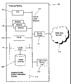
[0021] Figure 1 is a diagram showing the invention and interoperating
equipment
within an imaging facility.
[0022] Figure 2 is a diagram showing multiple imaging facilities and a
regional
data center.
CA 02799736 2012-12-27
WO 2012/000073 PCT/CA2010/001005
6
[0023] Figure 3 is an example of a modality temporal utilization report.
[0024] Figures 4a-4d show an example of data that may be stored in a facility
database.
[0025] Figure 5 is an example of an exam record summary.
[0026] Figure 6 is a drill-down report that may be displayed when a user
clicks on
the report shown in Figure 3.
[0027] Figure 7 is an example of a regional report.
DETAILED DESCRIPTION OF THE INVENTION
[0028] The system, in a preferred embodiment, which may be referred to as an
imaging facility device information system 110, as shown in Figure 1, consists
of a
facility imaging modality analysis processor (facility IMAP) 104 and a
facility
database 105, which is connected to and managed by the facility IMAP 104. The
facility IMAP 104 is connected to a facility local network 107. The system is
located at an imaging facility 100, such as a hospital, typically having a RIS
101, a
PACS 103 and one or more modalities 102. Each modality 102 is a class of DI
devices, such as computed tomography (CT), and includes at least one device of
that modality, referred to as a DI modality device or a diagnostic imaging
modality
device. For example, a hospital may have one MR device, two CT devices and
five Ultrasound (US) devices. The RIS 101, modalities 102, and PACS 103 are
all
connected to the facility local network 107. The imaging facility 100 may also
have
a facility firewall 106 that connects the facility local network 107 to a wide
area
network 108, such as the internet. In addition to collecting data and
populating the
facility database 105, the facility IMAP 104 also produces facility reports
based on
the data in the facility database 105. Such facility reports reflect the real-
time
status of the DI modality devices, and may be requested via a GUI by an
authorized facility user 109 on the facility local network 107.
[0029] Facility reports may also be generated showing statistics on an
aggregated basis, such as for all devices of a DI modality 102.
[0030] The RIS 101 maintains patient information for each patient, such as a
unique patient identifier, the patient's name, address and date of birth, the
admitting physician's name and a list of all accession numbers for the patient
CA 02799736 2012-12-27
WO 2012/000073 PCT/CA2010/001005
7
along with the corresponding procedure numbers for each associated exam or
group of exams that has been ordered for that patient. Each exam may include
one or more studies using a particular imaging modality, each study having a
unique procedural code. A study may include a number of image series, each of
which is a series of one or more images of a patient acquired, or to be
acquired,
on a DI modality device, sequentially in time.
[0031 ] An image series complying with the DICOM standard for file transfer
and
sharing is composed of two segments. The first segment is the DICOM header,
which consists of patient demographic information and a number of data fields
identifying the technical specifications required to read the study images and
present them in an appropriate arrangement, window and level of greyscale,
including the time of first image capture, time of last image capture, and
where to
start to read the data set to display the images, and other technical factors.
The
second segment of the DICOM file is the binary image data that is the image
series. This segment of the DICOM file can be very large as it can in some
cases
include thousands of images for a single image series.
[0032] The RIS 101 may employ a standard radiology information system
protocol, such as HL7, for storing patient information and transmitting
patient
information on the facility local network 107. The RIS, using HL7, has a
number
of types of messages it can use to send information to other devices on the
network. HL7 employs the technique of broadcasting messages across the
network, which permits any device connected to the network to listen and use
the
data. Two such messages are named ORU and ORC, which are for the transfer of
data. The types of data transferred may consist of messages containing
information such as the patient ID number, patient number, gender, date of
birth,
referring physician, area the patient is being referred in the hospital from
(inpatient, outpatient, emergency department), the type of study requested,
the
procedural code for the requested study, and the time and location of the
study to
be completed. A message may also be a confirmation that the study has been
reported by a radiologist and is now available through the hospital
information
system (HIS) for viewing by other clinicians providing healthcare to the
patient.
CA 02799736 2012-12-27
WO 2012/000073 PCT/CA2010/001005
8
[0033] Within the facility IMAP 104 the information collected and put into the
facility database 105 includes information such as PatientlD, PatientName,
OrderEnteredBy, PatientAccountNumber, ExamCode, AccessionlD,
PatientArrivalDateTime, PatientReleaseDateTime, OrderStatus,
ObservationDateTime, LoggedDateTime, TakenDateTime, DraftDateTime,
Resulted DateTime, Ordering Physician, Tech nologistAssigned1,
TechnologistAssigned2, TechnologistAssigned3, TechnologistAssigned4, and
RadiologistAssigned. Figures 4a-4d show an example of a complete set of fields
within the Master Exam Table (MET) stored in the facility database 105. This
is
also referred to as the database schema.
[0034] The DI modality devices may transmit image series information, such as
the start and end time of the series, using a standard imaging system
protocol,
such as DICOM. This is used to transmit an image series to the PACS, but such
a
message may also be captured and interpreted by the facility IMAP 104. A
technique known as port mirroring may be employed to listen on the network to
capture all the DICOM information being sent to the PACS archive. Port
mirroring
refers to the duplication of an Internet Protocol (IP) address in a network
switch to
send the same data to two different devices at the end of a single but
duplicated
IP address, which ensures network traffic is not increased. It ensures that
the
facility IMAP 104 receives all the DICOM information being sent to the
archive.
[0035] The collected DICOM information that is inserted into the database
includes information such as DICOM AETitle, DICOM_StudyDateTime, and
DICOM_LastlmageDateTime.
[0036] DICOM and HL7 are two different international standards that are not
interoperable because the objectives and functions of the data being shared
are
for two different purposes. The objective of HL7 data is to pass patient
demographics around the healthcare enterprise with the prime function being
the
tracking of patient Admission, Discharge and Transfer (ADT). HL7 broadcasts
all
messages across the network, permitting any device the opportunity to listen
to
the message. There is no confirmation that all information has been received
and
few if any fields are required to be filled in. In contrast, the objective of
the DICOM
file standard is to transfer patient image data between key devices within the
CA 02799736 2012-12-27
WO 2012/000073 PCT/CA2010/001005
9
Diagnostic Imaging department, and there is a series of DICOM messages to
confirm that a patient study has been sent and that all data has been
received.
DICOM, unlike HL7, uses a point-to-point communication protocol to confirm it
is
speaking with the equipment it is programmed to share patient studies with. An
initial handshake between devices confirms authentication of devices and is
repeated at other times during the transfer of data, thus adding additional
time for
the transfer of data. Due to the differences in objectives and protocols for
sharing
information, these two international standards are not interoperable and, as a
result, have left a void for years in the development of hybrid databases,
which the
imaging facility device information system 110 fills.
[0037] The facility IMAP 104 receives image series information transmitted by
the
DI modality devices and patient information broadcast by the RIS 101. The
image
series information includes the accession number which is used to match up
patient information with image series information. The facility IMAP 104
extracts
portions of this information to create patient records and stores them in the
facility
database 105. This includes all the information required to produce facility
reports
and exam record summaries (discussed later) for use by an imaging device
regional information system.
[0038] The matching of patient HL7 data with the DICOM study data is achieved
via the following method. Orders for patient exams are generated and entered
into
the RIS through a number of different methods. For example, each area of the
hospital may have access to the hospital scheduling function within the HIS. A
patient may be scheduled for a CT of the head. The order (requisition) for the
study will sit in a holding area until the night before the study is to be
completed.
At midnight all the orders may be transferred to the RIS. Alternatively, a
patient
can walk into the DI department with an imaging request in paper form from a
referring doctor and hand the paper to the clerical staff at the front desk.
An order
is then typed into the RIS for a patient study. In either case, once the order
is
received the RIS assigns an accession number for the study request. During the
generation of the order within the RIS, the RIS assigns an accession number to
the specific imaging study request and matches the patient's demographics to
the
accession number and the study code for that imaging study request. This
information is then broadcast on the network and if, for example, the imaging
CA 02799736 2012-12-27
WO 2012/000073 PCT/CA2010/001005
study is for a CT Head, the CT device will pick this data up and, using a
DICOM
work list, create a record indicating that the patient is to have a CT Head
study
today, and populate the DICOM header with the patient's demographics, patient
ID number, accession number, study type and specific study code. In
interpreting
5 this data the CT device may make errors in the populating of the DICOM
header,
and, as a result, this may create a "Broken Study" as the information in the
RIS is
different than in the DICOM file. The "Broken Study" is identified when the
PACS
archive checks the DICOM file information against the information as received
from the RIS and it is found to be different. The PACS system administrator is
10 then required to go into either the RIS or PACS archive and make the
appropriate
corrections. In the case when the data is transferred correctly, the facility
IMAP
104 takes in data from the HL7 messages first and starts a new record in the
database. When the first DICOM data is transferred to the PACS archive, the
facility IMAP 104 then compares the patient demographics and accession number
and patient ID number to confirm it is the same person as received before.
Once
this is confirmed the facility IMAP 104 will update the record with the
incremental
data received from the DICOM file header messages and then execute
calculations to fill in other fields within the database. In the case where
the patient
demographics or identification do not match for one of many different reasons,
the
facility IMAP 104 then considers this to be a Broken Study and will isolate
the data
and report the studies that are considered to be broken, which may be done in
a
daily report and electronic dashboard. Users of the RIS, PACS, and modality
can
then check this list and make appropriate corrections in the HL7 records or
DICOM Header Records and thus repair the study. When this has been
completed and the data are re-broadcast or sent, the facility IMAP 104 will
see the
changes and will update the original record with the new correct information
to
confirm it is not broken and confirm it has the most recent correct data.
[0039] Figure 3 shows an example of one type of facility report, which is a
modality temporal utilization report, displayed in the browser window 300 of
an
authorized facility user 109 on the facility local network 107. The report
displayed
in the browser window 300 shows the temporal utilization of a number of DI
modality devices in the form of a digital dashboard, with the temporal
utilization
expressed as percentages. For example items 301 and 302 show the temporal
CA 02799736 2012-12-27
WO 2012/000073 PCT/CA2010/001005
11
utilization of one radio fluoroscope (RF) each, using a color coded scale 303
showing the temporal utilization and an indicator 304 showing the current
utilization rate. For example, the current temporal utilization rate of device
C-
ARM2-RM229 is shown in item 302 to be approximately 79%. This may be the
temporal utilization rate for the current day, or for some other time period
specified
by the authorized facility user 109 who. requested this facility report. It
may be
computed by calculating the total time for which the DI modality device was
used
during that time period and dividing it by the duration of the time period.
Other
statistics may also be optionally reported, such as patient wait times, number
of
exams booked, in progress, completed, to be reported and reported in a given
time period, and total workload units (actual, standard, or hospital specific
standard) in a given time period. A time period might be, for example, the
last
hour, the current day, the previous day, the current or previous shift, or the
year to
date. The temporal utilization of a DI modality device may be calculated using
the
actual, CIHI guideline, or hospital specific workload units.
[0040] The report shown in Figure 6 is a drill down report displayed when a
user
clicks on the report shown in Figure 3. This is a temporal report showing
additional
information on the utilization for a specific modality, in this case CT. The
Modality
Utilization 500 is the same display as seen repeatedly in Figure 3. This gauge
represents, as a percentage, the amount of time the unit has been busy
capturing
images since the start of the work shift of image capture. To the right is
Exams
Resulted 501, which breaks down into two numbers: exams resulted on the day
the report is generated and exams resulted year to date for this specific
modality.
To the right of this key performance indicator are three gauges entitled
Workload
Units 502. The top gauge 503 is the actual time in minutes the modality has
been
busy capturing images since the start of the work shift. The gauge 504 below
is
the total number of work load units for the case mix the modality has done on
the
day the report is generated for the current shift as per the CIHI guidelines.
The
final gauge 505 is the total number of workload units for the case mix the
modality
had done on the day the report is generated for the current shift as per the
hospital's dictionary for studies. In many cases these two gauges 504, 505
will
appear to be very close in number. To the left of these gauges on the bottom
row
is a Patient Waiting gauge 506. This displays how many patients are in the
waiting
CA 02799736 2012-12-27
WO 2012/000073 PCT/CA2010/001005
12
room waiting for that modality, the number of patients that are in the exam
room
and having the study completed, and the number of patient's studies completed
by that specific modality today. Double clicking on any of these three
coloured
areas will pop-up a list of the specific patient names and demographics
relevant
information to the study. The final gauge 507 is the accession numbers to be
reported. This is a thermometer gauge indicating the number of patient studies
the
radiologists need to report for this modality device. When a patient is taken
off this
list it is added to the Exams resulted today and Exams Resulted YTD gauge 501.
[0041] The above describes the digital real-time dashboard displays within the
facility IMAP 104. Numerous other reports are available for other types of
operational and statistical analysis.
[0042] The facility reports are produced using the patient records in the
facility
database 105. The content of the patent records is tailored to provide all the
information required to produce those reports, but exclude unnecessary
information, such as diagnostic image data.
[0043] The imaging facility device information system 110 can be sold as a
Software as a Service (SaaS) model, which includes all software, software
updates, software upgrades, software service agreements, schedule of benefits
updates, hospital dictionary, CIHI dictionary updates and hardware refreshes.
Clients may e-mail, call in, or iChat with customer service personnel.
Customer
support personnel may remotely log-on to support the client's needs, diagnose
reported problems and download fixes or call for hardware vendors to come and
replace any defective parts.
[0044] In another embodiment, as shown in Figure 2, which may be referred to
as
an imaging device regional information system, the system may comprise two or
more imaging facility device information systems 110, located at
geographically
separated imaging facilities 100, and a regional IMAP 201, which is connected
to
and maintains a regional database 202. The regional IMAP 201 and regional
database 202 may be located at a regional data center 200 having a regional
center network 205 that connects the regional IMAP 201 to the wide area
network
108 via a regional center firewall 203. A service and support 204 system may
also
be connected to the regional center firewall 203.
CA 02799736 2012-12-27
WO 2012/000073 PCT/CA2010/001005
13
[0045] The regional IMAP 201 receives facility-independent exam record
summaries from each facility 1MAP 104. The exam record summaries are
designed to exclude data that is not necessary for the production of regional
reports, such as personal health information (PHI) of patients, such as their
names and addresses. PHI is generally subject to various stringent privacy
laws
and policies and generally may not be disclosed other than to specifically
authorized personnel. An authorized facility user 109 or remote authorized
user
206 who has authorization to see PHI for a specific imaging facility 100 may
be
able to request that the facility IMAP 104 generate facility reports that
include PHI.
[0046] The facility-independent exam record summaries may be sent
periodically,
such as once per day, or alternatively may be sent more frequently to provide
real-time, or near real-time, status information to the regional IMAP 201 to
allow it
to produce regional reports that are up to date to the time at which they are
requested. An authorized regional user 208 may be able to specify via a menu a
temporal basis for comparing imaging facilities 100 with each other.
[0047] An example of an exam record summary is shown in Figure 5, which
summarizes the utilization of the ultrasound modality device (US1) at one
hospital
or imaging facility 100.
[0048] The facility IMAPs 104 create the exam record summaries from the
information stored in the facility databases 105. Since each imaging facility
100
may use its own customized exam dictionary, the procedure codes used in each
facility are not directly comparable. Each facility IMAP 104 makes the exam
record
summaries facility-independent through the use of a unique standardized
dictionary, referred to the Gold Code. The Gold Code includes reimbursement
and
workload unit codes and is mapped against each hospital's own exam dictionary.
Because it is mapped to each imaging facility's exam dictionary, it
standardizes
the imaging facility's exam codes to a common mapping definition which allows
an
apple-to-apple comparison of productivity in terms of Workload Units, Exam
Counts, and Technical Fee Revenues.
[0049] A Gold Code dictionary of DI exam procedures is used to map codes from
different imaging facilities to a uniform set of codes to permit meaningful
inter-
facility comparisons. Each exam procedure listing contains the appropriate
CA 02799736 2012-12-27
WO 2012/000073 PCT/CA2010/001005
14
workload unit code mappings of payment plans for technical and professional
reimbursement codes and fees. Each individual imaging facility's DI Exam
dictionary is mapped against the Gold Code dictionary using the exam
dictionary.
Where there are differences found between a facility's exam dictionary and the
gold code dictionary, these are noted in a code log of differences. When a
facility's
numbers of procedures are compared against another facility's numbers of
procedures the "Gold Code" is applied to calculate the exam (study) volumes,
workload units, the technical fees and professional fees, for example. This
results
in a standard basis of comparison.
[0050] The regional IMAP 201 then uses the facility-independent exam record
summaries to produce regional reports for inter-hospital comparison in
response
to requests from authorized regional users 208 or remote authorized users 206.
These reports may show, for example, a user-selected number of imaging
facilities side by side, modality by modality, on the same chart to facilitate
comparisons. Each imaging facility's identity may be masked and anonymized
except for the imaging facility 100 associated with the user who is requesting
the
report.
[0051] Figure 7 shows an example of a portion of a regional report showing
three
different sites that compares their revenue by modality type and volume of
studies
for the current month. Below the table that displays the numbers for each
hospital
are pie charts (only one is shown in Figure 7). The first chart 601 is the
revenue
pie chart for the first hospital. Other reports exist and can be configured to
display
whatever comparison users select, for example, by dragging and dropping
database elements, to compare them in pivot tables and/ or other types of
charts
over various time periods as requested.
[0052] Remote authorized users 206 may be able to request the generation of
facility reports and regional reports. A remote authorized user 206 may have
authorization to request facility reports from one or more facility IMAPs 104
via a
web browser in the same manner that an authorized facility user 109 may. The
facility IMAP 104 may act as a web server for this purpose and allow the user
to
obtain reports by a secure mechanism, such as a VPN 207 or via an
authenticated HTTPS interface. Similarly the regional IMAP 201 may act as a
web
CA 02799736 2012-12-27
WO 2012/000073 PCT/CA2010/001005
server to allow a remote authorized user 206 or an authorized regional user
208 to
request and obtain regional reports.
[0053] A facility report, such as a modality temporal utilization report, may
be
used by administrative staff of the imaging facility 100 to assess the need
for
5 changes to operations of the imaging facility 100. For example, where such a
report indicates that a particular Dl modality device is used less than a
certain
amount, i.e. is under-utilized, corrective action may be required. For
example,
from prior experience, it may be known that a device of a certain modality
should
be used at least 60% of the time in the absence of problems such as too few
10 patients requiring imaging series using that modality device and technical
problems with the device, or insufficient number of imaging facility staff
attending
the device. If a modality temporal utilization report shows that a particular
device
has a temporal utilization rate below 60% it may indicate that one of these
problems exists. In the absence of a lack of patients requiring image series
using
15 that modality and technical problems with the device, this may indicate
that the
imaging facility has an insufficient number of staff members capable of
operating
and available to operate, or attend, that DI modality device. Thereby it may
be
determined that the number of attending imaging facility staff needs to be
increased, either by training additional staff to operate the device or by
adding
additional trained staff members, and the imaging facility 100 administrative
staff
may take such action. As another example, it may be determined that the low
temporal utilization of the device is due to some intermittent technical
problem,
which may then result in technicians being called in to investigate and repair
the
device.
[0054] In the instance of a hospital not meeting a benchmarked target for
workload units when compared against peer hospitals, the daily volume of
workload units may fall short of the required targets due to one of many
reasons.
This may be because, for example, the study code dictionary does not have
correct workload units assigned for each study code or the staff is not
including
additional workload units when doing an extra option on the study. For
example,
when an Ultrasound study is being completed, the sonographer may decide to
add a Doppler flow to the study. If the sonographer neglects to modify the
study
code to include this, the associated workload units are not included,
resulting in a
CA 02799736 2012-12-27
WO 2012/000073 PCT/CA2010/001005
16
shortfall for the daily total. Being able to see dynamically during the day
how the
unit and the department is tracking toward the required workload units along
with
retraining of staff may ensure users do not leave workload units uncounted and
provide a realistic representation of the true amount of work completed by the
hospital.
[0055] The ability for inter-hospital benchmarking on a true apple-to-apple
basis
enables authorized users to compare like-sized hospitals to see where
operational
excellence is being achieved and where remedial assistance is required. For
example, Hospital A may be performing at a higher level than any other
hospital in
the area of CT utilization, but hospital A may be in the bottom quartile in
Ultrasound utilization. Wait times may be excessively long in hospital A for
ultrasound due to their poor schedule management. The Local Healthcare
Integrated Network (LHIN) may review this and decide to shift two ultrasound
units
from hospital A to hospital C, which is nearby and operating in the top
quartile of
peer Ultrasound departments. This may be done because the cost per study in
hospital C is less than performing the same exam in hospital A, and physicians
may be instructed to direct their referrals to hospital C. In response to this
action,
the LHIN may adjust the funding to hospital A by increasing funding for the
global
budget as a result of the CT utilization and reducing the funding as a result
of the
Ultrasound utilization. By shifting the physical assets to hospital C which
can
perform the exams for the least cost, the funding for hospital C can be
adjusted for
the increased volume, but not by the amount saved by taking it away from
hospital
A, resulting in a overall lower cost to provide healthcare within the LHIN.
[0056] The computer-based processors described herein may be run on a single
computer system comprising a processor, network interface for accessing the
local area network, storage means such as semiconductor memories and hard
disk drives, and software running on the processor to cause it to perform the
described functions. Such an application may alternatively be run on a
distributed
system including multiple processors communicating via a communication
network. Such a system may alternatively be a purpose-built processor, or
network of processors, comprising computer hardware designed to perform the
functions described herein. In all cases, each embodiment of these systems and
CA 02799736 2012-12-27
WO 2012/000073 PCT/CA2010/001005
17
subsystems is a particular machine that performs the described functions in
the
manner described herein as they would be understood by a skilled person.
[0057] It will be appreciated that the above description relates to the
described
embodiments by way of example only. Many variations on the system and method
for delivering the invention without departing from the spirit of same will be
clear to
those knowledgeable in the field, and such variations are within the scope of
the
invention as described and claimed, whether or not expressly described.