Note : Les descriptions sont présentées dans la langue officielle dans laquelle elles ont été soumises.
CA 02803845 2012-12-21
WO 2012/006153 PCT/US2011/042353
TISSUE RETRACTOR ASSEMBLY
CROSS-REFERENCE TO RELATED APPLICATIONS
This application claims the benefit of co-pending, commonly assigned U.S.
Provisional Patent Application Nos. 61/398,612, 61/398,633, 61/398,645 and
61/398,657,
each of which was filed on June 29, 2010. The entire contents of the foregoing
provisional
patent applications are incorporated herein by reference.
BACKGROUND
1. Technical Field
The present disclosure is directed generally to tissue retractor assemblies
and, more
particularly, to tissue retractor assemblies for single incision laparoscopic
surgery.
2. Background Art
Single port laparoscopic surgery is a surgical procedure that may provide
fewer risks,
less patient trauma and/or reduced surgical time. In a typical single port
procedure, a port is
introduced through the umbilicus to gain access to internal organs and/or
desired anatomical
region(s). Retraction of the gall bladder or other organs is generally
required during single
port procedures. However, retraction is difficult with single port access
because the port
location is often caudal to the organs and provides limited access for an
additional retraction
instrument. Retraction is also an important issue in other port-based
procedures, even when
multiple ports are employed. Thus, tissue and/or organ retraction is generally
required in a
host of surgical procedures including, inter alia, gall bladder, appendix,
colon, bariatric,
hysterectomy and other surgical procedures.
Thus, a need exists for an organ retraction system that may be delivered in a
minimally invasive manner, e.g., through a 5 mm laparoscopic port, without
requiring
additional abdominal incision(s) and/or ports to facilitate introduction of
the organ retraction
1
CA 02803845 2012-12-21
WO 2012/006153 PCT/US2011/042353
system. A further need exists for an organ retraction system that is
atraumatic, e.g., reducing
the risk of organ damage and/or puncture in connection with tissue engagement
and/or
retraction. Reduction in such risks is important because, inter alia, organ
trauma and/or
puncture may cause infection, e.g., release of bile from the gall bladder may
cause infection
in the peritonea] space and increase patient risk. An additional need exists
for an organ
retraction system wherein the tension or traction of the organ can be adjusted
during the
procedure extracorporeally, e.g., without removing and/or reintroducing the
delivery device.
Still further, a need exists for an organ retraction system which can grasp
varied anatomical
presentations of target organs. These and other needs are addressed by the
assemblies of the
present disclosure.
SUMMARY
In accordance with embodiments of the present disclosure, tissue retractor
assemblies
are disclosed that are particularly advantageous for use in single port and
multi-port
laparoscopic surgery procedures or similar operations. Generally, the
disclosed tissue
retractor assemblies are laparoscopic surgical assist devices which facilitate
the retraction of
various organs and/or structures intracorporeally. In exemplary embodiments,
the tissue
retractor is a multi-component device configured and dimensioned to be
delivered through a
5 mm laparoscopic or other minimally invasive access device, and provides an
atraumatic
means to grasp and hold an organ or other anatomical structure, e.g., the gall
bladder.
In accordance with embodiments of the present disclosure, exemplary tissue
retractor
assemblies are disclosed which are adapted to retract an organ or other
anatomical structure
through cooperative interaction between an atraumatic grasper and an anchored
guide
member/suture subassembly, e.g., a suture that passes through an anchor
positioned or
otherwise secured with respect to a fixed position (e.g., the abdominal wall).
More
particularly, the disclosed tissue retractor assemblies may function by (i)
placing or securing
2
CA 02803845 2012-12-21
WO 2012/006153 PCT/US2011/042353
an anchor with respect to an anatomical structure, e.g., the abdominal wall,
(ii) associating a
suture with the anchor (either before or after securing the anchor with
respect to the
anatomical structure), (iii) engaging, attaching and/or securing a deployable
atraumatic
grasper with respect to an organ, tissue or other anatomical structure, and
(iv)
managing/manipulating the suture that is attached or otherwise secured with
respect to the
grasper and routed through the anchor, thereby allowing the
organ/tissue/anatomical structure
to be retracted, moved or otherwise manipulated, e.g., by tensioning the
suture. Of note, the
suture is advantageously passed through the abdominal wall, e.g., through a
port, and is
generally manipulated by a surgeon/user from such external location.
In exemplary implementations, the tissue retractor assembly is adapted for
introduction and use through an abdominal wall, e.g., through a 5mm port, and
includes a
cannula that defines an axis and a distal end. A grasper and an anchor are
detachably secured
with respect to the distal end of the cannula. A suture is cooperatively
associated with the
grasper and the anchor for movement/manipulation of the grasper relative to
the anchor. The
grasper generally is configured and dimensioned to be extended axially from
the cannula, and
includes first and second legs for gripping an organ, tissue and/or other
structure. The
grasper further includes a tubular member that is axially movable so as to
move the legs/jaws
of the grasper relative to each other, e.g., through a cinching or ramming
action. The tubular
member may function as or otherwise cooperate with a locking mechanism that is
configured
and dimensioned to be advanced/pushed distally to grasp and lock the first and
second
leg/jaw in engagement with a target structure, e.g., organ, tissue and/or
other structure.
The anchor generally is configured and dimensioned to be deployed from the
distal
end of the cannula. The anchor may be advantageously secured relative to an
anatomical
location/structure within the abdominal cavity, e.g., the anchor may be
attached to an
abdominal wall adjacent to an organ, tissue or other structure of interest.
The anchor is
3
CA 02803845 2012-12-21
WO 2012/006153 PCT/US2011/042353
generally defined by a body, e.g., a cylindrical body, and at least two
sharpened legs that
extend relative thereto. In exemplary embodiments, the sharpened legs are
fabricated from a
resilient material, e.g., nitinol or stainless steel, and formed/shaped so as
to resiliently move
between first and second positions/orientations. More particularly, the legs
are
advantageously fabricated so as to move between first positions (e.g., during
abdominal
introduction) and second positions (e.g., after deployment through the cannula
within the
abdominal cavity) to provide anchoring functionality. Thus, the at least two
legs may
automatically deploy into the abdominal wall, e.g., in a substantially
crossing configuration,
to effect fixation of the anchor relative to the abdominal wall. The sharpened
ends of the legs
facilitate tissue penetration and the arcuate orientation upon deployment
resists withdrawal of
the anchor from the abdominal wall.
According to the present disclosure, a suture may be introduced to the
abdominal
cavity, e.g., through the cannula, and passed through (i) the body of the
anchor (or through an
extension associated with the body) and (ii) a cooperative aspect of the
grasper. Typically,
the suture is pre-associated with the grasper and anchor before abdominal
introduction. The
suture may be manipulated by the surgeon/user from a position external to the
abdominal
cavity and, based upon passage through the anchor, a desired level of tension
and/or
directional force may be delivered to the grasper. Thus, in exemplary
implementations, the
position of the grasper relative to the anchor may be remotely adjusted
through suture
manipulation, e.g., by introducing an additional length of suture to the
abdominal cavity or
withdrawing a length of suture from the abdominal cavity.
The disclosed tissue retractor assembly, therefore, includes an anchor which
provides
a robust attachment to a desired substrate, e.g., a peritoneal structure
associated with the
abdominal wall, that is not possible with non-piercing anchors. In addition,
the disclosed
tissue retractor assembly may be used to transfer a force to a grasper
positioned within the
4
CA 02803845 2012-12-21
WO 2012/006153 PCT/US2011/042353
abdominal cavity, e.g., through manipulation of a length of suture or fiber
that extends from
the abdominal cavity, thereby enabling a surgeon/user to tension and/or
reposition the grasper
relative to a substantially fixed point defined by the anchor. In exemplary
implementations,
the suture/fiber passes through a port to effectuate and/or support minimally
invasive surgical
procedures. The disclosed assembly thus enables introduction and manipulation
of an
advantageous grasper through a minimally invasive point of access, e.g., a 5
mm port.
In accordance with further embodiments of the present disclosure, the first
and second
legs/jaws of the grasper are fabricated from stainless steel or other material
providing the
requisite strength/resilience. The legs/jaws are generally formed into a
desired initial shape.
At least one of the first and second legs/jaws of the grasper may further
include serrations on
a distal end thereof in order to better grasp and/or capture tissue. The
locking mechanism
associated with the grasper may be defined by a locking ring that is
configured and
dimensioned to be pushed or otherwise advanced distally relative to the
grasper so as to pass
over/around proximal portions of the first and second legs/jaws, thereby
causing the first and
second legs/jaws to clinch shut over the tissue, organ or other anatomical
structure, e.g.,
through a camming action effectuated by the distally locking ring.
In accordance with further embodiments of the present disclosure, one or both
of the
first and second legs/jaws of the grasper may include a rubber or other
coating applied to the
distal ends thereof (in whole or in part) in order to better grasp and capture
tissue in an
atraumatic manner.
In accordance with further embodiments of the present disclosure, an exemplary
tissue retractor assembly is disclosed that is adapted to (i) fire an anchor
with a retractable
sharp tip and retractable barbs into an abdominal wall or other anatomical
structure, (ii)
deploy a wire form to grasp an organ or other structure to be retracted or
otherwise
manipulated, and (iii) facilitate management of a suture that is attached or
otherwise secured
5
CA 02803845 2012-12-21
WO 2012/006153 PCT/US2011/042353
relative to the wire form and is routed through the anchor, thus allowing the
organ/structure
to be retracted or manipulated by tensioning the suture. Specifically, an
exemplary tissue
retractor assembly includes a cannula which is configured and dimensioned to
house or
otherwise detachably secure an anchor, a wire form and optionally a grasper.
The anchor is
configured and dimensioned to be deployed from the cannula and attached
relative to an
abdominal wall adjacent to an organ or other structure of interest, e.g.,
anterior thereto.
The anchor may be defined by an outer tube and a central shaft, and may
further
include at least two barbs which are configured to be deployed when the
central shaft is
pulled axially. The wire form may be defined by a coiled spring configured and
dimensioned
to deploy out of a distal end of the cannula and expand radially, whereby the
wire form may
be secured and adjusted relative to the anchor by a length of suture or other
fiber. In
accordance with further embodiments of the present disclosure, the wire form
further
includes surface roughness or barbs along the interior surface of the wire
form to enhance the
gripping of tissue. A grasper may be provided that is configured and
dimensioned to extend
out of the distal end of the cannula and through the wire form, such grasper
being effective to
grasp tissue and retract it into the wire form.
The disclosed tissue retractor assembly, therefore, while utilizing known
medical
technologies and current laparoscopic techniques, advantageously provides an
anchor which
may be used to transfer a force that is delivered from a position external to
the abdominal
cavity. The force may be delivered through manipulation of a length of suture
or fiber
relative to the anchor, thereby allowing the surgeon/user to tension or
otherwise manipulate a
grasper positioned within the abdominal cavity. Indeed, according to the
present disclosure, a
surgeon may manipulate a grasper relative to an anchor point established in
the peritoneal
wall or other location within the abdominal cavity.
6
CA 02803845 2012-12-21
WO 2012/006153 PCT/US2011/042353
In accordance with embodiments of the present disclosure, an exemplary tissue
retractor assembly is disclosed which may function to deploy a suture around
an organ or
other anatomical structure, e.g., in a looping manner. The tissue retractor
assembly may
further be adapted to (i) fire an anchor with an angled sharp tip into an
abdominal wall, and
(ii) manage a suture that is attached to the loop of suture and routed through
the anchor, thus
allowing the organ/structure to be retracted by tensioning the suture, e.g.,
through a port that
passes through the abdominal wall. Specifically, the exemplary tissue
retractor assembly
includes a cannula which houses an anchor and a grasper. The grasper is
defined by a loop of
suture with a one way locking toggle that is configured and dimensioned to be
released/advanced distally from the cannula, the loop of suture being
configured and
dimensioned to grasp an organ or other anatomical structure, and to retract
and tighten around
the organ/structure.
In accordance with further embodiments of the present disclosure, the loop
suture
may include small cuts or barbs to increase the friction of the loop of suture
relative to the
organ, thereby reducing the possibility of slippage therebetween. The one way
locking toggle
may be defined by a molded plastic part which allows the loop of suture to be
pulled through
in one direction, but stops the loop of suture from loosening. Additionally,
the anchor may
be defined by a substantially symmetrical structure.
The anchor may be defined by a back span, torsion springs and an axial
connection
between the back span and torsion springs. The anchor may further include two
sharpened
legs configured and dimensioned to deploy from a distal end of the cannula.
The grasper
may be adjusted and/ or manipulated relative to the anchor by a length of
suture that passes
therethrough. The disclosed tissue retractor assembly, therefore, while
utilizing known
medical technologies and current laparoscopic techniques, provides a spring
clip anchor
which allows penetration of the abdominal tissue with a reduced chance for
clinical injury
7
CA 02803845 2012-12-21
WO 2012/006153 PCT/US2011/042353
and may be used to transfer a force from a location external to the abdominal
cavity, A
length of suture or fiber may be used to enable tensioning of the organ
grasper, such
suture/fiber passing through the anchored point and ultimately passing through
the abdominal
wall, e.g., through an access port. The disclosed delivery system may
advantageously
facilitate introduction through a 5 mm port, and permit interaction with both
the grasper and
the anchor to achieve the clinical results described herein. The disclosed
system also
generally facilitates management and routing of the suture from the delivery
port and
permits/facilitates removal of the clip from the abdominal wall.
In accordance with embodiments of the present disclosure, an exemplary tissue
retractor assembly is disclosed which fires sequentially deployed 5 mm Raney
clips. The
Raney clips may be adapted to exit the end of the cannula and are therefore
fired axially as
opposed to the traditional transverse method. The Raney clip may be
atraumatically applied
to an organ or structure according to the present disclosure and a second clip
may be applied
which functions as an anchor in the abdominal wall. A suture may be attached
to a grasper
and routed through the second clip/anchor, thus allowing the organ/structure
to be retracted
by tensioning the suture, e.g., from an external location based on the suture
passing through
the abdominal wall, e.g., through a port.
Specifically, in exemplary embodiments, the tissue retractor assembly may
include a
cannula which houses a first grasper and a second grasper. The first grasper
is defined by a
first clip configured and dimensioned to be deployed axially from a distal end
of the cannula,
and is further defined by a C-shaped form after deployment from the cannula.
The second
grasper is defined by a second clip configured and dimensioned to be deployed
axially from a
distal end of the cannula, and is further defined by a C-shaped form after
deployment from
the cannula. The first grasper is advantageously adapted to be secured and
adjusted relative
to the second grasper by a length of suture.
8
CA 02803845 2012-12-21
WO 2012/006153 PCT/US2011/042353
In accordance with further embodiments of the present disclosure, the first
clip and
second clip are metal, plastic or a combination of metal and plastic. The
first clip and second
clip are further defined by teeth at an open tip or on an inside surface of
the first clip and
second clip to aid in gripping tissue. The first clip and second slip may
further include rubber
coating to aid in atraumatically grasping tissue.
The disclosed tissue retractor assembly, therefore, provides both organ and
anchor
attachment with the same type of clip and deployment technique. The disclosed
retractor
assembly thus allows the possibility of deployment of multiple clips if
advantageous to the
procedure, an anchor which may be used to transfer a force, a length of suture
or fiber to
enable tensioning from the organ grasper through the anchored point, and a
delivery system
to enable introduction through a 5 mm port. The disclosed system permits
attachment/securement of both the grasper and the anchor within the abdominal
cavity and
permits the surgeon/user to manage/manipulate the grasper through interaction
with a suture
that passes through the abdominal wall, e.g., through a delivery port.
Moreover, the disclosed
tissue retractor assembly permits removal of the clip/anchor from the
abdominal wall on an
as-desired basis.
Additional features, functions and benefits of the disclosed tissue retractor
assemblies
will be apparent from the detailed description which follows, particularly
when read in
conjunction with the appended figures.
BRIEF DESCRIPTION OF FIGURES
To assist those of skill in the art in making and using the disclosed tissue
retractor
assemblies, reference is made to the accompanying figures wherein:
FIGURE 1 is a perspective view of an exemplary tissue retractor cannula
housing a
grasper and anchor.
9
CA 02803845 2012-12-21
WO 2012/006153 PCT/US2011/042353
FIGURES 2A-C are perspective views of an exemplary tissue retractor at
progressive
stages of grasping a tissue or organ.
FIGURE 3 is a perspective view of an exemplary grasper in an open position.
FIGURE.4 is a side view of an exemplary grasper in an open position.
FIGURE 5 is a side view of an exemplary grasper in an initial closed position.
FIGURE 6 is a side view of an exemplary grasper in a final closed position.
FIGURE 7 is a perspective view of an exemplary anchor in a released position.
FIGURE 8 is a perspective view of an exemplary anchor with a different
configuration in a released position.
FIGURE 9 is a perspective view of an exemplary grasper and anchor in
operation.
FIGURE 10 is a perspective view of an exemplary tissue retractor cannula
housing an
anchor, a wire form and a grasper.
FIGURES 11A-C are perspective views of an exemplary tissue retractor at
progressive stages of deploying an anchor.
FIGURES 12A-E are perspective views of an exemplary tissue retractor at
progressive stages of deploying a wire form.
FIGURE 13 is a perspective view of an exemplary anchor and wire form in
operation.
FIGURE 14 is a perspective view of an exemplary tissue retractor cannula
housing an
anchor and a grasper.
FIGURE 15 is a partial section view of an exemplary tissue retractor cannula
housing
an anchor and a grasper.
FIGURES 16A-D are perspective views of an exemplary tissue retractor at
progressive stages of grasping an organ or tissue.
FIGURES 17A-D are perspective views of an exemplary tissue retractor at
progressive stages of deploying an anchor.
CA 02803845 2012-12-21
WO 2012/006153 PCT/US2011/042353
FIGURE 18 is a perspective view of an exemplary anchor and grasper in
operation.
FIGURE 19 is a partial section view of an exemplary tissue retractor cannula
housing
a first grasper and a second grasper.
FIGURES 20A-E are perspective views of an exemplary tissue retractor at
progressive stages of deploying the first grasper and second grasper.
FIGURE 21 is a perspective view of an exemplary first grasper and second
grasper in
operation.
DESCRIPTION OF EXEMPLARY EMBODIMENT(S)
In accordance with embodiments of the present disclosure, tissue retractor
assemblies
are disclosed that generally involve tissue retractors for use in minimally
invasive procedures,
e.g., in procedures where abdominal access is gained through a single port or
multiple ports.
Specifically, the tissue retractor assemblies are laparoscopic surgical assist
devices which
facilitate the retraction of various organs or tissues intracorporeally. The
tissue retractor
assemblies generally take the form of multi-component devices configured and
dimensioned
to be delivered through an abdominal wall, e.g., through a 5 mm laparoscopic
port, and to
provide an atraumatic means to grasp and hold an organ or other anatomical
tissue/structure,
e.g., the gall bladder. The dimensional characteristics of the disclosed
tissue retractor
assemblies/systems are generally adapted for use through a 5 mm cannula
commonly
encountered in the use and operation of laparoscopic surgical tools.
With reference to FIG. 1, an exemplary embodiment of a tissue retractor
assembly is
depicted in accordance with the present disclosure in the form of a tissue
retractor assembly
100. The tissue retractor assembly 100 includes a cannula 101 which houses a
grasper 102
and an anchor 103. The grasper 102 is configured and dimensioned to be
extended axially
from a distal end of the cannula 101 and includes a first leg 102a and a
second leg 102b for
gripping tissue. The first leg 102a and second leg 102b may be fabricated from
flat sheet
11
CA 02803845 2012-12-21
WO 2012/006153 PCT/US2011/042353
metal or plastic. Further, first and second leg 102a and 102b may optionally
be coated with a
rubber and may have surface features or a shape that is/are advantageous to
grasping without
damaging the organ. The grasper 1 02 further comprises a locking ring 104
which is
configured and dimensioned to be pushed distally by a pusher cannula 125 to
grasp and lock
the first leg 102a and second leg 102b around an organ or tissue.
The tissue retractor assembly 100 further includes the anchor 103 which is
configured
and dimensioned to be deployed from the cannula 101 and attached to an
abdominal wall
anterior to an organ. The anchor 103 is further defined by a cylindrical body
105 attached to
at least two sharpened thin legs, 106a and 106b, respectively, that lie along
an axis 115 of the
cylindrical body 103. The at least two sharpened thin legs 106a and 106b are
preformed into
a preformed shape that when the anchor 103 is deployed by the cannula 101, the
at least two
sharpened thin legs 106a and 106b return to the preformed shape to increase a
pull out force
of the anchor 103. As shown in FIG. 1, legs 106a and 106b may rest on an
exterior surface of
cannula 130. Cannula 130 may advantageously define a "D-shape" such that the
relatively
flattened sides of cannula 130 effective interact with and support legs 106a,
106b until
deployment thereof.
With reference to FIGS. 2A-B, the tissue retractor assembly 100 is depicted at
progressive steps of grasping an organ or tissue 107 after the tissue
retractor assembly 100
has been introduced into an access port (not shown). With specific reference
to FIG. 2A, the
tissue retractor assembly 100 is depicted with the grasper 102, which had been
loaded into the
cannula 101 for purposes of introduction into the port, and the first leg 102a
and second leg
102b have been extended out of a distal end of the cannula 101. Once inside
the port, and at
the organ 107 to be grasped, the grasper 102 is pushed out of the distal end
of the cannula
101, e.g., by a hook member 135 attached to the proximal end of the grasper
102. Generally,
the clinician has a multipurpose 5 mm grasper in the surgical field during the
procedure for
12
CA 02803845 2012-12-21
WO 2012/006153 PCT/US2011/042353
managing the tissue of the organ in question. As depicted in FIG. 2A, the
first leg 102a and
the second leg 102b of the grasper 102 have been extended from the distal end
of the cannula
101 and are used to surround the organ 107 to be grasped.
With reference to FIG. 2B, the grasper 102 has been positioned sufficiently
around
the organ 107 to be grasped and the locking ring 104 is utilized to lock the
grasper 102
around the organ 107. Specifically, the locking ring 104 is pushed distally by
the pusher
cannula 125 while the interior hook member 135 holds the grasper 102 in
position. Thus, the
locking ring 104 clenches the first leg 102a and second leg 102b around the
organ 107.
With reference to FIG. 2C, once the grasper 102 is locked, the cannula 101 is
retracted and lifted (or otherwise manipulated) to release the hook member
135. As depicted
in FIG. 2C, the tissue retractor assembly 100 further includes a suture 108
which secures and
allows adjustment of the grasper 102 relative to the anchor 103. Specifically,
the suture 108
is looped relative to the apex of the first and second legs 102a and 102b of
grasper 102, and
the cannula 101 trails the suture from the distal end thereof.
Turning now to FIGS. 3-6, an alternate grasper 110 is depicted for providing a
stronger force for gripping an organ or tissue. With reference to FIG. 3, the
grasper 110 is
depicted in an "open" position and includes a first leg 112a and second leg l
12b and a
locking sleeve 111. The first and second leg 112a and 112b may be made of
stainless wire
and may be formed into shape. Additionally, at least one of the first and
second legs 112a
and 112b may have surface features, e.g., serrations, on a distal end of the
first leg 113a
and/or a distal end of the second leg 113b. The first leg 112a additionally
may include a
tissue capture area 114 between the distal end of the first leg 113a and the
distal end of the
locking sleeve 111. Specifically, the tissue capture area 114 includes angled
bends up and
down relative to the surface of the first leg 112a, wherein the angled bends
may be
approximately 45 and thus provide a space for capturing tissue when the first
leg 112a and
13
CA 02803845 2012-12-21
WO 2012/006153 PCT/US2011/042353
second leg 112b are clinched shut. Further with reference to FIG. 3, the
locking sleeve 11 I is
depicted and may be formed from a long tube.
With reference to FIG. 4, an additional side view of the alternate grasper 110
in an
"open" position is depicted for providing a clearer view of the tissue capture
area 114. The
second leg 112b may be either formed in a straight manner or may contain a
curve in order to
provide a stronger grip on the organ or tissue when the first leg 112a and
second leg 112b are
clinched shut.
With reference to FIG. 5, the grasper 110 is depicted in a "closed" position.
As the
locking sleeve 111 is pushed distally by a pusher cannula, e.g., pusher
cannula 125, in the
direction of the distal end of the first and second leg 113a, 113b, the first
leg 112a and second
leg 112b clinch shut. Thus, the clinching of first and second legs 112a, 112b
is achieved by
relative axial motion of the pusher cannula and the legs of the grasper, e.g.,
the pusher
cannula moves distally while the grasper legs are maintained in a fixed axial
position.
Alternatively, the grasper legs could be moved proximally relative to a fixed
cannula, but
such relative motion would cause proximal movement of the grasper legs
relative to the
tissue/organ of interest which is likely undesirable in clinical use.
With reference to FIG. 6, the grasper 110 is depicted in a "closed" position
where the
locking sleeve 111 has been pushed distally by a pusher cannula, e.g., pusher
cannula 125, to
an appropriate point of closure, i.e., to achieve a desired grasping force. It
should be
understood by those with ordinary skill in the art that as the locking sleeve
111 is advanced
distally relative to first and second legs 113a and 113b, the first leg 112a
and second leg 112b
would be pushed together with an increasing force, thereby providing the
clinician a range of
forces which may be applied by the grasper 110 in order to sufficiently grip
the organ or
tissue.
14
CA 02803845 2012-12-21
WO 2012/006153 PCT/US2011/042353
Turning now to FIG. 7, an exemplary embodiment of the anchor 103 is depicted.
The
tissue retractor assembly 100 is used to deploy the anchor 103 to an abdominal
wall. The
anchor 103 may be fabricated from a preformed shape memory nitinol staple
which could be
formed from a single wire form or cut from a tube. The fundamental structure
of the anchor
103 is a cylindrical body 105 attached to at least two sharpened thin legs
106a and 106b that
lie along an axis 115 of the cylindrical body 105. The at least two sharpened
thin legs 106a
and 106b are sharpened to allow them to penetrate tissue. The preformed nature
of the at
least two sharpened thin legs 106a and 106b allows the at least two sharpened
thin legs 106a
and 106b to return to their preformed shape in order to increase the pull out
force of the
anchor. As depicted in FIG. 1, the anchor 103 is initially loaded over a "D"
or specifically
shaped cannula 101 that helps to constrain the at least two sharpened thin
legs 106a and 106b
and allows the anchor 103 to be deployed by a simple tube over a tube push
mechanism.
With reference to FIG. 7, the anchor 103 is depicted in a "released" position,
wherein the at
least two sharpened thin legs 106a and 106b were preformed to fold in towards
the axis 115
and center of the cylindrical body 106.
With reference to FIG. 8, an alternate exemplary embodiment of the anchor 203
is
depicted. Unlike the anchor 103 in FIG. 7, the anchor 203 in FIG. 8 includes
the at least two
sharpened thin legs 206a, 206b, 206c and 206d which were preformed to fold
away from the
axis 208 and the center of the cylindrical body 205. As in FIG. 7, the anchor
203 of FIG. 8
also has cylindrical body 205 as the fundamental structure of the anchor 203,
and the
cylindrical body 205 is attached to the at least two sharpened thin legs 206a,
206b, 206c and
206d. The anchor 203 may also include grooves 207 in the cylindrical body 205
for purposes
of guiding the suture 108.
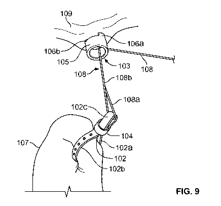
With reference to FIG. 9, the exemplary anchor 103 or 203 is attached by
positioning
the distal tip of the D-shaped cannula 130 on the abdominal wall 109, the
clinician's hand
CA 02803845 2012-12-21
WO 2012/006153 PCT/US2011/042353
usually palpates on the outside of the abdominal wall 109, the cannula 101
pushes the anchor
103 or 203 axially distal off of the D-shaped cannula 130 as the anchor 103 or
203 penetrates
the tissue 109. As the anchor 103 or 203 is released from the D-shaped cannula
130, the at
least two sharpened thin legs 106a and 106b or 206a, 206b, 206c and 206d curve
in or away
from the center of the cylindrical body 105 or 205 and thus provide a
sufficient pull out force
as well as protect the clinician from injury.
FIG. 9 further depicts the exemplary grasper 102 and anchor 103 in operation.
Specifically, the grasper 102 has clinched the organ 107 and has been locked
in position by
the locking mechanism 104. Additionally, the anchor 103 has been released from
the cannula
101 and the preformed at least two sharpened thin legs 106a and 106b have
returned to their
preformed shape in order to provide a sufficient pull out force. It should be
noted that the
grasper 102 is movably secured to the anchor 103 by the suture 108. The
cannula 101 is
retracted from the port trailing the suture 108 which keeps all the components
tethered and
allows the clinician to retract the organ 107 by increasing the tension on the
suture 108. As
shown therein, grasper 102 is fixed with respect to tissue/organ 107 with
suture 108 looped
through a U-shaped extension region 102c thereof. In the exemplary
implementation of
FIG. 9, suture 108 defines a loop region 108a that interacts with U-shaped
extension region
102c of grasper 102, such loop region extending to suture junction 108b. A
single suture
strand 108 extends from suture junction 108b and extends through anchor 103
which is fixed
relative to a second tissue location 107a, e.g., a peritoneal wall of the
patient. The legs of
anchor 103 are positioned within second tissue location 107a, e.g., in a
crossing orientation.
The free end of suture 108 generally extends through the abdominal wall, e.g.,
through an
access port (not pictured), and permits manipulation of tissue/organ 107
relative to anchor
103. The deployment tool has been withdrawn from the surgical field. The
suture 108 can be
secured outside the port with a clamp or other appropriate means. At the end
of the surgery,
16
CA 02803845 2012-12-21
WO 2012/006153 PCT/US2011/042353
the grasper 102 will be removed with the organ 107 (in the case of a gall
bladder removal).
The anchor 103 can be removed by gripping it with a 5 mm grasper (not shown)
and pulling
along the axis of the anchor 103 to remove it from the abdominal wall. Both
parts of the
tissue retractor assembly 100 can be removed through the abdominal incision
created by the
introduction of the port.
Now turning to FIG. 10, an alternate tissue retractor assembly 300 is depicted
in
accordance with the present disclosure. In the exemplary embodiment of FIG.
10, the tissue
retractor assembly 300 includes a cannula 301 which houses an anchor 302, a
wire form 303
and a grasper 304. The anchor 302 is configured and dimensioned to be deployed
from the
cannula 301 and attached to an abdominal wall anterior to an organ. The anchor
302 includes
an outer tube 314 and a central shaft 309, the central shaft 309 further
including at least two
barbs 308a and 308b, depicted in FIG. 11B, which are configured to be deployed
from the
outer tube 314 when the central shaft 309 is axially pulled. Additionally, the
anchor 302
includes a retractable sharp tip 306 which retracts into the outer tube 314
when the central
shaft 309 is pulled axially to deploy the at least two barbs 308a and 308b.
The wire form 303
may be fabricated as a coiled spring configured and dimensioned to deploy out
of a distal end
of the cannula 301 and expand radially. Further, the wire form 303 is secured
and adjusted
relative to the anchor 302 by a length of suture 311. Lastly, the grasper 304
is configured and
dimensioned to extend out of the distal end of the cannula 301 and through the
wire form
303, grasp tissue 313 (depicted in FIG. 13), and retract into the distal end
of the cannula 301
to pull tissue 313 into the wire form 303. The grasper 304 may be a pediatric-
type grasper
with specialized jaws 305.
Still with reference to FIG. 10, the tissue retractor assembly 300 is based
upon a 5 mm
cannula 301 commonly used in the design of laparoscopic surgical tools. The
cannula 301
contains both the anchor 302 and the wire form 303 used to grasp the organ or
tissue 313.
17
CA 02803845 2012-12-21
WO 2012/006153 PCT/US2011/042353
The components of the cannula 301 are arranged coaxially with the anchor 302
in the center
with a fully functional 3 mm grasper 304 proximal to the anchor 302. Separated
by a cannula
wall 315, the wire form 303 is compressed into an annular ring 316 surrounded
by the outer
cannula wall 317.
The anchor 302 is constructed in two pieces, the outer tube 314 which forms
the body
of the anchor 302 and internal to the outer tube 314, the central shaft 309
which includes the
retractable sharp tip 306 and at least two barbs 308a and 308b integrated that
can be deployed
by pulling the central shaft 309 of the anchor 302 proximal to the retractable
sharp tip 306.
The anchor 302 may be fabricated from metal or plastic.
With reference to FIGS. I IA-C, the tissue retractor assembly 300 is depicted
at
progressive steps of securing the anchor 302 to the abdominal wall 312 after
the tissue
retractor assembly 300 has been introduced into an access port (not shown).
Initially, the
distal tip of the cannula 301 is positioned on the abdominal wall 312. The
clinician's hand
usually palpates on the outside of the abdominal wall 312. A specially
designed 3 mm
grasper 304, commonly used in pediatric laparoscopic procedures, is utilized
to push the
anchor 302 axially distal to penetrate the tissue 312. The 3 mm grasper 304
then retracts in
order to retract the sharp tip 306 and deploy the at least two barbs 308a and
308b, as depicted
in FIGS. 11A and 11B. Specifically, the at least two barbs 308a and 308b
deploy through
openings 307a and 307b in the outer tube 314 of the anchor 302. The at least
two barbs 308a
and 308b dramatically increase the holding force of the anchor 302 in the
abdominal wall
312. As depicted in FIG. 11 C, the anchor 302 further has a suture 311
attached to the
proximal end and the cannula 301 trails the suture 311 from the distal tip.
The suture 311
may be attached with respect to the proximal end of the central shaft 309 of
the anchor 302
by a ring 310 or similarly shaped component.
18
CA 02803845 2012-12-21
WO 2012/006153 PCT/US2011/042353
With reference to FIGS. 12A-E, the tissue retractor assembly 300 is depicted
at
progressive steps of grasping an organ 313 after the tissue retractor assembly
300 has been
introduced into the port and the anchor 302 has secured to the abdominal wall
312.
Generally, a clinician has a multipurpose use 5 mm grasper (not shown) in the
surgical field
during the procedure, The 5 mm grasper manages the tissue of the organ in
question during
the grasping of the organ. The wire form 303 is in essence a specifically
designed coiled
spring which may have features such as surface roughness or barbs along the
interior wire
surface to enhance the gripping of the organ 313. As depicted in FIG. 12A, the
wire form
303 is pushed out of the distal end of the cannula 301 and expands radially to
enable a larger
diameter profile which can accommodate more tissue of the organ 313. The 3 mm
grasper
304 is then extended from the distal end of the cannula 301 to reach through
the wire form
303, grasp the organ 313 and retract back into the distal end of the cannula
301 to pull tissue
of the organ 313 into the wire form 303, which will grip the organ 313 by
virtue of the forces
generated between the surfaces of the wire form 303 and organ 313. With
reference to FIG.
12E, once the wire form 313 has been secured around the organ 313, the wire
form 303 is
secured and adjusted relative to the anchor 302 by a length of suture 311.
Specifically, the
length of suture 311 is attached to the wire form 303, extends to the ring 310
of the anchor
302, as depicted in FIG. 11C, and is attached to the cannula 301.
With reference to FIG. 13, the exemplary wire form 303 and anchor 302 are
depicted
in operation. Once the wire form 303 and anchor 302 are attached to the organ
313 and
abdominal wall 312, respectively, the cannula 301 is retracted from the access
port trailing
the length of suture 311, which keeps all the components tethered and allows
the clinician to
retract the organ 313 by increasing the tension on the length of suture 311.
The length of
suture 311 can be secured outside the port with a clamp or other appropriate
means (not
shown). At the end of the surgery, the grasper 303 will be removed with the
organ 313 (in
19
CA 02803845 2012-12-21
WO 2012/006153 PCT/US2011/042353
the case of a gall bladder removal). Removal of the anchor 302 will require
the
reintroduction of the cannula 301, which contains the 3 mm grasper 304. A 5 mm
grasper
could be used to grip the outer tube 314 of the anchor 302, while the 3 mm
grasper 304 is
used to attach to the central shaft 309 of the anchor 302 and push distally to
retract the at least
two barbs 308a and 308b to allow the anchor 302 to be removed from the
abdominal wall
312. The anchor 302 could be retracted into the cannula 302 or removed through
the 5 mm
port individually. Both components of the cannula 301 may also be removed
through an
abdominal incision created by the introduction of the access port as both are
tethered to the
length of suture 311.
Now turning to FIGS 14 and 15, an alternate tissue retractor assembly 400 is
depicted
in accordance with the present disclosure. FIG. 15 provides a partial view of
the alternate
tissue retractor assembly 400 for a more convenient depiction of the internal
components of
the tissue retractor assembly 400. In the exemplary embodiment of FIGS. 14 and
15, the
tissue retractor assembly 400 includes a cannula 401, which houses an anchor
405 and a
grasper 403. The grasper 403 is comprised of a loop of suture 403a with a one
way locking
toggle 402, configured and dimensioned to be released distally from the
cannula 401.
Specifically, the loop of suture 403a is configured and dimensioned to grasp
tissue of an
organ 406, retract into the distal end of the cannula 401 and tighten around
the tissue of the
organ 406. The anchor 405 includes a back span 412, torsion springs 409 and an
axial
connection between the back span 412 and torsion springs 409. The anchor 405
further
includes two sharpened legs 405a and 405b configured and dimensioned to deploy
from a
distal end of the cannula 401. The grasper 403 is secured and adjusted
relative to the anchor
405 by a length of suture 411.
Further with reference to FIGS. 14 and 15, the tissue retractor assembly 400
is based
upon a 5 mm cannula 401 commonly used in the design of laparoscopic surgical
tools. The
CA 02803845 2012-12-21
WO 2012/006153 PCT/US2011/042353
cannula 401 contains both the grasper 403 to attach to an organ 406 and an
anchor 405, as
well as a system to deploy each. The components of the cannula 401 are
arranged with both
the grasper 403 and anchor 405 along the axis of the cannula 401 with the
grasper 403 below
the anchor 405, which may be fabricated as a spring clip. The cannula 401 may
further
include a slot to allow the deployment of the loop of suture 403a. The cannula
401 further
includes features to aid in the delivery and firing or deployment of the
grasper 403 and
anchor 405.
With reference to FIGS. 16A-D, the tissue retractor assembly 400 is depicted
at
progressive steps of grasping an organ 406 after the tissue retractor assembly
400 has been
introduced into an access port (not shown). The grasper 403 is a suture based
organ grasper
including a loop of suture 403a with a one way locking toggle 402. The one way
locking
toggle 402 may be fabricated as a small molded plastic part which allows the
loop of suture
403a to be pulled through in one direction, but stops the loop of suture 403a
from loosening.
The loop of suture 403a may also be a ribbon or similar structure to increase
friction or
distribute force more evenly. Additionally, the loop of suture 403a may have
surface
features, i.e., small cuts or barbs, on its diameter to increase the friction
of the loop of suture
403a to the organ 406 and reduce the possibility of slipping. The loop of
suture 403a is held
flat in the cannula 401 by a hook 404 which is in the loop of suture 403a and
holds tension on
the loop of suture 403a in the cannula 401.
Still with reference to FIGS. 16A-D, in order to attach the loop of suture
403a to the
organ 406, the cannula 401 would be introduced through an access port and
placed near the
attachment site. The loop of suture 403a would be moved distal in order to
produce slack in
the loop of suture 403a. Generally, the clinician has a multipurpose use 5 mm
grasper 413 in
the surgical field during the procedure. The 5 mm grasper 413 manages the
tissue of the
organ 406 in question during the grasping of the organ 406. The 5 mm grasper
513 would be
21
CA 02803845 2012-12-21
WO 2012/006153 PCT/US2011/042353
used to pull the tissue of the organ 406 through the loop of suture 403a. The
delivery portion
of the cannula 401 would pull the free end of the loop of suture 403a through
the one way
locking toggle 402 to tighten the loop of suture 403a around the tissue of the
organ 406. By
retracting the hook 404 and withdrawing the cannula 401, the one way locking
toggle 402
and grasper 403 assembly could be released from the cannula 401. The free end
of the loop
of suture, a length of suture 411, would be trailed out of the distal end of
the cannula 401
while approaching the attachment point for the anchor 405 to be attached to
the abdominal
wall 410.
With reference to FIGS. 17A-D, the tissue retractor assembly 400 is depicted
at
progressive steps of securing the anchor 405 to the abdominal wall 410 after
the tissue
retractor assembly 400 has been introduced into an access port (not shown) and
after the
grasper 403 has been secured around the organ 406. The anchor 405 may be
fabricated as a
wire form constructed from a single piece of wire. The wire is a form which
has a
substantially symmetrical structure, consisting of a back span 412, torsion
springs 409 and
axial connections between the elements. The anchor 405 has a structure similar
to the
normally closed springs used in the typical construction of cloths pins. The
anchor 405
further includes two sharpened legs 405a and 405b which are not connected by a
cross
member and are sharpened to facilitate tissue penetration. The anchor 405 is
normally closed
and resides in the cannula 401 in a tray 413, or similar structure, for
deployment.
Still with reference to FIGS. 17A-D, the deployment of the anchor 405 requires
that a
device internal to the cannula 401 push the anchor 405 distal enough that the
stripping feature
407 on the cannula 401 can wedge under the two sharpened legs 405a and 405b of
the anchor
405. The tray 413 is then retracted proximally, which positions the two
sharpened legs 405a
and 405b of the anchor 405 to penetrate the abdominal wall 410. Specifically,
the cannula
401 includes the stripping feature 407 and two slits 408a and 408b, which are
dimensioned
22
CA 02803845 2012-12-21
WO 2012/006153 PCT/US2011/042353
and configured to allow the two sharpened legs 405a and 405b of the anchor 405
to deploy
from the distal end of the cannula 401 by an internal retracting mechanism
when the anchor
405 has been partially deployed from the distal end of the cannula 401.
Therefore, while the
anchor 405 is normally closed in the tray 413, the two sharpened legs 405a and
405b can
deploy from the cannula 401 through the two slits 408a and 408b in order to
properly
penetrate and attach to the abdominal wall 410.
Further with reference to FIGS. 17A-D, the distal tip of the cannula 401 is
positioned
adjacent to the abdominal wall 410. The clinicians hand usually palpates on
the outside of
the abdominal wall 410. The clinician would push the cannula 401 anterior,
while pulling the
cannula 401 and anchor 405 proximally. This would cause the two sharpened legs
405a and
405b of the anchor 405 to snag and penetrate the abdominal wall 410. The
anchor 405 would
then be released from the cannula 401 by retracting the cannula 401 and
pushing the tray 413
distal. The closing action of the anchor 405 and the direction of the tension
applied by the
length of suture 411 would increase the holding force of the anchor 405.
With reference to FIG. 18, the exemplary anchor 405 and grasper 403 are
depicted in
operation. The cannula 401 is retracted from the access port trailing the
length of suture 311,
which keeps all parts tethered and allows the clinician to retract the organ
406 by increasing
the tension on the length of suture 411. The length of suture 411 can be
secured outside the
port with a clamp or other appropriate means. At the end of the surgery, the
grasper 403 may
be removed with the organ 406 (in the case of a gall bladder removal). The
grasper 413,
depicted in FIG. 16C, may further be used to grip the back span 412 of the
anchor 405 and
push away from the entry direction, thereby allowing the anchor 405 to be
easily removed.
The normally closed nature of the anchor 405 would render the two sharpened
legs 405a and
405b safe in the abdominal cavity. Both the anchor 405 and grasper 403 of the
tissue
23
CA 02803845 2012-12-21
WO 2012/006153 PCT/US2011/042353
retractor assembly 400 may also be removed through the abdominal incision
created by the
introduction of the access port as both are tethered to the suture.
Now turning to FIG. 19, an alternate tissue retractor assembly 500 is depicted
in
accordance with the present disclosure. In the exemplary embodiment of FIG.
19, the tissue
retractor assembly 500 includes a cannula 501, which houses a first grasper
502 and second
grasper 503. The first grasper 502 may be fabricated as a first clip
configured and
dimensioned to be deployed axially from a distal end of the cannula 501 and is
defined by a
C-shaped form after deployment from the cannula 501. The second grasper 503
may be
fabricated as a second clip configured and dimensioned to be deployed axially
from the distal
end of the cannula 501 and is also defined by a C-shaped from after deployment
from the
cannula 501. The first grasper 502 is further secured and adjusted relative to
the second
grasper 503 by a length of suture 504, which is pre-threaded through the first
and second
grasper 502 and 503 and into the cannula 501.
The tissue retractor assembly 500 of FIG. 19 is based upon a 5 mm cannula 501
commonly used in the design of laparoscopic surgical tools. The cannula 501
deploys the
first and second grasper 502 and 503, respectively, by pushing them
sequentially out of the
distal tip of the cannula 501. The first and second grasper 502 and 503 would
be pushed
forward by a rod or cannula 501 sliding axially and with a force supplied by a
screw or gear
driven mechanism (not shown). The first and second grasper 502 and 503 may be
fabricated
from metal, plastic or a combination of materials that are formed in either a
"C" or "U" shape
which is normally closed, i.e., Raney type clips. The first and second grasper
502 and 503
would have a first and second back span 505 and 506, respectively, for guiding
or attaching a
length of suture 504. The first and second grasper 502 and 503 may
additionally have
gripping features, i.e., teeth, points, chevrons, 502a, 502b, 503a and 503b,
at the open tips or
on the inside surface to aid in gripping tissue. The first and second grasper
502 and 503 for
24
CA 02803845 2012-12-21
WO 2012/006153 PCT/US2011/042353
the organ 507 may further be coated with a rubber, have surface features or a
shape that is
advantageous to grasping without damaging the organ 507. Additionally, the
second grasper
503, which is to be attached to the abdominal wall 509, may have more
aggressive gripping
features 503a and 503b, i.e., aggressive teeth or sharp points, to attach to
the abdominal wall
509. The first grasper 502, however, which is to be used to grasp the organ
507, may have
atraumatic teeth at the gripping features 5 02a and 5 02b, in order to prevent
damage to the
organ 507.
Still with reference to FIG. 19 and further with reference to FIGS. 20A-E, the
tissue
retractor assembly 500 is depicted at progressive steps of securing the first
grasper 502 to the
organ 507 and securing the second grasper 503 to the abdominal wall 509 after
the tissue
retractor assembly 500 has been introduced into an access port (not shown).
The first and
second grasper 502 and 503, as they reside in the cannula 501, would be fully
opened such
that they are nearly flat. The first and second grasper 502 and 503 are loaded
into the cannula
501 for purposes of introduction into an access port. Once inside the port and
at the organ
507 to be grasped, the first and second grasper 502 and 503 may be deployed.
Generally, the
clinician has a multipurpose use 5 mm grasper 508, depicted in FIG. 20A, in
the surgical field
during the procedure. The 5 mm grasper 508 manages the tissue of the organ 507
in
question. The distal end of the cannula 501 is placed near the organ 507 and
the first grasper
502 is pushed out of the cannula 501 distally by a rod or shaft which is
driven by a screw or
gear mechanism (not shown). The first grasper 502 will be pushed out
approximately
halfway to allow the clinician to position the first grasper 502 and then the
first grasper 502
would be deployed. The shape of the cannula 501 and features at the tip would
help to
manage the dynamic nature of the first grasper 502 deployment. This design
also offers the
possibility of deploying multiple graspers onto the organ 507 as necessary
(not shown). The
CA 02803845 2012-12-21
WO 2012/006153 - - --PCT/US2011/042353
cannula 501 trails a length of suture 504 that is tethered to the first
grasper 502 placed on the
organ 507.
Further with reference to FIGS. 20A-E, the second grasper 503 is deployed next
to
allow the organ 507 to be retracted. The second grasper 503 may have the same
overall
shape and function as the first grasper 502. The second grasper 503 may
include more
aggressive features on the gripping features 503a and 503b of the insufflated
abdominal wall
509. The gripping features 503a and 503b may also be sharpened to the pint of
forming
penetrating features. The cannula. 501 is advanced to the abdominal wall 509
and the general
use 5 mm grasper 508 is used to manage the tissue of the abdominal wall 509.
The second
grasper 503 is deployed in a substantially similar method as the first grasper
502 attached to
the organ 507.
With reference to FIG. 21, the exemplary first grasper 502 and second grasper
503 are
depicted in operation. The cannula 501 is retracted from an access port
trailing the length of
suture 504 which keeps all the components tethered and allows the clinician to
retract the
organ 507 by increasing the tension on the length of suture 504. The length of
suture 504
may be secured outside the port with a clamp or other appropriate means. At
the end of the
surgery, the first grasper 502 on the organ 507 may be removed with the organ
507 (in the
case of a gall bladder removal). Depending on the geometry of the second
grasper 503,
removal of the second grasper 503 may require a specific tool which would be
integrated into
the cannula 501 or be a separate tool. If integrated into the cannula 501, the
tool could be
reintroduced to engage the second grasper 503 in order to remove it without
damaging the
tissue of the abdominal wall 509. Both the first and second grasper 502 and
503 may be
removed through the abdominal incision created by the introduction of the
access port.
Although the present disclosure has been described with reference to exemplary
embodiments and implementations, it is to be understood that the present
disclosure is neither
26
CA 02803845 2012-12-21
WO 2012/006153 PCT/US2011/042353
limited by nor restricted to such exemplary embodiments and/or
implementations. Rather,
the present disclosure is susceptible to various modifications, enhancements
and variations
without departing from the spirit or scope of the present disclosure. Indeed,
the present
disclosure expressly encompasses such modifications, enhancements and
variations as will be
readily apparent to persons skilled in the art from the disclosure herein
contained.
27