Note : Les descriptions sont présentées dans la langue officielle dans laquelle elles ont été soumises.
- 1 -
METHOD FOR CONTROLLING AND ACCELERATING DIFFERENTIATION
OF STEM CELLS USING GRAPIIENE SUBSTRATES
RELATED APPLICATION
This application claims the benefit of U.S. Provisional Application No.
61/362,506, filed July 8, 2010.
BACKGROUND OF THE INVENTION
Stem cell scaffolds, which can be both 2D and 3D in nature, have been
fabricated to mimic the intrinsic characteristics of natural substrates such
as muscle,
bone and cartilage. (Jaiswal, N., et al., J. Cell Biochem. 1997, 64, 295;
Engler, A. J.,
et al., Cell 2006, 126, 677; Reilly, G. C., et al., J Biomech. 2010, 43, 55).
Recently,
both the lithographic patterning of suitable surfaces such as
polydimethylsiloxane
(PDMS), polymethyl methacrylate (PMMA), self-assembled titanium dioxide (TiO2)
rod arrays and functionalized carbon nanotubes have been explored. (Kim, S.
J., et
al., J. Mater. Sci: Mater. Med. 2008, 19, 2953; Dalby, M. J., etal., Nat.
Mater.
2007, 6, 997; Oh, S., et al., Proc. Natl. Acad. Sci. USA 2009, 106, 2130;
Nayak, T.
R., et al., ACS Nano, 2010, 4, 7717). While there have been tremendous
advances in
this field, many challenges still remain. In particular in the field of bone
tissue
engineering, almost all artificial materials require the administration of
multiple
growth factors to promote human mesenchymal stem cell (hMSC) differentiation
and bioactive implants still suffer from severe limitations including
potential
pathogenic infections, low availability and high costs. In addition, many
modem
approaches also face further challenges when it comes to scalability and
compatibility with implants.
Therefore, there remains a significant need for development of more
biocompatible scaffolds that allow for better scalability of the biocompatible
scaffold materials and compatibility with implants.
CA 2804647 2017-11-02
- 2 -
SUMMARY OF THE INVENTION
In a first main aspect, the invention relates to a method of directing stem
cell
differentiation in the absence of growth factors or external stimulation,
comprising:
placing stem cells on a graphene substrate and exposing to a culture media for
a
period of time sufficient to allow the stem cells to differentiate in cells of
interest in
the absence of growth factors or external stimulation. The graphene can be
single layer,
multi-layer, two dimensional or three dimensional. In one embodiment, the
culture
media is an osteogenic medium.
In one embodiment, the stem cells are mesenchymal stem cells. In another
embodiment, the stem cells are progenitor cells.
In another aspect, the invention relates to a method of repairing and
improving
bone tissue function comprising directing stem cell differentiation by placing
the stem
cells on graphene, e.g., single layer or multi-layer, in the absence of growth
factors or
external stimulation and transplanting the graphene with the stem
cells in the tissue at the site of repair.
In a further aspect, the invention relates to a composition for accelerating
differentiation of human mesenchymal stem cells comprising single layer
graphene on
an implantable, biocompatible scaffold for support of tissue growth.
In another aspect, the invention relates to the use of graphene as a substrate
for stem cell differentiation.
The present invention provides graphene as a low cost, biocompatible
scaffold that does not hamper the proliferation of human stem cells and
accelerates
their specific differentiation into various cell types.
BRIEF DESCRIPTION OF THE DRAWINGS
The foregoing will be apparent from the following more particular
description of example embodiments of the invention, as illustrated in the
accompanying drawings in which like reference characters refer to the same
parts,
throughout the different views. The drawings are not necessarily to scale,
emphasis
instead being placed upon illustrating embodiments of the present invention.
CA 2804647 2019-11-26
- 3 -
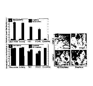
FIG. 1 (a) is a graph of cell viability of hMSCs grown on different substrates
including silicon wafer with 300nm SiO2 (Si/SiO2), polyethylene terephthalate
(PET),
and polydimethylsiloxane (PDMS) in percentage normalized to cover slips used
as a
reference. NG is no graphene and G is graphene. FIG. 1 (b-i) and inset show
cell
morphology of hMSCs grown on standard cover slips and on glass slide, Si/S102,
PET
and PDMS with or without graphene. Scale bars are 1001.1.m.
FIG. 2 shows Raman analyses of (a) graphene on Si/SiO2 after removal of cells
and cleaning with acetone, (b) graphene on Si/SiO2 after removal of cells and
(c)
Si/SiO2 after removal of cells.
FIG. 3 shows immunostaining of cells growing on Si/SiO2, PDMS and PET
without BMP-2 growth factor. Cells are stained with DAPI and either CD-44,
MAP2, Desmin or Osteocalcin (OCN) as indicated. (a-d) Cells growing on
Si/SiO2,
without graphene showing presence of CD-44, and with graphene showing presence
of
OCN. (e-h) Cells growing on PDMS without graphene showing some MAP2
immunostaining , and with graphene showing staining of OCN. (i-1) Cells
growing on
PET without graphene showing some staining of desmin, and with graphene
showing OCN immunostaining. Scale bars are 100 pm.
FIG. 4 (a) is an optical image of a 1 xl cm graphene coated Si/SiO2, showing
the graphene boundary. FIG. 4 (b) shows highly fluorescent osteocalcin (OCN)
marker indicating bone cell formation on the same chip only on the graphene
coated
area.
FIG. 5 shows a quantitative, functional proof of graphene-mediated hMSCs'
differentiation into osteocytes via Alizarin Red assay, N in the presence
(dark gray
bars) or absence (black bars) of graphene, (h) in the absence (a) or presence
(b) of
additional growth factor BMP-2. NG is no graphene and G is graphene.
Conventional
plain cover slips were used as a positive control. (c-f) Qualitative staining
via alizarin
red assay of calcium deposits on PET substrates due to osteogenesis. (c) PET
without
BMP-2 and without graphene; (d) PET without BMP-2 and with graphene; (e) PET
with BMP-2 and without graphene; (f) PET with both BMP-2 and graphene. Scale
bars are 100 tun.
CA 2804647 2019-11-26
-3a-
FIG. 6 shows time-dependent immunostaining of hMSCs growing on Si/SiO2
substrates either treated with BMP-2 or coated with graphene. Experiments were
performed from 1 hour to 15 days. (Left) CD-44, marker for stem cells,
decreased
over time and completely disappeared by Day 7. (Center) (31-integrin, marker
for
CA 2804647 2019-11-26
CA 02804647 2013-01-07
WO 2012/005699 PCT/SG2011/000247
- 4 -
cell-substrate adhesion, increased over time, reaching its peak by Day 15.
(Right)
OCN, marker for bone cells, became visible at Day 4 and very intense by DAY 7.
Scale bars are 100 m.
DETAILED DESCRIPTION OF THE INVENTION
The invention pertains to methods of directing differentiation of stem cells
when cultured on graphene, in the presence of osteogenic medium that does not
require further supplementation of additional growth factors or replenishment
of
growth factors. The methods and compositions of the invention may be used for
repairing or improving tissue function. The invention is based, in part, upon
data
reported herein showing that graphene provides a biocompatible scaffold that
does
not hamper the proliferation of stem cells in stem cell medium and directs the
stem
cells to specifically differentiate into bone cell types once cultured in
osteogenic
medium.
Results showed that mono-atomic graphene coated substrates accelerated cell
differentiation to a higher extent than un-coated substrates or cover slips.
In contrast
to other materials, graphene does not require additional chemical inducers
(e.g.,
growth factors including BMP-2) to be continuously added or replenished to the
osteogenic medium to achieve cell differentiation. In fact, direct comparison
of the
effects of graphene and growth factors on stem cell differentiation showed
that
differentiation rates with graphene were comparable to the ones achieved with
common growth factors.
In one aspect, the invention pertains to a method for directing the
differentiation of stem cells into cells of interest using graphene as a
scaffold for
accelerated differentiation. The term "directing differentiation of a stem
cell" as
used herein is taken to mean causing a stem cell to develop into a specific
differentiated cell type. The stem cells are grown on a graphene substrate in
an
appropriate culture medium under conditions that do not require implementation
with growth factors or external stimulation, or combinations thereof. In
certain
embodiments of the invention, the stem cells or progenitor cells on graphene
are
grown and differentiated in vitro.
CA 02804647 2013-01-07
WO 2012/005699 PCT/SG2011/000247
- 5 -
In another aspect, the invention pertains to a method for accelerating stem
cell differentiation by culturing stem cells on the graphene substrate. The
term
"acceleration" as used herein means acceleration of stem cell differentiation
on
graphene and in the presence of osteogenic media as compared to
differentiation
only in osteogenic media.
The results reported herein show that graphene provides unique properties
that enhance the differentiation of stem cells into cells of interest,
particularly bone
cells. These differentiated cells on the graphene substrate can be
incorporated into
bioimplants having improved biocompatibility.
Graphene is a two dimensional sheet of carbon that has highly desirable
physical properties for use in tissue regeneration and medical devices.
Graphene is
the strongest material known having a Young's modulus of 0.5 ¨ 1 TPa, yet it
is
extremely flexible and not brittle. Graphene can be transferred onto any flat
or
irregular shaped surface and graphene-coated, flexible, supporting substrates
can be
easily bent into any shape required. Being only one atom thick, yet fully
continuous
it also introduces the minimum amount of non-biodegradable material preventing
inflammatory or other immune responses seen with other non-biologic materials.
Graphene also serves as an impenetrable gas barrier and can hermetically seal
the
substrate or implant material, protecting it from any degradation due to
external
factors. As a result, graphene may significantly strengthen bone structures or
eventual implants in addition to serving as a substrate for tissue
regeneration and/or
repair.
High-quality, continuous graphene sheets can be produced on a large scale
through chemical vapor disposition on copper foil. (Bae, S., et al., Nat.
Nano, 2010,
5, 574). "Chemical vapor deposition (CVD)" refers to a chemical process used
to
produce high-purity, high-performance solid materials where substrate is
exposed to
one or more volatile precursors, which react and/or decompose on the substrate
surface to produce the desired deposit. For example, graphene can be produced
by
exposing copper foils to hydrogen and methane at high temperatures which react
to
form single layer graphene that is deposited on the metal surface. Graphene
can be
directly deposited onto any substrate, without the need to intercalate any
additional
CA 02804647 2013-01-07
WO 2012/005699
PCT/SG2011/000247
- 6 -
material between graphene and substrate. These substrates include, but are not
limited to, quartz, polydimethylsiloxane (PDMS), polyethylene terephthalate
(PET),
and silicon wafer with 300nm SiO2 (Si/SiO2). In terms of biomedical
applications,
the substrate of interest could consist of the metal implant or the defective
tissue
itself.
Substrates that may be used for growing graphene include, but are not
limited to, copper (Cu), nickel (Ni), silicon carbide (SIC) and may include
also non-
metal or non-oxide substrates. Substrates are not limited to planar substrates
but can
be three dimensional forms of nickel, copper or any other material
facilitating the
growth of graphene.
Classifications of graphene include, but are not limited to, mechanical
exfoliation of graphene, CVD grown graphene, chemically derived graphene
oxide,
reduced graphene oxide, functionalized graphene, hydrogenated graphene,
fluorinated graphene, chemically modified graphene, embedded graphene, silicon
carbite based graphene, two-dimensional graphene and three-dimensional
graphene.
In one embodiment, the graphene is three-dimensional graphene.
"Chemically modified graphene" is graphene whose structure has been
chemically altered or modified. Chemical modifications can include, but are
not
limited to, covalent or ionic linking of agents to the graphene structure or
addition or
substitution of substituents that may alter the properties of graphene.
Examples of
agents that may be linked to the graphene include, but are not limited to,
growth
factors, drugs (e.g., anticoagulants, such as heparin, antibiotics),
antibodies, steroids,
proteins, amino acids, hormones, peptides or enzymes. Such agents can augment
of
enhance the healing process or tissue repair.
"Embedded graphene" is intended to embrace any type of graphene where a
biochemical agent is incorporated into the graphene during the coating of the
substrate or thereafter. Examples of biochemical agents that can be embedded
into
the graphene are those described above.
The graphene substrate useful in the present invention consists of many
micrometer ripples and wrinkles and has a high Young's modulus. The ripples
themselves provide local curvature further enhancing the reactivity of the
graphene
CA 02804647 2013-01-07
WO 2012/005699 PCT/SG2011/000247
- 7 -
sheets while the high Young's modulus provides the flexibility for the out-of
plane
deformations which contribute to graphene's cellular differentiation
properties. As a
result, the ripple and wrinkles lead to the large scale disorder that plays a
role in
protein adsorption, cell adhesion, proliferation and differentiation.
In one embodiment, grapheme is multi-layer graphene. The term "multi-layer
graphene" refers to graphene that has multiple layers of single atomic
graphene on
individual graphene flakes. In one non-limiting embodiment, the graphene has
ten to
twenty layers. In another embodiment, the graphene has five to ten layers. In
yet
another embodiment, the grapheme has one to five layers. In another embodiment
of
the invention, the graphene is single layer graphene. As used herein, the term
"single
layer graphene" refers to a graphene monoatomic sheet that has less than or
about
5% two or three layer graphene. For example, graphene grown on copper is self
terminating producing single layer graphene that has less than 5% two and
three
layer grapheme flakes. In one non-limiting embodiment, the graphene has about
5%
two and three layer graphene. In another embodiment, graphene has less than 5%
two and three layer graphene.
According to the invention, a stem cell is cultured in the presence of
graphene. In one embodiment, the graphene is in direct contact with the cells.
In
another embodiment, the graphene is in contact with the culture media, and in
direct
contact with the cells. For example, stem cells are seeded on graphene coated
substrate and then placed in culture media.
A variety of stem cells of various types and stages of differentiation can be
used in the invention and include but are not limited to, for example,
totipotent,
pluripotent, multipotent and unipotent stem cells. In one embodiment of the
invention, the stem cell is an embryonic stem (ES) cell. In another embodiment
of
the invention, the stem cell is a progenitor stem cell. In yet another
embodiment, the
stem cell is a mesenchymal stem cell.
Of particular interest are mesenchymal stem cells (MSCs) which can
differentiate in vitro, in a variety of connective tissues or progenitor
cells, including,
but not limited to, mesodermal (osteoblasts, chondrocytes, tenocytes, myocytes
and
adipocytes), ectodermal (neurons, astrocytes) and endodermal (hepatocytes)
derived
CA 02804647 2013-01-07
WO 2012/005699 PCT/SG2011/000247
- 8 -
lineages. The term "mesenchymal stem cell" and "marrow stromal cell" are often
used interchangeably, so it is important to note that MSCs encompass
multipotent
cells from sources other than marrow, including but not limited to, muscle,
denial
pulp, cartilage, synovium, synovial fluid, tendons, hepatic tissues, adipose
tissue,
umbilical cord, and blood, including cord blood.
While stem cells exemplified herein are differentiated into bone cells,
differentiation into any desired "cell of interest" is contemplated. Examples
include,
but are not limited to, osteocytes, chondrocytes, adipocytes, muscles cells,
nerve
cells and cardiac myocytes. In one embodiment, the differentiated cell is a
chondrocyte. In another embodiment, the differentiated cell is an osteocyte.
In
another embodiment, the differentiated cell is a cardiac myocytes. In a
further
embodiment, the differentiated cell is a muscle cell. In yet another
embodiment, the
differentiated cell is a nerve cell. In another embodiment, the differentiated
cell is an
osteoblast. In another embodiment, the differentiated cell is an adipocyte. In
another
embodiment, the differentiated cell is a hepatocyte.
The invention also applies to a variety of stem cells of various types and
stages of differentiation, and cultured in media that promotes differentiation
toward
a particular type of cell. The term "culture media" as used herein means any
liquid
or solid preparation made specifically for the growth, storage or transport of
microorganisms or other types of cells. The variety of media that exist allow
for the
culturing of specific organisms and cell types, such as differential media,
selective
media, test media and defined media. In one embodiment, the culture medium is
chondrogenic. In another embodiment the culture medium is osteogenic. In
another
embodiment, the culture medium is myogenic. In another embodiment, the culture
medium is neurogenic. In another embodiment, the culture medium is adipogenic.
In
another embodiment, the culture medium is hepatogenic. For example, human
mesenchymal stem cells (hMSCs) can be placed on graphene in osteogenic media
to
obtain osteogenic differentiation.
Conventional osteogenic medium contains dexamethasone, which can lead to
osteogenic differentiation. However, it is usually administered in combination
with
other agents, growth factors or external stimulants to achieve differentiation
through
CA 02804647 2013-01-07
WO 2012/005699 PCT/SG2011/000247
- 9 -
a synergistic effect since differentiation in osteogenic medium occurs over
prolonged periods of time. "Growth factors" include naturally occurring
substances
capable of stimulating cellular growth, proliferation and cellular
differentiation. For
example, bone morphogenetic protein-2 (BMP-2) is a growth factor that plays an
important role in the differentiation of cells into bone and cartilage. As
used herein,
"external factors" or "external stimulants" are external sources of
mechanical,
acoustic or electromagnetic energy that can stimulate cellular proliferation
and
differentiation. For example, radiowaves or electromagnetic radiation can be
used to
supply cells with the sufficient energy needed to promote cellular growth.
According to the invention, the graphene can be employed not only in tissue
culture, but wherever it is desired to stimulate growth and/or repair of bone,
cartilage, muscle, or nervous tissue in a host. The stem cells can be cells
already
present at a particular location, or implanted, or injected. In one
embodiment, stem
cells are stimulated on graphene in vitro. In a further embodiment, progenitor
cells
are stimulated directly using graphene. In certain embodiments, the stem cells
seeded on graphene are implanted as part of a tissue or prosthesis or
treatment of
structures so destined for insertion or implantation into a host.
One example of such a structure is a matrix for bone or cartilage growth or
regeneration. Examples include, but are not limited to a demineralized bone
matrix
(e.g., composed primarily of collagen and non-collagenous proteins),
devitalized
cartilage matrix, or other artificial matrix for bone or cartilage repair.
Other porous
scaffolds (ceramics, metals, polymers and nano-reinforced) are osteoconductive
and
promote bone ingrowth, with osteoinductive properties provided by
incorporation of
peptides, hydroxyapetite and cytokines known to influence bone cells.
In one embodiment, collagen, particularly collagen type II, is used to
promote chondrogenic differentiation of stem cells on graphene. In another
embodiment, osteogenic matrix is used to promote osteogenic differentiation of
stem
cells on graphene. In another embodiment, graphene seeded with stem cells is
implanted at the regeneration site. In another embodiment, stem cells on
graphene
are incorporated into an implant or prosthesis. In yet another embodiment,
progenitor cells on graphene are incorporated into an implant or prosthesis.
In
CA 02804647 2013-01-07
WO 2012/005699
PCT/SG2011/000247
- 10 -
another embodiment, the implant is coated with graphene and osteogenic
differentiation is promoted at the implant site. In another embodiment, an
implant
made of TiO2 or any other medical implant material, is coated with graphene
and
osteogenic differentiation are promoted at the implant site. In another
embodiment,
the graphene is grown on the implant and differentiation is promoted at the
implant
site. In another embodiment, the graphene is a three-dimensional scaffold
serving as
implant material and differentiation is promoted by the -graphene implant. In
another
embodiment, graphene can be used as bonefilling material. Medical implant
materials include, but are not limited to, graphene, metal, metal alloy (e.g.,
stainless
steel or Cobalt Chrome), metal oxide (e.g., TiO2), oxide, ceramic, composite
materials and plastics.
Preferably, graphene would be directly implanted at the site of defective
tissue, to provide mechanical support while promoting stem cells growth and
proliferation in a particular cell lineage. Graphene offers the potential to
be further
functionalized and/or embedded with biochemical agents to enhance healing
process
and tissue repair. Also, graphene can be used as a temporary scaffold to
direct cell
differentiation into a specific lineage, after which, it could be separated
from the
differentiated cells and completely discharged.
Thus, the matrices can include bone- or cartilage-specific matrix components
and are populated with bone or cartilage progenitor cells, which are
stimulated
according to the invention.
The invention also provides for a composition for stimulating and/or
differentiating stem cells or progenitor cells. The compositions are suitable
for cell
growth and comprise stems cells on a graphene substrate exposed to culture
media.
In one embodiment, the composition comprises graphene coated or placed on a
biocompatible material. In another embodiment, the composition comprises stem
cells on a graphene coated biocompatible material. Biocompatible materials can
include natural or synthetic materials used to replace part of a living system
(e.g.,
tissue or organ replacement) or to function in intimate contact with living
tissue.
The method of the invention is also applied to the manufacture and use of
medical implants, such as an orthopedic or a dental implant. The implant can
be a
CA 02804647 2013-01-07
WO 2012/005699 PCT/SG2011/000247
- 11 -
metal implant, such as an artificial hip, knee, or shoulder, to which the bone
must
meld. Other examples include dental implants. The implants are prepared with
graphene attached surfaces that are to be fused to bone, providing an improved
surface that enhances growth of bone forming cells. The implant can also be
made of
a composite material such as a fiber composite. For example, orthopedic
implants
can be made from composite material strengthened by the addition of graphene.
The
implants can be implanted directly, or incubated with oSteoblasts from the
recipient
prior to implantation.
When implanted or injected, stem cell development is often governed by the
site of implantation or the site in the body to which the stem cell is home.
According
to the invention, differentiation of stem cells and progenitor cells can also
be
directed in vitro by selection of media components and/or matrix components.
For
example, cytokines, and growth factors that promote osteogenic differentiation
include various isoforms of bone morphogenetic protein (BMP) such as BMP-2, -
6,
and -9, interleukin-6 (IL-6), growth hormone and others. (See, e.g., Heng et
al.,
2004, J. Bone Min. Res. 19, 1379-94). Cytolcines and growth factors that
promote
chondrogenesis include various isoforms of TGF-(3 and bone morphogenetic
protein,
activin, FGF and other members of the TGF-13 superfamily. Osteogenesis of
chondrogenesis is favored by naturally occurring or synthetic cartiliage
extracellular
matrix (ECM) material. For example chondrogenesis is favored by naturally
occurring or synthetic ECM. Such an ECM can comprise collagenous proteins such
as collagen type II, proteoglycans such as aggrecan, other proteins and
hyaluronin.
(See, e.g., Heng et al., 2004, Stem Cells 22, 1152-67). Phenotypic markers
expressed
=
by cells of the various lineage are well known in the art.
The invention further provides kits for differentiating stem cells. The kits
comprise graphene for controlled and accelerated differentiation of the stem
cells.
The graphene can be provided separately from the stem cells or coated on the
containers used for culturing stems cells. In another embodiment, the kit
contains
graphene incorporated onto a support, such as a scaffold on or within which
stem
cells or progenitor cells are stimulated and/or differentiated. In a further
embodiment, the kits contain instructions on how to use the invention to
obtain
CA 02804647 2013-01-07
WO 2012/005699 PCT/SG2011/000247
- 12 -
stimulated or differentiated cells using graphene and the appropriate culture
media.
Optionally, the kits further contain media formulations selected to promote
differentiation to osteocytes, chondrocytes, or other differentiated cell
types.
Suitable media include, but are not limited to, adipogenic media, osteogenic
media,
chondriogenic media, myogenic media, neurogenic media, hepatogenic media
In one embodiment of the invention, the differentiated stem cells are used to
identify and/or isolate biological compounds, including but not limited to
proteins
and nucleic acids characteristic of the stimulated or differentiated state of
the cells.
Such biological compounds are useful for example, as markers of
differentiation and
as targets for antibodies and other agents. Fluorescent antibodies, specific
for
immunostaining of typical proteins produced by defined cell lines, can be used
to
confirm whether differentiation has occurred or not. A few examples are the
fluorescent antibody for CD-44 (which is typical of MSCs), or DESMIN (D-33,
specific for muscle cells), or antibody for MAP-2 (used as a marker for
neurons) or
OCN (specific for osteocytes) or131-integrin (protein produced when cells have
increased adhesion to the substrate underneath). As an example, hMSCs
incubated in
osteogenic media for 14 days, show the ability to bind OCN only in the
presence of
graphene-coated substrates, while they immunostain for CD-44 on cover slips or
uncoated substrates.
EXPERIMENTAL PROCEDURES
Substrate preparation
Graphene was grown on copper foils by chemical vapor deposition at
1000 C in a mixture of hydrogen and methane as discussed elsewhere. (Li, X.,
etal.,
Science 2009, 324, 1312). The graphene film was mechanically supported by a
thin
film of polymethyl methacrylate (PMMA) (Microchem) and the copper foil was
etched in a weak solution of ammonium persulfate (Sigma). The graphene coated
with PMMA was transferred to deionized water to remove residues and the
transfer
was completed by gently contacting graphene with the substrate and lifting it
out of
the water. To avoid any residues from the transfer process the samples were
left in
warm acetone for 12 hours followed by 3 hours in isopropanol. In a final step
the
Si/SiO2 substrates were annealed in Ar/H2 90/10 wt% for 7 hours at 300 C to
further
CA 02804647 2013-01-07
WO 2012/005699
PCT/SG2011/000247
- 13 -
reduce impurities in the graphene layer. However, note that Si/SiO2 without
the
additional step of annealing showed the same cell viability and induced stem
cell
differentiation at the same rate (data not shown).
Large-scale graphene used in this study was synthesized by the chemical
vapor deposition method on copper foils. After growth, copper was etched and
the
same batch of graphene was transferred to four distinct substrates used in
this study
according to methods discussed elsewhere. (Li, X., et al., Science 2009, 324,
1312)
The influence of graphene on stem cell growth was studied by investigating
four
distinct substrates with widely varying stiffness and surface roughness: (1)
polydimethylsiloxane (PDMS), (2) polyethylene terephthalate (PET), (3) glass
slide
and (4) silicon wafer with 300nm SiO2 (Si/SiO2). Plain cover slips without
graphene
were used as a control or reference for normalization. Atomic Force Microscopy
(AFM) was used to analyze the surface roughness of the various substrates with
and
without graphene coating.
Transferred to PET, PDMS, and Si/SiO2, the graphene sheet exhibits nano-
ripples with high density compared to graphene on glass slide. Despite being
only
one atom thick, on Si/SiO2 substrates with well-defined oxide thickness,
graphene
can be easily seen with a simple conventional optical microscope. First cell
viability
was studied with cells cultured in normal stem cell medium. Next, stem cell
differentiation was examined in cells cultured on conventional osteogenic
media.
Cell lines and markers
Human mesenchymal stem cells (hMSCs) were purchased from ATCC and
cultured in low-glucose Dulbecco's modified eagle medium (Sigma) supplemented
with 10% FBS (Invitrogen), 1% penicillin/streptomycin (Gibco), 1% Non-
essential
amino acids (Sigma) and 1% sodium pyruvate (Sigma). hMSCs at passage 2 were
used in this study. Osteogenic medium consisting of DMEM basal medium (Sigma)
added with dexamethasone, L glutamine, ascorbic acid and Beta-glycerophosphate
was prepared according to a known procedure. (Fahmi, H., et al.,
Osteoarthritis and
Cartilage 2002, 10, 845). FITC-Goat anti mouse antibody was purchased from
Biolegend, San Diego, California (USA). Markers (osteocalcin (OCN), CD44,
CA 02804647 2013-01-07
WO 2012/005699 PCT/SG2011/000247
- 14 -
Desmin (D33), MAP-2, 01-integrin) were purchased from Acris Antibodies GmbH
(Germany).
Cell viability and morphology mesenchymal cells in the presence of graphene.
hMSCs differentiation into osteogenic lineage.
hMSCs (20,000 cells/well (24 well plate)) were seeded on uncoated (control)
and graphene coated (test) chips and cultured in normal stem cell medium. Post
confluence (2 weeks), cells growing on each chip were transferred to new well
plate
and washed 3 times with 2 ml of PBS. 1 ml of PBS was added to each well
followed
by 51.1 of linM Calcein acetoxymethyl ester (Calcein AM) and incubated at room
temperature for 15 minutes. After removing the unbound stains, the chips were
inverted on to glass slides mounted with vectashield with 4,6'diamidino-2-
phenylindole (DAPI) (H 1200, Vector labs) and visualized under fluorescence
microscope (Nikon AZ-100 multipurpose microscope). Pictures were taken at 4
different positions of the chips and processed by image J software to count
the
number of viable cells to the number of nucleus as determined by staining with
DAPI. Cell viability was measured by comparing the cell numbers for each
substrate
with the cells counted on cover slips. In addition, (3-(4,5-Dimethylthiazol-2-
y1)-2,5-
=
diphenyltetrazolium bromide (MT) assays were carried out, in which
cytotoxicity
evaluation was based on the activity of enzymes to reduce MTT to formazan
dyes,
giving a purple colour. (Mosmann, T., J. Immun. Met. 1983, 65, 55).
Experiments
were carried out in triplicates, following the procedure reported in
supporting
document. The morphology of the hMSCs on different substrates was compared
according to the image as seen in the form of calcein AM staining (FIG. 1).
Cell cytotoxicity of graphene was tested by comparing cell counts for all four
substrates with and without graphene coverage and found that graphene does not
hamper stem cells' normal growth over the whole period of investigation (1-18
days). On the contrary, MTT assay showed that cells grew better on graphene
covered substrates in particular on the softest substrates, i.e. PDMS and PET.
From
FIG. 1 (a) it can be seen that, independent of the substrate, there is no
significant
difference (p>0.05) in cell viability between graphene-coated and uncoated
substrates. mn assays were also performed to confirm the cell viability data.
- 15 -
Again, regardless of the substrate, there was no difference (p>0.05) between
uncoated
and graphene-coated substrates, demonstrating that cell growth was indeed not
affected
by the presence of graphene. Note that cell viability is lower on PET and PDMS
independent of the presence of graphene.
A similar conclusion can be reached by just comparing cell morphology with
and without graphene (FIG. 1 (b-i)). In general, the presence of graphene did
not influence
the shape of the cells in comparison to uncoated substrates. Mesenchymal stem
cells
maintained their spindle-shape across glass slides and Si/SiO2 after 15 days
of incubation.
Here stem cells presented the usual elongated structure with noticeable
filopodia
extensions and cellular propagation fronts. In the case of PET and PDMS, cells
showed
rounded or irregular morphology, most probably due to poor adhesion to the
substrate.
This suggests that graphene does not hamper the normal growth of stem cells
and that the
incorporation of this material in implants or injured tissues would not affect
the
physiological conditions of the microenvironment. In fact, Raman measurements
and visual
inspection of the samples after cell incubation and subsequent removal clearly
showed that
the graphene sheets remained largely intact.
Raman spectra of graphene on Si/SiO2 after cell removal and subtraction of
the background, clearly show the G and 2D peaks, which represent the Raman
"fingerprints of graphene" (FIG. 2). Note also, that the absence of the D-peak
at
1350cm-1 indicates the lack of defects to the graphene crystal lattice
(Ferrari et al. PRL,
2006, 97, 187401). Optical images also clearly shows that the graphene sheet
remains
largely intact (images not shown).
Immunofluorescence of hMSCs
hMSCs at 20,000 cells/well (24 well plate) were seeded, osteoinduced and
incubated up to confluence (2 weeks) as reported above. The cells on all the
chips were
fixed by treating them with ice cold 50%/50% methanol/acetone. After 5
minutes,
methanol/acetone was removed and the chips were left open inside the laminar
hood to
be air dried. After the chips were completely dried, the fixed cells were
treated with 10%
FBS (blocking agent) in PBS for 20 minutes. The blocking agent was aspirated
out and 5
I of different antibodies to cellular markers (CD-44
CA 2804647 2019-11-26
CA 02804647 2013-01-07
WO 2012/005699 PCT/SG2011/000247
- 16 -
for hMSCs, OCN for osteoblasts, Desmin for muscle cells and MAP2 for neuronal
cells) were added on to separate chips (previously marked). After 1 hour the
cells on
the chips were extensively washed in Millie water for 5 minutes and then
rinsed
in PBS 1X for 5 minutes. After that, 100 ill of diluted (1/100) FITC-goat
antimouse
antibody were added on to each chip and incubated at room temperature. After
30
minutes the cells were washed with Millie water for 5 minutes and then rinsed
in
PBS 1X for 5 minutes. The chips were inverted on to glass slides mounted with
vectashield with DAPI (H 1200, Vector labs) and visualized under fluorescence
microscope (Nikon AZ-100 multipurpose microscope).
Osteogenic differentiation was evaluated over a time frame of two weeks.
Uncoated substrates were subjected to BMP-2 (75 ng/mL, added every 3 days) and
compared to graphene coated substrates at 1 hour and at Day 1,4, 7, 10 and 15
in
terms of binding to CD-44 (which stains hMSCs), 131-integrin (which indicates
cell-
substrate adhesion) and OCN (which indicates bone cells). The above mentioned
procedure was followed for the immunofluorescence and imaging purposes.
Next, specific markers were used to determine the conversion of hMSCs into
specific cell types when cultured in osteogenic media. Note that conventional
osteogenic medium does contain dexamethasone, which can lead to osteogenic
differentiation by itself. However, it is usually administered in combination
with
other agents and growth factors such as BMP-2 to achieve differentiation
through a
synergistic effect. In none of the un-coated substrates studied here, the
osteogenic
medium alone was sufficient to lead to osteogenic differentiation over the
whole
duration of the experiment (15 days). In the absence of graphene, stem cells
on
cover slips, on glass slides and on Si1Si02 did not differentiate: this was
demonstrated by immunofluorescent staining of two typical protein markers,
namely
CD-44 for hMSCs and osteocalcin (OCN) for osteoblasts (FIG. 3). These three
substrates showed a CD-44-positive staining and the absence of OCN. However,
once these stiff substrates were coated with graphene, hMSCs lost their
ability to
bind the fluorescent antibody specific for CD-44 expression, suggesting they
underwent a different fate. In fact, hMSCs immunostained for OCN, indicating
osteogenic differentiation. On uncoated PDMS, hMSCs did not stain CD-44 but
they
CA 02804647 2013-01-07
WO 2012/005699
PCT/SG2011/000247
- 17 -
showed a weak expression of MAP2 (typical neuronal marker). On the other hand,
in the case of uncoated PET, desmin (D33, a muscle cell marker) staining but
not
CD-44 was observed. However, once coated with graphene, hMSCs growing also
on these softer substrates bound specifically to OCN only, demonstrating that
graphene is the driving force of bone cell formation, regardless of the
underlying
substrate.
This is most clearly seen in the immtmofluorescent staining of cells on a
Si/SiO2 wafer, which are cultured in osteogenic medium but only partially
covered
by graphene. Despite the stiffness of the substrate, specific immunostaining
for OCN
was only observed in the area covered by graphene. The boundary separating the
graphene coated region from the uncoated region is clearly visible even from
the
immunofluorescent image (FIG. 4).
Alizarin red staining and quantification
hMSCs (20,000 cells/ well (24 well plate)) were seeded in to the control and
the test well plate. After 24 hours, osteogenesis was induced by replacing the
original medium with osteogenic medium, which was changed every 3 days up to
confluence (2 weeks).
Alizarin red staining was performed using the protocol adapted from
Chemicon Mesenchymal Stem cell Osteogeneis kit Cat. No. SCR028. Briefly, the
medium was aspirated out from each well and cells were fixed with ice cold 70%
ethanol for 1 hour at room temperature. Then the cells were rinsed twice with
MilliQTm water followed by addition of 2 ml of alizarin red (Sigma) solution
for
each well and incubated for 30 minutes. Finally the unstained alizarin red was
washed with MilliQmt water and the chips were visualized under microscope
(Nikon
eclipse TE2000-U, Japan). Cells with calcium deposits due to bone nodule
formation were stained red. Alizarin red quantification was done using a
previously
reported procedure. (Tataria, M., et al., J. Pediatr. Surg. 2006, 41, 624).
Alizarin Red assay is used to assess the presence or absence of calcium
deposits due to bone nodule formation.
The extent of calcium deposition on each substrate was compared using the
alizarin red staining results, with and without graphene coating, in the
absence of the
CA 02804647 2013-01-07
WO 2012/005699
PCT/SG2011/000247
- 18 -
typical growth factor BMP-2 (FIG. 5). A strong increase in calcium deposit
with
graphene coating is observed for all substrates. While the effect is more
pronounced
with the stiffer substrates, surprisingly graphene had a similar effect also
on the
softer substrates PET and PDMS. It should be noted that in the absence of
growth
factors both PDMS and PET are known to be less favorable towards osteoblasts.
(Konttinen, Y. T., et al., Clin. Orthop. Relat. Res. 2005, 430, 28). Yet the
presence
of graphene induced a drastic change of their natural behavior similar to what
has
been observed with apatite coating on such polymers.(Kawai, T., etal., Biomat.
2004, 25,4529; Kim, H.-M., etal., J. Mat. Sci. Mater. Med. 2000, 11, 421; Kim,
H.-
.. M., etal., J. Biomed. Mater. Res. 1999, 46, 228). Osteogenic medium alone
was not
sufficient to induce differentiation: within the 15 day time frame of the
experiment,
the control represented by cover slips in osteogenic medium without graphene,
i.e.
hMSC cultured on ordinary tissue culture plate, did not show any calcium
deposition.
The impact of graphene on softer substrates such as PET became even more
evident in a parallel study, where graphene's influence to that of BMP-2 was
directly compared after 15 days of incubation (FIG. 5). In the absence of both
graphene and BMP-2, no bone nodule formation was observed as indicated by
negative alizarin red staining. As expected, positive staining with identical
samples
after the addition of BMP-2 was observed. On the other hand graphene-coated
PET
showed a positive staining even without BMP-2. Experiments were also performed
where both graphene coating and BMP-2 treatment were combined. In the case of
PET and PDMS, significant enhancement of calcium deposits were observed
compared to the above-mentioned samples, which were either only coated with
graphene or only treated with BMP-2. This enhancement was specific to soft
substrates, and much less evident on the stiffer glass slides and Si/SiO2.
FAGS analysis (flow cytometry experiments)
The hMSCs grown on different substrates (i.e. cover slips, uncoated-Si/SiO2
and graphene-coated Si/SiO2) were subjected to differentiation with osteogenic
medium (in the presence or absence of BMP-2) and analyzed after 14 days by
fluorescent-activated cell sorting (FACS). The harvested cells were fixed with
4%
- 19 -
paraformaldehyde by incubating for 20 minutes. After centrifugation at 1500
RPM
for 5 minutes and washing with PBS, the cell pellets were suspended in 100 mM
glycine for 10 minutes to quench. The cells were then again centrifuged and
washed
with PBS and permeabilized by incubating in 50 pl of 0.1% TritonTm X for 30
minutes. Subsequently, the cells were washed with PBS and were incubated with
mouse antihuman osteocalcin antibody for 30 minutes at room temperature. The
cells were further washed with PBS and incubated with FITC conjugated goat
anti
mouse IgG for another 30 minutes. Finally the cells were washed 2-3 times with
PBS and were analyzed using BD LSR II flow cytometcr (Becton Dickinson).
FACS histogram confirmed negligible osteocalcin positive cells in case of
hMSCs on substrates incubated in normal medium. The expression of osteocalcin
was maximal for all the substrates in osteogenic media with both graphene and
BMP-2. This is similar to the results obtained with the alizarin red
quantification and
confirms the synergistic effect when both graphene and BMP-2 were concurrently
present. Interestingly, osteogenic medium with graphene, but in the absence of
BMP-2, reached almost the same levels of cell differentiation (83%) as those
in
osteogenic medium with both graphene and BMP-2 (100%).
Time dependence of differentiation.
An important parameter for practical applications is also the time a material
takes to induce bone cell differentiation. To that purpose a study was
conducted to
see how fast cells on graphene-coated Si/SiO2 substrates differentiate over a
time
frame of 15 days in comparison to cells growing on uncoated Si/Si02, but
treated
with BMP-2 (FIG. 6). These samples were studied at specific time points of 1
hour
and 4, 7, 10 and 15 days. Interestingly, both BMP-2-treated and graphene
coated
substrates were able to induce cell differentiation at the same rate. More
precisely,
hMSCs on neither substrate showed any sign of osteoblast formation until Day
4.
This is demonstrated by the intensity of fluorescence due to CD-44 marker,
which is
characteristic for stem cells and clearly visible already after one hour of
incubation.
Conversely, fluorescence due to CD-44 decreased remarkably by DAY 4 and
completely disappeared by DAY 7. On the other hand, a progressive enhancement
of
fluorescence was observed due to OCN (indication of terminal osteogenic
CA 2804647 2017-11-02
CA 02804647 2013-01-07
WO 2012/005699
PCT/SG2011/000247
- 20 -
differentiation) and 131-integrin, a protein indicating cell-substrate
interaction. The
results confirmed a successful differentiation into bone cells with a strong
adhesion
to the substrates by DAY 7 for both types of samples. Si/SiO2 substrates
treated
with a) only BMP-2 and b) only graphene were able to accelerate cell
differentiation
at the same rate over a period of 15 days of incubation. Equally important, in
contrast to graphene, BMP-2 needed to be administered every three days during
the
course of the experiment due to the very short half-lives of BMP-2 again
showing
graphene as a worthy replacement of biochemical growth factors. (Balmayor, E.
R.,
et al., Clin. Orthop. Re/at. Res. 2009, 467, 3138; Dragoo, J. L., etal., J
Orthop. Res.
2003, 21, 622).
Control Experiments.
To confirm that graphene is critical for the observed stem cell
differentiation,
control experiments were performed with both amorphous carbon thin films and
highly oriented pyrolytic graphite (HOPG) samples. Following identical
experimental protocols, it was observed that while both types of samples did
support
cell proliferation, none of them led to cell differentiation.
Cells were cultured on graphene or HOPG in osteogenic medium. After 4
days the fluorescence deriving from the antibody specific for CD-44 expression
was
significantly lower for cells grown on graphene than for cells on HOPG. At the
same
time, specific immunostaining for OCN was already detectable with cells grown
on
graphene, while only the DAPI stained nuclei were visible for cells on HOPG.
The observed effect is almost certainly due to a complex interplay of
mechanical, chemical and electrical properties of graphene and the
interactions
between graphene and cells, as well as graphene and supporting substrates. The
disparities between the results obtained with graphene and HOPG point towards
mechanical properties and surface morphology as the decisive factors. AFM
images
of graphene and HOPG clearly show the difference in their topography. While
locally (-100nm) the two systems have comparable surface morphology, on a
larger
scale they look very different. CVD graphene consists of many ripples and
wrinkles
on the micron scale. Such localized out-of plane deformations are completely
absent
- 21 -
in HOPG graphite, the surface of which consists instead of a large number of
micron
size terraces.
The fact that (HOPG) graphite is made out of weakly bound graphene planes
may be equally important. In the presence of lateral forces such materials
easily
shear off and are therefore, commonly used in lubricants. In the specific
context of
cell adhesion and in view of the (lateral) contractual forces cells exert on
the surface,
this effect may hamper strong cell adhesion. Note that cells can mechanically
"sense" lower lying layers down to several tens of micrometers. In the case of
graphene, the cells sense the underlying (amorphous) substrate instead.
Conclusions
To summarize, the presence of graphene did not influence the shape and the
growth of the cells in normal stem cell media, demonstrating biocompatibility
and
suggesting that the incorporation of this material in implants or injured
tissues would
not affect the physiological conditions of the microenvironment. In the
presence of
an osteogenic medium, graphene-coating helped by remarkably accelerating the
differentiation of hMSCs at a rate comparable to differentiation under the
influence
of BMP-2. This represents a critical aspect to its successful use for stem
cell-based
regenerative medicine strategies. In contrast to other substrates, graphene is
neither
brittle nor does require further nanoscale patterning or functionalization. In
addition
it is scalable and provides a cost effective way to prepare scaffolds for
biological
tissues. Currently graphene is only available in form of sheets and we
envision a
promising role of graphene located between implants and the surrounding
tissues.
However, the conditions under which graphene is grown arc being constantly
improved. There is for example a strong effort in establishing graphene growth
at
much lower temperatures. Thus, growth on alternative biocompatible and
biodegradable surfaces, potentially even without the need to resort to
catalytic metal
films, seems feasible. Even the growth on 3D scaffolds has recently been
demonstrated.(Chen, Z., etal., Nat. Mater., 2011, doi:10.1038/nmat3001).
CA 2804647 2017-11-02
CA 02804647 2013-01-07
WO 2012/005699 PCT/SG2011/000247
- 22 -
While this invention has been particularly shown and described with
references to example embodiments thereof, it will be understood by those
skilled in
the art that various changes in form and details may be made therein without
departing from the scope of the invention encompassed by the appended claims.