Note : Les descriptions sont présentées dans la langue officielle dans laquelle elles ont été soumises.
R1794475
METHODS FOR INCREASING ISOLATION YIELDS OF CELLULAR PRODUCTS
CROSS-REFERENCE TO RELATED APPLICATION
[00011 This nonprovisional application laims the benefit of U.S. Provisional
Application No. 61/365,103 filed July 16,2010.
BACKGROUND
100021 In modern medicine, cellular therapies, regenerative medicine and
tissue
engineering all involve technologies for harvesting, expanding, modifying and
re-implanting
live viable cells and tissues. A primary example is the transplantation of
isolated pancreatic
islets of Langerhans for the treatment of Type I (insulin dependent) diabetes.
Ever since the
first experimental attempts to ameliorate Type I diabetes by transplantation
of allograft donor
islets the field has been challenged by the need for improved methods of
retrieving islets from
donor pancreata. In fact, there is a considerable worldwide effort to further
develop the
concept for treating Type I diabetes by transplanting islets, but clinical
application of the
techniques developed in animal models is fraught with many challenges. The
field of islet
transplantation generally relies upon enzymatic digestion processes that
destroys the
extracellular matrix of the tissue, releasing the entrapped islets for further
processing and
purification. This widely practiced procedure has drawbacks due principally to
the difficulty
of controlling the digestive process to yield an optimum quantity of viable
cells.
[00031 The source of the islets also remains a primary concern, and isolation
from
donor pancreases demands resolution of questions concerning the source,
supply, and
condition of the donor organs. Reliance upon an adequate supply of human
organs for this
purpose is considered futile, such that alternative sources are actively been
sought (Bonner-
Weir, S. et at, New sources of pancreatic beta-cells, Nat. Bioteclmol. 23857-
861, 2005;
Hering, B. J. et al., Prolonged diabetes reversal after intraportal
xenotransplantation of wild-
type porcine islets in immunosuppressed nonhuman primates, Nat. Med., 12:301-
303, 2006;
Inada, A.; Bonner-Weir, S. et al., How can we get more beta cells?, Cum Diab.
Rep., 6:96-
101,2006).
(00041 Various mammals are considered optimal candidates for xenogeneic islet
transplantation. Of the potential mammals, pigs are considered the donor
species of choice
for xenogeneic islet transplantation for a number of compelling reasons. Pigs
share many
physiological similarities to humans and porcine insulin has demonstrated
clinical efficacy for
1
CA 2805717 2017-10-27
CA 02805717 2013-01-16
WO 2012/009651 PCT/US2011/044210
many years. Pigs are raised as a food source and provide an ethical source of
donor islets by
being housed in a controlled environment to ensure safety for porcine islet
xenotransplantation. However, experiences in many laboratories over the past
10 years show
that isolation of porcine islets appears to be more difficult (Finke, E., et
al., Large scale
isolation, function, and transplantation of islets of Langerhans from the
adult pig pancreas.
Transplant. Proc. 23:772-773,1991; Giannarelli, R. et al., Preparation of
pure, viable porcine
and bovine islets by a simple method. Transplant. Proc., 26:630-631,1994;
Marchetti, P. et
al., Automated largescale isolation, in vitro function and xenotransplantation
of porcine islets
of Langerhans, Transplantation 52:209-213,1991; O'Neil, S. S. et al., The
isolation and
function of porcine islets from market weight pigs. Cell Transplant., 10:235-
246,2001; Toso,
C. et al., Isolation of adult porcine islets of Langerhans. Cell Transplant.,
9:297-305,2000),
compared with the isolation of human (Kenmochi, T. et al., Improved quality
and yield of
islets isolated from human pancreas using two-step digestion method, Pancreas
20:184-190,
2000), bovine (Figliuzzi, M. et al., Influence of donor age on bovine
pancreatic islet isolation,
Transplantation, 70:1032-1037,2000), or rodent islets (Shapiro, A. M. et al.,
High yield of
rodent islets with intraductal collagenase and stationary digestion¨a
comparison with
standard technique, Cell Transplant., 5:631-638,1996).
[0005] Porcine islets are less compact and tend to fragment during the
isolation
procedure and during prolonged periods of in vitro culture (Ricordi, C. et
al., A method for
the mass isolation of islets from the adult pig pancreas, Diabetes, 35:649-
653,1986).
Moreover, the age, and even the strain, of the donor pig has been documented
by several
groups to markedly influence the islet isolation process, with young, so-
called market size
pigs (<6 months old) proving to be particularly difficult as a source of
transplantable islets
(Bottino, R. et al., Isolation outcome and functional characteristics of young
and adult pig
pancreatic islets for transplantation studies, Xenotransplantation, 14:74-
82,2007; Dufrane,
D. et al., Impact of porcine islet size on cellular structure and engaftment
after
transplantation: Adult versus young pigs, Pancreas 30:138-147,2005; Toso, C.
et al.,
Isolation of adult porcine islets of Langerhans. Cell Transplant., 9:297-
305,2000). Islets
from adult pigs (>2 years old) offered not only higher yields, but retained
the ability to
preserve intact morphology during the isolation process and culture, in
association with
higher functional properties after transplantation. Despite the challenge
encountered by many
groups attempting to isolate islets from young pigs, donor pigs of market
weight (<80 kg =
<12 months old) are preferred to retired breeders (>200 kg = >2 years old) due
to their
2
CA 02805717 2013-01-16
WO 2012/009651 PCT/US2011/044210
abundance, lower animal and husbandry costs, and they are more suitable to
meet regulatory
guidelines for donor tissue for xenotransplantation. The methods of this
disclosure may
improve the cellular product yield from donor tissues and improve the efficacy
of
hypothermic machine perfusion (HMP) of donor tissues, such as pancreata, prior
to use, such
as during islet isolation.
[00061 The scientific basis for hypothermic perfusion preservation of organs
is
founded upon the effect of temperature on all biologic and chemical processes,
which are
fundamentally slowed by a reduction of temperature. Hence the deleterious
consequences of
ischernia and anoxia can be attenuated by the application of hypotheimia,
which has provided
the cornerstone of most of the effective methods of organ preservation in
common use today.
Hypothermic perfusion preservation is based upon the fundamental premise that
devices can
be designed to facilitate the replacement of blood in the circulation of an ex
vivo organ with
specially designed fluids to maximize the protective effects of hypothermia on
the ischemic
tissue.
[00071 Since the advent of clinical organ transplantation in the 1960's, a
variety of
perfusion machines have been developed principally for kidney preservation,
but until
recently these were not employed clinically due to the relatively high cost
and complexity
compared with simple cold storage techniques. Today, there is a growing use of
machine
perfusion for donor kidney preservation due to the reported effect of improved
outcome using
so-called "marginal" or "expanded criteria" donor organs. This technique
therefore has a
major potential impact upon increasing the numbers of organs available for
transplantation.
One of the commercially available machines (LifePort0; LifeLine Scientific)
approved for
clinical use for kidneys may be utilized in the methods associated with the
present application
improving the cellular product yield from donor tissues either with or without
hypothermic
preservation.
[00081 Earlier studies have demonstrated that hypothermic preservation of
organs,
such as the pancreas, by machine perfusion is feasible and may be safely
extended to 24 and
48 h (Alteveer, R. J. et al., Hemodynarnics and metabolism of the in vivo
vascularly isolated
canine Pancreas, Am. T. Physiol., 236:E626¨E632, 1979; Florack, G. et al.,
Preservation of
canine segmental pancreatic autografts: Cold storage versus pulsatile machine
perfusion, J.
Surg. Res., 34:493-504, 1983; Leeser, D. B. et al., Pulsatile pump perfusion
of pancreata
before human islet cell isolation, Transplant, Proc. 36:1050-1051, 2004;
Tersigni, R. et al.,
Pancreaticoduodenal preservation by hypotheinne pulsatile perfusion for twenty-
four hours,
3
CA 02805717 2013-01-16
WO 2012/009651 PCT/US2011/044210
Ann. Surg., 182: 743-748,1975;Toledo-Pereyra, L. H., Hypothermic pulsatile
perfusion: Its
use in the preservation of pancreases for 24 to 48 hours before islet cell
transplantation, Arch.
Surg., 115:95-98,1980; Moors, C. et al., Machine perfusion or cold storage in
deceased-
donor kidney transplantation, N. Engl. J. Med., 360:7-19,2009; Rakhorst, G. et
al., Revival
of machine perfusion: New chances to increase the donor pool? Expert Rev. Med.
Devices
2:7-8,2005; Reznik, 0. N. et al., Increasing kidneys donor's pool by machine
perfusion with
the LifePort-pilot Russian study, Ann. Transplant, 11:46-48,2006; Taylor, M.
J. et al.,
Current state of hypothermic machine perfusion preservation of organs: The
clinical
perspective, Cryobiology, in press).
[0009] Dedicated renal perfusion systems may be employed by the methods of the
present disclosure after appropriate modifications are made to accommodate the
characteristics of the respective organ, such as, for example, the physiologic
low flow and
pressure needs of the pancreas. The latter helps avoid excessive organ edema
that
postsegmental transplantation and reperfusion has been documented to result in
subcapsular
bleeding, hemorrhagic necrosis, venous congestion, and hemorrhagic
pancreaticoduodenal
secretions.
10010] Transplantation of cellular products has been previously reported. For
example, transplanted islets isolated from 24-h perfused dog pancreata have
been reported to
result in 60% recipient survival post transplantation, providing similar
outcome to fresh islets
implantation. Islets isolated from human pancreas after 13 h of cold static
storage and 4 h of
hypothermic pulsatile perfusion on a Waters RM3 system were characterized by
higher viable
yield and stimulation index relative to cells isolated from organs that
sustained more than 8 la
of static storage alone (Gondolesi, G. E. et al., Reduction of ischemia-
reperfusion injury in
parenchymal and nonparenchymal liver cells by donor treatment with DL-alpha-
tocopherol
prior to organ harvest, Transplant. Proc., 34:1086-1091,2002).
100111 These studies clearly provide the basis for a major clinical/commercial
impact for new technologies that provide desperately needed improved methods
of pancreas
preservation to produce better yields of high quality islets. Clearly, islet
transplantation is
emerging as a viable option for the treatment of insulin-dependent diabetes
mellitus, and
clinical trials are under way at many centers around the world (Alejandro, R.
et al., 2008
update from the Collaborative Islet Transplant Registry, Transplantation
86:1783-1788,
2008; and Shapiro, A. M. et al., Intethational trial of the Edmonton protocol
for islet
transplantation. N. Engl. J. Med. 355:1318-1330,2006). Accordingly, the demand
for donor
4
81794475
islets is escalating and will continue to grow. Thus, there is a need for
higher quality and
quantities of islets.
[0012] Despite many efforts to improve the technique of islet isolation, the
field remains
constrained by the limitations and vagaries of enzymatic digestion of a gland
that comprises less
than 5% endocrine tissue. Consequently, harvesting islets from a single donor
pancreas often
yields insufficient islet mass to reverse diabetes in a recipient, such that
multiple donors often
have to be considered for treating a single recipient.
[0013] The potential for xenotransplantation to relieve the demand on an
inadequate
supply of human pancreases depends upon the efficiency of techniques for
isolating islets from
the source pancreases (Hering, B. J. et al., The International
Xenotransplantation Association
consensus statement on conditions for undertaking clinical trials of porcine
islet products in type 1
diabetes¨ executive summary, Xenotransplantation 16:196-202, 2009). However,
at this time,
procurement of donor pancreases for islet isolation and transplantation is not
yet widely practiced
due in part to concerns about postmortem ischemia upon functional islet
yields.
SUMMARY
[0014] Methods are disclosed for isolating cellular products by application of
hypothermic machine perfusion (HMP) and the development of interstitial edema
while
preserving the integrity of the cellular products, such as islets, which
greatly increases the amount
and quality of cellular products that may be retrieved compared with
conventional methods
applied to nonperfused donor tissues (i.e., fresh or static cold stored donor
tissues).
[0014a] In an embodiment, the invention relates to a method of isolating a
cellular
product, comprising: connecting a perfusion apparatus to a donor tissue having
desired and
undesired cells ex vivo to allow fluid communication between the donor tissue
and the perfusion
apparatus, perfusing the donor tissue with a perfusion solution, developing
edema during
perfusion of the donor tissue to form a swelled tissue, monitoring an
extracellular space in the
donor tissue by microdialysis, satisfying the 02 demand of the donor tissue
throughout a
preservation interval/process occurring from the time the perfusion apparatus
is connected to the
donor tissue to a time when the perfusion apparatus is disconnected from the
donor tissue, and
separating the desired cells from undesired cellular material to obtain a
cellular product; wherein
the 02 content of the perfusion solution is replenished during perfusion.
CA 2805717 2018-10-16
81794475
[0015] In embodiments, a cellular product may be isolated by methods
comprising
developing edema during perfusion of the donor tissue. In such embodiments,
developing edema
during perfusion of the donor tissue may occur by increasing a first flow rate
of the perfusion
solution through the tissue to achieve a second flow rate, increasing a first
perfusion pressure
applied by the perfusion apparatus to the tissue to achieve a second perfusion
pressure, and/or
selecting a composition of the perfusion solution that causes edema of the
tissue.
[0016] In embodiments, a cellular product, such as islets, hepatocytes, or
cardiomyocytes, may be isolated by methods comprising: providing a donor
tissue, developing
edema during perfusion of the donor tissue to form a swelled tissue, and
separating the desired
cells from undesired cellular material to obtain a cellular product.
5a
CA 2805717 2017-10-27
CA 02805717 2013-01-16
WO 2012/009651 PCT/US2011/044210
[0017] In embodiments, development of edema may occur by increasing a first
flow
rate of the perfusion solution through the tissue to achieve a second flow
rate, increasing a
first perfusion pressure applied by the perfusion apparatus to the tissue to
achieve a second
perfusion pressure, and/or selecting a composition of the perfusion solution
that causes edema
of the donor tissue, where the extent of edema may be assessed by monitoring
buoyancy of
the donor tissue, monitoring surface area of the donor tissue, monitoring a
circumference of
the donor tissue, monitoring weight and/or mass of the donor tissue, and or
monitoring
volume of the donor tissue.
[0018] hi embodiments, a cellular product may be isolated by methods
comprising
providing a tissue having desired cells that are less prone to destructive
freezing and
undesired cells that are more prone to destructive freezing, freezing the
tissue, disrupting the
tissue, warming the tissue, and separating the desired cells from undesired
cellular material to
obtain the cellular product.
[0019] In embodiments, the cellular product may be isolated by methods
comprising
pre-treating a tissue such that desired cells are less prone to destructive
freezing and undesired
cells are more prone to destructive freezing, freezing the tissue, disrupting
the tissue,
warming the tissue, and separating the desired cells from undesired cellular
material to obtain
the cellular product.
[0020] In embodiments, the cellular product that retains sufficient functional
integrity to be useful as a transplantation resource may be isolated by
methods comprising
surgically preparing an ex vivo tissue for vascular and ductal cannulation,
cooling the tissue,
equilibrating tissue with a cryoprotective agent, optionally freezing the
tissue to a temperature
from about -I0 C to about -200 C, optionally mechanically disrupting the
tissue while
keeping the tissue frozen, optionally thawing the tissue, filtering the
tissue, washing the
tissue, purifying the cellular product, such as by gradient purifying, and/or
optionally
culturing the cellular product.
[0021] In embodiments, the tissue may be pancreatic tissue and the cellular
product
comprises pancreatic islets. In embodiments, islets of a pancreas may be
isolated by methods
comprising infusing islet tissue with a cryoprotectant solution comprising a
cryoprotective
agent (CPA) via a vascular system, infusing the acinar tissue with an aqueous
solution via a
ductal system, freezing the pancreas, disrupting the pancreas, warming the
pancreas, and
separating the islets. In embodiments, pancreatic islet tissue retains
sufficient functional
integrity to be useful as a transplantation resource.
6
CA 02805717 2013-01-16
WO 2012/009651
PCT/US2011/044210
[0022] In embodiments, the donor tissue may be from the liver and the cellular
product comprises hepatocytes. In embodiments, the donor tissue may be from
the heart and
the cellular product comprises cardiomyocytes.
[0023] In
embodiments, developing edema during perfusion of the donor tissue
comprises: increasing a first flow rate of the perfusion solution through the
tissue to achieve a
second flow rate, increasing a first perfusion pressure applied by the
perfusion apparatus to
the tissue to achieve a second perfusion pressure, and or selecting a
composition of the
perfusion solution that causes edema of the tissue, In embodiments, the
methods of the
present disclosure comprise: monitoring buoyancy of the donor tissue to assess
the extent of
edema, monitoring surface area of the donor tissue to assess the extent of
edema, monitoring
a circumference of the donor tissue to assess the extent of edema, monitoring
mass of the
donor tissue to assess the extent of edema, and/or monitoring volume of the
donor tissue to
assess the extent of edema.
[0024] Additional features and advantages of the present invention are
described in,
and will be apparent from, the following detailed description of embodiments.
BRIEF DESCRIPTION OF THE DRAWINGS
[0025] Figure 1 is an illustration of a diagram showing the pancreas excised
with a
segment of the descending aorta for cannulation of the celiac trunk (CT) and
superior
mesenteric artery (SMA);
[0026] Figures 2A-D are photographs depicting the cannulation of an excised
pig
pancreas, (A) excised pig pancreas with attached duodenal section to preserve
the superior
and inferior pancreaticoduodenal arteries, (B) cut-down arterial vessel (CT)
illustrating early
side branches that may be occluded when straight cannulas; (C) illustrates the
openings of the
CT and SMA on an aortic patch clamped inside the seal-ring cannula, (D) a pig
pancreas
immersed in perfusion solution in an organ cassette and perfused via a seal-
ring carmula
installed on the LifePort0 perfusion machine, the scale bar in each panel is 2
cm;
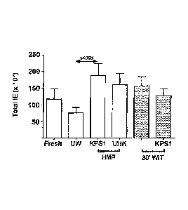
[0027] Figures 3A and B are graphical representations of islet yields
expressed as
both islet equivalents (IEQ) per gram of pancreas (A) and total IEQ (B) where
the data for
each group are expressed as the mean (ISEM);
[0028] Figures 4A-F depict light micrographs (100x magnification) showing the
relative purity of the respective islet preparations at the end of the
digestion phase (A, C, E)
7
CA 02805717 2013-01-16
WO 2012/009651 PCT/US2011/044210
and after density gradient purification (B, D, F), all panels are shown at the
same
magnification and represented by the 100 um scale bar shown in the top left
hand corner;
[0029] Figures 5A-F illustrate the histology of pancreatic biopsies sampled
for each
of the treatment groups: (A) Fresh control pancreas; (B) 24 h cold storage in
UW-Viaspan;
(C) 24 h HMP perfused with KPS1 (WIT = 0); (D) 24 h HMP perfused with Unisol-
UHK
(WIT = 0); (E) 24 h perfused with KPS1 (WIT = 30 min); (F) 24 h HMP perfused
with KPS1
(WIT = 30 min). Scale bars: 10 um;
[0030] Figure 6 are photographs illustrating hypothermic perfusion
preservation of a
porcine pancreas on a LifePort machine; the lower panel shows the principal
features of the
LifePort0; the middle panel shows the details of a pig pancreas installed in
the perfusion
cassette and hooked up to the perfusion inlet line via a seal-ring cannula;
this proprietary
cannula allows simultaneous perfusion of the celiac trunk (CT) and superior
mesenteric artery
(SMA) by way of an aortic patch clamped in the seal-ring cannula (see circular
inset); the
inset photo shows the opening of the CT and SMA in the aortic patch (AP) which
was
exposed for viewing by opening the seal-ring cannula;
[0031] Figures 7A-F are light micrographs illustrating the effect of HMP on
subsequent islet isolation at various magnifications, as indicated, showing
the presence of
isolated islets at different stages in the processing of both Fresh (panels A-
C) and HMP
pancreases (panels D-F); islets are identified by dithizone staining and
appear purple-red in
contrast to the unstained exocrine tissue which appears grey-brown; the
pancreatic digest
stained during the enzymatic processing shows a typically more uniform digest
and isolated
cleaved islets in the perfused pancreas (D) compared with the more non-
homogeneous digest
observed using freshly isolated pancreas (A); the more homogeneous digest
typically derived
using perfused pancreases often resulted in a cleaner separation of isolated
islets on the
density gradient yielding a more highly purified preparation of islets (E)
compared with the
either fresh (B) or statically cold stored pancreas (not shown); this
differential separation
during gradient purification was also manifest in examination of the gradient
residual, which
in the case of fresh pancreas often included many trapped or embedded islets
(C) compared
with perfused pancreases which showed a "clean" residual fraction with very
few identifiable
islets (F); this apparent differential effect on islet separation and
purification was also
manifest in the yield of islets obtained as an end-product (see data in Table
5, below);
[0032] Figure 8 is a graphical representation of exemplary pancreas perfusion
parameters monitored continuously during perfusion;
8
CA 02805717 2013-01-16
WO 2012/009651 PCT/US2011/044210
[00331 Figures 9A-D are photographs depicting variations in the method of
cannulation for pancreas perfusion on the LifePort Transporter; A. Exemplary
method of
cannulation for juvenile pig pancreas using the proprietary seal-ring cannula
(LifeLine
Scientific), which avoids the need to insert cannulas into the individual
arteries by allowing
perfusion via the openings of the CT and SMA on an aortic patch clamped inside
the seal-ring
cannula (see also Figure 6); B. Dual seal-ring cannulas each supporting the
openings of the
SMA and CT on individual aortic patches and linked via a coupler, This
arrangement is
useful and necessary when the openings of these two main arteries are spaced
too far apart to
be accommodated in a single seal-ring cannula; C. Straight cannulation of a
large pig
pancreas, or pancreatic lobe using insertion cannulas coupled together via a"
T" connector
for linking with the infusion port; D. A combination of a seal-ring cannula on
one artery
linked to a straight insertion cannula on the other artery, this configuration
can be used as
variant of the arrangement in B for larger pig pancreases or those in which
the openings are
anatomically too far apart for a single seal-ring cannula to be used;
[0034] Figure 10 is a photograph depicting hypothermic perfusion of human
pancreas on the LifePort i transporter; and
[00351 Figure 11 is a photograph depicting a vascular cut-down to illustrate
anatomical variants with early diverging side branches; successful perfusion
of the pancreas,
especially from young pigs, via the SMA and celiac trunk requires extreme care
to avoid
occlusion of early side branches by inserted cannulas such as those
illustrated here. These
anatomical constraints are prevalent in young pig pancreata as illustrated by
the vessel cut-
down shown here. The risk of undesirable occlusion of these side branches is
avoided by use
of a seal-ring cannula as described in the text and illustrated in Figure 6.
DETAILED DESCRIPTION OF EMBODIMENTS
[00361 In embodiments, methods are disclosed for isolating cellular products
by
application of hypothermic machine perfusion (FIMP) and the development of
interstitial
edema while preserving the integrity of the cellular products, such as islets,
which greatly
increases the amount and quality of cellular products that may be retrieved
compared with
conventional methods applied to nonperfused donor tissues (i.e., fresh or
static cold stored
donor tissues).
[00371 "Edema" is used herein to refer to an accumulation of an excessive
amount
of watery fluid in cells, tissues, or serous cavities.
9
81794475
[0038) "Tissue or organ" is used herein to refer to any natural or engineered
biological tissue or organ, including, but not limited to, cardiovascular
tissue, neuronal tissue,
periodontal tissue, glandular tissue, islets of Langerhans, hepatocytes,
cardiomyocytes, organ
tissue, and organs, such as pancreas, bladder, kidney, breast, liver,
intestine, heart and
sections or pieces thereof. Such tissue may be obtained from any organism,
such as a
mammal, for example, humans or otherwise, including heart-beating donors, or
non-heart-
beating donors. Tissues may be used in whole or in-part, such as tissues that
have been cut or
sliced.
[0039) As used herein, the term "perfusion" means the flowing of a fluid
through
the tissue. Techniques for perfusing organs and tissues are discussed in, for
example, U.S.
Patent No. 5,723,282 to Fahy at at.
[0040] The excision of a tissue for transplantation means that ischemia is
total and
inevitable even though the period may be brief. An immediate consequence of
cessation of
blood supply to an organ is deprivation of the supply of oxygen to the
tissues, but anoxia
(total) or hypoxia (partial) is only one of the many consequences of a lack of
blood supply. A
multifactorial cascade of events ensues following the initiation of ischemia.
The pivotal
event is ATP depletion, which occurs within the first few minutes of oxygen
deprivation.
This early event leads immediately to a shift from aerobic to anaerobic
metabolism, which
very quickly becomes self-limiting with the production of lactate and protons.
Cell
depolarization also occurs very early in the cascade leading to a breakdown of
ion
homeostasis, and a concatenation of other intracellular and membrane-
associated events that
eventually culminate in cell death by either apoptosis or necrosis.
[00411] The basic principle of cellular preservation for clinical application
is to
minimize the deleterious effects of ischemia and anoxia during the
preservation interval. This
can either be achieved pharmacologically by using a wide variety of
eytoproteetive drugs,
and/or by reducing temperature. Interestingly, conventional wisdom teaches us
that there is
no single drug, or cocktail of drugs, that can so safely and effectively
suppress metabolism
and provide ischemic protection for multiple tissues and organs as the
application of
hypothermia can. Accordingly, the focus changes to control the environment of
cells to
optimize hypothermic preservation.
100421 fn embodiments, the methods disclosed herein implement a new approach
that utilizes advances in perfusion technology and optionally combines those
advances with
hypothermic blood substitute solutions to improve Ordelivery by means of PFC-
CA 2805717 2017-10-27
CA 02805717 2013-01-16
WO 2012/009651 PCT/US2011/044210
augmentation. This approach circumvents several recognized shortcomings in the
present
modes of clinical organ storage, the most notable of which is the demonstrated
low
penetration of PFC and oxygen using the conventional two layer method (ILM).
[0043] In the specific case of pancreas preservation prior to islet isolation,
a salutary
effect of HMP on islet yield in a juvenile porcine model has emerged. However,
given the
vulnerability of islets to even short periods (<10h) of cold ischemia, the new
approaches
described herein extend tolerance to ischemia by circumventing the constraints
recognized in
conventional techniques of pancreas preservation. The innovation revolves
around the
application of one or more of three individually important components of organ
preservation,
namely machine perfusion for inducing development of edema; hypothermic blood
substitution, and improved oxygen delivery by PFC augmentation.
[0044] Hypothermic Machine Perfusion (HMP): Conventional methods of organ
preservation for transplantation rely principally upon static cold storage on
ice, a relatively
simple and economic technique that has been used for several decades. However,
modern day
demands for increasing the numbers of organs available for transplant has led
to a resurgence
of interest in hypothermic perfusion preservation (HPP) of organs because
perfusion
techniques provide significant advantages over static cold storage. In this
context FIPP is
based upon the fundamental premise that devices can be designed to facilitate
the replacement
of blood in the circulation of an ex vivo organ with specially designed fluids
to maximize the
protective effects of hypothermia on the ischemic tissue. This approach has
the potential, and
has already been shown in many applications, to circumvent some of the
recognized
shortcomings of conventional cold storage. However, in the field of pancreas
preservation,
particularly as it applies to source organs for islet isolation, static cold
storage imposes severe
restrictions upon the yield and quality of islets obtained from a single donor
pancreas. For
example, introducing a perfluorochemical layer to purportedly increase the
supply of oxygen
to the ischemic organ has failed in static cold storage methods to provide the
added
protection.
10045] In embodiments, the methods disclosed herein utilize a combination of
technologies in HMP and FIBS along with the merits of PFC oxygenation to
generate a new
hybrid technique that solves the problems of static cold storage methods
having a
perfluorochemical layer. Selection of the baseline medium or perfusate in
which to deliver
the PFC as an emulsion also demands consideration of what will be optimal for
the respective
cell (e.g., pancreatic cells, cardiac cells, etc.,) preservation under
hypothermic conditions. To
11
81794475
this end, this disclosure includes the preparation of preservation solutions
designed as
hypothermic blood substitutes.
[0046] Hypothermic blood substitutes as preservation media: Traditionally, a
variety of organ preservation solutions have been developed.
[0047) U.S. Patents Nos. 5,643,712, 5,699,793, 5,843,024 to Brasile and
Nos. 5,599,659, 5,702,881 to Brasile et al., describe separate resuscitation
and preservation
solutions for tissues and organs. The Brasile patents disclose compositions
that may be used
in methods in this disclosure.
[0048] Taylor at al. have formulated and evaluated two solutions designated
HypotherinosolTm-purge (HTS-P) and Ilypothermosolm-maintenance (IITS-M). Some
aspects of these solutions are described in U.S. Patents Nos. 5,405,742 and
5,514,536 to
Taylor. The Taylor patents disclose compositions that may be used in methods
of this
disclosure.
[0049) The protective properties of solutions such as the UnisolV family of
solutions (as described in US. Patent Nos, 6,492,103 and 6,994,954, entitled
''System for organ and tissue preservation and hypothermic
blood substitution" to Taylor) may be use in methods of
this disclosure. In embodiments, Unisol may be utilized as the vehicle
solution for
emulsifying PFCs to significantly increase its oxygen delivery capacity, in
addition to
eroprotective additives.
[0050] In embodiments, the principal solution may be a hyperkalemic,
"intracellular-type" solution designed to "maintain" cellular integrity during
hypothermic
exposure at the nadir temperature (<10 C).
[0051] increasing oxygen delivery to tissues during hypothermic storage and
the
role ofPFCs: The Unisole "maintenance" solution was developed and tested at
temperatures
in the range of 740 C, which conforms with the temperature range in which ATP
reserves
can be re-established if an adequate supply of 02 is maintained by continuous
perfusion. For
example, numerous investigations have suggested that oxygen supply is
essential during
hypothermic preservation of livers.
[0052] The rapid depletion of adenine nucleotides during cold storage of
organs at
0-2 C (e.g. conventional static cold ice-storage) may be suggestive that
mitochondria]
12
CA 2805717 2017-10-27
CA 02805717 2013-01-16
WO 2012/009651 PCT/US2011/044210
function is severely impaired by hypothermia. These levels of 02 may need to
be sustained
during perfusion to ensure the highest quantify and quality cellular products,
such as islets,
and the use of PFCs allows for this to be accomplished.
[0053] PFCs are hydrocarbons in which all or most of the hydrogen atoms are
replaced with fluorine (e.g., perfluorocarbons), They have twice the density
of water and a
high capacity for dissolving respiratory gases. The solubility of dissolved
oxygen in PFC is
approximately 25 times greater than in blood or water. The ability of PFCs to
release oxygen
in accordance with Henry's Law is not significantly influenced by temperature,
making them
ideal for delivering oxygen during hypothermic organ preservation. This is
also supported by
recent demonstrations that the gas-dissolving and gas-unloading properties of
pm-fluorocarbon
were necessary in a peritoneal perfusion application for systemic oxygenation
since the same
effect was not obtained when saline solution alone was employed as the
perfusate. However,
the use of perfluorocarbon under hypothermic conditions has been limited.
[0054] In embodiments, the methods of the present disclosure comprise
preventing
anaerobic glycolysis in the donor tissue. In embodiments, preventing anaerobic
glycolysis in
the donor tissue may comprise introducing perfluorochemicals into the
perfusion solution
and/or preventing oxygen deprivation/depletion in the donor tissue. For
example, preventing
oxygen deprivation/depletion in the donor tissue may comprise introducing
perfluorochemicals into the perfusion solution and oxygenating the perfusion
solution.
[0055] In embodiments, the methods of the present disclosure comprise
perfusion
with a perfusion solution, where the perfluorochemicals represent from about
10% to about
90% of the total weight of the perfusion solution, the perfluorochernicals
represent-from
about 10% to about 80% of the total weight of the perfusion solution, the
perfluorochemicals
represent from about 20% to about 70% of the total weight of the perfusion
solution, or the
perfluorochemicals represent from about 300/0 to about 60% of the total weight
of the
perfusion solution.
[0056] In embodiments, the methods of the present disclosure may comprise
satisfying the 02 demand of a donor tissue throughout a preservation
interval/process
occurring from the time the perfusion apparatus is connected to the donor
tissue to the time
perfusion apparatus is disconnected from the donor tissue. Such methods may
comprise
replenishing 02 content in the perfusion solution during perfusion, increasing
02 content in
the perfusion solution during perfusion, and/or decreasing CO2 content in the
perfusion
13
CA 02805717 2013-01-16
WO 2012/009651 PCT/US2011/044210
solution during perfusion. The levels of 02 and CO2 in the tissue and/or
perfusion solution
may be monitored by any known method.
[0057] In embodiments, the methods of the present disclosure comprising
monitoring the extracellular space in the donor tissue by microdialysis. For
example,
monitoring the extracellular space in the donor tissue by rnicrodialysis may
comprise
implanting a dialysis probe into the donor tissue and assessing the
concentration of interstitial
fluid components. Such a dialysis probe may comprise a semi-permeable bio-
compatible
membrane as the active part. In embodiments, in the methods of the present
disclosure, the
concentration of interstitial fluid components is assessed periodically.
Exemplary interstitial
fluid components include any analyte contained in the perfusion solution or
tissue, including
one or more selected from the group consisting of glucose, lactate, pyruvate,
glycerol, ATP,
02 and CO2. In embodiments, the methods of the present disclosure include
assessing the
oxygen consumption rate of the donor tissue before the perfusion apparatus is
connected to
the donor tissue, assessing the oxygen consumption rate of the donor tissue
after the perfusion
apparatus is connected to the donor tissue, and/or monitoring the oxygen
consumption rate of
the donor tissue after the perfusion apparatus is connected to the donor
tissue.
[0058] As discussed below, the application of one or more of these three organ
preservation strategies outlined above minimizes damage and cell death in the
donor tissue or
organ, such as the pancreas, which may promote an increase the overall islet
yield. This
strategy has the potential of significant benefits for other transplantable
organs all of which
suffer ischemic injury during cold storage.
[00591 As discussed above, procurement and preservation of panereata is
important
for islet isolation as a prelude to islet transplantation as an option for the
treatment of Type I
diabetes mellitus. Pancreas perfusion can further be applied for the
preservation of organs
exposed to warm ischemia prior to islet isolation and to optimize pancreas
preservation
solution for a better islet yield and quality. The above-mentioned organ
preservation prior to
islets isolation may allow for more time for proper donor-recipient matching
and quality
control of isolated cells, and offers the possibility of banking cells for
increased availability to
Ilypotherrnic machine perfusion provides an answer to the pancreas shortage
for
transplantation by improving flow and reducing vascular resistance and
allowing for pancreas
quality evaluation prior to transplantation.
[0060] Physiologically, the pancreas is a low flow organ. In embodiments, the
methods of this disclosure may comprise pancreas perfusion, which may be based
on a low
14
81794475
constant pressure (about I mmHg or less, such as from about brining to about
lOmmHg )
driven flow. The present design of the LifePort may not accommodate infusion
pressures of
less than lOmmHg. Thus, lower pressure values may be installed in order to
reach the desired
infusion pressures of less than 10mInHg for the methods disclosed herein for
preserving
pancreata for transplantation without inducing irreversibly high levels of
edema that may be
detrimental to the organ and/or recipient. Controlled development of edema and
better
perfusion outcome for both islets isolation and whole pancreas transplantation
may be better
attained by employing a constant flow regime as opposed to constant pressure.
In -
embodiments, the driven flow rate values may be selected in accordance with
organ
characteristics and quality (such as warm ischemia exposure, size, species,
etc.).
100611 fn alternative embodiments, the methods of perfusion may be based on a
high (about 10intnHg or more, such as from about I mmHg to about 60mmHg)
constant
pressure driven flow.
(00621 in embodiments) the methods described herein employ an apparatus for
peribsing one or more organs or tissue (hereinafter generally referred to as
donor tissues). An
exemplary apparatus is described in 'U.S. Patent Application No. 12/379,239,
which is a
division of U.S. Patent Application No. 11/075,690, filed March 10, 2005
(issued as U.S. Patent No. 7,504,201 on March 17, 2009).
In embodiments, the methods described herein
employ the LifePort() platform transporter or a modified LifePort platform
transporter in
order to accomplish hypothermic machine perfusion (HMP) of a donor tissue
(such as the
pancreas).
(00631 in embodiments, IIMP may result in uniform fluid accumulation within
the
donor tissue that in turn may provide a disrupted extracellular space with
beneficial effects for
islet isolation without compromising islet viability and function. The methods
of this
disclosure, described herein with respect to juvenile porcine pancreata, may
be easily applied
to human and adult porcine donor pancreases, the latter being regarded as the
source of choice
for xenogeneic islet transplantation, and/or other various donor tissues of
interest, such as the
heart and/or liver. The successful methods described herein rely strongly upon
the details of
pancreas surgical procurement, cannulation and perfusion on the LifePort .
Based on these
methods, pancreas hypothermic perfusion optimization may be achieved for
development of
methods of organ evaluation and quality control during perfusion in order to
reliably select
high quality pancreases for clinical transplantation.
CA 2805717 2017-10-27
CA 02805717 2013-01-16
WO 2012/009651 PCT/US2011/044210
[0064] A major technological issue to be addressed in applying the established
"LifePort" kidney perfusion technique to the pancreas is the different
perfusion parameters
required by the pancreas since this is a low flow, low pressure organ compared
with the
kidney. Typically, the optimum perfusion parameters for a kidney on a LifePort
machine,
which by design is a pressure-controlled device, are a set perfusion pressure
of 25-40 mmHg
(typically produces a flow rate of 100-150 ml/min). These perfusion parameters
may impact
the fluid exchange between the vascular and interstitial compartments of the
organ and hence
the degree of edema sustained during the perfusion interval. The method
described here
demonstrates the adaptation of the LifePort() machine for pancreas perfusion
with an
emphasis on developing a specific amount of Edema. In embodiments, the
LifePort
machine for pancreas perfusion may be adapted to operate at a low pressure
setting (about
lOmmHg or less, such as in the range from about 10mning to about 2mmHg, or in
the range
from about 8mmHg to about 4mmHg)-controlled perfusion of porcine pancreas as a
prelude
to pancreas processing for islet isolation.
[0065] In embodiments, the LifePort machine for pancreas perfusion may be
adapted to operate a flow rate of less that 150 ml/mm, such as less than 100
nil/min, or from
about 10 ml/min to about 100 ml/min, such as from about 15 ml/min to about 50
ml/min, or
from about 20 ml/min to about 30 ml/min.
[0066] In embodiments, the LifePort machine for pancreas perfusion may be
adapted to operate at a high pressure setting {about lOmmHg or more, such as
in the range
from about lOmmHg to about 60mmHg, or in the range from about 20mrnHg to about
50mmHg)-controlled perfusion of porcine pancreas as a prelude to pancreas
processing for
islet isolation. In embodiments, the LifePort machine for pancreas perfusion
may be
adapted to operate a flow rate of less that 200 rnlimin, such as less than 150
ml/min, or from
about 10 ml/min to about 150 ml/min, such as from about 50 ml/min to about 120
ml/min, or
from about 60 ml/min to about 110 ml/min.
[0067] In embodiments, the LifePort machine may ensure proper cold static
storage of the donor tissue or organ if the pump fails and the fluid transport
through the organ
stops. For example, inside the closed transporter, a properly filled ice
container may be
maintained at a temperature below about 6 C for more than 24h, without ice
replenishment.
The LifePort transporter may be programmed to allow for re-circulation of a
desired
perfusate for under predetermined conditions, for example, the transporter may
be
16
CA 02805717 2013-01-16
WO 2012/009651 PCT/US2011/044210
programmed to allow one liter of perfusate re-circulation at 5-7 C by a
pulsatile
(30pu1seslmin) constant low pressure (about 1 mmHg) flow.
100681 The LifePorto pulsatile perfusion system has been successfully employed
for
small pig pancreas hypothermic ex-vivo perfusion. The system is designed and
FDA cleared
for kidney hypothermic perfusion/preservation for clinical transplantation.
Using the kidney
system the whole pancreas of young porcine donors (25-32kg, 2 months old) may
be
continuously perfitsed in a closed loop while being completely immersed in the
perfusion
solution inside the organ bath. The latter also serves as a solution
reservoir, the perfusatc
being drawn out by the pump, forced to go through the filter, bubble trap and
the infusion port
before returning to the pancreas and organ cassette. Pancreas submersion in
the temperature-
controlled perfusate helps eliminate temperature gradients across the organ
surface and ensure
high quality hypothermic preservation.
[0069] Embodiments of the invention may provide an improved method of
isolating
cellular products, which may be more consistent and reliable than conventional
methods that
rely on enzyme digestion. Embodiments may also provide methods that yield an
optimum
quantity of desired cells that retain sufficient functional integrity to be
useful as a
transplantation resource.
[0070] In embodiments, methods disclosed herein may be used to isolate any
cellular product for therapeutic use and research, as long as the desirable
and undesirable cells
have, or can be treated to promote, development of edema. Such methods may
allow the
preservation of the integrity of the islets in addition to greatly
facilitating islet isolation to the
extent that the yield of cellular product may significantly increase (in some
situations at least
about double the yield or even triple the yield) compared with the yield of
cellular product
obtained from nonperfused tissues and even fresh tissues.
[0071] In embodiments, a cellular product may be isolated by methods
comprising
developing edema during perfusion of the donor tissue by increasing a first
flow rate of the
perfusion solution through the tissue to achieve a second flow rate,
increasing a first perfusion
pressure applied by the perfusion apparatus to the tissue to achieve a second
perfusion
pressure and/or selecting a composition of thc perfusion solution that causes
edema of the
tissue.
[0072] In embodiments, development of edema in donor tissues to form a swelled
tissue may occur by application of hypothermic machine perfusion (HMP). The
application
of donor tissue HMP, such as the pancreas HMP, as a prelude to islet isolation
also
17
CA 02805717 2013-01-16
WO 2012/009651 PCT/US2011/044210
capitalizes upon the some of benefits of HMP demonstrated for other various
organs
(principally the kidney) as a means of better preservation during extended
periods of storage,
especially for sub optimum organs. In addition, an unexpected salutary effect
of machine
perfusion applied to the application of cellular product harvesting, such as
islets, has
emerged.
[0073] The progressive development of edetna during extended machine perfusion
of organs is a phenomenon that is generally regarded as undesirable. In fact,
steps are usually
taken to minimize the problem by adjusting the mechanical perfusion parameters
such as flow
and pressure, as well as the composition of the perfusate, to minimize the
development of
interstitial edema. In resolving a technical problem with respect to
cannulation of the donor
tissues (in this case the pancreas) that affects the efficiency of perfusion,
it was determined
that 24 h of HMP resulted in moderate edema in the gland compared to the
controls that were
simply flushed with and immersed in cold UW-Viaspan solution. However,
contrary to
expectations, development of edema, such as up about 280% (i.e., a 180% gain
in the
particular parameter that is monitored to assess the extent of edema, such as,
for example,
weight, mass, circumference, buoyancy, arid/or volume), or up to about 250%,
or up to 150%
to did not prove deleterious to cellular product harvesting, but was observed
to be of
considerable benefit by correlating with a more efficient disruption of the
pancreas during
enzymatic digestion to yield a significantly greater number of islets.
[0074] In embodiments, developing edema during perfusion of the donor tissue
to
fowl a swelled tissue may result in a swelled tissue exhibiting a weight,
mass, circumference,
surface area, buoyancy, and/or volume about 110% (i.e., gain in weight, mass,
circumference,
surface area, buoyancy, and/or volume of about 10%) of that of the initial or
original non-
perfused donor tissue, such as from about 120% to about 280% (i.e., gain in
weight, mass,
circumference, surface area, buoyancy, and/or volume of from about 20% to
about 180%), or
from about 130% to about 250% (i.e., gain in weight, mass, circumference,
surface area,
buoyancy, and/or volume of from about 30% to about 150%). In further
embodiments, the
swelled tissue has a mass that is less than 300% of an initial non-perfused
mass of the donor
tissue, the swelled tissue has a volume that is at least 110% of an initial
non-perfused volume
of the donor tissue, the volume of the swelled tissue is from about 150% to
about 250% of the
volume of the donor tissue, the volume of the swelled tissue is from about
120% to about
280% of the volume of the donor tissue, wherein the volume of the swelled
tissue is from
18
81794475
about 130% to about 250% of the volume of the donor tissue, or the swelled
tissue has a
volume that is less than 300% of an initial non-perfused volume of the donor
tissue.
[00751 It is believed the presence of a predetermined amount of edema causes
sufficient disruption to the extracellular matrix and architecture of the
pancreatic gland that
the subsequent distension and digestion of the gland proceeds more
effectively. This is
evidenced by significantly shorter digestion times (Table 3; below), a more
homogeneous
digestion product (Fig. 4), and better gradient Purification resulting in
higher yields and purity
of the final islet preparation. The structure and function of the islets per
se did not appear to
be compromised by the level of tissue edema encountered in these studies.
Concerns that a
change in the hydration of the isolated islets due to IIMP might alter the
buoyant density of
the islets And thereby alter their ability to be separated from exocrine
tissue on a density
gradient did not appear to be a-problern. This may presumably be due to the
fact that any
inherent edema in the islets is counteracted by the pregradient incubation in
TJW solution,
which is a hypertonic medium that would dehydrate the Islets during the 30-
rnia cold
incubation prior to loading on the density gradient for purification, which is
generally used in
islet isolation protocols (Lakey, I. R. T., Technical aspects of islet
preparation and
transplantation, Transpl. InL, 16:613-632,2003; Lakey, J. It. T.; Current
human islet
isolation protocol, Chuo-ku, Osaka: Medical Review Co. Ltd., 2004).
[00761 The morphological integrity of the islets in situ in the preserved
pancreata
may be evaluated by taking wedge biopsies at the end of a preservation
interval. Changes
associated with ischemia and the mode of preservation are illustrated and
discussed with
respect to Figure 5. Dithizone staining ofboth the digest samples and
purification fractions
may he used to evaluate the gross structure, purity and numbers of islets in
the respective
samples. Figure 4F shows the typical appearance of the highest purity
preparations obtained
from the }IMP-treated pancreases. The islets have an irregular cluster shape
that has been
described as "grape-like" (Rijkelijkhuizen, J. K., et at., Pretransplant
culture selects for high
quality porcine islets, Pancreas 32:403-407,2006) and this appearance may be
characteristic
of islets isolated from young pigs, reflecting the irregular shape observed in
the endogenous
pancreas prior to isolation (Bottino, R. et at., Isolation outcome and
functional characteristics
of young and adult pig pancreatic islets for transplantation studies,
Xertotransplantation
14:74-82,2007). This characteristic irregular, fragmented appearance of islets
from young
pigs contrasts sharply with the more normal regular rounded shape of islets
from adult pigs
19
CA 2805717 2017-10-27
CA 02805717 2013-01-16
WO 2012/009651 PCT/US2011/044210
and may not a reflection of the method of preservation. Figure 4B shows that
islets from the
fresh control pancreases have the same morphology.
[0077] The unanticipated mechanical benefit of HMP described above may be
achieved without compromising the quality of the harvested islets. The data
presented in the
Examples section demonstrates that the functional ability, in terms of their
insulin secretory
index, of the islets isolated from the perfused pancreases in which a moderate
amount of
edema has been developed is equivalent to that of the controls including fresh
pancreas.
Moreover, the insulin content was significantly higher than the control group
comprising
pancreases stored statically in cold UW-Viaspan solution, which is currently
the standard
method employed clinically. These effects and standards of preservation may be
achieved
using either of two proprietary solutions, KPS l and Unisol-UHK.
[0078] Further improvements and benefits to this technique may occur by
optimizing the composition of these baseline perfusates by adding
cytoprotective agents
design to minimize preservation and reperfusion injury, and/or PFCs.
For example, cytoprotective additives may be additives displaying efficacy
during low
temperature preservation and therefore a high probability they will have a
positive impact on
the quality of pancreas preservation during hypotheilnic machine perfusion,
such as
antioxidants, anti-apoptotic agents and trophic factors.
[0079] In embodiments, the methods of the present disclosure comprise
perfusing
the organ and/or tissue with a perfusion solution comprises cytoprotective
additives, such as
one or more antioxidants, anti-apoptotic agents and trophic factors. Such a
perfusion solution
may be any perfusion solution, such as any perfusion solution described in the
present
disclosure, including hypothermic blood substitutes, including those
comprising: one or more
cytoprotective agents, and perfluorochemicals.
[00801 In embodiments, the methods of the present disclosure may comprise a
step
of increasing the ATP levels in the donor tissues during perfusion and/or a
step of introducing
cytoprotective agents during perfusion of the donor tissue for preventing cold-
induced cell
death of the donor tissue. In embodiments, the methods of the present
disclosure may
comprise a step of introducing cytoprotective agents during perfusion of the
donor tissue for
preventing cells of a donor tissue, such as a pancreas, from entering
destructive pathways. For
example, the methods may comprise introducing cytoprotective agents during
perfusion of the
donor tissue for inhibiting mitochondrial dysfunction in cells of a donor
pancreas.
CA 02805717 2013-01-16
WO 2012/009651 PCT/US2011/044210
[0081] Antioxidants: Oxygen-derived free radicals (ODFR) have been the focus
of
attention as mediators of various tissue injuries and particularly
rnicrovascular injury. It is
possible for the production of injurious free radicals to be enhanced during
cold storage, it is
important to appreciate that the resultant cell damage may not occur entirely
at the low
temperature. On the contrary there is a growing body of evidence that
reintroduction of
oxygenation via a regular blood supply upon rewarming and reperfusion provides
a powerful
impetus for further oxidative stress. A principal pathway is the stimulation
of enzymically
driven radical reactions such as the xanthine/xanthine oxidase system
involving the
interaction of ATP catabolic products with molecular oxygen. Vascular
endothelial cells are
thought to be particularly vulnerable to this type of injury mediated by free
radical generation
by this so-called "respiratory burst" mechanism. Nevertheless, low
concentrations of
molecular oxygen such as that dissolved in organ preservation solutions may be
sufficient to
support the generation of free radicals during prolonged storage. Therefore,
without the
proper balance of antioxidants, cold exposure may set the stage for a
progressive development
of tissue injury as a result of reactions and processes that occur during
hypothefinia.
[0082] In embodiments, the antioxidants may be present in a sufficient amount
to
substantially eliminate cellular damage and/or oxidative stress.
[0083] Whilst cells employ a number of repair mechanisms to recover from
injuries
occurring as a result of free radical activity, cell survival depends upon
whether salvage
pathways are overwhelmed or whether a point of irreversible damage is reached
during the
storage/reactivation process such that cell death becomes inevitable.
Accordingly, in
embodiments, the antioxidants, and amounts thereof, are selected to circumvent
oxidative
stress and reperfusion injury under both hypothermic and normotherrnic
conditions.
Exemplary antioxidants may include dibutyryl-cAMP (db-cAMP), a-tocopherol
(Vitamin E),
TroloxT", and hwothermosol plus both EDTA and Vitamin E.
[0084] Anti-Apoptotie Agents: While many of the diverse stresses known to
cause
necrotic cell death have also been reported to induce apoptosis in a variety
of cells, the role of
low temperatures as a possible stimulus of programmed cell death has only
recently begun to
emerge. It is now established that apoptosis plays an integral role in cell
death induced by the
rigors of both hypothermia and cryopreservation. More specifically, apoptosis
has been
identified to be directly associated with delayed-onset cell death (DOCD).
This is defined as
death associated with cold exposure that is not apparent immediately upon
rewarrning, but
extending over the post-exposure recovery period. Recent research into the
causative
21
CA 02805717 2013-01-16
WO 2012/009651 PCT/US2011/044210
apoptotic and necrotic pathways responsible for low temperature induced DOCD
has
identified the contribution of multiple apoptotic pathways, including receptor-
and
rnitochondrial-induced apoptosis. Investigations into these pathways, their
progression, and
their induction stressors has begun to facilitate new methods for improving
preservation
efficacy through the modulation of the cellular and molecular responses of a
cell undergoing
preservation (both hypothermic and cryopreservation).
100851 Incorporation of specific apoptotic protease inhibitors in preservation
media
has now been reported to markedly improve the survival of a variety of cells
and tissues.
Furthermore, investigation into the modification of the carrier medium from
that of standard
extracellular-type culture media with, or without cryoprotectants, to that of
specifically
designed intracellular-type preservation solutions such as UnisolTM, or its
predecessor
Hypothermosol, have led to studies showing significant improvement in
preservation
efficacy.
100861 Anti-apoptotic agents may be selected from those that possess
recognized
antioxidant activities and hence implied anti-apoptotic activity. For example,
reduced
glutathione is a component of both formulations as a multifaceted molecule
that is also
known to fulfill a natural role in the regulation of apoptosis, bongkrekic
acid (BA) has been
shown to be a potent inhibitor of mitochondrial permeability transition (PT)
pores that form
during apoptosis. In addition, BA can inhibit cytochrome c release that is
influenced by Bax,
a pro-apoptotic protein 85. BA, a stable inhibitor of PT, has been shown to
increase cell
viabilities and protein production levels following virus. infection. With
respect to the
inhibition of caspases, a variety of compounds have been shown to be effective
for
mammalian cells in culture. Other exemplary compounds include, P35, which
confers
irreversible inhibition to a large number of caspases, and Z-VAD.fmk (or its
latest broad-
spectrum counterpart, Q-VD-OPH), which has the ability to inhibit both the
intrinsic and
extrinsic pathways.
[0087] Traphic Factors: Many cell signaling pathways retain activity at very
low
temperatures and can be affected by trophic factor administration. Trophic
factor deprivation
disrupts many aspects of cell function and is well known to induce apoptosis
and cell death in
a wide variety of cultured cells. Trophic factor supplementation (TFS) leads
to a markedly
improved outcome in kidney storage an influence cold ischernic injury by
interaction with the
tissue during cold storage and not merely by being present during rewarming
and reperfusion.
Exemplary tropic factors, which may be employed include, for example, Insulin-
like growth
22
81794475
factor-1 (IGF-l) Epidermal growth factor (EGF), Bovine neutrophil peptide-I
(BNP-1), also
referred to as bactenecin 98, Substance P (SP), which has mitogenic effects
for a variety of
cell types and stimulates DNA synthesis in ocular cell lines, EGF, a
polypeptide growth factor
(its effects may be additive or synergistic with other growth factors and
cytokines), and
polypeptide growth factors (IGFs), such as IGF-1.
[0088) In embodiments, the PFCs may possess one or more of the following
qualities: (1) the ability to dissolve large quantities of many gases, (2) can
transport these
gases to diffuse across distances, (3) are non-toxic, (4) biologically inert,
(5) biostatic liquids
at room temperature. In embodiments, PFCs with densities of about 1.5-2.0
g/m1., and high
solubilities for. oxygen and carbon dioxide may be selected.
100891 In embodiments, the cellular product may be isolated by pre-treating a
tissue
such that desired cells are less prone to destructive freezing and undesired
cells are more
prone to destructive freezing as described in U.S. Application Serial No.
12/654,147, entitled
"Method for Isolating Cellular Products by Cryopreservation," to Michael J.
Taylor et al.
[00901 In embodiments, cryopreservation may be applied to selectively preserve
the
desired cells and/or destroy the undesired cells. Cryopreservation is a
complex process of
coupled heat and mass transfer, generally executed under non-equilibrium
conditions. Simply
freezing cells or tissues generally results in dead, nonfunctional materials.
[009I) In embodiments, the method comprises pre-treating donor tissues such
that
(1) the desired cellular product is less susceptible to events resulting in
cell destruction, such
as destructive freezing, and/or (2) the undesired tissue is made more
susceptible to events
resulting in cell destruction, such as destructive freezing. For example, when
the donor tissue
is a pancreas, the pre-treatment may occur by differential perfusion such that
the destruction
of ac-mar tissue is maximized while islet tissue is preserved. In such
embodiments, islet tissue
may be infused with a cryoprotectant solution comprising a cryoprotective
agent (CPA) via a
vascular system, such as through celiac trunk and superior mesenteric artery;
after adequate
equilibration of islet tissue, acinar tissue may be infused with an aqueous
solution through
pancreatic ducts.
100921 In embodiments, the pretreatment the donor tissue may occur under
controlled conditions to preferentially equilibrate the cellular product with
in within the
tissue. For example, pre-treating the pancreas may occur under controlled
conditions to
preferentially equilibrate the islet tissue within the pancreas gland at a
temperature of from
23
CA 2805717 2017-10-27
CA 02805717 2013-01-16
WO 2012/009651
PCT/US2011/044210
about 2 C to about 35 C. Furthermore, perfusion may be maintained sufficiently
long to
allow equilibration of the islet tissue, but not the whole gland, with the
permeating CPA. For
example, perfusion may be maintained for a period of about 20 min. to about 70
min., such as
about 25 to 35 min. or about 30 min. The rationale for this step is to deliver
sufficient CPA
to the islet tissue to protect it against freezing injury during subsequent
cooling or freezing of
the pancreas, which may occur during preservation and/or transport.
[0093] In embodiments, for a variety of reasons, such as preservation,
transport,
and/or disruption, the donor tissue may be cooled to a sufficient temperature
to attenuate
metabolism, such as a temperature of from about 15 C to about -20 C, such as
from about
C to about -10 C, or from about 10 C to about 0 C. In embodiments, for a
variety of
reasons, such as preservation, transport, and/or disruption, the donor tissue
may be frozen to a
temperature of from about -10 C to about -200 C, such as from about -40 C to
about -170 C,
or from about -80 C to about -130 C. In embodiments, the cooling rate may be
from about
0.5 C/min. to about 5 C/min. In embodiments, freezing and/or cooling the donor
tissue may
occur at a cooling rate of from about l'Clmin. to about 20 C/min., such as
from about
6 C/min. to about 15 C/min.
[0094] In embodiments, the rate of cooling and/or freezing the donor tissue
coupled
with a rapid wat __________________________________________________ uling rate
(such as the above rates for cooling and freezing multiplied by a
factor of at least 1.5, such as a factor in the range from 1.5 to 10, such as
a factor of 2, or 3, or
4, or 5) during warming of the donor tissue may provide optimum conditions for
recovery of
functional islet tissue. Warming of the donor tissue may be achieved by, in
embodiments,
direct immersion in a warm medium, such as an osmotically-buffered medium.
[0095] In embodiments, the extent of equilibration with CPA may or may not
reach
completeness, which may be beneficial because the conditions for full
equilibration of islets
in situ may not be easily determined in relation to the requirement for
minimal permeation of
the CPA into the exocrine cells.
[0096] In embodiments, the donor tissue may be divided into smaller pieces,
fractured, and/or fragmented. In order to enhance fracturing of a donor
tissue, such as the
pancreas, volumetric warming may be combined with the addition of a compressed-
air heat
exchanger immersed in a hot water bath. In embodiments where the donor tissue
is cooled or
frozen on a preservation or transport platform, this may enable thawing of the
donor tissue
without the need to remove it from the platform. Donor tissue dividing or
fracturing may
24
CA 02805717 2013-01-16
WO 2012/009651 PCT/US2011/044210
occur at any time before exposure to the digestive enzyme, such as during
warming of the
donor tissue.
E00971 In embodiments, it may be advantageous to expose the donor tissue to
various doses of a digestive enzyme to assist in connective tissue dispersion
to allow release
of the cellular product, such as islets (which optionally may be cryoprotected
islets) from the
disrupted tissue.
100981 In embodiments, after the extent of edema has reached a predetermined
level, such as a level of edema where the there is gain in weight, mass,
circumference, surface
area, buoyancy, andlor volume of up to about 200% (i.e., if the initial or
original weight,
mass, circumference, surface area, buoyancy, and/or volume of the tissue is X
(such as 100
grams), a gain of about 200% would result in a final weight of 3X (300 grams),
such as a gain
of up to about 150%, or a gain of up to about 100%, the donor tissue may be
disrupted to
release cellular product from the disintegrated donor tissue. In embodiments,
disrupting the
donor tissue may occur while the donor tissue is frozen, while the donor
tissue is warming,
and/or after the tissue reaches room temperature. In embodiments, the
disruption may be
achieved by mechanical stress, thermo-mechanical stress induced by
differential expansion,
thermo-mechanical stress induced by steep temperature gradients, and thenno-
mechanical
stress induced by volume change upon freezing, via a digestive enzyme, or a
combination
thereof.
100991 Thermo-mechanical stress may be the outcome of the tendency of material
to
contract upon freezing, which may be driven by three different effects: volume
change upon
freezing as described above, steep temperature gradients, and differential
expansion in
composite materials. In practice, two or more of the above effects may be
acting in concert.
[01001 In other embodiments, disrupting the donor tissue may be achieved by
mechanically fracturing a frozen donor tissue. For example, this may be
accomplished in two
stages. The first stage may be to physically split the frozen donor tissue
into pieces, for
example, with a hammer and chisel. The second stage may be to grind the frozen
tissue
pieces while immersed in warm water or isotonic medium, for example, by using
an electric
ice crusher or blender. This may also serve to effect rapid warming and
dilution of a
cryoprotectant, if included, at the same time as mechanically grinding the
tissue.
10101] In embodiments, the method further comprises separating the cellular
product from the undesired donor tissue material. Separation of the cellular
product may be
achieved, for example, by filtration, density gradient separation, tissue
culture, or a
CA 02805717 2013-01-16
WO 2012/009651 PCT/ES2011/044210
combination thereof. Filtration may be performed using a filtration apparatus,
such as a
stainless steel mesh (tea strainer). Separation may include washing the
filtered donor tissue
with a medium containing a protease inhibitor, such as PEFABLOC , and a
deoxyribonuclease, such as PULMOZYMEO, such that harmful endogenous proteases
and
DNA from lysed exocrine tissue are removed. In embodiments, the filtered donor
tissue may
be stained with an indicator for identifying the cellular product, such as
dithizone for staining
islets, and examined under the microscope for the presence of intact cellular
product.
[0102] The separated cellular product may not be cleanly cleaved from the
donor
tissue and not all of the cellular product may be completely intact. For
example, with respect
to islets, some islet tissue may have a diffuse or loose structure that could
reflect osmotic
shock due to direct immersion into an aqueous medium during any warming of a
frozen
pancreas. In embodiments, such a problem may be averted by employing osmotic
buffering
during elution of the CPA from the islet tissue during or after thawing of a
frozen pancreas.
Utilizing the osmotic buffering technique in embodiments may protect the
structure of the
islet tissue and minimize osmotic swelling and lysis during efflux of the
permeating CPA. In
contrast, in such embodiments, osmotic buffering does not impact the
simultaneous
destruction and lysis of the acinar cells because these cells have not been
protected by CPA
permeation.
[0103] In embodiments, sufficiently clean cleavage of islet tissue may be
obtained
by a eryoisolation method, in embodiments, in combination with a mild enzyme
digestion to
purify the islet tissue. Another approach may be to use tissue culture as a
modality for the
"clean-up" process since the residual acinar tissue injured during the cryo-
isolation process
will die and disintegrate in culture.
[01041 Examples are set forth hereinbelow and are illustrative of different
compositions and conditions that can be utilized in practicing embodiments.
All proportions
are by weight unless otherwise indicated. It will be apparent, however, that
the disclosure can
be practiced with many types of compositions and can have many different uses
in accordance
with the disclosure above and as pointed out hereinafter. For example, these
Examples will
be readily recognized by those having ordinary skill in the art as also being
applicable to
isolating human islets because pig pancreas is an art-recognized model for
human pancreas.
[0105] Pig pancreas is a useful model for at least the following reasons: (I)
pig
pancreas is a large animal model, (2) pigs are regarded as the most promising
source of islets
for future clinical xenografting. For example, in view of the current
resurgence of clinical
26
CA 02805717 2013-01-16
WO 2012/009651 PCT/US2011/044210
interest in hypothermic perfusion preservation of organs for transplantation,
the pig provides
the model of choice for large-animal preclinical evaluation of hypotheiuiic
machine perfusion
(HMP) technology for segmental donor pancreas preservation. Secondly, the
special case of
pancreas preservation prior to islet isolation is of high significance in view
of both the
worldwide interest in islet xenotransplantation and our data that HMP can
facilitate improved
islet yields without compromising islet function.
101061 Moreover, the consensus strategic plans recently published by the
International Xenotransplantation Association for considering clinical trials
of porcine islet
products for type 1 diabetes emphasizes the need and importance of sterile,
disease free
environment for the source pigs and the products. To this end, the LifePort0
system provides
a convenient sterile environment for transport of the source pancreas from the
site of
procurement to the islet processing facility. This approach is applied to the
preservation and
procurement of viable islets after hypothermic perfusion preservation of
porcine pancreata
because pigs are now considered the donor species of choice for xenogeneic
islet
transplantation for a number of compelling reasons (O'Neil, J. J. et al., The
isolation and
function of porcine islets from market weight pigs, Cell Transplant. 10:235-
246; 2001).
[0107] The age of the donor pig has proved to be a significant factor in the
islet
isolation process with young, socalled market size pigs (<6 months old),
proving to be
particularly difficult as a source of transplantable islets (7,10,50).
Nevertheless, despite these
challenges young pigs are favored over retired breeders (>2 years old) due to
their abundance,
lower animal and husbandry costs, and are more suitable to meet regulatory
guidelines for
donor tissue for xenotransplantation. The following examples demonstrate the
efficacy of
hypothermic machine perfusion of pancreata from young pigs prior to islet
isolation. Data
regarding the details of the surgical model that was developed in light of
special
considerations to achieve uniform perfusion of the porcine pancreas during
2411 of
hypothermic perfusion at 7 C are also presented.
(0108) The success of porcine pancreas hypothermic perfusion for islets
isolation
may strongly be influenced by the surgical procedure of organ procurement and
pancreas
cannulation for ex-vivo machine preservation. The development of porcine
pancreas surgical
recovery method has not been an obvious procedure. Initially, the lack of
detailed pig
pancreas anatomy documentation has led to improper organ vasculature
preservation during
pancreas procurement. Inadequate organ procurement has resulted in
inconsistent and
incomplete pancreas machine perfusion, thus low islet yield and viability.
27
81794475
101091 Until recently, the anatomy of the pig pancreas was not well
documented.
Physiologically and topographically the pig and human pancreata are considered
similar. The
pancreas is an elongated retroperitoneal gland as shown in Figure 1. In both
pigs and
humans, the pancreas head is closely related to the proximal duodenum, but for
pigs the
pancreatic duct opening is found on the duodenum distal and separate from the
common bile
duct (Swindle, M. M.; Smith, A. C. Comparative anatomy and physiology of the
pig. Scand.
J. Lab. Anim. Surg. 23:1-10; 1997). There are a variable number of vessels
originating from
the splenic, hepatic, gastroduodenal, superior mesenteric, and celiac arteries
that on an
individual basis have irregular configuration of blood supply to the pancreas.
Commonly,
blood to the head is supplied by the posterior and anterior arcades arising
from the
gastroduodenal and superior mesenteric arteries (Fig. 1). In pigs, the head
does not surround
the pancreaticoduodenal arteries and veins¨the latter lie between the head and
duodenum
with the branches to the pancreas easily identifiable. The neck and the body
of the pancreas
are usually vascularized by the dorsal and inferior pancreatic arteries. The
former can
originate from the either the splenic, hepatic, or directly from the celiac
arteries. The inferior
pancreatic artery may begin from the superior mesenteric artery (SMA) under
the neck of the
pancreas and course toward the tail along the posterior inferior margin of the
pancreatic
surface in intimate contact with the gland. It can communicate with a varying
number of
sple ic artery branches. The neck of the pancreas is also the site of the
portal vein at the
confluence of the splenic and superior mesenteric veins. The pancreas tail
receives its blood
supply mainly from the splenic artery.
101101 In preparation for attaching the pancreas to the LifePort perfusion
machine,
all exposed arterial branches on the margin of gastroduodenal and hepatic
sides of the
pancreas were meticulously identified and ligated to ensure unifoim perfusion
throughout the
gland and allow the effluent to emerge only from the portal vein. This
surgical approach
proved optimal ibr pancreas perfusion/ preservation for islet isolation, as
described by Taylor,
M. J., et al., in Hypothermic perfusion of pancreas: Emphasis on preservation
prior to islet
isolation. In: Lee, C. Y., ed., Organ perfusion preservation. Boston, MA: Meth
House
Publisher; 2010.
[01111 As described by Taylor et al., an exemplary surgical approach may
include
the following: 1. A team of two operators for pancreas procurement; 2. Follow
surgical
facility requirements for dress code and personal protection equipment; 3.
Verify with the OR
veterinary technician that the pig is intubated and under general anesthesia
(i.e., ketamine
28
CA 2805717 2017-10-27
CA 02805717 2013-01-16
WO 2012/009651 PCT/US2011/044210
22mg/kg, acepromazine 0.2 mg/kg, and atropine 0.025mg/kg); confirm with the OR
veterinary technician of pig anesthesia maintenance and proper ventilation; 4.
Verify with the
OR veterinary technician that all vital signs are monitored (ECG, heart rate,
oxygen saturation
level, body temperature, etc); 5. Verify that an electrical knife, a suction
line and canisters are
available. 6. Verify that the OR back field surgical table has been properly
prepared (surgical
instruments, lap sponges, gauze, cold saline, umbilical tape, etc.); 7. Verify
that 2L of cold
Lactated Ringer's solution have been placed on ice; 8. Verify that an 1.V.
pole is available
near the operating table and its height is appropriate for the gravity driven
in-situ flushing of
the organs (about 6 to about 6.5 feet); 9. Obtain permission from the OR
veterinary technician
to proceed with the surgery; 10. Minimize pancreas exposure to warm ischemia
to 3 minutes,
unless otherwise desired; 11. When permission has been granted, perform a
midline incision
from the xiphoid cartilage to just above the pelvis and expose the abdominal
cavity; 12.
Instruct the OR veterinary technician to administer heparin to the pig (about
150U/kg), allow
at least three minutes to pass before starting in-situ flushing; 13. Move and
keep aside the
bladder and the intestines (with the help of lap sponges) and identify the
descending aorta; 14.
Dissect, below the kidneys, a segment (about 3cm) of the aorta apart from the
surrounding
tissue/vessels, place umbilical tape ties around the aortic segment; cut a
small opening into
the aorta between the two ties while the surgery assistant applies pressure on
the aortic walls
to prevent blood from squirting out; 15. Insert aortic cannula into the
opening and tie it in
place (make sure the umbilical tape tie is securely placed over the collar of
the cannula); 16.
Insert the two spikes of the irrigation set into the appropriate infusion
ports of the two bags of
Lactated Ringer solution (make sure the roller clamp is closed to prevent
solution loss); 17.
Hang the bags of Lactated Ringer solution on the I.V. pole and flush the
irrigation set tubing
to properly remove all the air; close the roller clamp; 18. Cross-clamp the
inferior vena cava
and the aorta above the diaphragm; 19. Connect the inlet opening of the
cannula to irrigation
set outflow port and open the roller clamp to initiate the gravity driven in-
situ flushing; 20.
Cut-open the inferior vena cava above the diaphragm, downstream from the clamp
for blood
outflow; 21. Immediately place plenty of ice inside the abdominal cavity
around the pancreas
and liver for organs maintenance/protection at low temperature; 22. Use the
suction tubing
and containers to collect the wash-out blood; 23. Make sure the solution flow
from the bags,
through the cannula into the aorta is not obstructed and that there is outflow
from the inferior
vena cava; 24. When empty, remove the bag of Lactated Ringer solution from the
IV. pole
and hang the bag of SPS-1 solution (previously kept on ice), use only half of
the SPS-1
29
CA 02805717 2013-01-16
WO 2012/009651 PCT/US2011/044210
solution volume to flush the organs; 25. Instruct the OR veterinary technician
to euthanize the
pig using a lethal dose of 5% sodium pentobarbital administered intravenously
(accepted form
of euthanasia according to the American Veterinary Medical Association Panel
on Euthanasia
(AVMA) guidelines) and complete in-situ flushing; 26. Transfer the second half
of the SPS-1
solution bag to the pancreas transportation biohazard bag and place the latter
on ice; 27.
Carefully and rapidly (less than 15 minutes) proceed to expose and dissect
apart the pancreas
from the surrounding tissue and organs (add ice around the visceral organs as
needed), make
sure pancreas capsule and integrity are maintained; 28. Keep a segment of
proximal
duodenum (from near pylorus and inclusive of most the duodenum second
descending part)
attached to the pancreas head; make sure the duodenum segment includes the
opening of the
pancreatic duct (Figure 1); 29. Ligate the splenic vein and artery prior to
spleen detachment;
30. Keep an about 5 to about 7cm long aortic segment attached to the pancreas
for future
organ cannulation; the aortic segment should include the openings of both
superior
mesenteric artery (SMA) and celiac trunk (CT); 31. Remove pancreas from the
body, and
with the aortic cannula attached, quickly wash off the blood from the pancreas
outer surface
using cold saline; immerse the pancreas in the SPS-1 solution inside the
transportation bag;
32. Place the bag with the pancreas on ice, inside the pancreas cooler for
transportation to the
islet isolation laboratory.
[0112] Exemplary methodology for pancreas cannulation for machine perfusion
may include the following: 1. A team of two operators is recommended for
pancreas cleaning
and cannulation; 2. Perform pancreas cannulation at the isolation laboratory
in order to reduce
static cold ischemia damage prior to machine perfusion; 3. Minimize pancreas
exposure to
static cold ischernia to less than 2 hour, static cold ischemic time is the
time elapsed from the
initiation of in-situ flushing to the beginning of machine perfusion; 4.
Transfer the pancreas
from the transporting cooler to the stainless steel surgical tray; place the
latter on ice and
dispense about 20-30mL of SPS-1 solution from the transporting bag into the
tray to help
keep the pancreas moist and cold; 5. Remove the aortic cannula; clean away all
miscellaneous
tissue while paying attention to maintaining pancreas integrity; identify and
expose the SMA
and CT vessels; 6. Dissect the aortic segment at midline to expose the
orifices of SMA and
CT, at this point the SMA and CT orifices should be clearly seen positioned
apart on the
aortic cuff (1.5cm x 4cm); 7. Place and secure in place the appropriate size
seal-ring cannula,
the correct size should enclose both SMA and CT orifices without obstruction
and clearly
allow for their visualization through the top clear wall of the cannula; 8.
Test for leaks; fill a
CA 02805717 2013-01-16
WO 2012/009651 PCT/ES2011/044210
20cc syringe with the solution to be used for perfusion, attach the syringe to
one end of the
cannula, remove the air inside the cannula and cap the other end of the
cannula, gently infuse
the solution into the pancreas and identify any leaks from exposed vessels; 9.
Meticulously
identify and ligate all exposed leaking arterial branches on the margin of
gastroduodenal and
hepatic sides of the pancreas (use umbilical tape and/or silk ties
appropriately); 10. Cumulate
the pancreatic duct; remove the needle from the surflo-winged infusion set and
use its tubing
as the duct cannula; using the micro-surgery scissors cut an opening into the
pancreatic duct
at its originating location on the duodenum and insert the cannula; secure the
latter in place by
tie suturing it to the duodenum wall; 11. Measure and record pancreas weight
(subtract
cannula weight), mass (subtract cannula mass), volume, circumference, and/or
buoyancy.
101131 The identification and tight ligation of all exposed vessels on the
hepatic and
gastroduodenal sides of the pancreas are of high importance. Usually about 12
to about 14
vessels are tied prior to perfusion to eliminate the possibility for a pathway
of 'least
resistance' for the flow throughout the organ and to allow the effluent to
emerge only from
the portal vein. Leaks from open exposed vessels compromise the uniformity of
the organ
perfusion that in turn can lead to pressure and temperature gradients across
organ surface and
suboptimal pancreas preservation.
[01141 Exemplary methodology for the application of pancreas machine perfusion
may include the following (Figure 6): 1. Fill up the ice container with a
mixture of ice and
cold water (consult LifePortTM operation manual), place the container in the
transporter main
enclosure; 2. Place the organ cassette inside the cassette well, install the
perfusion circuit tube
frame on the pump deck and close the aluminum locking arm, connect the
pressure sensor to
the pressure transducer; 3. Press POWER to turn On the user controls of the
transporter and
follow the directions of the outer display to get the transporter ready for
perfusion; 4. Add 1L
of cold perfusion solution to the organ cassette; set the infusion pressure to
about I OnimHg
on the control panel, verify that the ice container temperature, as indicated
by the outer
display, is below about 8 C; 5. Press WASH to start the pump and circulate
the perfusate
throughout the circuit; make sure all the air from the circuit is removed; 6.
Place the pancreas
(with the duodenum attached) inside the cassette, and position the organ
cannula in the
cannula mount of the cradle, connect the cannula inlet port to the infusion
line and open the
cannula outlet port; 7. Press PRIME to remove the air from the cannula and
infusion line, and
then cap the cannula; the latter will stop the flow and the pump based on the
detected
resistance; 8. Press STOP; press INFUSE to initiate the pancreas perfusion
mode, watch for
31
CA 02805717 2013-01-16
WO 2012/009651 PCT/US2011/044210
the pump to begin rotating and to increase its speed until the pressure set
point is reached (e.g.
about lOmm Hg); 9. Ensure real time visualization and recording of flow
parameters on both
outer display and data station, the perfusion parameters as displayed on the
outer panel are:
pressure set point (systolic pressure, namHg), flow rate (mL/min), resistance
(mmHg/(mL/min)), temperature ( C, within the insulated cold section of
transporter, i.e., ice
container), to read the infusion temperature ( C) and diastolic pressure
(mmHg) press the
scrolling arrows on the right side of the outer display to sequentially toggle
through these
additional parameters; 10. Allow pancreas perfusion for the desired amount of
time, such as
less than about 24h (or in the range from about 4h to about 24h, such as about
8h to about
16h), or about 24h or more, or in the range from about 24h to about 48h; stop
the pump and
save the data file (includes the dynamics of all perfusion parameters); 11.
Remove the
pancreas from the cassette; measure post-perfusion pancreas weight, mass,
circumference,
buoyancy, volume and record it; determine the level of fluid accumulation
within the organ
(edema, %).
EXAMPLES
[0115] The inclusion of the duodenum segment along with the pancreas head
allows
for consistent perfusion. Leaks from the small vessels diverging from the
pancreaticoduodenal arteries (the two loops around the head in Fig. 1, which
are between the
head and duodenum in a pig) may be eliminated by maintaining the vessels
integrity and thus
allowing for a uniform perfusion of the pancreas head and neck. Moreover, the
opening of
the pancreatic duct into the duodenum may be preserved. This procedure may
considerably
facilitate pancreatic duct cannulation, by avoiding difficulties encountered
with retracted duct
identification and cannulation, and preserved early duct branches. The latter
may be necessary
to ensure good organ distension for gland digestion and islet isolation. The
identification and
tight ligation of all exposed vessels on the hepatic and gastroduodenal side
of the pancreas
may be of high importance. Prior to perfusion it may be necessary to eliminate
the
posSibility for a pathway of "least resistance" (by tying certain vessels) for
the flow
throughout the organ, which may result in inconsistent organ perfusion,
pressure, and
temperature gradients across organ surface and suboptimal pancreas
preservation.
[0116] Pancreas perfusion on the LifePort may be monitored using various
parameters, such as perfusion pressure (mmHg, systolic and diastolic),
perfusate flow rate
(ml/rnin), vascular resistance (mmHg/(mL/min)), and temperature ( C). The
temperatures of
the perfusate and the insulated cold section of the transporter (ice
container) may be
32
CA 02805717 2013-01-16
WO 2012/009651 PCT/US2011/044210
measured. All these parameters may be recorded and displayed in real time by
the data
recording station as illustrated in Figure 8. During perfusion each one of
these parameters
dynamics may be visualized and later correlated with the perfusion and/or
islet isolation
outcome. Hypothermic machine perfusion of young pig pancreata may performed at
an
infusion temperature between about 5 C and about 7 C. The ice container is
specifically
designed to accommodate this temperature range through the volume of ice/water
mix and the
heat exchange characteristics. Also, the pump may programmed to stop if the
temperature of
the ice container rises above a predetermined temperature, such as about 8 C,
as read by the
temperature sensor located outside the container in the main enclosure and in
intimate contact
with container wall. Under these circumstances the preservation reverts to
static cold storage
for the remaining duration unless there is operator intervention to restart
the pump.
10117] A properly perfomied organ in-situ flushing and limited pancreas
ischemia
exposure prior to perfusion may result in low organ vascular resistance. The
latter may be
illustrated by immediate organ perfusion initiation and/or a constant
reduction in vascular
resistance and increase in flow rate throughout the duration of perfusion
(Figure 8). For open
flow circuits, with leaking pancreata, tubing and fittings, the transporter
fails to maintain the
imposed infusion pressure, thus resulting in erroneous perfusion and pump
inactivation. This
event may be remedied by operator intervention to identify and correct any
leaks responsible
for the low vascular resistance status.
[0118] Materials
[0119] Surgical procurement of pig pancreas: Animal designated research
surgical
facility (the OR should provide adequate environment and instrumentation to
ensure proper
pig anesthesia, ventilation and vital signs monitoring during pancreas
procurement); domestic
Yorkshire male farm pigs, 25 to 32 kg; pancreas recovery cooler containing:
one aortic
cannula (size 18, Brad), a two spikes 'Y' irrigation set (Medlinc), one
sterile biohazard bag,
1L of cold UW solution (SPS-1, Organ Recovery Systems); pack cooler half way
with ice for
organ transportation from the OR to the isolation lab; Lactated Ringer's
solution, 2L (B
Braun 'Medical.).
[0120] Pancreas caimulation for machine perfusion: Surgical tray and
instruments
(Mayo and Metzenbaum scissors, DeBa.key forceps, curved and straight
hemostatic forceps,
micro-surgery spring scissors, needle holders); gauzes (4"x 4") and umbilical
tape
(10"segments); sterile suture, coated Vicryl, 4-0, RB-I, 17mrn, 1/2e taper
needle (Ethicon);
sterile ties, 0 (3.5 metric) silk, black braided (Ethicon); surflo-winged
infusion set, 21Gx3/4",
33
CA 02805717 2013-01-16
WO 2012/009651 PCT/US2011/044210
12" tubing, V=0.45mL (Temino); sterile needles (16G, 18G) and 20ec syringes;
LifePort
disposable 10x35rnm and 7x20mm sealring cannulae (Organ Recovery Systems);
LifePort
disposable 3, 5, 8mm straight cannulae and coupler (Organ Recovery Systems);
perfusion
solution, 1L, (KPS-1, UHK, Organ Recovery Systems, Inc.); tissue weighing
scale; and trays.
[0121] Pancreas machine perfusion: LifePortTM Kidney Transporter, pulsatile
configuration (includes insulating cover, ice container, power and data
acquisition cable,
batteries, Organ Recovery Systems); organ cassette (includes vented dual leads
and organ
cradle with CarlDula mount, Organ Recovery Systems); perfusion circuit frame
with built-in
pressure sensor (includes filter and compliance chamber, Organ Recovery
Systems); data
recording station (computer and data station software).
[0122] Small farm pigs (Domestic Yorkshire, male, 25-32 kg; Hambone Farms,
SC) were used as pancreas donors. Following induction of general anesthesia
with ketamine
(22 mg/kg), acepromazine (0.2 mg/kg), and atropine (0.025 mg/kg), and
anesthesia
maintenance with isoflurane in oxygen, the animals were intubated and
connected to a
ventilator. The abdominal cavity was opened through a midline incision from
the xiphoid
cartilage to just above the pelvis, and the descending aorta was identified
and eannulated
below the kidneys. The inferior vena cava and aorta were identified, isolated,
and
closeclamped above the diaphragm. An in situ gravity-driven flushing of the
pancreas was
initiated using 2 L of cold lactated Ringer's solution while for blood flow
the inferior vena
cava was cut open above the diaphragm, downstream from the clamp. The pig was
euthanized
through exsanguinations and a lethal dose of 5% sodium pentobarbital
administered
intravenously. The latter is an accepted form of euthanasia according to the
latest guidelines
from the American Veterinary Medical Association Panel on Euthanasia (AVMA).
All animal
care and handling complied with policies and approval of the Institutional
Animal Care and
Use Committee (IACUC) at the Medical University of South Carolina, where the
organ
procurements were carried out.
101231 Organ exposure to warm ischemia was kept below 3 min by using the cold
solution vascular flush and by placing ice inside the abdominal cavity during
surgical
excision of the pancreas. The pancreas was carefully and rapidly exposed and
dissected apart
from the surrounding tissue and organs. A segment of proximal duodenum
starting near the
pylorus and inclusive of most of the duodenum's second descending loop was
kept intact with
the pancreas to protect the superior and inferior pancreaticoduodenal arteries
(Fig. 1). The
common bile duct and pancreatic duct openings were included as part of the
duodenum
34
CA 02805717 2013-01-16
WO 2012/009651 PCT/US2011/044210
segment. This considerably facilitated pancreatic duct cannulation, by
avoiding the
difficulties encountered with retracted duct identification and cannulation,
and preserved
early duct branches. The latter were necessary to ensure good organ distension
for gland
digestion and islet isolation. The splenic vein and artery were ligated prior
to detachment of
the spleen (Fig. 1). A 57-cm-long aortic segment was left attached to the
pancreas for future
organ cannulation. The segment included the openings of both superior
mesenteric artery
(SMA) and celiac trunk (CT) vessels (Fig. 1). The pancreas was removed from
the body,
immersed in cold University of Wisconsin solution (UW; Viaspan, Fisher
Scientific), and
placed on ice for transportation from the operation room to the research
laboratory, a trip of
less than 30 min. Overall, from the initiation of in situ cold flushing to the
beginning of ex
vivo hypothermic perfusion, the pancreata exposure to static cold ischemia was
kept below
2h. Upon arrival at the lab all exposed arterial branches on the margin of
gastroduodenal and
hepatic sides of the pancreas were meticulously identified and ligated to
ensure uniform
perfusion throughout thegland and allow the effluent to emerge only from the
portal vein by
avoiding leaks from the many arterial branches.
[0124] Pancreas Cannulation and Perfusion
[0125] Due to anatomical configurations and variations of the vasculature in
the
pancreas from young pigs it proved difficult to achieve a consistent perfusion
preparation by
using direct cannulation of the SMA and celiac truck individually. This was
due to arterial
side branches that were easily blocked and impeded by the cannulas as
illustrated in Figure
2A and B. This problem was circumvented by employing a seal-ring cannula (10 x
35 mm;
Organ Recovery Systems), which has a geometric design that pemitted direct
access to the
openings of the SMA and CT via an aortic patch as illustrated in Figure 2C.
[0126] For the porcine pancreas, all flow problems are eliminated by using the
seal
ring cannula for perfusion. Its geometrical design allows for direct flow to
the pancreas CT
and SMA vessels without interference. The cannula is placed on the aortic
patch inclusive of
the two vessel openings, without obstructing the vessels as illustrated in
Figure 6 and Figure
9a. This contrasts with the use of insertion cannulas that enter the arterial
lumen and
potentially impede or occlude the openings to vascular side branches. The seal-
ring cannula
provides a sealed flow link between the pancreas and perfusion system and
ensures 24hour
continuous uniform perfusion without undesirable events.
[0127] The LifePofte perfusion machine provided a controlled closed loop
pulsatile
perfusion at a set systolic pressure of 10 mmHg. In order to hook up the
pancreas to this
CA 02805717 2013-01-16
WO 2012/009651 PCT/US2011/044210
machine for consistent 24-h uninterrupted perfusion, several methods of
cannulation were
evaluated. Initially, a 5-7-em-long aortic segment, inclusive of both the
superior mesenteric
artery (SMA) and celiac trunk (CT) arterial openings, was used by ligating one
end of the
aortic segment and inserting a straight cannula (6.25 mm OD connector) into
the other end.
The cannula was attached to the infusion port of the LifePort0 pump. However,
this
arrangement proved to be problematic due to the configuration of the clinical
LifePorte
machine, which was unable to reach and maintain the target perfusion pressure.
[0128] For the aortic cannulation, a 5-7cm long aortic segment, inclusive of
both
superior mesenteric and celiac trunk artery openings, is ligated at one end
and straight-
cannulated (6.25mm0D cannula) at the other end. The cannulated end is attached
to the pump
infusion port. Under this configuration, possibly due to aortic segment
elasticity, the pump
was unable to reach or sustain its targeted perfusion pressure. By design,
under these
circumstances the LifePort is configured to try to compensate by increasing
its speed until
the maximum allowed value is reached (240mL/min), thereafter the pump stops.
These
conditions of increased pump speed inevitably result in higher fluid
accumulation in the tissue
as reflected in a doubling of the glandular edema. At this point, usually
within 6-12hour from
perfusion onset, the pump stops and the organ preservation reverts to
conventional cold static
storage by fluid immersion only without circulating perfusate. It is presumed
that the inherent
compliance in the aortic segment relative to the vascular resistance of the
pancreas
contributed to this phenomenon.
[0129] Alternative modes of cannulation were evaluated involving direct
straight
cannulation of the SMA and celiac trunk individually using two 4 mm OD luer-to-
barb
connectors joined together with a coupler attached to the pump infusion port
(Fig. 2). The
success of this arrangement proved to be dependent on anatomical differences
from one pig
pancreas to another. Specifically, increased flow resistance and eventual pump
stalling with
incomplete perfusion is problematic and may occur due to occlusion of arterial
side branches
by the cannulas inserted into the SMA and CT as illustrated in Figure 2, which
occurred in
about one third of the cases. These flow problems were alleviated by using a
proprietary seal
ring cannula (10 35 mm; Organ Recovery Systems) illustrated in Figure 2C.
These cannulas
are designed to enclose the openings of the SMA and CT by clamping an aortic
patch as
shown in Figure 2C. In this way it provided a sealed flow link between the
pancreas and the
perfusion system without compromising the normal physiological flow even if
early side
branches were present. These constraints may be peculiar to the anatomy of
juvenile pigs but
36
CA 02805717 2013-01-16
WO 2012/009651 PCT/US2011/044210
the use of the seal ring cannula permitted consistent, trouble-free perfusion
for about 24 h,
and even about 48 h.
[0130] Using the straight-carmulation insertion method, the CT and SMA vessels
are individually cannulated with 4rmriOD luer-to-barb connectors that are
directly inserted
inside the two vessels (Figure 9c). The two connectors/cannulae are joined
together with
either a coupler (Figure 10), or a "T" connector (Figure 9c). The latter is
attached to the
pump infusion port. In the case of young pig pancreata, this approach is
dependent on organ
anatomy, and in many cases has provided inconsistent perfusion and increased
flow resistance
that ultimately leads to flow ending, pump stopping and incomplete organ
perfusion for
reasons that will now be discussed. In young pig pancreata, several small
vascular branches
diverge early from both the celiac trunk and superior mesenteric artery and
can be blocked by
the cannula tip (illustrated in Figure 11). Although the cannula is normally
advanced only
6mm inside the vessels (20mm long), obstructing the flow from the cammla to
the branches
leads to none, or differential perfusion across the organ surface. In marked
contrast, straight-
cannulation of the SMA and splenic artery of human pancreata is a simple,
basic procedure
that is not subject to the same anatomical constraints as the porcine
pancreas. As for the
porcine pancreas, the two cannulae are connected with a coupler that in turn
is attached to the
transporter infusion port (see Figure 10). The human pancreas may also
perfused with the
duodenal segment attached. The diameter of the straight cannulae vary
according to human
pancreas size and anatomy, normally they cover the range of from about 3 to
about lOmm.
[0131.] The LifePort0 pulsatile system was initially designed, and FDA
cleared, for
kidney hypothermic perfusion/preservation for clinical transplantation (Baicu,
S. C. et al.,
Interstitial fluid analysis for assessment of organ function, Clin.
Transplant. 18, Suppl.
12:16-21; 2004; Baicu, S. C., The role of preservation solution on acid-base
regulation
during machine perfusion of kidneys, Clin. Transplant. 20:113-121,2006; and
Moers, C. et
al., Machine perfusion or cold storage in deceased-donor kidney
transplantation. N. Engl. J.
Med. 360:7-19; 2009). Using the kidney system the pancreas was perfused in a
closed loop
while being completely immersed in the perfusion solution inside the organ
cassette, which
comfortably accommodated the whole pancreas from these young pigs (Fig. 2D).
The cassette
also served as a solution reservoir, the perfusate being drawn out by the
pump, was passed
through the filter, bubble trap, the infusion port before returning to the
pancreas and organ
cassette. Pancreas submersion in the temperature controlled perfusate helped
eliminate
temperature gradients across the organ surface and ensure high-quality
hypothermic
37
81794475
preservation. The selected perfusate (1 L) was maintained at 5-7 C. A
pulsatile (30
pulses/min) constant low pressure flow regime was imposed with a setting of 10
mmHg for
the systolic pressure. The perfusion pressure value of 10 mmHg was selected
based on the
fact that physiologically the pancreas is a low flow organ and all preliminary
experiments
performed to optimize the perfusion regime of juvenile panereata indicated a
need for either
low pressure, or low flow rate, driven perfusion preservation. The technical
features of the
already commercially available LifePorti10 system were able to support these
demands.
Perfusion flow rate and pressure, organ resistance, and perfusate temperature
were measured,
recorded, and displayed in real time. Organ weight was measured before and
after perfusion
and used to determine postpreservation.fluid Accumulation within the
organ.(edema). More
details of the development of the method of perfusion for the pancreas are
described in
Taylor, M. J., et al., Hypothermic perfusion of pancreas: Emphasis on
preservation prior to
islet isolation. In: Lee, C. Y., ed., Organ perfusion preservation. Boston,
MA: Axtech House
Publisher, 2010.
[0132j Pancreas Distension and Islet Isolation Islets were isolated from fresh
and
hypothermically preserved pancreata. Following either organ recovery or
preservation,
TM
dissociation enzyme (Liberase PI, Roche, Indianapolis, IN) was delivered to
the pancreas via
the pancreatic duct by direct syringe infusion. Prior to its and use the
Liberase (0.5 g) was
reconstituted to a final volume of 350 ml with 1113SS (Hank's balanced salt
solution, VWR,
Suwanee, GA) and permanently maintained on ice. Three different batches of
Liberase PI
were employed throughout the duration of the experiments reported here. The
average value
of collagenase activity for the three lots was 2192.4 * 114.6 Wunschs per vial
(500 mg) with
a standard deviation of 198.6 Wunsch. Following intraducta) pancreas
distension, all
extraneous tissue was removed and the pancreas was cut in seven to nine
pieces. The latter
were placed in a 1000 ml Ricordi chamber (BioRep Technologies Inc., Miami, FL)
containing
of nine stainless steel balls and a 500-um mesh screen. The dissociation
chamber, an integral
part of the Ricordi islet dissociation system (a 1200-ml pump driven
temperature controlled
flow circuit); was alreadyprimed (RPMI, Invitrogen, Carlsbad, CA) and brought
to
physiologic temperature (36 * 1 C). Through combined chamber mechanical
agitation and
enzymatic digestion the islets were liberated under controlled conditions of
temperature and
flow rate. With the aid of dithizone staining, periodic sampling of the tissue
digest allowed
visualization under the microscope of the progress of tissue digestion and the
percentage of
free islets, and guided the assessment of digestion end point. When the
digestion end point
38
CA 2805717 2017-10-27
81794475
was determined, the digestion was stopped and the dilution phase (with 4 L of
cold RPMI)
was initiated while the tissue digest containing free islets was collected and
placed on ice.
The tissue digest was washed three times (3 mm, at 4 C and 1000 rpm) with cold
10%
FCSHBSS, and the final tissue/islet pellet was consolidated in two 250-ml
conical tubes. The
packed cell volume was weighed and recorded (less than 20 g/tube), the islet
pellet was
resuspended in Inv% up to 100 ml per tube, and placed on ice for a cold
incubation of at least
30 mm. Periodic swirling of tubes was performed to avoid pellet compaction.
[0133] Islet Purification
[0134) At the completion of cold incubation in1.IW solution, islet
purification based
on density gradient centrifugation was performed using the COBB 2991 (Gambro
I3CT,
TM
Lakewood, CO). A continuous Ficoll gradient of 1.108 and 1.069 densities was
employed to
separate the cells at 2400 rpm for 5 mm. The purified Ficoll islet fractions
were collected
sequentially in six predetermined 250 ml conical tubes (prefilled with 2.5%
FCS-M199
media). The remaining content of purification bag was also retrieved in the
seventh tube. All
tubes were centrifuged at 4 C and 1500 rpm for3 min. Following appropriate
supernatant
removal, the fractions were sampled (0.5 ml sample in 2 ml of dithizone
solution) to
determine under the microscope the purest fractions. Dithizone solution (50 mg
diphenylthiocarbazone and 5 ml dimethyl sulfoxide in 45 ml phosphate buffer
solution) was
used to stain the islets for their identification and quantification. Images
of all fractions were
recorded for comparison purposes. The fractions containing islets were
recombined as found
appropriate, properly labeled, and assigned for islet counting and/or
viability testing.
, [01351 Islet Quantification and Assessment
[0136] Following islet isolation and purification the total number of islets
was
determined using conventional techniques (for example, see Ricordi, C. et al.,
Pancreatic islet
cell transplantation. Austin, TX: R. G. Landes; 1992:132-142). Briefly, a
volume of 100 ttl
of islet fraction was placed in 250 p.1 of dithizone solution inside a 35 x 10
mm tissue dish
with grid. Thus, islets were stained, counted, and converted to islet
equivalents (IB)
according to standard convention. Counts were performed in duplicate by two
independent
observers. The purity of the islet preparation was also assessed by comparing
dithizone-
stained tissue to unstained exocrine tissue.
[0137) islet Insulin Content and Stimulated Secretion Assay
101381 Islet insulin release upon exposure to low and high glucose
concentrations
was determined following an initial recovery of 1 h at 37 C in low (2 iriM)
glucose (in RPMI-
39
CA 2805717 2017-10-27
CA 02805717 2013-01-16
WO 2012/009651 PCT/US2011/044210
1640) solution. Then consecutive 30-min islet incubation periods (37 C water
bath shaker) in
2, 20, and 2 mM glucose solution, respectively, were performed, each followed
by
supernatant removal and freezing (0.5 ml), and islet resuspension in the next
glucose
concentration solution. The insulin content and glucose stimulated insulin
response
assessments were carried out immediately following islet purification and
quantification,
without prior incubation/culturing. Based on the purest fraction(s) islet
yield, small volume
aliquots of islet suspension containing 25 1E were distributed to each one of
the 12 x 1.5 ml
conical tubes containing 1 ml of the corresponding glucose solution. Gravity
driven
sedimentation of islets within the 1.5-ml conical tubes was used prior to
removal of
supernatant (0.5 ml per tube) at the end of each glucose stimulation phase.
[0139] From the purest fraction(s) islet suspension, two samples of 0.5 ml
were
removed and subsequently frozen to later determine the insulin and amylase
content,
respectively. The insulin release upon glucose stimulation of the frozen
supernatants and the
insulin content of the purest fraction(s) samples were quantitatively
determined using the
Insulin Porcine ETA kits (Alpco Diagnostics, Windham, NH). The latter is a
solid phase two-
site enzyme immunoassay based on the direct sandwich technique. According to
manufacturer's protocol, two monoclonal antibodies are directed against
separate antigenic
determinants on the insulin molecule, the bound enzyme labeled antibody to the
insulin
molecule is detected by a reaction with the 3,3',5,5'-tetramethylbenzidine and
the end point is
read spectrophotometrically (Spectra Max Plus 384; Molecular Devices, 450
rim). The
results were normalized to islet equivalent and expressed as ng/m1/1E. The
EnzChek Ultra
Amylase Assay kit (Molecular Probes, Carlsbad, CA) was used to measure the
amylase
content, as per manufacturer's specifications.
[0140] Islet Viability
[0141] Glutathione and ATP were measured as indices of tissue injury and
energy
status, respectively. For this, from the purest fraction(s) islet suspension
samples were
removed, 1 and 0.5 ml for glutathione and ATP measurements, respectively, spun
and
immediately immersed in liquid nitrogen after complete supernatant removal.
These two
volumes satisfy the analysis requirements for the two assays employed for
glutathione and
ATP quantification using the Glutathione Fluorimetric Assay kit (Sigma, St.
Louis, MO) and
Viability-ATP Assay kit (Dojindo Molecular Technologies, Gaithersburg, MD),
respectively.
The sample analysis was performed in accordance with the manufacturer's assay
instructions;
glutathione and ATP were determined, normalized to IE and expressed in nMillE.
CA 02805717 2013-01-16
WO 2012/009651 PCT/US2011/044210
[0142] Structural Analysis
[01431 A protocol for pancreas tissue processing was developed to visualize
morphological changes induced by the organ preservation methods. In this
protocol, all
wedge biopsies were fixed overnight in 2% glutaraldehyde/ 0.1 M Sorenson's
buffer solution.
Following this, the samples were rinsed (0.1 M Sorenson's buffer) and placed
in 2%
Osmium/0.1 M Sorenson's buffer solution for 1 h. After another rinsing the
samples were
dehydrated using a graded series of acetone solution and infiltrated with
Araldite 502 resin
using initially a 1:1 resin/acetone mixture. After 30 min the samples were
moved to a 9:1
resin/acetone mixture, placed on a vertical rotator, and left overnight. The
next day, the
samples were transferred to 100% resin containing molds ensuring no air
bubbles were
present and polymerized at 60 C for 24 h. Thick sections (2 pm) were cut using
an
ultramicrotome, stained with toluidine blue, and viewed with the aid of a
light microscope.
[0144] The following data demonstrate the effects of both the nature of the
perfusate and prior warm ischemia on islet isolation from juvenile pig
pancreases. The
experimental groups in relation to the conditions of preservation are
summarized in Table 1.
Table I. Experimental Groups
Warm Cold
Ischemia Ischemia
Group Storage Condition
Time Time
1. Fresh None 0 <2 h 7
control
1 Static cold Static cold storage, 2-4 C after flush 0 24 h 9
storage with UW-Viaspan
3. Machine perfusion with KPS1 or 0 24 h 7
Hypotheimic Unisol, pressure = mmHg,
machine temperature =5-7 C
perfusion
4.Hypothennic Machine perfusion with KPS1 or 30 min 24 h 7
machine Unisol, pressure = 10 mmHg,
perfusion temperature = 5-7 C
101451 Preservation Solutions
101461 Three solutions were used for pancreas hypothermic preservation: (i) L-
W
(Viaspan, Barr), for static cold storage; (ii) KPS-1 (Organ Recovery Systems),
FDA cleared
for kidney machine perfusion; and (iii) Unisol-UHK, part of the Unisoll'm
proprietary family
of solutions (Organ Recovery Systems and Cell and Tissue Systems, Charleston,
SC) (See
41
81794475
U.S. Patent No, 6,492,103). Currently Viaspari, considered the "gold standard"
solution for
organs hypothermic preservation, is the most commonly used solution in
clinical organ
transplantation. KPS-1, a hybrid "intracellulartextracellular" solution, is
the current industry
standard for machine perfusion of kidneys (23; Szust, J. et at, A comparison
of OPOpulsatile
machine preservation practices and results. S. Transpl. Coord. 9:97-100,
1999).
[01471 The Unison"' family of solutions, of which Unisol-UHK is a component,
has
been designed as a universal solution system for optimum cell, -tissue, and
organ preservation.
UPIK, the Unisonm intracellular base solution, was designed for application at
profound
hypothermic temperatures (<15 C). Table 2 shows the chemical formulations for
the
solutions used in this application. The UHK solution, prior to its use, was
supplemented with
fresh reduced glutathione (3 niNf), in accordance with its chemical
formulation (Baicu, S. C.
et al., The rote of preservation solution on acid-base regulation during
machine perfusion of
kidneys, Clin. Transplant. 20:113-121, 2006; Baicu, S. C. et al, Modulating
biochemical
perturbations during 72-hour machine perfusion of kidneys: Role of
preservation solution,
Cryobiology, 54:114-120,2007; U.S. Patent No. 6,492,103; Taylor, M. 3.,
Biology of cell
survival in the cold: The basis for biopreservation of tissues and organs. In:
Baust, J. G.,
Baust, S. M., eds., Advances in biopreservation, Boca Raton,LA: CRC Press,
2007:15-62;
Taylor, M. S. et at, Design of Preservation Solutions for Universal Tissue
Preservation in
vivo: Demonstration of efficacy in pre-clinical models of profound hypothermic
cardiac
arrest. Transpl. Proc. 37: 303-307, 2005). KPS-1 solution contains the same
amount of
glutathione, but was added at the time of solution manufacture.
42
CA 2805717 2017-10-27
CA 02805717 2013-01-16
WO 2012/009651 PCT/US2011/044210
Table 2. Preservation Solution Formulations
Chemical UW KPS-1 UHK
Components (mM) (Visapan) (Belzer-MPS) (Uniso1TM4 base)
Ionic
Na 30.0 100.0 62.5
K+ 125.0 25.0 70.0
CA2+ 0.5 0.05
me+ 5.0 5.0 15.0
Cl 1.0 30.1
So4 5.0
PH Buffers
FI,P0- 4 25.0 25.0 2.5
HCO3 5.0
HEPES 10.0 35.0
impermeants
Lactobionate- 100.0 30.0
Raffinose 30.0
Sucrose
Mannitol 30.0 25.0
Glucose 10.0 25.0
Gluconate 85.0 70.0
Ribose 0.5
Adenosine 5.0 2.0
Colloids
HES 5% 5%
Dextran 40 6%
Osmolality 320 300 350
(mOsm/kg)
[0148] Each pancreas was assigned to one of six preservation treatment groups:
fresh controls-processed immediately (cold ischemia <1 h) (G1, n = 7); static
cold storage-
flushed with cold UW-Viaspan and stored in UW-Viaspan at 2-4 C for 24 h with
no prior
WIT (G2, n = 9); IIMP perfused on a LifePort0 machine at 4-6 C and low
pressure (10
mmHg) for 24 h with either KPS1 solution (G3, n - 7) or Unisol-UHK (G4, n -=
7).
Additional treatment groups to evaluate the effects of prior warm ischemia
examined islet
isolation after 30 min WIT in situ without (G5, n = 6) or with subsequent 24-h
HMP with
KPS1 (G6, n = 7). The pancreas was intraductally distended with Liberase PI
enzyme and
nonnothermically digested. The isolated islets were purified by a continuous
density-gradient
centrifugation. Perfusion-induced glandular edema was G3 = 138 19%, G4 = 160
16%,
and G6 = 127 22%. Islet yield (1PQ/g of pancreas) varied between the groups:
G1 = 1,425
43
CA 02805717 2013-01-16
WO 2012/009651 PCT/US2011/044210
610, G2 = 1,002 262, G3 = 2,242 449 (p <0.05 vs. 02), G4 = 1,901 420 (p
<0.05 vs.
G2), G5 = 1,756 329, and G6 = 1,396 243.
[0149] The method of preservation had a significant impact on the extent of
digestion time and the amount of free islets released from the pancreatic
digest. Data are
summarized in Table 3 and illustrated in Figure 3. Microscopic examination of
the different
preparations using dithizone staining for islets showed a consistently more
uniform digestion
of the pancreata from G3 and G4 compared with G1 and G2, with greater
separation of the
tissue and less entrapped islets (Fig. 4). Tissue digest from both fresh (G1)
and SCS control
group (G2) pancreata showed more mantled (incompletely cleaved islets with
adherent
exocrine tissue) and entrapped islets (Fig. 4A¨D) in comparison to perfused
organs (Fig. 4E
and F). Islet sampling during the process of digestion revealed early free
islets and a more
homogenous digest, without fragments of exocrine tissue, for the machine
perfused pancreata
(Fig. 4E). The islet retrieval data are summarized in Figure 3, which shows
that pancreas
perfusion, resulted in a high yield of islets that was statistically
significantly (p <0.05) when
compared to the experimental control cold flush group (G2). Machine perfusion
allowed the
remnant blood to be washed out and also, based on the amount of water
accumulation
(edema), provided a disrupted extracellular space without a negative impact on
the ductal
distension. =These ultimately helped rapidly free more islets and a
correlation between edema
and digestion time exists with shorter digestion times in pancreases with
higher edema (Table
3). The slightly negative edema observed in the cold flush group (-2.8 0.7%)
appears to
have been due to the hypertonicity of the UWViaspan solution.
44
Table 3. Pancreas Preservation and Islet Isolation Indices
GI: Fresh G2: Control G3: HMP 04: HMP
G5: 30 WIT 06
Pancreas/Islet Characteristics (Untreated (Viaspa.n,
(KPS-1, (UHK, N=7) N=6 30WIT/11MP w
o
Control, N=7) N=9 N=7
(KPS4, N=7) t..e
,
=
Postpreservation edema (%) - -2.8 0.7 138 19 106 16
127 21
o
o
Pancreas weight at procurement (g) 115 7 118 5 107 8 103 3
111 2 110 6 u,
Undigested tissue (%) 17.8 2.9 21.9 - 3.2@t 29.4 4.5 28.3
33 28.6 2.2 24.2 2.7
Digestion time (s) 757 61 707 39 638 27 553 231
335 30 426 181f
Insulin stimulation index 4.59 1.33 2.45 0.37 2_88 0.44
3.26 0.34 611 2.18 4.17 0.43
High-glucose insulin 0.33 0.15 0.20 0.05 0.23 0.08
0.27 0.03 0.22 0.02 0.36 0.05
(ng/multilayerLE)
Insulin content (ng/IE) 4.25 1.84 2.37 0.5 5.9 1.89#
5.02 1.02 2.70 0.38 3.28 0.53 a
Amylase content (lig) 51.06 29.55 6.60 1.07 14.1 3.67
22.21 6.40 2.95 0.72 9.26 3.39 0
i.,
CD
Insulin/amylase (%) 4.71 1.13 " 11.54 1.89 25.53 5.02 25.49
8.02 74.92 22.72 16.24 4.3 0
u-,
-..,
-.,
tN=6.
K,
0
p < 0.05 versus Gl, G2 (Anova, Tukeys posttest).
w
i
p <0.01 versus 01, 02, 03, 04 (ANOVA, Tukeys' posttest)
0
H
I
if p < 0.01 versus GI, G2, 03 (ANOVA, Tukeys posttest).
1--,
0,
#p <0.05 versus G2.
** p <0.01 versus 02-06.
ro
n
.i
c7)
k.)
,-,
,
.6.
4=.
N
I-,
0
CA 02805717 2013-01-16
WO 2012/009651 PCT/US2011/044210
101501 Following purification, for all the experimental groups, the purified
islets
were found in two fractions; frequently one fraction had more islets than the
other one, which
often contained bigger islets and less exocrine tissue. The fractions were
labeled
chronologically, in the order of collection, from 1 to 7. Most of the free
pure islets in the
preps from perfused pancreata, with or without 30 min of warm ischemia
exposure (G3, G4,
G6), were found in fractions 3 and 4. The purest islets of static stored
pancreata (G2) and
nonpumped warm ischemic (30 min) organs (G5) were usually seen in fractions 2
and 3.
However, in the case of fresh and Viaspan control group pancreata, free islets
were also
contained by fractions 5 and 6, for these group pancreata the prep revealed
entrapped islets
within exocrine tissue fragments that were unable to migrate in the density
gradient.
101511 In the HMP group, where significantly shorter digestion times were
needed
and more uniform preps of separated islets and exocrine tissue were seen, a
density gradient
separation was more efficient, with a higher yield and purity of islets (see
Fig. 4). Fraction 7,
which contained the remnants from the density gradient bag, had no islets for
perfused
pancreata while it contained entrapped islets from control and fresh organs
(Fig. 413, right).
The purity of the islet preparations after density gradient purification,
measured as the ratio of
insulin (from islets) to amylase (from exocrine cells), was also increased in
the perfused
groups compared with both fresh and static cold storage (Table 3). The size
distribution of the
islets harvested in the different groups is summarized in Table 4, which shows
that a very
high percentage (>90%) of the islets harvested from preserved pancreases was
in the range
50-100 lam irrespective of the mode of preservation. This was not
significantly different
(ANOVA) to the size distribution obtained from control untreated pancreases
obtained from
these young pigs. The islet counts given in Table 4 represent the yield
expressed as absolute
islet numbers irrespective of their size (>50 Inn) and is distinct from the
yields shown in
Figure 3, which are expressed in terms of "islet equivalents" using the
standard convention
(34). The size distribution of islets obtained from these juvenile pigs
concurs with previous
reports in the literature comparing adult and young porcine donors (Dufrane,
D. et. al., Impact
of porcine islet size on cellular structure and engraftment after
transplantation: Adult versus
young pigs. Pancreas 30:138-147, 2005; Jay, T. R., et al., The distribution of
porcine
pancreatic betacells at ages 5, 12 and 24 weeks. Xenotransplantation 6:131-
140, 1999; Jay, T.
R., The distribution of porcine pancreatic betacells at ages 5, 12 and 24
weeks.
Xenotransplantation 6:131-140, 1999; Toso, C. et al., Isolation of adult
porcine islets of
Langerhans. Cell Transplant., 9:297-305, 2000; Ulrichs, K. et al.,
Histomorphological
46
CA 02805717 2013-01-16
WO 2012/009651
PCT/US2011/044210
characteristics of the porcine pancreas as a basis for the isolation of islets
of langerhans.
Xenotransplantation 2:176-187, 1995).
Table 4. Islet Size Distribution
N Total Islet Islet Size Islet Size -- Islet Size --
Islet Size
Count Range 50- Range Range 151-
Range
Group (x1,000) 100 pm(%) 101-150
200 .t.m >200
(%) (%) (%)
GI. Fresh control 7 1,220 450 89.0 2.4 -- 9.4 1.6 -- 1.2 0.7 --
0.3 0.2
G2. SCS (UW) 9 577 152 91.8 1.8 7.6 1.7 0,6 0.2
0.04 0.04
G3. HMP-KPS1 7 825 188 90.9 1.0 8.5 1.0 0.6 0.2
0.03 0.03
G4. HMP-UHK 7 848 176 96.0 0.5 3.7 0.5 0.3 0.1 -
- 0
05. 30 min WIT 6 844 135 96.7 0.4 2.9 0.3 0.4 0.1
0
G6. IIMP-30 min 7 621 130 95.4 -F 1 . 1 4.2 1.0 -- 0.5 0.2 -- 0
WIT
Total islet count represents the number of individual islets with a diameter
greater than 50
p.m.
101521 Perfusion of pancreata from large adult porcine donors (more than 2
years
old and over 4001b) may also be performed using the LifePort transporter, In
order to
accommodate a tissue from a donor with higher organ resistance, such as adult
porcine
donors, the following adjustment may be made to the Li fePort transporter:
(i) higher
perfusion pressure values may be set to balance higher organ resistance and
increased
vasculature (by design, the transporter can regulate the infusion pressure
between 10 and 65
mmHg), (ii) the cradle may be removed from the organ cassette to allow for the
entire donor
tissue, such as the pancreas, immersion in the cold perfusate, for proper
temperature control,
and/or (iii) pancreas lobular perfusion (i.e,, head, tail, etc, can be
separated and individually
perfused) may be employed. Straight eannulation (discussed below) of the main
vessels of
the selected pancreas segment may be considered for lobular perfusion.
101531 In embodiments, lobular perfusion is an alternative option to whole
donor
tissue perfusion when the donor tissue or organ size exceeds cassette volume.
For example,
with respect to the pancreas, following recovery from the body and cleaning,
the pancreas
lobes are identified and visually delimited from the surrounding tissue.
Ferrer et al. have
recently documented in great details the anatomy of the pig pancreas and the
variations in its
vascular and ductal configuration (Ferrer J., Scott W.E., HT, Weegman B.P. et
al., Pig
pancreas anatomy: implications for pancreas procurement, preservation, and
islet isolation.
Transplantation 2008, 86:1503-1510). For lobular perfusion, the major
vessel(s) of each
pancreas lobe/segment may be individually straight-cannulated (carmula
inserted into the
47
CA 02805717 2013-01-16
WO 2012/009651 PCT/US2011/044210
vessel lumen). If more than one cannula is used, a coupler is employed to join
all cannulac
and to connect them to the infusion port as illustrated in Figure 9. For
example, the SMA, or
its branch, and the splenic artery are recommended for machine perfusion of
the pancreas tail
using straight-eannulation.
[0154] The effect of prior warm ischemia on islet yield is also shown in
Figure 3.
Islet retrieval from young porcine pancreases was not compromised after 30 min
of warm
ischemia (G5) and was further maintained after an additional 24 h of
hypothermic machine
perfusion (G6). Islet Integrity Table 3 summarizes the data for islet function
in terms of
insulin content and the ability to respond to respond to a secretory glucose
challenge. The
latter is expressed as the Stimulation Index determined by comparing the
insulin released
during sequential exposure to a low (nonstimulatory, 2 mM) and high
(stimulatory, 20 mM)
concentration of glucose. The mean insulin content of islets isolated from
perfused pancreata
was significantly higher than that of the UW-Viaspan cold stored control group
and was not
significantly different to the mean values from fresh tissue. Moreover, the
stimulation indices
showed that the insulin secretory function of the islets isolated from
perfused pancreata was
not compromised when compared with the control groups even after 30 min prior
warm
ischemia. Ischemia alone, without subsequent perfusion, produced greater
variability in the
secretory function (G5) compared with all of the other groups as reflected in
the standard
error that was an order of magnitude greater. However, the imposition of HMP
after 30 min
WIT (G6) appeared to stabilize this response and insulin secretory function
was not
significantly differentia controls. The energy status of the isolated islets,
in terms of ATP
content, was also preserved during the 24-h perfusion technique.
[01551 Histological integrity of the pancreases was evaluated from wedge
biopsies
taken at the end of the preservation interval and examples from the control
and experimental
groups are shown in Figure 5. Figure 5A shows the typical morphology of fresh
tissue with
an intact islet, which stains more lightly with toluidine blue than the
surrounding acini that
are characterized by the abundance of zymogen granules. In marked contrast,
tissue stored
for 24 h in UW-Viaspan shows some degenerative changes characteristic of
ischernic injury
(Fig. 5B). These include budding, rounding, and vacuolated cells. Breakdown of
the acini is
also apparent with separation of cells and degranulation. Comparable
micrographs prepared
from pancreases perfused for 24 h with KPS1 or Unisol-UHK are shown in Figure
5C and D,
respectively. Figure SC shows an intact islet surrounded by acinar tissue that
clearly shows
changes in the acini compared with fresh tissue (Fig. 5A). The exocrine cells
of the acini
48
CA 02805717 2013-01-16
WO 2012/009651 PCT/US2011/044210
appear to have a looser structure consistent with the moderate edema that
developed during
perfusion. Figure 5D shows an islet in pancreas perfused with Unisol-UHK
having intact
morphology surrounded by acinar tissue. In this specimen the exocrine tissue
appears better
preserved with less disruption than that detected in either the 24-h cold
storage group (Fig.
5B) or the group perfused with KPS1 (Fig. 5C). Figure 5E and F illustrates the
morphology of
islets and acinar tissue in sections of pancreas perfused for 24 h with KYS1
after 30 min prior
warm ischemia. Again some acinar disruption is apparent consistent with both
tissue edema
and warm ischemic changes, but the islets have an intact morphology comparable
with those
from islets subjected to hypothermic perfusion without prior warm ischemia.
101561 Islet stimulation indices were equivalent between the groups and
similar to
controls (GI). Insulin content (ng/m1/IEQ) was different between the treatment
groups with
the highest insulin content in islets harvested from HMP pancreata. Dithizone
staining for
islets consistently showed more uniform digestion of the perfused organs, with
greater
separation of the tissue, less entrapped islets, and higher islet yield and
purity. The salutary
effects of HMP for 24 h were also manifest after 30-min prior warm ischemia.
We conclude
that 24 h of HMP is well tolerated, leading to moderate edema but no loss of
function of the
harvested islets. The edema appears to aid in enzymatic digestion, producing a
greater yield
and purity of islets compared with pancreas subjected to 24 h of static cold
storage.
101571 Juvenile pig pancreata recovered, cannulated and perfused using the
aforementioned methods are successfully preserved for up to 24hour on the
LifePort
transporter. As shown in Table 5, prolonged hypothermic perfusion results in
uniform fluid
accumulation within the organ (136 12%, n=19) even at low perfusion pressure
(10mmHg).
The edema proves to be advantageous for islet isolation. It provides a
disrupted extracellular
space that helps free rapidly more islets during subsequent enzymatic
digestion and generates
a more homogeneous digest, with less mantled and entrapped islets, in
comparison to fresh
and static stored pancreata (see Figure 7 and Table 5). The hypothermic
perfusion also
preserves islet function and viability (Table 5).
49
CA 02805717 2013-01-16
WO 2012/009651 PCT/US2011/044210
Table 5. Islet Yield and Function Indices
FRESH- &CS
PANCREAS/ISLET (untreated. mazpan.) HU?' /
= CHARACTERISTIC'S contro0 [N=
'101
D.! EN=91
Pancreas Weght (gl 118t5. 101+2
FAust-ipTes,amatiOrt -2.8 G.7 136 12
edema MI
"fatal rIet:yid
(1E0 xl 147 31e:PEI t55-!-20*
Stimutti6in .R= =
Ir-de5.11t1.1 2.5 0.4
High-glmoseinailin
fiRgiMIP:Eq Ø27 0.1 0.2aa.c5 0.251;0.04
c.c.riteW
fRglirlf:EQ.] 035 3.1 425 1.00
p<0.05 vs. Static f,-.;',01d Stomee Group (SC)
[01581 Hypothermic perfusion of human pancreata may be performed following the
steps of young pig whole pancreas perfusion method discussed above, with the
exception of
the cannuiation site and cannula type. Under normal clinical recovery
protocols, human
pancreata are procured without the aortic patch, but with the duodenal segment
attached and
with intact vasculature. In this case the SMA and splenic artery are
individually straight -
cannulated (as discussed below) and simultaneously perfused during pancreas
machine
preservation (Figure 10). The transporter organ cassette without modification
can
accommodate the human pancreas. However, in comparison to the pig pancreas,
the human
pancreas may be highly fibrotic, which may need to be considered along with
donor medical
history for optimizing the perfusion pressure of the human pancreas.