Note : Les descriptions sont présentées dans la langue officielle dans laquelle elles ont été soumises.
WO 2012/012213 CA 02806035 2013-01-18 PCT/US2011/043520
METHOD AND INTERNAL APPARATUS FOR DETERMINING FINAL
POSITION OF DENTATE SKELETON IN ORTHOGNATHIC SURGERY
FIELD OF THE INVENTION
[0001] The present invention relates generally to orthognathic,
reconstructive jaw, craniofacial or maxillofacial surgery. In particular, the
invention is directed to a system and method for positioning a skeletal
structure of a patient during orthognathic surgery.
BACKGROUND OF THE INVENTION
[0002] Traditional operative orthognathic surgery involves cutting
(osteotomizing) a skeletal structure of an upper and/or lower jaw (maxilla and
mandible), repositioning the osteotomized dentate skeletal segments with
teeth into pre-operatively determined positions, and then fixing the mobile
dentate skeletal jaw segments into the pre-determined position with a
combination of wires, plates, and screws (rigid fixation) and inter-maxillary
fixation (wiring teeth together).
[0003] Once the jaw(s) are cut (osteotomized) the dentate skeletal
segments are loose and lose a positional relationship relative to each other
and to a remaining stable, uncut, facial skeleton. One challenge of successful
orthognathic surgery is determining where to position and fix the osteotomized
dentate skeletal segments with respect to the remaining, uncut facial
skeleton.
[0004] Typically, preoperative planning is utilized to predict required
changes to the original position of the teeth and jaws of the patient.
Anticipated changes to dentate skeletal segments in orthognathic surgery can
be determined through various documented methodologies including clinical
evaluation of the patient's face and head, cephalometric x-rays, photographs,
and through dental models mounted on a dental articulator. More accurately,
pre-surgical planning in a digital environment can be performed using
computed tomography (e.g. CT/CBCT) and a laser dental (occlusal) scan.
Regardless of the methodology utilized in a pre-operative planning, there is
1
WO 2012/012213 CA 02806035 2013-01-18 PCT/US2011/043520
currently no accurate technique for translating planned skeletal movements to
intra-operative stable skeletal reference points or landmarks.
[0005] lntra-operative surgical occlusal splints are important traditional
devices for determining the relationship of the teeth of the upper and lower
jaws during orthognathic surgery. The occlusal surgical splints can be
manufactured from computed tomography and digital dental models or from
traditional plaster dental models. Regardless of how dental occlusal splints
are manufactured, the splints secure only changes in dental relationships,
thereby providing no reference for final intra-operative repositioning of the
dentate skeletal segments relative to the uncut facial skeleton. While splints
assist in determining changes in dental or occlusal relationships, lacking is
a
method to precisely determine the final skeletal position of the osteotomized
. dentate skeletal segments in orthognathic surgery.
[0006] Specifically, when a dentate skeletal segment of the face is cut or
osteotomized, the loose mobile skeletal segment must be related back to a
stable (uncut) facial skeleton assuming a final post-osteotomy position. For
example, in the most common orthognathic procedure performed, LeFort I
maxillary osteotomy, (horizontal cut across the maxilla at the approximate
level of the nasal floor) following completion of the upper jaw transverse cut
or
osteotomy, the dentate skeletal segment is loose or mobile.
[0007] Traditionally, the mobile maxillary (LeFort I) dentate skeleton
segment is related to the uncut lower jaw or mandible for its required stable
relationship by wiring the maxillary dentition or teeth to the dentition or
teeth of
the mandible through a dental occlusal splint, and then rotating the entire
maxillary/mandibular complex upward until it abuts with the stable facial
skeleton above. Unfortunately, the mandible itself is inherently unstable due
to its rotational and translational relationship to two highly mobile joints
(temporomandibular joints or TMJs). Stability of the mandible is attempted by
manually placing the mandibular condyles (joint heads of the mandible) into a
specific location within the joint space of the skull base (TMJ fossa).
[0008] The relation of the mandible when the mandibular condyles are in
the most posterior superior unrestrained positions in the temporal bone
2
WO 2012/012213 CA 02806035 2013-01-18PCT/US2011/043520
glenoid fossa is known as Centric Relation (CR). With the mandible in CR
(held manually typically by an assistant), the mandible is rotated with the
attached maxillary segment to relate the osteotomized maxilla or loose
dentate skeleton to the remaining facial skeleton using an "estimated" centric
relation. The complex of bone and teeth are manually held in this position
while the maxillary skeletal segment is fixated (wires/plates/screws) to the
uncut facial skeleton. The entire process can be wrought with errors and
problems.
[00091 In an anesthetized supine (lying on one's back) patient, the TMJ
joints are lax and precise joint position is difficult to determine.
Asymmetric
positioning of the mandible can easily occur with only one of the TMJs in CR.
Many patients undergoing orthognathic surgery have abnormal or dysplastic
or absent TMJs making centric relation determinations difficult or impossible.
Because CR relationships are determined manually (by feel only; the joint
spaces are not visualized directly) the technique is very susceptible to
error.
This susceptibility is exacerbated by the fact that the CR position needs to
be
manually maintained for an extended time period intra-operatively while
applying fixation to the maxilla in its new position. Shifting of the hand
while
holding the mandible, even slightly, can dramatically alter CR positioning. In
addition, CR positioning of the mandible/maxillary complex also has no
determination in the final vertical positioning of the maxilla. Final vertical
placement of the osteotomized dentate skeletal segment of the maxilla is
done in an entirely subjective fashion based on the surgeon's clinical
determination in an anesthetized, supine, and facially swollen patient.
[00101 When a two jaw surgery (upper and lower, bimaxillary, double jaw)
is performed, the maxilla is initially repositioned through the method
described
above. Next, the lower jaw or mandible is cut and repositioned based on the
new maxillary position. This procedure is complicated by all of the problems
related above with single jaw surgery, then compounded with the addition of
the second jaw.
[00111 As an example, U.S. Pat. No. 6,726,479 and U.S. Pub. Appl. No.
2004/0166469 to Tremont disclose a technique utilizing a modified RED I
3
WO 2012/012213 CA 02806035 2013-01-18 PCT/US2011/043520
device (KLS Martin, L.P., Jacksonville, Florida) intra-operatively in order to
determine skeletal relationships. Through a series of out rigging devices, an
external bite is registered on a halo and compared to planned surgical
changes. The halo is an externally mounted neurosurgical halo device which
is very bulky and risks brain perforation with penetrating scalp pins. The
registration bite with the RED I device can slip or torque easily, completely
eliminating all accuracy. The halo itself is not completely rigid and can slip
or
torque easily, completely eliminating all accuracy. The device is expensive
and impractical for intra-operative use in traditional orthognathic surgery.
[0012] U.S. Pat. No. 4,639,220 to Nara discloses a complicated technique
utilizing a bulky, external halo frame similar to that of the Tremont patent
(U.S.
Pat. No. 6,726,479). The device described by Nara is complicated and is
deficient for at least the same reasons as Tremont, discussed herein above.
[0013] As a further example, an article by Perkins et al., (Perkins, S., et.
al.: "A Modified Boley Gauge for Accurate Measurement During Maxillary
Osteotomies". J Oral Maxillofac Surg 50:1018-1019, 1992) discloses a
modified Boley gauge (Walter Lorenz) where a caliper is attached to a K-wire
drilled into the skull of the patient intra-operatively. Changes in jaw
position
during surgery are measured, indicating change from original positions. The
method described in the article by Perkins et al. is deficient because it only
measures changes in dental positions. Furthermore, a minimal movement or
torque of the long external wire required by the method of Perkins et al.,
completely eliminates accuracy of all measurements involved.
[0014] It would be desirable to have a simplified apparatus, system, and
method for positioning a skeletal structure during orthognathic surgery,
wherein disadvantages of the prior art are overcome by eliminating a
dependency on Centric Relation (CR) for dentate skeletal repositioning.
SUMMARY OF THE INVENTION
[0015] Concordant and consistent with the present invention, an
apparatus, system, and method for positioning a skeletal structure during
orthognathic surgery, wherein disadvantages of the prior art are overcome by
4
WO 2012/012213 CA 02806035 2013-01-18
PCT/US2011/043520
eliminating a dependency on Centric Relation (CR) for dentate skeletal
repositioning, has been discovered.
[0016] In one embodiment, a skeletal positioning apparatus comprises: a
splint having a main body coupled to a skeletal structure of a patient; and a
positioning guide having a skeletal footplate, a splint footplate, and an arm
coupling the skeletal footplate and the splint footplate, the skeletal
footplate
including an aperture formed therein and the splint footplate including a
means for releasably coupling the positioning guide to the splint, wherein a
relative position and orientation of the skeletal footplate and the splint
footplate is pre-determined to align a portion of the skeletal footplate with
a
pre-defined portion of the skeletal structure of the patient.
[0017] The invention also provides methods for positioning a skeletal
structure of a patient. .
[0018] One method comprises the steps of: coupling a splint to a portion
of
the skeletal structure of the patient; coupling a pre-osteotomy positioning
guide to the splint; designating a skeletal reference point based upon a
position of the pre-osteotomy positioning guide relative to the skeletal
structure; decoupling the pre-osteotomy positioning guide from the splint;
performing a surgical operation on the skeletal structure of the patient;
coupling a post-osteotomy positioning guide to the splint; and aligning a
portion of the post-osteotomy positioning guide with the skeletal reference
point to position the skeletal structure of the patient in a pre-determined
post-
osteotomy position.
[0019] Another method comprises the steps of: generating a pre-
osteotomy model of the skeletal structure of the patient; generating a post-
osteotomy model of the skeletal structure of the patient; forming a splint
based upon at least one of the pre-osteotomy model and the post-osteotomy
model; forming a pre-osteotomy positioning guide based on the pre-
osteotomy model of the skeletal structure; forming a post-osteotomy
positioning guide based on the post-osteotomy model of the skeletal structure;
coupling the splint to a portion of the skeletal structure of the patient;
coupling
the pre-osteotomy positioning guide to the splint; designating a skeletal
5
WO 2012/012213 CA 02806035 2013-01-18 PCT/US2011/043520
reference point based upon a position of the pre-osteotomy positioning guide
relative to the skeletal structure; decoupling the pre-osteotomy positioning
guide from the splint; performing a surgical operation on the skeletal
structure
of the patient; coupling the post-osteotomy positioning guide to the splint;
and
aligning a portion of the post-osteotomy positioning guide with the skeletal
reference point to position the skeletal structure of the patient in a pre-
determined post-osteotomy position.
BRIEF DESCRIPTION OF THE DRAWINGS
[0020] The above, as well as other advantages of the present invention,
will become readily apparent to those skilled in the art from the following
detailed description of the preferred embodiment when considered in the light
of the accompanying drawings in which:
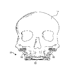
[0021] FIG. 1 is a front elevational view of a surgical positioning apparatus
and system coupled to a skeletal structure according to an embodiment of the
present invention;
[0022] FIG. 2 is a side elevational view of the surgical positioning
apparatus and system of FIG. 1;
[0023] FIG. 3 is a bottom plan view of the surgical positioning apparatus
and system of FIG. 1;
[0024] FIG. 4 is a bottom plan view of an occlusal splint of the surgical
positioning apparatus and system of FIG. 1;
[0025] FIG. 5 is a side elevational view of the occlusal splint of FIG. 4;
[0026] FIG. 6 is a bottom plan view of a pre-osteotomy positioning guide of
the surgical positioning apparatus and system of FIG. 1;
[0027] FIG. 7 is a front elevational view of the pre-osteotomy positioning
guide of FIG. 6;
[0028] FIG. 8 is a rear elevational view of the pre-osteotomy positioning
guide of FIG. 6;
[0029] FIG. 9 is a partially exploded front elevational view of a surgical
positioning apparatus and system coupled to a skeletal structure according to
another embodiment of the present invention;
6
WO 2012/012213 CA 02806035 2013-01-18 PCT/US2011/043520
[0030] FIG. 10 is a side elevational view of the surgical positioning
apparatus and system of FIG. 9;
[0031] FIG. 11 is a bottom plan view of a post-osteotomy positioning guide
of the surgical positioning apparatus and system of FIG. 9;
[0032] FIG. 12 is a front elevational view of the post-osteotomy positioning
guide of FIG. 9;
[0033] FIG. 13 is a rear elevational view of the post-osteotomy positioning
guide of FIG. 9;
[0034] FIG. 14 is a front elevational view of a surgical positioning
apparatus and system coupled to a skeletal structure according to another
embodiment of the present invention;
[0035] FIG. 15 is a side elevational view of the surgical positioning
apparatus and system of FIG. 14; =
[0036] FIG. 16 is a bottom plan view of an occlusal splint of the surgical
positioning apparatus and system of FIG. 14;
[0037] FIG. 17 is a bottom perspective view of a pre-osteotomy positioning
guide of the surgical positioning apparatus and system of FIG. 14;
[0038] FIG. 18 is a front perspective view of the pre-osteotomy positioning
guide of FIG. 17;
[0039] FIG. 19 is a side elevational view of the pre-osteotomy positioning
guide of FIG. 17;
[0040] FIG. 20 is a partially exploded front elevational view of a surgical
positioning apparatus and system coupled to a skeletal structure according to
another embodiment of the present invention;
[0041] FIG. 21 is a side elevational view of the surgical positioning
apparatus and system of FIG. 20;
[0042] FIG. 22 is a bottom perspective view of a post-osteotomy
positioning guide of the surgical positioning apparatus and system of FIG. 20;
[0043] FIG. 23 is a front perspective view of the post-osteotomy positioning
guide of FIG. 22;
[0044] FIG. 24 is a rear perspective view of the post-osteotomy positioning
guide of FIG. 22;
7
WO 2012/012213 CA 02806035 2013-01-18 PCT/US2011/043520
[0045] FIG. 25 is a side elevational view of the post-osteotomy positioning
guide of FIG. 22; and
[0046] FIG. 26 is a schematic flow diagram of a method form positioning a
skeletal structure of a patient according to an embodiment of the present
invention.
DETAILED DESCRIPTION OF EXEMPLARY
EMBODIMENTS OF THE INVENTION
[0047] The following detailed description and appended drawings describe
and illustrate various embodiments of the invention. The description and
drawings serve to enable one skilled in the art to make and use the invention,
and are not intended to limit the scope of the invention in any manner. In
respect of the methods disclosed, the steps presented are exemplary in
nature, and thus, the order of the steps is not necessary or critical.
[0048] FIGS. 1-3 illustrate a surgical positioning apparatus and system 10
for providing three dimensional (3-D) positioning and fixation of the jaws
(maxilla and mandible) and teeth (dentate skeleton) of a patient during
orthognathic (maxillofacial) surgery. As shown, the system 10 is in an initial
(pre-osteotomy or prior to a cutting procedure) configuration and includes an
occlusal splint 12 coupled to a skeletal structure 14 of the patient and a
pair of
initial or pre-osteotomy positioning guides 16 releasably coupled to the
splint
12 for marking or designating a plurality of skeletal reference points 17. The
term osteotomy is used as an illustrative example. However, it is understood
that the system 10 can be used in other operations and procedures.
[0049] As more clearly shown in FIGS. 4-5, the occusal splint 12 includes a
main body 18 molded to include a plurality of dental markings 19 to fit an
upper arch of the teeth of the patient. It is understood that the splint 12
can
be molded for the lower arch of teeth. The main body 18 includes a pair of
spaced apart connectors 20 disposed on an anterior and a posterior portion of
the splint 12, bilaterally. Each of the connectors 20 extends from the main
body 18 and includes a rounded exterior contour. A hollow cut-out or cavity
22 is formed in each of the connectors 20. As a non-limiting, the cavity 22 of
8
WO 2012/012213 CA 02806035 2013-01-18 PCT/US2011/043520
each of the connectors 20 has a generally square cross-section for precise
fitting of an associated retaining insert. However, the connectors 20 and
cavities 22 can have any size, shape, and contour. In certain embodiments,
the connectors 20 are manufactured to be easily removed from the main body
18 to minimize intraoral irritation to the patient after the surgical
operation is
complete.
[0050] As more clearly shown in FIGS. 6-8, each of the pre-osteotomy
positioning guides 16 includes a skeletal footplate 24, a splint footplate 26,
and an arm 28 coupling the skeletal footplate 24 to the splint footplate 26.
[0051] The skeletal footplate 26 is designed to substantially follow a
contour of the maxillary/malar skeleton. However, it is understood that the
skeletal footplate 24 can have any shape and size based on the specific
needs of the surgical scenario. The skeletal footplate 26 contains round
perforations 30 or apertures used as locators and guides to mark or designate
the skeletal reference points 17. As a non-limiting example, the skeletal
reference points 17 are surgically created by drilling into the skeletal
structure
14 using the perforations 30 as an exact guide. It is understood that the
drilling can be assisted by the use of commercially available "surgical
guides".
[0052] The splint footplate 26 includes a plurality of retaining inserts 32 or
male connectors for insertion into the cavity 22 formed in each of the
connectors 20 attached to the occlusal splint 12. As a non-limited example
the retaining inserts 32 have a generally square cross-section to
substantially
match the shape of an associated one of the cavities 22. It is understood that
the retaining inserts 32 can have any size and shape. It is further understood
that other means for selectively and releasably coupling the splint footplate
26
to the splint 12 can be used. As a non-limiting example, the splint footplate
26
can include a plurality of cavities (not shown) and the splint 12 can include
a
plurality of associated retaining inserts or extensions (not shown).
[0053] The arm 28 is coupled to the skeletal footplate 24 and the splint
footplate 26 to provide a pre-defined relative positioning therebetween. As a
non-limiting example a relative position and orientation of the skeletal
footplate 24 and the splint footplate 26 is pre-determined to align a portion
of
9
WO 2012/012213 CA 02806035 2013-01-18 PCT/US2011/043520
the skeletal footplate 24 with a pre-defined portion (e.g. skeletal landmarks)
of
the skeletal structure 14 of the patient. As a further non-limiting example,
the
arm 28 is designed to closely fit the anatomical contours of a facial skeleton
of
the patient. It is understood that the arm 28 can have any size and shape.
[0054] FIGS. 9-10 illustrate a surgical positioning apparatus and system
100 according to another embodiment, wherein the surgical positioning
system 100 is similar to the system 10 except as described herein below. As
shown, the system 100 is in a final (post-osteotomy or after a
cutting/sectioning procedure) configuration and includes the occlusal splint
12
coupled to an osteotomized skeletal segment 14' (e.g. below a LeFort I cut
line 102) of the skeletal structure 14 of the patient and a pair of final or
post-
osteotomy positioning guides 104 releasably coupled to the splint 12. The
term osteotomy is used as an illustrative example. However, it is understood
that the system 100 can be used in other operations and procedures.
[0055] As more clearly shown in FIGS. 11-13, each of the post-osteotomy
positioning guides 104 includes a skeletal footplate 106, a splint footplate
108,
and an arm 110 coupling the skeletal footplate 106 to the splint footplate
108.
[0056] The skeletal footplate 106 is designed to substantially follow a
contour of the maxillary/malar skeleton. However, it is understood that the
skeletal footplate 106 can have any shape and size based on the specific
needs of the surgical scenario. The skeletal footplate 106 includes a
plurality
of recessed regions 112 with a through-hole or aperture 114 formed therein.
It is understood that the apertures 114 are used as locators to align the
skeletal footplate 106 with the skeletal reference points 17.
[0057] The splint footplate 108 includes a plurality of retaining inserts 116
or male connectors for insertion into the cavities 22 formed in the connectors
20 attached to the occlusal splint 12. As a non-limited example the retaining
inserts 116 have a generally square cross-section to substantially match the
shape of the cavities 22. It is understood that the retaining inserts 116 can
have any size and shape. It is further understood that other means for
selectively and releasably coupling the splint footplate 108 to the splint 12
can
be used. As a non-limiting example, the splint footplate 108 can include a
10
WO 2012/012213 CA 02806035 2013-01-18
PCT/US2011/043520
plurality of cavities (not shown) and the splint 12 can include a plurality of
associated retaining inserts or extensions (not shown).
[0058] The arm 110 is coupled to the skeletal footplate 106 and the splint
footplate 108 to provide a pre-defined relative positioning therebetween. As a
non-limiting example a relative position and orientation of the skeletal
footplate 106 and the splint footplate 108 is pre-determined to align a
portion
of the skeletal footplate 106 with the skeletal reference points 17. As a
further
non-limiting example, the arm 110 is designed to closely fit the anatomical
contours of a facial skeleton of the patient. It is understood that the arm
110
can have any size and shape.
[0059] FIGS. 14-15 illustrate a surgical positioning apparatus and system
200 according to another embodiment of the present invention similar to the
system 10, except as described below. As shown, the system 200 is in an
initial (pre-osteotorny) configuration and includes an occlusal splint 202
coupled to a skeletal structure 204 of the patient and a pair of initial or
pre-
osteotomy positioning guides 206 releasably coupled to the splint 202 for
marking or designating a plurality of skeletal reference points 207. The term
osteotomy is used as an illustrative example. However, it is understood that
the system 200 can be used in other operations and procedures.
[0060] As more clearly shown in FIG. 16, the occlusal splint 202 includes a
main body 208 molded to include a plurality of dental markings 209 to fit both
an upper arch and lower arch of the teeth of the patient. The main body 208
includes a pair of spaced apart connectors 210 disposed on an anterior and a
posterior portion of the splint 202, generally bilaterally. Each of the
connectors 210 extends from the main body 208 and includes a rounded
exterior contour. A hollow cut-out or cavity 212 is formed in each of the
connectors 210. As a non-limiting, the cavity 212 of each of the connectors
210 has a generally square cross-section for precise fitting of an associated
retaining insert. However, the connectors 210 and cavities 212 can have any
size, shape, and contour. In certain embodiments, the connectors 210 are
manufactured to be easily removed from the main body 208 to minimize
intraoral irritation to the patient after the surgical operation is complete.
11
WO 2012/012213 CA 02806035 2013-01-18 PCT/US2011/043520
[0061] As more clearly shown in FIGS. 17-19, each of the pre-osteotomy
positioning guides 206 includes a skeletal footplate 214, a splint footplate
216,
and an arm 218 coupling the skeletal footplate 214 to the splint footplate
216.
[0062] The skeletal footplate 126 is designed to substantially follow a
contour of the mandible skeleton. However, it is understood that the skeletal
footplate 214 can have any shape and size based on the specific needs of the
surgical scenario. The skeletal footplate 216 contains round perforations 220
or apertures used as locators and guides to mark or designate the skeletal
reference points 207. As a non-limiting example, the skeletal reference points
207 are surgically created by drilling into the skeletal structure 204 using
the
perforations 220 as an exact guide. It is understood that the drilling can be
assisted by the use of commercially available "surgical guides".
[0063] The splint footplate 216 includes a plurality of retaining inserts 222
or male connectors for insertion into the cavity 212 formed in each of the
connectors 210 attached to the occlusal splint 202. As a non-limited example
the retaining inserts 222 have a generally square cross-section to
substantially match the shape of an associated one of the cavities 212. It is
understood that the retaining inserts 222 can have any size and shape. It is
further understood that other means for selectively and releasably coupling
the splint footplate 216 to the splint 202 can be used. As a non-limiting
example, the splint footplate 216 can include a plurality of cavities (not
shown)
and the splint 202 can include a plurality of associated retaining inserts or
extensions (not shown).
[0064] The arm 218 is coupled to the skeletal footplate 214 and the splint
footplate 216 to provide a pre-defined relative positioning therebetween. As a
non-limiting example a relative position and orientation of the skeletal
footplate 214 and the splint footplate 216 is pre-determined to align a
portion
of the skeletal footplate 214 with a pre-defined portion (e.g. skeletal
landmarks) of the skeletal structure 204 of the patient. As a further non-
limiting example, the arm 218 is designed to closely fit the anatomical
contours of a facial skeleton of the patient. It is understood that the arm
218
can have any size and shape.
12
WO 2012/012213 CA 02806035 2013-01-18 PCT/US2011/043520
[0065] FIGS. 20-21 illustrate a surgical positioning apparatus and system
300 according to another embodiment, wherein the surgical positioning
system 300 is similar to the system 200 except as described herein below. As
shown, the system 300 is in a final (post-osteotomy) configuration and
includes the occlusal splint 202 coupled to an osteotomized skeletal segment
204' (e.g. between a pair of cut lines 302) of the skeletal structure 204 of
the
patient and a pair of final or post-osteotomy positioning guides 304
releasably
coupled to the splint 202. The term osteotomy is used as an illustrative
example. However, it is understood that the system 300 can be used in other
operations and procedures.
[0066] As more clearly shown in FIGS. 22-25, each of the post-osteotomy
positioning guides 304 includes a skeletal footplate 306, a splint footplate
308,
and an arm 310 coupling the skeletal footplate 306 to the splint footplate
308.
[0067] The skeletal footplate 306 is designed to substantially follow a
contour of the mandible skeleton. However, it is understood that the skeletal
footplate 306 can have any shape and size based on the specific needs of the
surgical scenario. The skeletal footplate 306 includes a plurality of recessed
regions 312 with a through-hole or aperture 314 formed therein. It is
understood that the apertures 314 are used as locators to align the skeletal
footplate 306 with the skeletal reference points 207.
[0068] The splint footplate 308 includes a plurality of retaining inserts 316
or male connectors for insertion into the cavities 212 formed in the
connectors
210 attached to the occlusal splint 202. As a non-limited example the
retaining inserts 316 have a generally square cross-section to substantially
match the shape of the cavities 212. It is understood that the retaining
inserts
316 can have any size and shape. It is further understood that other means
for selectively and releasably coupling the splint footplate 308 to the splint
202
can be used. As a non-limiting example, the splint footplate 308 can include a
plurality of cavities (not shown) and the splint 202 can include a plurality
of
associated retaining inserts or extensions (not shown).
[0069] The arm 310 is coupled to the skeletal footplate 306 and the splint
footplate 308 to provide a pre-defined relative positioning therebetween. As a
13
WO 2012/012213 CA 02806035 2013-01-18 PCT/US2011/043520
non-limiting example a relative position and orientation of the skeletal
footplate 306 and the splint footplate 308 is pre-determined to align a
portion
of the skeletal footplate 306 with the skeletal reference points 207. As a
further non-limiting example, the arm 310 is designed to closely fit the
anatomical contours of a facial skeleton of the patient. It is understood that
the arm 310 can have any size and shape.
[0070] FIG. 26 illustrates a method 400 for positioning the osteotomized
skeletal segment 14', 204' of the skeletal structure 14, 204 of the patient
during an orthognathic operation according to an embodiment of the present
invention.
[0071] Initially, a skeletal data is obtained from a computerized
tomography (CT) scan or a cone beam computerized tomography (CBCT)
scan (referred to herein as scans). The data is relied upon,to generate a pre-
osteotomy model of the skeletal structure 14, 204 of the patient and a post-
osteotomy model of the skeletal structure 14, 204 of the patient, as shown in
steps 402 and 404. As a non-limiting example, the pre-osteotomy model
includes stable and identifiable skeletal landmarks that can be designated by
the skeletal reference points 17, 207. In certain embodiments, the skeletal
reference points 17, 207 are pre-defined by a computer based upon skeletal
landmarks in the pre-osteotomy model. As a further non-limiting example, the
pre-osteotomy model can be manually manipulated or automatically
manipulated based upon a surgical plan to generate the post-osteotomy
model and the post-osteotomy position of the skeletal structure 14, 204.
[0072] As shown in step 406, the occlusal splint 12, 202 is formed based
upon at least one of the pre-osteotomy model and the post operation model.
As a non-limiting example, the splint 12, 202 can be fabricated using
stereolitography technology. It is understood that the splint 12, 202 can be
customized for any patient.
[0073] As shown in step 408, the pre-osteotomy positioning guides 16, 206
are generated based on the features and contours of the pre-osteotomy
model. As a non-limiting example, the pre-osteotomy positioning guides 16,
206 are fabricated using stereolitography technology. As a further non-
14
WO 2012/012213 CA 02806035 2013-01-18 PCT/US2011/043520
limiting example, the pre-osteotomy positioning guides 16, 206 are formed
from a photo cured plastic. As another example, the pre-osteotomy
positioning guides 16, 206 are designed based on a digital model of at least
one of a pre-osteotomy (uncut) maxilla and mandible.
[0074] As shown in step 410, the post-osteotomy positioning guides 104,
304 are generated based on the features and contours of the post-osteotomy
model. As a non-limiting example, the post-osteotomy positioning guides 104,
304 are fabricated using stereolitography technology. As a further non-
limiting example, the post-osteotomy positioning guides 104, 304 are from a
photo cured plastic. As another example, the post-osteotomy positioning
guides 104, 304 are designed based on a digital model of at least one of a
post-osteotomy (cut) maxilla and mandible.
, [0076] At surgery, after exposure of the facial skeleton, the occlusal
splint
12, 202 is coupled (e.g. wired) to a portion of the skeletal structure 14, 204
(e.g. an upper arch of teeth), as shown in step 412. The pre-osteotomy
positioning guides 16, 206 are coupled to the splint 12, 202 on opposite sides
thereof, as shown in step 414. Using the apertures 30, 220 as a guide, the
skeletal reference points 17, 207 are designated (e.g. marked, drilled,
burred,
or the like) as shown in step 416. As a non-limiting example, the skeletal
reference points 17 are disposed superior to the LeFort I osteotomy line 102
on both sides of the facial skeleton. As a further non-limiting example, the
skeletal reference points 17, 207 demarcate pre-defined skeletal landmarks of
the skeletal structure 14, 204 of the patient. In certain embodiments, a tool,
sleeve, or drill guide (plastic or metal) is placed into the apertures 30, 220
of
the skeletal footplate 24, 214 to assist in the exact designation/marking.
[0076] In step 418, the pre-osteotomy positioning guides 16, 206 are
decoupled from the occlusal splint 12, 202. In step 420, a surgical operation
is performed on the skeletal structure 14, 204. As a non-limiting example, a
skeletal osteotomy or bone cut is performed accordingly to a pre-surgical
plan.
[0077] In step 422, the splint footplate 108, 308 of the post-osteotomy
positioning guide 104, 304 is coupled to the splint 12, 202. As a non-limiting
15
WO 2012/012213 CA 02806035 2013-01-18 PCT/US2011/043520
example, the post-osteotomy positioning guide 104, 304 is held in position by
inserting a "positioning jig" (not shown) through the apertures 114, 314 and
into the previously drilled skeletal reference points 17, 207. With the
apertures 114, 314 of the skeletal footplate 106, 306 aligned with the
skeletal
reference points 17, 207, the osteotomized skeletal segment 14', 204' is
accurately positioned in a post-osteotomy position relative to a stable
portion
of the skeletal structure 14, 204, as shown in step 424. The osteotomized
skeletal segment 14', 204' is secured with known rigid fixation techniques to
maintain the post-osteotomy position dictated by the alignment of the post-
osteotomy positioning guide 104, 304.
[0078] It is understood that interrnaxillary wire fixation (IMF) is
unnecessary and a final maxillary three-dimensional skeletal positioning and
fixation is performed independent of the mandible and determination of CR.
[0079] It is further understood that the systems 10, 100, 200, 300 and the
method 400 of the present invention can be applied to orthognathic surgery of
a lower jaw or mandible. Positioning of a proximal segment of the mandible is
also determined through skeletal reference points drilled into the skeletal
structure 14, 204 of the patient before osteotomizing the bone at the anterior
(ramus) border of the mandible. After the mandible is osteotomized, the distal
(dentate) segment of the mandible is repositioned, while preserving the pre-
surgical natural CR of the mandibular condyles. In a case in which only the
mandible is moved, there is only the need for one mandibular "guide".
[0080] The systems 10, 100, 200, 300 and the method 400 of the present
invention eliminate the need for external reference points, pins, wires, bulky
and expensive head gear and calipers, for positioning teeth and jaws in
orthognathic surgery. The systems 10, 100, 200, 300 and method 400 of the
present invention simplify orthognathic surgery and minimize a time needed
for orthognathic surgery by eliminating the intra-operative need for wiring
teeth together. The systems 10, 100, 200, 300 and the method 400 of the
present invention minimize error in orthognathic surgery by eliminating the
need for knowing (intra-operatively) the CR of the mandible when performing
surgery on the upper jaw and teeth. The systems 10, 100, 200, 300 and the
16
WO 2012/012213 CA 02806035 2013-01-18 PCT/US2011/043520
method 400 of the present invention allow for single splint surgery,
eliminating
the need for intermediate and final splints when performing double jaw
surgery.
[0081] The systems 10, 100, 200, 300 and the method 400 of the present
invention allow a surgeon to make precise skeletal changes to an original
position of a patient's teeth and jaws (dentate skeleton) without external
references and independent of centric relation (CR). The systems 10, 100,
200, 300 and the method 400 of the present invention anatomically and
accurately solve the problems inherent to the prior art. The invention is
reproducible, affordable, and easily applicable in the operative field,
greatly
reducing operative time.
[0082] From the foregoing description, one ordinarily skilled in the art can
easily ascertain the essential characteristics of this invention and, without
departing from the spirit and scope thereof, make various changes and
modifications to the invention to adapt it to various usages and conditions.
17