Note : Les descriptions sont présentées dans la langue officielle dans laquelle elles ont été soumises.
DEVICES, SYSTEMS, AND METHODS FOR IMPROVING ACCESS TO CARDIAC
AND VASCULAR CHAMBERS
FIELD
[00021 The present disclosure provides devices and methods for improved
cardiac and vascular
access to allow minimally invasive replacement or repair of cardiac and
vascular structures.
Such devices and methods rely on increasing the strength of the heart tissue
or vascular wall to
allow safer manipulation and reduced potential for catastrophic side effects.
BACKGROUND
[0003] Various types of surgical procedures are currently performed to
investigate, diagnose,
and treat certain cardiovascular disorders. Such procedures include repair and
replacement of
mitral, aortic, and other heart valves, repair of atrial and ventricular
septal defects, pulmonary
thrombectomy, treatment of aneurysms, electrophysiological mapping and
ablation of the
myocardium, and other procedures in which interventional devices are
introduced into the
interior of the heart or a vascular structure.
[0004] Using current techniques, many of these procedures require a gross
thoracotomy to gain
access into the patient's thoracic cavity and cardiac or vascular structures.
A relatively large
opening into the thoracic cavity is created through which the surgical team
may directly visualize
and operate upon the heart and other thoracic contents. Open-chest valve
replacement surgery
has the benefit of permitting the direct implantation of the replacement valve
at its intended site.
This method, however, is highly invasive and often results in significant
trauma, risk of
complications, as well as an extended hospitalization and a painful recovery
period for the
patient.
[0005] Minimally invasive valve replacement procedures have emerged as an
alternative to
open-chest surgery. Two types of minimally invasive valve procedures that have
emerged are
percutaneous valve procedures and trans-apical valve procedures. Percutaneous
valve
1
CA 2813592 2017-12-05
CA 02813592 2013-04-03
WO 2012/048005 PCT/US2011/054932
procedures pertain to making small incisions in the skin to allow direct
access to peripheral
vessels or body channels to insert catheters. Trans-apical valve procedures
pertain to making a
small incision in or near the apex of a heart to allow valve access. Because
minimally invasive
approaches require smaller incisions, they generally allow for faster patient
recovery with less
pain and bodily trauma. This, in turn, reduces the medical costs and the
overall disruption to the
life of the patient.
[0006] Minimally invasive trans-apical valve replacement procedures have
emerged as an
alternative to both open chest surgery and percutaneous valve surgeries. The
use of minimally
invasive approaches, however, highlights certain complexities in the surgery.
Unlike open heart
surgery, minimally invasive heart surgery offers a small surgical field that
greatly reduces the
surgeon's field of view and, consequently, the ability of the surgeon to
detect complications as
they arise. U.S. Patent Publication No. 2005/0240200 to Bergheim et al.
presents certain
methods and systems for the repair, removal, and/or replacement of heart
valves through the
apex of the heart. Similarly, U.S. Patent Publication No. 2007/0112422 to
Dehdashtian provides
a delivery system and method for delivering heart valves via a device that
passes through the
apex of the left ventricle.
[0007] Although there are a number of methods and devices available to assist
in these
procedures, the incidence of complications remains high, particularly in high
risk and elderly
populations. See for example, Hsieh, et al. 2010 Circulation: Arrhyth.
Electrophys. 3:178-185,
Pasic, et al. (2010) J. Am. Coll. Cardiol. 56:813-20 and Walther et al (2008)
Ann Thorac Surg.
85(3): 1072-1073.
SUMMARY
[0008] There remains a need for improved methods and devices that allow the
surgical
manipulation through trans-apical or trans-cardiac wall access while reducing
the likelihood that
a patient's heart muscle will weaken or tear and release the access device. If
such methods and
devices can provide security from bleeding and tissue disruption during and
after the period of
access to the cardiac chambers or vascular structure, then such access may not
require even a
minimally invasive incision or thoracotomy, but may be performed
percutaneously, potentially
under local anesthesia.
2
CA 02813592 2013-04-03
WO 2012/048005 PCT/US2011/054932
[0009] In some embodiments, the disclosure relates to the use of certain
devices, compositions
and methods for enhancing the strength of tissue at a cardiac and/or vascular
chamber access site
(also referred to access channel). In some embodiments, the disclosure
provides methods,
systems and devices that are configured or structured to stabilize the access
channel by
mechanically enhancing or strengthening the access channel. In some
embodiments, the
methods, systems, and devices relate to delivering energy to stabilize the
access channel. In
other embodiments, the methods, systems, and devices relate to delivering a
tissue-stabilizing
composition to stabilize the access channel in addition to or in alternative
of delivering energy to
stabilize the access channel.
[0010] In some embodiments, the disclosure relates to heart access devices and
systems. The
heart access devices and systems may be configured or structured to provide a
mechanically
enhanced access channel within tissue or muscle of the heart. According to
some embodiments,
the heart access device may include a sheath including an open channel
configured or structured
to accept an interventional device, the sheath being configured to be inserted
into the access
channel. The heart access device may further include at least one energy-
transducing component
configured to deliver energy to tissue surrounding the sheath, the energy-
transducing component
being configured to cause the surrounding tissue to stabilize around the
sheath. The heart access
device may include a plurality of energy-transducing components, the plurality
of energy-
transducing components being disposed in a pattern. In some embodiments, the
energy-
transducing component may be configured to heat a tissue-stabilizing
composition.
[0011] In some embodiments, the heart access device may include a sleeve. The
sleeve may
include the at least one energy-transducing component. The sleeve may be
configured to
surround the sheath. The sleeve may be configured to be movable with respect
to the sheath.
[0012] In other embodiments, the device may include an introducer configured
to form the
access channel. In some embodiments, the sleeve may be configured to surround
the introducer.
[0013] In some embodiments, the device may include a sheath including an open
channel
configured to accept an interventional device; and an introducer including at
least one least one
energy-transducing element configured to delivery energy. The open channel of
the sheath may
be configured to accept the introducer, the introducer being movably disposed
with respect to the
sheath. The open channel may also be configured to accept another device
configured to
3
CA 02813592 2013-04-03
WO 2012/048005 PCT/US2011/054932
manipulate a valve or inner cardiac chamber of the heart. The introducer may
include an inner
channel configured to accept a guidcwirc.
[0014] In some embodiments, the introducer may include a section that has a
cross section that is
equal or slightly less than a diameter of the inner channel of the sheath. The
introducer may
include a puncture tip. In some embodiments, the introducer may include a
guide member
having a cross section that is larger than an inner diameter of the inner
channel.
[0015] In some embodiments, the device may further include a power source, the
power source
configured to deliver power to the at least one energy-transducing element. In
some
embodiments, the sheath may include at least one energy focusing or dispersing
element, the at
least one energy focusing element being configured to focus the energy on at
least one of
surrounding tissue or a tissue-stabilizing composition surrounding a portion
of the heart access
device. The at least one energy focusing component may correspond to or
compliment the
energy-transducing component. In some embodiments, the energy-transducing
component may
be configured to deliver microwave, ultrasound, radiofrequency (RF), or heat
energy.
[0016] In some embodiments, the device may further include a sealing device
configured to seal
the access channel. The sealing device may be a plug. In some embodiments, the
sealing device
may be of a wound bioabsorbable material and /or a pre-formed hydrophilic
material. In further
embodiments, the sealing device may further include a base. The base may
include an open
channel configured to be disposed on a sealing device delivery device, such as
an introducer. In
some embodiments, the sealing device may further include extending members
that extend from
an elongated section constructed or made of a wound bioabsorbable material and
/or a pre-
formed hydrophilic material. The extending members may be constructed or made
of a
bioabsorable material. In some embodiments, the sealing device may further
include a clip
member. The clip member may be constructed or made of a memory shape alloy.
[0017] In some embodiments, the device may further include a sealing device
introducer, the
sealing device introducer configured to be anchor the sealing device within
the access channel.
The sealing device introducer may include external threads and the sheath may
include internal
threads, the threads of the sealing device introducer and the threads of the
sheath being
complementary. The sealing device introducer may further include a release
mechanism
configured to release the sealing device within the access channel.
4
CA 02813592 2013-04-03
WO 2012/048005 PCT/US2011/054932
[0018] In some embodiments, the sealing device may further include at least
one sensor. The
sensor may be configured to monitor cardiac conduction currents in myocardium.
The sealing
device may be configured to be anchored to the surrounding tissues by a
fastener. The fastener
may include one or more needles mounted in a pattern.
[0019] In some embodiments, the device may further include an energy source,
the energy
source configured to deliver energy through the heart access device. The heart
access device
may be configured to focus the energy on at least one of surrounding tissue or
a tissue-stabilizing
composition surrounding a portion of the heart access device.
[0020] In some embodiments, the disclosure provides a heart access device that
may include a
sheath including an open channel configured to accept an interventional
device; and a delivery
device configured to deliver a tissue-stabilizing composition into, around, or
adjacent to the
tissue surrounding the sheath, the tissue-stabilizing composition configured
to mechanically
enhance the tissue surrounding the sheath. In further embodiments, the heart
access device may
include an energy-transducing element configured to deliver energy to at least
one of the tissue
surrounding the sheath and the tissue-stabilizing composition, the energy-
transducing component
being configured to cause the surrounding tissue to stabilize around the
sheath.
[0021] In some embodiments, the disclosure provides a device configured to
provide access to
the chambers of a beating heart or a vascular conduit. The device may include
a sheath including
an open channel configured to be inserted into a muscle of the heart or a wall
of the vascular
conduit to access an inner chamber of the heart or a vascular lumen of the
vascular conduit, and
configured to accept an interventional device. The device may include an
introducer including
atleast one energy-transducing element configured to deliver energy to the
tissue surrounding the
sheath for stabilization and strengthening. The device may also include
sealing device
configured to be delivered into the access channel.
[0022] The sealing device may be configured to close the access channel
permanently or
reversibly so that the access channel may be accessed at a later time.
[0023] In some embodiments, the sheath may include a section constructed of a
material that
conducts energy than remainder of the sheath. The sheath may include a section
that has a
different thickness than the remainder of the sheath. The sheath may include a
section that is
configured to focus energy to a specific location in the tissue surrounding
the sheath.
CA 02813592 2013-04-03
WO 2012/048005 PCT/US2011/054932
[0024] The device may further include a sleeve. The sleeve may include at
least one energy-
dispersing element or energy-transducing element. The sleeve may be structured
to surround the
sheath. The sleeve may be movable with respect to the sheath. The device may
further include
an sealing device configured to be inserted into the open channel of the
sheath, the introducer
being disposed with respect to the sheath. The introducer may include a
section having at least
one or more energy-dispersing elements or energy-transducing elements. The
energy-dispersing
elements or energy-transducing elements being disposed in a pattern. The
introducer may
include an access channel.
[0025] The sleeve may include at least one energy-dispersing element or energy-
transducing
element configured to surround the introducer. The energy-transducing element
may be
configured to deliver a plurality of forms of energy, the forms may include
heat, radio-frequency,
ultra-sound or microwave. The sealing device may include a first section that
is configured to
close or seal the access channel and a second section configured to enable
releasably attachment
to a delivery introducer. The sealing device may be constructed of one of or
any combination of
a biological material, a biocompatible polymer, or a metal.
[0026] The device may further include a sealing device introducer configured
to deliver the
sealing device through the sheath into the access channel. The sealing device
introducer may
include threads on an outside surface and the sheath may include the threads
within the channel.
The threads of the sealing device introducer and the threads of the sheath may
be
complementary.
[0027] In certain embodiments, the disclosure provides a heart access device
that allows
insertion through a heart wall and includes an energy-transducing element on
at least one portion
of an insertion sleeve. In certain embodiments, the heart access device may
include a sheath
with an open channel configured to accept an interventional device. In further
embodiments, the
device may include a sleeve that is attached to or makes up at least a part of
the sheath, wherein
the sleeve is configured to provide energy to a surrounding tissue. In certain
embodiments, the
sleeve may be configured to heat the surrounding tissue. In other embodiments,
the energy-
transducing element (device) may be introduced separately from the sleeve
during the procedure.
In some embodiments, the heart access device may include a sheath with an open
channel
configured to accept an interventional device, and a sleeve that is attached
to or makes up at least
a part of the sheath, wherein the sleeve is configured to provide a tissue
strengthening
6
CA 02813592 2013-04-03
WO 2012/048005 PCT/US2011/054932
composition to surrounding tissue. Typically, the sleeve may be a sheath, tube
or cannula. The
sleeve is typically configured so as to provide a rigid channel through the
wall of the heart.
Typically, the sleeve may include an energy-transducing element on its tip. In
certain
embodiments, the element may be a heating element. In some embodiments, the
element may
surround the sleeve. The element may include coils that surround the sleeve.
The element may
be typically configured to surround the sleeve for at least 3mm, or at least
6mm, or at least 9mm,
or at least 12mm, or at least 15mm. Typically, the element may surround the
sleeve for a
distance sufficient to contact a portion of the tissue at the wall of a heart,
but does not expand
beyond the wall of the heart.
[0028] The energy-transducing element may be a mechanism for providing high
frequency
energy, which can include radiofrequency, ultrasound or microwave energy. In
some
embodiments, the sleeve may include a tissue contacting member that includes
an array of
electrodes which can penetrate the tissue surrounding the sleeve. Typically,
the electrodes may
include a radiofrequency electrode, a focused ultrasound electrode (i.e.
transducer) or a
combination of these. In some embodiments, the sleeve may include a tissue
contacting coil for
generating heat. The term "heating element" as used herein encompasses
elements that apply
energy thereby inducing heat in the tissue as well as to elements that apply
heat to the tissue. In
a preferred embodiment, the tissue may be heated to a temperature in the range
of about 40
degrees Celsius to about 110 degrees Celsius, more preferably about 60 degrees
Celsius to about
65 degrees Celsius.
[0029] In some embodiments, the disclosure provides a method of accessing a
cardiac chamber
including: (i) providing an access channel into the chamber; (ii) providing an
energy-transducing
element configured to provide heat or to cool tissue surrounding the access
channel; and (iii)
applying energy to the tissue. In certain embodiments, the method may further
include
modifying the tissue with a strength-enhancing compound (may also be referred
to as a -tissue-
stabilizing composition" or "tissue-stabilizing compound") prior to applying
the energy. In some
embodiments, the energy-transducing element may heat the tissue.
[0030] In some embodiments, the disclosure provides a method for accessing a
cardiac chamber
or a vascular conduit. The method may include: providing an access channel
into tissue of the
chamber or the conduit; providing an energy-transducing element configured to
provide heat
within the access channel; and applying energy to the tissue or a tissue-
stabilizing composition
7
CA 02813592 2013-04-03
WO 2012/048005 PCT/US2011/054932
injected into the tissue to mechanically enhance the access channel. The
energy may be applied
to the tissue, and the energy heats the tissue. In some embodiments, the
method may further
include delivering the tissue-stabilizing composition prior to applying the
energy. The energy
may additionally or alternatively be applied to the tissue-stabilizing
composition.
[0031] In other embodiments, the disclosure provides a method of accessing a
cardiac chamber
including: (i) providing an access channel into the chamber; (ii) modifying
the tissue surrounding
the access channel with a strength-enhancing compound; and (iii) closing the
access channel.
[0032] In some embodiments, the method may further include delivering a tissue-
stabilizing
composition prior to applying the energy. The energy may be applied to the
tissue-stabilizing
composition. In further embodiments, the method may further include
positioning or inserting a
sealing device into the access channel.
BRIEF DESCRIPTION OF THE FIGURES
[0033] The disclosure can be better understood with the reference to the
following drawings and
description. The components in the figures are not necessarily to scale,
emphasis being placed
upon illustrating the principles of the disclosure.
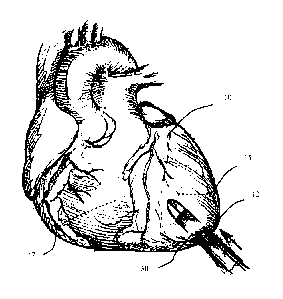
[0034] Figure 1 is a diagram of the heart, showing an access port or channel
through the apex.
[0035] Figures 2(A) ¨ (D) are diagrams detailing the Seldinger method for
providing tissue
access according to embodiments. (A) shows a guidewire being inserted through
a trocar into
the tissue. (B) shows retraction of the needle. (C) shows the rotational
insertion of a catheter
over the guidewire to increase the diameter of the access port. (D) shows
insertion of the sheath
into the access port.
[0036] Figure 3 shows the initial insertion of the sheath of the disclosure.
[0037] Figure 4 is a diagram of a heart access device according to
embodiments.
[0038] Figure 5 shows the insertion of a heart access device into the tissue
of the heart according
to embodiments.
[0039] Figure 6 shows a detail of injection of a tissue-stabilizing
composition into the tissue
surrounding the access port (also referred to as "access channel")..
[0040] Figure 7 shows a different embodiment detail of injection of a tissue-
stabilizing
composition into the tissue surrounding the access port.
[0041] Figure 8 shows inflation of a balloon to retract the sleeve into the
appropriate position.
8
CA 02813592 2013-04-03
WO 2012/048005 PCT/US2011/054932
[0042] Figure 9 shows insertion of a sealing to close the access port, and
removal of the
remainder of the sheath from the sleeve.
[0043] Figures 10 and 11 show a heart access device including a fastener
according to
embodiments.
[0044] Figure 12 shows an introducer according to embodiments.
[0045] Figure 13 shows a sheath according to embodiments.
[0046] Figure 14 shows a heart access device according to embodiments.
[0047] Figures 15(A)-(C) show a heart access device according to different
embodiments.
[0048] Figures 16(A)-(D) show sealing devices according to embodiments.
[0049] Figure 17 shows a sheath according to embodiments.
[0050] Figure 18 shows a sealing device introducer according to embodiments.
[0051] Figure 19 shows a detail of identifying the location for performing a
method according to
embodiments.
[0052] Figure 20 shows a detail of inserting an access device according to
embodiments.
[0053] Figure 21 shows a detail of positioning the access device according to
embodiments.
[0054] Figure 22 shows a detail of applying energy to surrounding tissue
according to
embodiments.
[0055] Figure 23 shows a detail of positioning the sheath to perform medical
procedures
according to embodiments.
[0056] Figure 24 shows a detail of introducing a sealing device introducer
according to
embodiments.
[0057] Figure 25 shows a detail of positioning the sealing device according to
embodiments.
[0058] Figure 26 shows a detail of retracting the access device according to
embodiments.
[0059] Figure 27 shows the removal of the access device according to
embodiments.
[0060] Figure 28 shows an example of a heart access device according to
embodiments.
[0061] Figures 29 through 34 show details of the method according to
embodiments performed
on a pig heart.
[0062] Figures 35 through 39 show results of the method shown in Figures 29
through 34.
9
CA 02813592 2013-04-03
WO 2012/048005 PCT/US2011/054932
DETAILED DESCRIPTION
[0063] The present disclosure provides methods, devices, and systems for
providing access to
the heart or heart vessels to perform cardiovascular surgery. The access may
be provided by
forming an access channel through the heart tissue or muscle (the myocardium).
[0064] The methods may include the step of, and devices, and systems may be
configured or
configured to provide stable access to the heart tissue or muscle. The
methods, devices, and
systems are configured or structured to stabilize the heart muscle or tissue
surrounding the access
channel so as to prevent the release of an access device. The methods,
devices, and systems
include a heart access device that includes a sheath. The methods, devices,
and systems are
configured or structured to stabilize the heart muscle or tissue surrounding
the sheath so as to
prevent the release of an access device. The methods, devices, and systems are
configured or
structured to mechanically enhance (strengthen) the tissue of the access
channel.
[0065] In some embodiments, the methods may include the step, devices and
systems may be
configured or structured to deliver heat or energy passively to the heart
tissue through the sheath
causing the heart tissue to shrink tightly around the sheath and seal the
tissue around the sheath.
This results in stable access to the channel inside the sheath to perform
interventional and
diagnostic procedures. The energy may be applied by transducers provided on a
wall of the
sheath (directly or indirectly by a sleeve) or by a heating element or energy
source (e.g., high-
energy focused ultrasound) built into an introducer.
[0066] In other embodiments, the methods may include the steps of, and
devices, and systems
may be configured to deliver a tissue-stabilizing composition. in addition to
or in alternative to
delivering energy, to the surrounding tissue. The tissue-stabilizing
composition may further
mechanically enhance or stabilize the access channel.
[0067] According to embodiments, because the tissue surrounding the sheath has
been stabilized
(mechanically enhanced), it is possible to seal the access channel with a
sealing device.
General Method to Access the Heart
[0068] Transapical cardiac surgery is not a new procedure. Levy and Lillehei
described a
technique for percutaneous direct cardiac catheterization in 1964 (Levy and
Lillehei (1964)
NEJM 271:273-280). The technique has been used since then, however
percutaneous venous
access is typically preferred. U.S. Patent Publication No. 2007/0112422
describes a general
CA 02813592 2013-04-03
WO 2012/048005 PCT/US2011/054932
method and device for transapical heart valve delivery system. The method
generally includes
inserting an instrument through the subject's chest wall and through the heart
wall. The
instrument carries on its distal end a movable element which is manipulated to
grasp a valve
leaflet and hold it while a needle mechanism punctures the valve leaflet and
loops a suture
around a portion of the valve leaflet.
[0069] A general method of introducing a stent or sleeve into a heart apex is
diagrammed in
Figure 1. The access system 30 is shown penetrating through the apex 12 of the
heart 10. The
moving direction of the access system is indicated by the arrow. The access
system may enter
either the right ventricle 17 or the left ventricle 15. To access the aortic
or mitral valve, the
access system typically passes through the left ventricle. This yields direct
access to the aortic or
mitral valve. To access the pulmonary or tricuspid valve, the access system
would typically pass
through the right ventricle.
[0070] The access system is diagrammed in Figure 2. Typically, the technique
used is the
Seldinger technique for progressive dilation of the access channel. The access
channel may be
formed in a blood vessel as shown in Figure 2 or in a cavity, such as a heart
chamber as shown in
Figure 1. An access system 25 (here shown as a typically sharp hollow needle
called a trocar)
may be used to puncture the desired vessel or cavity, with ultrasound guidance
if necessary to
form an access channel. A round-tipped guidewire 22 may then advance through
the lumen of
the trocar, and the trocar is withdrawn. A blunt cannula or introducer 29 may
then be passed
over the guidewire into the cavity or vessel to increase the size of the
opening. In the alternative,
an introducer having a puncture tip at the end may be used to puncture the
desired vessel or
cavity. After the opening is of the appropriate size, a tube or sheath 40 may
be introduced, in
this instance including a sleeve as described herein and the guidewire may be
withdrawn.
[0071] The tube or sheath 40 may be used to introduce catheters or other
devices to perform
endoluminal (inside the hollow organ) procedures, such as angioplasty.
Fluoroscopy may be
used to confirm the position of the catheter and to maneuver it to the desired
location. Injection
of radiocontrast may be used to visualize organs. Interventional procedures,
such as
thermoablation, angioplasty, embolization or biopsy, may be performed. Upon
completion of the
desired procedure, the access port may be closed as described herein and the
tube or sheath may
be withdrawn. In certain embodiments, a sealing device may be used to close
the hole made by
the procedure.
11
CA 02813592 2013-04-03
WO 2012/048005 PCT/US2011/054932
[0072] In addition to grasping and needle mechanisms, instruments used for
repair procedures
often include fiber optics that provide direct visual indication that the
valve leaflet is properly
grasped. A set of illuminating fibers terminate at the distal end of the
instrument around the
needle mechanism in close proximity to a set of sensor fibers. The sensor
fibers may convey
light from the distal end of the instrument to produce an image for the
operator. When a valve
leaflet is properly grasped, light from the illuminating fibers may be
reflected off the leaflet
surface back through the sensor fibers. On the other hand, if the valve
leaflet is not properly
grasped, the sensor fibers may sense or view blood.
[0073] The present disclosure provides methods, devices and systems for
performing
cardiovascular surgery, wherein access to the heart or great vessels may be
provided through the
heart muscle. In preferred embodiments, access may be provided through the
apical area of the
heart. The apical area of the heart is generally the blunt rounded inferior
extremity of the heart
formed by the left and right ventricles. In normal healthy humans, it
generally lies behind the
fourth or fifth left intercostal space in the mid-clavicular line.
[0074] The unique anatomical structure of the apical area permits the
introduction of various
surgical devices and tools into the heart without significant disruption of
the natural mechanical
and electrical heart function. While access to the heart through peripheral
(e.g. femoral, jugular,
etc.) vessels in percutaneous methods are limited to the diameter of the
vessel (approximately 1
to 8 mm), access to the heart through the apical area may be significantly
larger (approximately 1
to 25 mm or more). Moreover, apical access is dramatically closer to
intracardiac structures than
access through peripheral vessels. Thus, apical access to the heart permits
greater flexibility with
respect to the types of devices and surgical methods that may be performed in
the heart and great
vessels.
[0075] It should be noted that while reference is made herein of trans-apical
procedures, it is
intended for such procedures to encompass access to the heart through any wall
thereof, and not
to be limited to access through the apex only. While the apical area is
particularly well suited for
the purposes of the present disclosure, for certain applications, it may be
desirable to access the
heart at different locations, all of which are within the scope of the present
disclosure.
12
CA 02813592 2013-04-03
WO 2012/048005 PCT/US2011/054932
DEVICES AND SYSTEMS
[0076] According to embodiments, the access devices and systems may be
configured or
structured to strengthen a patient's heart muscle so as to reduce the
likelihood that the patient's
heart muscle will weaken and release an access device.
[0077] In some embodiments, the access device may include at least one sheath.
In some
embodiments, the access device may include one sheath. In other embodiments,
the access
device may include one sheath that has more than one section. In certain
embodiments, the
access device may include more than one sheath. Each sheath may have one or
more than one
section.
[0078] Referring to Figures 3 and 4, in some embodiments, the access device
400 may include
the sheath 40. The sheath 40 may be of any shape. The sheath 40 may be in the
form of an
elongated tube. The sheath 40 may include an interior bore or channel 42
extending between its
proximal and distal ends.
[0079] In certain embodiments, the sheath 40 may include more than one
section. In some
embodiments, the sheath 40 may include a first section that is relatively
stiff and a second
section that is relatively flexible. In certain embodiments, the sheath 40 may
include a relatively
stiff wall section extending from its distal end 41 to juncture 43 (also
referred to as "distal
section"), and a relatively limber wall section throughout the rest of the
sheath (also referred to
as "proximal section"). Alternatively, the sheath may have stiffness
throughout its entirety.
[0080] The sheath 40 may be arranged so that at least one section is
"torquable." That is, at least
the proximal section of the sheath may be arranged to transmit torsional
motion about its axis.
Thus, by turning the proximal end of the sheath, the distal end of the sheath
may also be rotated
about an axis.
[0081] In some embodiments, the access device 400 may be configured to receive
or accept and
introduce medical instruments for procedures to be performed such
interventional and diagnostic
procedures to a chamber of the heart. The inner channel 42 of the sheath 40
may be configured
to receive or accept medical instruments. In other embodiments, an end of the
access device 400
may be configured to connect to an interventional and/or diagnostic device.
The access device
400 may be configured to connect to a connecting device.
[0082] In some embodiments, the access device may be configured to introduce
catheters or
other devices to perform endoluminal (inside the hollow organ) procedures,
such as angioplasty.
13
CA 02813592 2013-04-03
WO 2012/048005 PCT/US2011/054932
Fluoroscopy may be used to confirm the position of the catheter and to
maneuver it to the desired
location. Injection of radiocontrast may be used to visualize organs.
Interventional procedures,
such as thermoablation, angioplasty, embolization, biopsy, or deployment of
stents or
replacement or repair valve devices may be performed.
[0083] The device 400 may be configured to receive or accept imaging devices.
The system
may also further include imaging devices. In addition to grasping and needle
mechanisms,
instruments used for repair procedures often include fiber optics that provide
direct visual
indication that the valve leaflet is properly grasped. A set of illuminating
fibers terminate at the
distal end of the instrument around the needle mechanism in close proximity to
a set of sensor
fibers. The sensor fibers convey light from the distal end of the instrument
to produce an image
for the operator. When a valve leaflet is properly grasped, light from the
illuminating fibers is
reflected off the leaflet surface back through the sensor fibers. On the other
hand, if the valve
leaflet is not properly grasped, the sensor fibers see blood.
[0084] The heart access device 400 may further include a manipulating
instrument 44 that is
slidably mounted thereon and that may be configured to be manipulated. The
manipulating
instrument 44 may be slidably mounted onto the sheath. The manipulating
instrument 44 may be
a guidewire.
[0085] An expandable balloon may be advanced over the guide wire into the
cardiac chamber,
illustratively in the form of balloon 80. When inflated, as depicted in Figure
8, the balloon may
generally be in the form of a surface of revolution about a central axis
coincident with proximal-
to-distal axis of the catheter. The diameter of the balloon may typically be
about 20 mm and
may usually be formed from a polymer such as nylon with a wall thickness of
about 8 microns to
about 30 microns. When deflated, the balloon may collapse inwardly to form a
relatively small
diameter structure. The balloon may be fabricated by blow-molding using
techniques that are
known in the art. Typically, the balloon may be advanced after insertion of
the sheath and
expanded within the ventricle. After inflation, the balloon may then be pulled
on to move the
sleeve 45 so that the distal end of the sleeve is coincident with the interior
of the tissue. This
positioning of the sheath may be accomplished either before or after the
tissue strengthening
procedure.
[0086] In some embodiments, the access device may include at least one energy-
transducing
component or element. The energy-transducing component (also referred to as
"energy-
14
CA 02813592 2013-04-03
WO 2012/048005 PCT/US2011/054932
transducing element) may be configured to heat and thus denature collagen
fibers and other
proteins within the surrounding tissue. The energy-transducing element may be
a mechanism for
providing high frequency energy, which may include radiofrequency, ultrasound
or microwave
energy. The energy-transducing element may be a heating element configured to
heat and thus
denature collagen fibers within the surrounding tissue. In some embodiments,
the energy-
transducin g element may be configured to deliver energy to the surrounding
tissue as described
herein. In further embodiments, the energy-transducing element may be
configured to convert
radio-frequency into heat. The energy-transducing component may be configured
to transmit
energy when connected to a power source.
[0087] In some embodiments, the access device 400 may include a sleeve 45. The
sleeve 45
may include may have an interior bore or channel 47 extending between its
proximal and distal
ends.
[0088] In some embodiments, the sheath 40 may include the sleeve 45 that has
an energy-
transducing component 48, as shown in Figure 4. In some embodiments, the
sleeve 45 may be
provided on all or a portion of the sheath 40. In other embodiments, the
sleeve 45 may be
attached to or extended from the sheath 40.
[0089] Figures 15(A)-(C) show another example of an access device including a
sheath. In some
embodiments, the energy-transducing component(s) may be disposed on a sleeve
configured to
be movably disposed around a sheath, as shown in for example, Figure 15.
[0090] Figures 15(A) - (C) show an access device 1500 having a sheath 1510
with a sleeve 1520.
The sleeve 1520 may include a plurality of energy-transducing components 1522.
As shown in
Figures 15(A) and 15(B) show how the sleeve 1520 may be configured to be
movable with
respect to the sheath 1510. Figure 15(C) shows an example of the access device
1500 being
positioned within an access channel in the myocardium 1530.
[0091] In other embodiments, the energy-transducing component(s) may be
integrated with the
sheath.
[0092] In some embodiments, the sleeve 45 may include one energy-transducing
component. In
other embodiments, the sleeve may include more than energy-transducing
components. The
sleeve may be typically made out of a thromboresistant, biocompatible material
such as pyrolytic
carbon. Pyrolytic carbon is a turbostratic carbon, which are materials that
are structurally similar
CA 02813592 2013-04-03
WO 2012/048005 PCT/US2011/054932
to graphite but have greater durability. The sleeve may also be made out of
some material that
can withstand heat.
[0093] In some embodiments, the energy-transducing component may include an
ultrasonic
transducer. In some embodiments, the heart access device may include a
tubular, cylindrical
ultrasonic transducer. The sleeve typically may include a tubular, cylindrical
ultrasonic
transducer. The transducer may be mounted on the sleeve. Such a transducer may
be coaxial or
nearly coaxial with the sleeve, and arranged so that the transducer extends
axially over at least
part of the sleeve. Merely by way of example, the transducer may have an axial
length of about
6 mm and an outside diameter of about 2-3 mm. Typically, the proximal end of
the transducer
will not extend out of the tissue when the sleeve is anchored or placed within
the heart. In some
embodiments, the transducer may be formed from a ceramic piezoelectric
material. The tubular
transducer may have metallic coatings on its interior and exterior surfaces.
[0094] In some embodiments, the sheath 40 may include RF electrode wires
inside the sleeve 45,
and the electrodes are surrounded by an insulating sleeve axially moveable
thereon; the sleeve is
retracted to expose a predetermined portion of the electrode; and RF energy is
applied to the
tissue through the electrode to cause heating of the tissue. In other
embodiments, an RF
electrode wire is positioned on the sleeve 45 and is exposed to tissue upon
insertion of the sleeve.
[0095] In some embodiments, the heart access devices and systems may further
include a wiring
support tube 49. The wiring support tube 49 may be provided within along all
or a portion of the
inner channel of the sheath. In further embodiments, the wiring support tube
49 may be provided
within along all or a portion of the inner channel of the sleeve. In some
embodiments, the wiring
support tube may be connected to one of the sheath and/or the sleeve. The
wiring support tube
49 may be provided at an end of the heart access device.
[0096] The heart access devices and systems may further include a catheter
that is configured to
deliver the wires to the transducer. The wires may be configured to extend
through wiring
support tube 49 to the distal end of the catheter. These wires may extend
through the catheter to
the proximal end of the catheter, and may be configured to be connected to an
ultrasonic
excitation or power source. Metallic support tubes and transducers may be
typically configured
so that the interior surface of the tubular transducer is spaced apart from
the exterior surface of
the tube by a gap distance which corresponds to approximately one-half the
wavelength of the
ultrasonic energy to be applied, i.e., about 83 microns for 9 MHz ultrasonic
energy propagating
16
CA 02813592 2013-04-03
WO 2012/048005 PCT/US2011/054932
in water. This promotes efficient operation of the transducer, with ultrasonic
energy reflected at
the exterior surface of support tube reinforcing ultrasonic energy propagating
within the
transducer, so as to provide ultrasonic energy directed outwardly from
external surface of the
transducer.
[0097] With the sleeve in place and in contact with the tissue, the energy-
transducing element or
component 48 may be configured to be activated. The energy-transducing element
48 may be
activated by an energy or power source. In some embodiments, the energy-
transducing element
48 may be configured to provide energy to surrounding tissue when the energy
source is
provided within an inner channel of sheath. In further embodiments, the energy
source may be
additionally or alternative provided within an inner channel of the sleeve 45.
[0098] In some embodiments, the energy source may be configured for ultrasound
energy. In
one embodiment, an ultrasonic excitation source may actuate the transducer to
emit ultrasonic
waves. In another embodiment, electrodes may be inserted into the tissue from
the sleeve and
either radiofrequency or microwaves are transferred through the system. Merely
by way of
example, ultrasonic waves may have a frequency of about 1 MHz to a few tens of
MHz, most
typically about 9 MHz. The transducer typically may be driven to emit, for
example, about 10
watts to about 100 watts of acoustic power, most typically about 30 to about
40 watts. The
actuation may be continued for about 20 seconds to about a minute or more,
most typically about
40 seconds to about 90 seconds. Optionally, the actuation may be repeated
several times as, for
example, about 5 times. The frequencies, power levels, and actuation times may
be varied from
those given above. The ultrasonic waves generated by the transducer propagate
generally
radially outwardly from the transducer, outwardly through surrounding tissue.
The ultrasonic
waves impinge on the tissues of the heart surrounding the sleeve. The energy
applied by the
transducer is effective to heat and thus denature collagen fibers within the
surrounding tissue. It
is expected that, because the energy is dissipated and converted to heat
principally inside the
surrounding tissue, the procedure does not damage the surface of the heart
that is in contact with
the blood, and hence does not provoke thrombus formation.
[0099] When necessary, it is envisioned that the sheath may include a cooled
liquid circulation
system, such as a balloon, that will reduce the heat provided to the tissue
through the sleeve.
Circulation of the cooled liquid during the procedure helps to cool the
transducer and essentially
prevents direct heat transfer between the transducer and the epithelial lying
at the surface of the
17
CA 02813592 2013-04-03
WO 2012/048005 PCT/US2011/054932
tissue. However, it is typically expected that the regions of the epithelium
that arc not in contact
with the sleeve are cooled by blood flowing over them during the procedure
with continued
operation of the heart.
[0100] In some embodiments, the heart access devices and systems may further
include a wiring
support tube 49. The wiring support tube 49 may be provided within along all
or a portion of the
inner channel of the sheath. In further embodiments, the wiring support tube
49 may be provided
within along all or a portion of the inner channel of the sleeve. In some
embodiments, the wiring
support tube may be connected to one of the sheath and/or the sleeve. The
wiring support tube
49 may be provided at an end of the heart access device.
[0101] In some embodiments, the heart access devices and systems may further
include a
catheter that is configured to deliver the wires to the transducer. The wires
may be configured to
extend through wiring support tube 49 to the distal end of the catheter. These
wires may extend
through the catheter to the proximal end of the catheter, and are configured
to be connected to an
ultrasonic excitation source. Metallic support tubes and transducers may be
typically configured
so that the interior surface of the tubular transducer is spaced apart from
the exterior surface of
the tube by a gap distance which corresponds to approximately one-half the
wavelength of the
ultrasonic energy to be applied, i.e., about 83 microns for 9 MHz ultrasonic
energy propagating
in water. This promotes efficient operation of the transducer, with ultrasonic
energy reflected at
the exterior surface of support tube reinforcing ultrasonic energy propagating
within the
transducer, so as to provide ultrasonic energy directed outwardly from
external surface of the
transducer. In some embodiments, the heart access devices and systems may
further include an
introducer. The introducer may be configured to puncture the tissue to create
a channel. The
introducer may be configured to be inserted into a sheath according to
embodiments described
herein.
[0102] According to some embodiments, a heart access device may include a
sheath and an
introducer that includes an energy-transducing component. Figures 12-14 show
an example of a
heart access device according to these embodiments.
[0103] The introducer 1200 may include a first end 1210 and an opposing,
second end 1220
(also referred to as proximal and distal ends, respectively), as shown in
Figure 12. In some
embodiments, the introducer 1200 may have the same or different diameters
along the length
(between the first end 1210 and the second end 1220). The diameter of the
introducer may be
18
CA 02813592 2013-04-03
WO 2012/048005 PCT/US2011/054932
decrease or taper from the first end 1210 to the second end 1220. In some
embodiments, the
introducer may include one section that has the substantially the same
diameter and another
section that has a diameter that tapers.
[0104] In some embodiments, the introducer 1200 may include a guide member
1212 extending
from or disposed at the proximal or first end 1210. The guide member 1212 may
include an
entrance to the interior bore or channel 1230. The guide member 1212 may have
a larger
diameter than the diameter of inner channel or bore of the sheath configured
to receive the
introducer 1200. The guide member 1212 may prevent the introducer from moving
further
within a sheath. The guide member 1212 may also be configured to form a tight
seal with the
sheath so as to prevent blood leakage.
[0105] In some embodiments, the introducer 1200 may include an inner bore or
channel 1230
configured to receive or accept instruments. The channel 1230 may be
configured to receive a
guidewire, such as guidewire 44 or guidewire 1410. The guidewire may be a
piggytail guidewire
like guidewire 1410 shown in Figure 14. The channel 1230 may also be
configured to receive or
accept an energy source.
[0106] In some embodiments, the channel 1230 may be along the entire length of
the introducer.
In other embodiments, the channel 1230 may be along a portion of the entire
length of the
introducer. In some embodiments, the channel 1230 may begin or have an
entrance at the first
end 1210. The guide member 1212 may also include an inner channel or bore that
corresponds
to channel 1230. The diameter of the channel 1230 may correspond to the
diameter(s) of the
introducer 1200. For example, the diameter of the channel 1230 may taper from
a point between
the first end 1210 and the second end 1220 towards the second end 1220.
[0107] The introducer 1200 may include a puncture tip 1222. The puncture tip
1222 may be
configured to puncture tissue, such as the myocardium, to create an access
channel within the
tissue. The puncture tip 1222 may be solid. In other embodiments, the puncture
tip 1222 may be
hollow.
[0108] In some embodiments, the introducer 1200 may further include at least
one energy-
transducing component or element 1240. The energy-transducing component 1240
may
correspond to any of the components described herein. The introducer 1200 may
include one or
more than one energy-transducing component 1240. In some embodiments, the
introducer 1200
may include at least two, three or four transducing components.
19
CA 02813592 2013-04-03
WO 2012/048005 PCT/US2011/054932
[0109] In some embodiments, the energy-transducing component(s) may be
integrated with the
introducer 1200. In other embodiments, the energy-transducing component may be
positioned
on a sleeve. The sleeve may be configured to be fixedly disposed around an
introducer as shown
in Figure 12.
[0110] The energy-transducing components may be provided in at least one
region or section of
the introducer or sheath that is configured to contact the tissue such as the
myocardium. The
energy-transducing region 1240 may be located near the distal end 1220 of the
introducer, as
shown in Figure 12.
[0111] In some embodiments, the introducer 1200 may further include at least
one marking
configured to assist with the placement of the introducer. In some
embodiments, the guide
member 1212 may include a marking 1214. In other embodiments, the introducer
may
additionally or alternatively include markings 1214 along all or part of the
length. In some
embodiments, the markings 1214 may be positioned along the length so that they
may be
counted to ensure proper adjustment of the introducer 1200 relative to a
sheath and/or the target
site.
[0112] In some embodiments, the heart access device may further include a
sheath 1300, as
shown in Figure 13. The sheath 1300 may be configured to accept or receive an
introducer. The
sheath 1300 may be similar to sheath 40. The sheath 1300 may be an elongated
tube.
[0113] . In some embodiments, the sheath 1300 may include an inner bore or
channel 1330
along the length configured to receive or accept instruments. The channel 1330
may be
configured to receive an introducer. The introducer may be the same or
different from the
introducers described herein.
[0114] The sheath 1300 may include a first end 1310 and an opposing, second
end 1320 (also
referred to as proximal and distal ends, respectively), as shown in Figure 13.
In some
embodiments, the sheath 1300 may have the same or different diameters along
the length
(between the first end 1310 and the second end 1320).
[0115] In some embodiments, the sheath 1300 may include a guide member 1312
extending
from or disposed at the proximal or first end 1310. The guide member 1312 may
include an
entrance to the interior bore or channel 1330. The guide member 1312 may have
a portion that
has larger diameter than the diameter of the length of the sheath. The guide
member 1312 may
prevent an introducer from moving further within the sheath.
CA 02813592 2013-04-03
WO 2012/048005 PCT/US2011/054932
[0116] In some embodiments, the sheath 1300 may include an inner bore or
channel 1330
configured to receive or accept instruments. The channel 1330 may be
configured to an
introducer. The channel 1330 may be along the entire length of the sheath. The
introducer 1200
may include a section that has a cross section that is equal or slightly less
than a diameter of the
channel 1330 of the sheath so that the channel may the introducer 1200 (but
for the guide
member).
[0117] In some embodiments, the length of the sheath 1300 may have a length
that is shorter
than or substantially equal to the length of the introducer 1200. As shown in
Figure 14, the
introducer 1200 is longer than the sheath 1300.
[0118] The sheath 1300 may include at least one valve to prevent the leakage
of the blood. In
some embodiments, the sheath 1300 may include a valve 1350 on the guide member
1312. The
valve 1340 may be a suction valve.
[0119] In some embodiments, the sheath 1300 may include more than one section.
In some
embodiments, the sheath 1300 may include an energy-dispersing or focusing
section 1340. The
energy-focusing section 1340 may be configured to contact or touch an outside
surface of the
introducer that includes the energy-transducing components.
[0120] The energy focusing section 1340 may include one or more than energy-
dispersing or
focusing elements 1342 (also referred to as components). The energy-dispersing
or focusing
elements 1342 may be configured to focus the energy from the energy-
transducing components
to a specific area of the tissue. The position and or pattern of the energy-
dispersing or focusing
elements 1342 may be based on the desired point(s) or location(s) of the
tissue at which energy
should be applied. The pattern of the energy-focusing elements may correspond
to the pattern of
the energy-transducing components provided on an introducer (in an associated
manner). As
shown in Figure 13, the pattern of the energy-focusing elements 1342 may
correspond to the
pattern of energy-transducing components 1240.
[0121] The energy-focusing elements 1342 may depend on the energy-transducing
components
to be used. For example, if the energy-transducing components are ultrasound,
the sheath does
not need energy focusing elements 1342 to transmit or disperse the energy to
the tissue. On the
other hand, if the energy-transducing components are heat, the sheath may
include energy
focusing elements 1342 to transmit or disperse the energy to the tissue.
21
CA 02813592 2013-04-03
WO 2012/048005 PCT/US2011/054932
[0122] In some embodiments, the thickness of the sheath may vary. In some
embodiments, the
energy focusing section 1340 may be thinner than the other sections of the
sheath.
[0123] Figure 14 shows an example of an assembly of a heart access device 1400
having the
introducer 1200 and the sheath 1300. As shown in Figure 14, the diameter of
the proximal or
first ends of the introducer 1200 and the sheath 1300 may correspond to each
other to prevent
leakage of blood. In some embodiments, the diameter of the guide members 1212
and 1312 that
may correspond to each other to prevent leakage of blood. The guide member
1212 of the
introducer may a cross section that is larger than an inner diameter of the
guide member 1312.
[0124] In some embodiments, the access devices and the systems may further
include a sealing
device configured to close the hole or channel made by the procedure. In some
embodiments,
upon completion of the desired procedure, the access port may be closed as
described herein and
all or parts the heart access device may be withdrawn. The sealing device may
include an
elongated section. The sealing device may be a plug.
[0125] In some embodiments, the sealing device may be configured to be a port
for further
procedures. In some embodiments, the port may be further configured to be
attached to a sealing
device delivery device, such as a sealing device introducer. In some
embodiments, the sealing
device may include a radiopaque marker. The radiopaque marker may be
configured to show the
sealing on a medical imaging device for later procedures. The medical imaging
device may
include but is not limited to X-ray, MRI and CT. In some embodiments, the
radiopaque marker
may be a balloon provided at one of the sealing device. The balloon may be
capable of being
expanded after implantation of the plug.
[0126] In some embodiments, the sealing device may further include at least
one sensor
configured to monitor the heart. For example, the cardiac conduction currents
in the
myocardium may be monitored.
[0127] In some embodiments, the sealing device may be one material. In other
embodiments,
the plug may be constructed or made of different materials. The sealing device
may be
constructed or made of a biocompatible material that expands upon insertion.
The sealing device
may be constructed or made of a dehydrated biocompatible material that expands
upon hydration
or exposure to biological fluids. The sealing device may be constructed or
made of a polymer or
material that has shape memory characteristics.
22
CA 02813592 2013-04-03
WO 2012/048005 PCT/US2011/054932
[0128] In some embodiments, the sealing device may include a base. The base
may be
configured to be removably attached to a delivery device, such as a sealing
device introducer.
The base may include an opening along all or part of the height (perpendicular
to the diameter)
to communicate with a delivery device. The base may be constructed or made of
a
biocompatible material, such as Teflon.
[0129] Figures 16(A) ¨ (D) show examples of different sealing devices. Figure
16(A) shows an
example of a sealing device 1610 composed of a wound bioabsorbable material
1612. The
bioabsorable material may have a hollow interior or center so as to promote
the formation of scar
tissue by allowing more absorption of the tissue and blood. The sealing device
1610 may
include a base 1614 having an opening 1616.
[0130] Figure 16(B) shows an example of a sealing device 1620 of a pre-formed
hydrophilic
material 1622. The hydrophilic material 1622 may be of any shape. The sealing
device 1620
may include a base 1624 having an opening 1626.
[0131] Figure 16(C) shows another example of a sealing device 1630 of a
bioabsorbable material
1632. The material may be wound as shown in, for example, Figure 16(a). The
sealing device
1630 may further include one or more than one extending member 1638 configured
to contact a
surface of the tissue so as to promote the flow of biological fluid and
formation of tissue. The
extending members 1638 may be flexible. The sealing device 1630 may include a
base 1634
having an opening 1636.
[0132] Figure 16(D) shows another example of a sealing device 1640 of multiple
materials. The
sealing device 1640 may include an elongated section 1642 constructed or made
of a pre-formed
hydrophilic material. The sealing device 1640 may further include a clip
section 1648
constructed or made of a memory shape alloy, such as Nitinol. The clip section
1648 may
include more than one extending member configured to open upon an application
of radial force.
The clip section 1648 may be configured to anchor the sealing device within
the access channel.
The sealing device 1630 may include a base 1644 having an opening 1646.
[0133] In some embodiments, the sheath may further include a mechanism to
promote linear
movement of the sheath with respect to a device within its interior bore or
channel. In some
embodiments, a sheath may include internal threads 1360, as shown in Figure
17. The internal
threads 1360 may be disposed adjacent to the proximal or first end 1310. The
internal threads
1360 may be configured or structured to cause the sheath to move distally when
a corresponding
23
CA 02813592 2013-04-03
WO 2012/048005 PCT/US2011/054932
device engages the internal threads. In some embodiments, the internal threads
may be female
threads. The mechanism may also be configured to gauge the location of the
corresponding
device with respect to the sheath and the access channel.
[0134] The corresponding device may be an introducer. In some embodiments, the
introducer
may be configured to deliver energy like the introducer shown in Figure 12. In
other
embodiments, the introducer may be configured to deliver and anchor a sealing
device in the
access channel in the tissue.
[0135] Figure 18 shows a sealing device introducer 1800 according to
embodiments. In some
embodiments, the introducer 1800 may include a first end 1810 and an opposing,
second end
1820 (also referred to as proximal and distal ends, respectively), as shown in
Figure 12. In some
embodiments, the introducer 1800 may have the same or different diameters
along the length
(between the first end 1810 and the second end 1820).
[0136] In some embodiments, the proximal or first end 1810 may include a guide
member 1812.
The guide member 1812 may include an entrance to the interior bore or channel
1830. The guide
member 1812 may have a larger diameter than the diameter of interior channel
or bore the sheath
configured to receive the introducer 1800. The guide member 1812 may prevent
the introducer
from moving further within a sheath. The guide member 1812 may also be
configured to form a
tight seal with the sheath so as to prevent blood leakage.
[0137] In some embodiments, the introducer 1800 may include an inner bore or
channel 1830.
In some embodiments, the channel 1830 may be along the entire length of the
introducer. In
other embodiments, the channel 1830 may be along a portion of the entire
length of the
introducer. In some embodiments, the channel 1830 may begin or have an
entrance at the first
end 1810. The guide member 1812 may also include an inner channel or bore that
corresponds
to the channel 1830.
[0138] In some embodiments, the introducer 1800 may include a release
mechanism configured
to release a sealing device 1840 disposed at the distal or second end 1820.
The introducer 1800
may include a spring mechanism that is disposed within the channel 1830. The
guide member
1812 may be configured to activate the release mechanism, for example, by
being depressed.
[0139] The second end 1820 may include a holding member 1822 configured to
releasably hold
a sealing device 1840. The holding member 1822 may be configured to releasably
hold and
mate a base of the sealing device. In some embodiments, the holding member
1822 may be a
24
CA 02813592 2013-04-03
WO 2012/048005 PCT/US2011/054932
protruding member that corresponds to the opening provided in the base as
shown in Figures
16(A) ¨ 16(D).
[0140] The sealing device 1840 is not limited to the sealing device shown and
may be any
sealing device including those described herein. The sealing device 1840 may
include an
elongated section 1842 constructed or made of a pre-formed hydrophilic
material and a clip
section 1844 constructed or made of a memory shape alloy, such as Nitinol. The
clip section
1844 may include more than one extending member configured to open upon an
application of
radial force (i.e., being released). The clip section 1844 may be configured
to anchor the sealing
device within the access channel.
[0141] In some embodiments, the sealing device introducer may further include
a mechanism
to promote linear movement of the sheath with respect to the introducer. The
mechanism may
also be configured or structured to gauge the location of the introducer with
respect to the sheath
and the access channel.
[0142] In some embodiments, the introducer may include external threads 1860.
The internal
threads 1860 may be disposed adjacent to the proximally end 1810. The threads
1860 may be
configured to cause the sheath to move away from the access channel
(proximally) when the
threads 1860 engage the internal threads 1360 of the sheath 1300. The threads
1860 may be
complimentary to the threads 1360 of the sheath 1300. In some embodiments, the
threads 1860
may be male threads. This mechanism may be configured to allow removal of the
sheath and
introducer after the plug is released and anchored into the channel.
[0143] In some embodiments, the access devices and the systems may be
configured to deliver a
tissue-stabilizing composition before or after the access device is properly
positioned. The
introducer may be configured to receive a tissue-stabilizing composition
delivery device
configured to deliver a tissue-stabilizing composition into, around, or
adjacent to the tissue. The
tissue-stabilizing composition may be a heat shapeable biomaterial formulated
for in vivo
administration in an area surrounding an access port or channel. The tissue-
stabilizing
composition is not limited to those described herein.
[0144] In some embodiments, the access devices and the systems may include a
tissue-
stabilizing composition delivery device. In some embodiments, the tissue-
stabilizing
composition delivery device may be a needle. The needle may have one or more
than one
opening configured to deliver the tissue-stabilizing composition. In further
embodiments, the
CA 02813592 2013-04-03
WO 2012/048005 PCT/US2011/054932
sheath may include a plurality of ports on the surface near the distal end to
deliver the tissue-
stabilizing composition into, around or adjacent to the surrounding tissue.
[0145] In some embodiments, all or at least one part of the heart access
device may be
configured to remain in the heart. In some embodiments, all or at least one
part of the heart
access device may be bioabsorbable. In other embodiments, all or parts of the
heart access
device may be configured to be removed from the heart.
[0146] In some embodiments, the access device may further include one or more
sensors. The
sensors may be provided on a surface of the access device. In some
embodiments, the sheath
may include at least one temperature sensors configured to measure the tissue
temperature. The
temperature sensor(s) may be provided on the surface of the sheath on the end
configured to be
inserted into the tissue to be heated.
Energy Transduction
[0147] In some embodiments, the systems, devices, and methods may provide
energy-
transducing elements configured or structured to apply energy to surrounding
tissue and/or a heat
shapeable biomaterial. In certain embodiments, the energy delivered may be
below a
temperature sufficient for effecting crosslinking of the biomaterial and
surrounding tissue. In
some embodiments, the energy may be delivered to the tissue needed for
treatment at or adjacent
a tissue structure. The energy may heat surrounding tissue and/or shapeable
biomaterial or cause
the temperature of the surrounding tissue and/or shapeable biomaterial to
rise. Examples of
energy sources and energy-transducing elements configured or structured for
energy transduction
are described herein.
[0148] In other embodiments, the energy being delivered reduces the
temperature of the
surrounding tissue. In these embodiments, the sheath is inserted into the
tissue and a cryo-probe,
capable of reducing the temperature of the tissue is provided.
[0149] Moderate heat is known to tighten and shrink the collagen tissue as
illustrated in U.S. Pat.
No. 5,456,662 and U.S. Pat. No. 5,546,954. It is also clinically verified that
thermal energy is
capable of denaturing the tissue and modulating the collagenous molecules in
such a way that
treated tissue becomes more resilient ("The Next Wave in Minimally Invasive
Surgery" MD &
DI pp. 36-44, August 1998). The general method according to embodiments
applies appropriate
heat to the tissues, and causes them to shrink and tighten. It may be
performed in a minimal
26
CA 02813592 2013-04-03
WO 2012/048005 PCT/US2011/054932
invasive fashion, which is often less traumatic than surgical procedures and
may be the only
alternative method, wherein other procedures are unsafe or ineffective.
Ablative treatment
devices have an advantage because of the use of a therapeutic energy that is
rapidly dissipated
and reduced to a non-destructive level by conduction and convection, to other
natural processes.
[0150] Radiofrequency (RF) therapeutic protocol has been proven to be highly
effective when
used by el ectrophysiologists for the treatment of tachycardia, atrial flutter
and atrial fibrillation;
by neurosurgeons for the treatment of Parkinson's disease; by otolaryngologist
for clearing
airway obstruction and by neurosurgeons and anesthetists for other RF
procedures such as
Gasserian ganglionectomy for trigeminal neuralgia and percutaneous cervical
cordotomy for
intractable pains. Radiofrequency treatment, which exposes a patient to
minimal side effects and
risks, is generally performed after first locating the tissue sites for
treatment. Radiofrequency
energy, when coupled with a temperature control mechanism, can be supplied
precisely to the
device-to-tissue contact site to obtain the desired temperature for treating a
tissue or for effecting
the desired shrinking of the host collagen or injected biomaterial adapted to
immobilize the
biomaterial in place.
[0151] Edwards et al. in U.S. Pat. No. 6,258,087 describes an expandable
electrode assembly
comprising a support basket formed from an array of spines for forming lesions
to treat
dysfunction in sphincters. Electrodes carried by the spines are intended to
penetrate the tissue
region upon expansion of the basket. Similarly, Tu in U.S. Pat. No. 6,267,781
teaches an
ablation device for treating valvular annulus or valvular organ structure of a
patient, comprising
a flexible elongate tubular shaft having a deployable spiral wire electrode at
its distal end
adapted to contact/penetrate the tissue to be treated and to apply high
frequency energy to the
tissue for therapeutic purposes. Tu et al. in U.S. Pat. No. 6,283,962
discloses a medical ablation
device system for treating valvular annulus wherein an elongate tubular
element comprises an
electrode disposed at its distal section that is extendible from an opening at
one side of the
tubular element, the energy generator, and means for generating rotational
sweeping force at the
distal section of the tubular element to effect the heat treatment and the
rotational sweeping
massage therapy for target tissues. U.S. Patent No. 5,980,563 describes
certain ablation methods
and apparatus for treating atherosclerosis. Similarly, U.S. Patent No.
6,882,885 to Solarant
describes certain heating methods for tissue contraction. U.S. Patent Nos.
6,485,489 and
27
CA 02813592 2013-04-03
WO 2012/048005 PCT/US2011/054932
6,306,133 describe certain ablation catheter systems for applying heat to an
annulus defect to
shrink or tighten the tissue.
[0152] Ultrasound is cyclic sound pressure with a frequency greater than the
upper limit of
human hearing. Although this limit varies from person to person, it is
approximately 20 kilohertz
(20,000 hertz) in healthy, young adults and thus, 20 kHz serves as a useful
lower limit in
describing ultrasound. The production of ultrasound is used in many different
fields, typically to
penetrate a medium and measure the reflection signature or supply focused
energy. The
reflection signature can reveal details about the inner structure of the
medium, a property also
used by animals such as bats for hunting. The most well known application of
ultrasound is its
use in sonography to produce pictures of fetuses in the human womb. There are
a vast number of
other applications as well. Ultrasound energy has two potential physiological
effects: it enhances
inflammatory response and it can heat soft tissue. Ultrasound energy produces
a mechanical
pressure wave through soft tissue. This pressure wave may cause microscopic
bubbles in living
tissues and distortion of the cell membrane, influencing ion fluxes and
intracellular activity.
When ultrasound enters the body, it causes molecular friction and heats the
tissues slightly. This
effect is typically very minor as normal tissue perfusion dissipates most of
the heat, but with high
intensity, it can also cause small pockets of gas in body fluids or tissues to
expand and
contract/collapse in a phenomenon called cavitation. Ultrasound has been used
successfully to
shrink the mitral valve annulus (see ReCor Medical press release, 2010).
[0153] Another mechanism that may be used to heat tissue in a localized for to
increase stability
is microwave radiation. Typically, thermal coagulation of tissue involves the
use of microwaves
to induce an ultra-high-speed (2450 MHz) alternating electric field, causing
the rotation of water
molecules. Although the use of microwaves for tissue ablation is not new, the
majority of the
clinical experience with this technique to ablate liver tumors comes from
Japan. Percutaneous
microwave ablation was first used as an adjunct to liver biopsy in 1986, but
it has since been
used for hepatic tumor ablation. As with RF ablation, microwave ablation
involves placement of
a needle electrode directly into the target tissue.
Tissue-stabilizing Compositions
[0154] Moderate heat is known to tighten and shrink the collagen tissue. The
same
shrinking/tightening techniques may also be applicable to stabilize injected
biomaterial that
28
CA 02813592 2013-04-03
WO 2012/048005 PCT/US2011/054932
allow stabilization of the tissue surrounding the apical access device wherein
the injectable
biomatcrial is suitable for penetration and heat-initiated
shrinking/tightening. The general
method applies appropriate heat to the tissues, and causes them to shrink and
tighten. It may be
performed in a minimal invasive fashion, which is often less traumatic than
surgical procedures
and may be the only alternative method, wherein other procedures are unsafe or
ineffective.
Ablative treatment devices may have an advantage because of the use of a
therapeutic energy
that is rapidly dissipated and reduced to a non-destructive level by
conduction and convection, to
other natural processes.
[0155] A variety of materials may be injected to improve the stability of the
tissue or improve
the sensitivity of the tissue to the heat treatment. In some embodiments, the
composition that is
administered is a gelatin-resorcinol-formaldehyde (GRF) glue (see Nguyen, et
al. (1999) Eur J
Carcliothorac Surg 15:496-500). In other embodiments, the material is a
gelatin-resorcinol-
formaldehyde-glutaraldehyde (GRFG) glue (see Nomori and Horio (1997) Ann
Thorac Surg
1997 63:352-355). In another embodiment, the composition or compound is a
gluteraldehyde/bovine serum albumin solution. The material may be a BioGlue
Surgical
Adhesive consisting of two components, a 10% glutaraldehyde solution and a 45%
bovine serum
albumin solution, which are kept separate until the time of application (see
BioGlue0 Product
Information. CryoLife, Inc, Kennesaw, GA, 1998).
[0156] In some embodiments, the material may be a biomolecular material
comprising at least
one biomolecule which has been mixed at high concentration with an aqueous
solvent. The
biomolecule(s) may be typically proteinaceous but it is envisaged that other
naturally occurring
biomolecules could be used as alternatives. Further, analogues of biological,
biodegradable
polypeptides may also be used. Analogues of biological, biodegradable
polypeptides useful in
the solders of the disclosure include synthetic polypeptides and other
molecules capable of
forming the material but which do not cause adverse reaction in the tissue
undergoing repair.
Examples of suitable proteins include albumins, collagen, fibrinogen and
elastin. Suitable
proteins are typically those which can be cross-linked to form a matrix and
which can be
resorbed by the body. Where combinations of proteins are used it is envisaged
that those
combinations will be of proteins having similar denaturation temperatures. An
example is the
combination of albumin and collagen. Use of different albumins is contemplated
including
bovine, horse, human, rat, ovine and rabbit albumin. The choice of a
particular albumin may be
29
CA 02813592 2013-04-03
WO 2012/048005 PCT/US2011/054932
made to reduce immunological reaction in the patient to the material. It is
envisaged that there
will be circumstances where the albumin used may be chosen to match the
patient's blood type
and possibly even more specifically with regard to histocompatibility markers
of the patient in
question. The solvent may be typically water but other aqueous solvents
including saline may be
used provided that any salt etc. present does not adversely affect the
material upon denaturation.
Various adjuvants may be added to the material to promote rapid or more
complete tissue
healing, e.g. fibrinogen (for blood vessels), growth factors, sodium
hyaluronate (for improved
viscous handling and possibly better healing), hormones, and/or
anticoagulants, such as heparin.
Various fibrous materials may be added to the material to improve the strength
(e.g. collagen or
polytetrafluoroethylene fiber (which is sold under the brand names goretex and
teflon) or
ceramic fibers). The fibers may typically be biocompatible polymers.
[0157] There are a variety of collagen-based compositions available that may
be used in the
present disclosure. These may include type I and type III injectable human
collagen product
derived from human sources (containing type I and type III collagen in a
proportion of 44:56)
(see e.g. Liu, et al. (2005) Semin Plast Sung. 19: 241-250). Similarly, Bovine
injectable collagen
(Zyderm Zyderm II , and Zyplast collagen implants; Inamed Corporation, Santa
Barbara,
CA) are readily available. U.S. Patent No. 4,837,285 describes certain
collagen-based
compositions for augmenting soft tissue, wound dressings, implants, injectable
formulations or
other drug delivery systems, comprising resorbable collagen matrix beads. U.S.
Patent No.
6,110,212 describes the use of certain elastin-based biomaterials for tissue
repair or replacement.
[0158] Fibrinogen compositions are also readily available. These may include
RiaSTAPTM, a
heat-treated, lyophilized fibrinogen (coagulation factor I) powder made from
pooled human
plasma. This composition may include fibrinogen, human albumin, L-arginine
hydrochloride,
sodium chloride and sodium citrate. In addition, Oss-Ronen and Seliktat
described certain
polymer-conjugated albumin and fibrinogen composite hydrogels in which serum
albumin was
conjugated to poly-(ethylene glycol) (PEG) and cross-linked to form mono-
PEGylated albumin
hydrogels.
[0159] Albumin compositions have also been used for tissue repair. In some
instances, the
albumin is stabilized with additional compounds or compositions, such as
genepin to increase
crosslinking. In addition, chitosan has been used to improve the malleability
of albumin
compositions, as well as to bind to collagen. Chitosan has also been modified
with lactobionic
CA 02813592 2013-04-03
WO 2012/048005 PCT/US2011/054932
acid and p-azidebenzoic acid, which can be cross-linked with UV light. U.S.
Patent No.
5,292,362 describes certain tissue bonding materials including albumins and
fibrinogens.
[0160] Biomaterials based upon elastin and elastin-derived molecules are
increasingly
investigated for their application in tissue engineering. This interest is
fuelled by the remarkable
properties of this structural protein, such as elasticity, self-assembly, long-
term stability, and
biological activity. Elastin can be applied in biomaterials in various forms,
including insoluble
elastin fibres, hydrolysed soluble elastin, recombinant tropoelastin
(fragments), repeats of
synthetic peptide sequences and as block copolymers of elastin, possibly in
combination with
other (bio) polymers. In this review, the properties of various elastin-based
materials will be
discussed, and their current and future applications evaluated. In certain
embodiments,
tropoelastin monomers and lysyl oxidase can be prepared and suspended an
aqueous solution
(e.g., water or saline) or in a lyophilized form and kept separate from each
other until right
before use. U.S. Patent No. 6,110,212 describes certain elastin-based
materials that can be useful
for stents. These materials may also be useful in the present disclosure.
[0161] Other compositions known in the art that allow strengthening of the
tissue can also be
used.
Detailed Methods of Providing Stable Access to a Heart Chamber & Delivering a
Prosthesis
[0162] In some embodiments of the present disclosure, methods for providing a
stable access to
a heart chamber for medical procedures are provided. In further embodiments,
methods for
delivering a prosthesis to a target site in or near a heart are provided.
[0163] In some embodiments of the present disclosure, a method of providing
access to a target
site in or near a heart is provided. The method includes (i) providing an
access channel into the
chamber; (ii) providing an energy-transducing element configured to provide
heat to tissue
surrounding the access channel; and (iii) applying energy to the tissue.
[0164] In some embodiments, the methods may include providing an access
channel into the
chamber. In certain embodiments, the steps of providing an access channel may
include
introducing a heart access device according to embodiments into a heart
chamber. In some
embodiments, the heart access device may be introduced by any known method. In
some
31
CA 02813592 2013-04-03
WO 2012/048005 PCT/US2011/054932
embodiments, the step of introducing a heart access device described herein
may include the
steps of the method shown and described with respect to Figures 2(A)-(D).
[0165] In some embodiments, the access channel may be disposed at or near the
apex of the
heart. In other embodiments, the access channel may at other areas of the
heart.
[0166] In some embodiments, the steps of introducing the heart access device
400 may include
inserting the sheath 40 into the apex of the heart and positioning the sleeve
45 inside the heart
tissue (see Figure 5).
[0167] In certain embodiments, in particular in patients in need of additional
stabilization of
heart tissue such as the elderly (above 65), the method may optionally include
a step of
administering or delivering a tissue-stabilizing composition. The tissue-
stabilizing composition
may be administered by threading a needle or other device sufficient to
administer a tissue-
stabilizing composition through (Figure 6) or alongside (Figure 7) the sheath.
In some
embodiments, a tissue-stabilizing composition may be administered into the
tissue surrounding
the sheath as shown in Figure 6. In other embodiments, a tissue-stabilizing
composition may be
administered to the tissue surrounding the access port as shown in Figure 7.
In some
embodiments, the tissue-stabilizing composition may be a heat shapeable
biomaterial formulated
for in vivo administration in an area surrounding an access port. The heat
shapeable biomaterial
may be formulated for in vivo administration in an area surrounding an access
port.
[0168] In some embodiments, the methods may further include the steps of
providing an energy
source 70 and applying energy. In some embodiments, the energy source may be
configured to
provide heat to tissue surrounding the access channel and energy may be
applied to the tissue.
Energy may be delivered to the tissue at a level sufficient to increase the
structural stability of
the tissue. In some embodiments, the energy source 70 may be provided by an
introducer.
[0169] In other embodiments, the energy source may be configured to
additionally or
alternatively heat an injected tissue-stabilizing composition. The tissue-
stabilizing composition
may have been injected into, around, or adjacent to the tissue. In some
embodiments, the
methods may include applying heat sufficient to shape the biomaterial and
immobilize the
biomaterial at about the access port after injection. In certain embodiments,
the heat delivered
may be below a temperature sufficient for effecting crosslinking of the
biomaterial and
surrounding tissue.
32
CA 02813592 2013-04-03
WO 2012/048005 PCT/US2011/054932
[0170] The methods may further include a step to anchor the access device so
that cardiac
procedures may be performed. In some embodiments, a balloon may be used to
position the
access system. In other embodiments, another positioning tool may be used. In
some
embodiments, the fasteners may be used to anchor the access device. The
fasteners may include
but are not limited to anchors, stent expansion, screws or biological glues.
As shown in Figure 8,
after stabilization of the tissue, an expandable balloon may be advanced over
the guide wire into
the cardiac chamber, illustratively in the form of balloon 80. The balloon 80
may be advanced
through the sheath and expanded into the ventricular space.
[0171] When inflated, as depicted in Figure 8, the balloon may generally be in
the form of a
surface of revolution about a central axis coincident with proximal-to-distal
axis of the catheter.
The diameter of the balloon may typically be about 20 mm and may usually be
formed from a
polymer such as nylon with a wall thickness of about 8 microns to about 30
microns.
[0172] The balloon may then pulled outwards to move the sleeve into a position
in which the
distal end 41 of the sleeve 45 is in line or coincident with the interior of
the tissue 82. The
balloon may then deflated and withdrawn. When deflated, the balloon may
collapse inwardly to
form a relatively small diameter structure. The balloon may be fabricated by
blow-molding
using techniques that are known in the art. This positioning of the sheath may
be accomplished
either before or after the tissue strengthening procedure.
[0173] The sleeve 45 may then be anchored into the tissue using any
biocompatible fasteners.
The fasteners may include but are not limited to anchors, stent expansion,
screws or biological
glues.
[0174] After the access system has been stabilized, an interventional
procedure may then be
performed by insertion of instruments through the sheath. In some embodiments,
this may
include implanting or delivering a prosthesis. The prosthesis may be any known
cardiac
prosthesis. The prosthesis may include but are not limited to replacement or
repair valve
devices. In some embodiments, after a stable access is provided, the methods
may further
include steps to deliver a prosthesis to a target site in or near a heart. The
methods may further
iv) introducing a delivery system into the heart through an access channel,
wherein a prosthesis
is disposed on the delivery member attached to the delivery system; (v)
advancing the prosthesis
to the target site; and (vi) disengaging the prosthesis from the delivery
member at the target site
for implantation.
33
CA 02813592 2013-04-03
WO 2012/048005 PCT/US2011/054932
[0175] After completion of the procedure, the sealing device 90 may be
inserted into the sheath.
The sealing device may be a plug. The plug may be typically made of a material
that expands
upon insertion, for example, a dehydrated material that expands upon
hydration. The surface of
the plug facing the cardiac chamber may be typically made of a polymer
material that
encourages rapid endothelialization. Normally, endothelial cells (EC) migrate
and proliferate to
cover denuded areas until confluence is achieved. Although the process of
recognition and
signaling to determine specific attachment receptor response to attachment
sites is incompletely
understood, regular availability of attachment sites, more likely than not,
would favorably
influence attachment and migration. There have been numerous attempts to
increase
endothelialization of devices such as implanted stents, including covering
with a polymeric
material (see e.g. U.S. Pat. No. 5,897,911), imparting a diamond-like carbon
coating (see e.g.
U.S. Pat. No. 5,725,573), covalently binding hydrophobic moieties to a heparin
molecule (see
e.g. U.S. Pat. No. 5,955,588), coating with a layer of blue to black zirconium
oxide or zirconium
nitride (see e.g. U.S. Pat. No. 5,649,951), coating with a layer of
turbostratic carbon (see e.g.
U.S. Pat. No. 5,387,247), coating with a thin layer of a Group VB metal (see
e.g. U.S. Pat. No.
5,607,463), imparting a porous coating of titanium or of a titanium alloy,
such as Ti--Nb--Zr
alloy (see e.g. U.S. Pat. No. 5,690,670), coating with a synthetic or
biological, active or inactive
agent, such as heparin, endothelium derived growth factor, vascular growth
factors, silicone,
polyurethane, or polytetrafluoroethylene, (see e.g. U.S. Pat. No. 5,891,507),
coating with a silane
compound with vinyl functionality, then forming a graft polymer by
polymerization with the
vinyl groups of the silane compound (see e.g. U.S. Pat. No. 5,782,908),
grafting monomers,
oligomers or polymers onto a surface using infrared radiation, microwave
radiation or high
voltage polymerization to impart the property of the monomer, oligomer or
polymer (see e.g.
U.S. Pat. No. 5,932,299). Any such materials, or others known in the art, may
be used to
manufacture all or part of the plug to be used in the present disclosure.
[0176] After insertion of the plug, the sleeve may be removed from the
remainder of the sheath
and the remainder of the sheath may be withdrawn from the body. As shown in
Figures 10 and
11, typically, the sleeve may be attached by a snap mechanism 1010 to the
remainder of the
sheath or may be attached via a screwtop mechanism 1110 to the remainder of
the sheath. Other
mechanisms that may be used to separate the sleeve from the remainder of the
sheath are also
envisioned herein.
34
CA 02813592 2013-04-03
WO 2012/048005 PCT/US2011/054932
[0177] In another embodiment of the disclosure, the sleeve may be removed
completely, leaving
the sealing device (the plug) in place. In some embodiments, the sealing
device (the plug) may
be fabricated from a biodegradable material, which may decrease the tendency
for infection that
is associated with any foreign body left in place.
[0178] According to some embodiments, the steps for creating or accessing an
access channel
may depend on the access device used and type of procedure performed. An
example of a
minimally invasive method of providing apical access to heart chamber for a
medical procedure
is shown in Figures 19 through 27. The method shown may also be performed
percutaneously
Figures 28 through 39 show an example of the method being performed on a pig
heart.
[0179] According to some embodiments, the step of creating the access channel
may include
identifying the location of apex with medical imaging, such as fluoroscopy or
echocardiography.
The location of the left-ventricle (LV) apex may be identified. In Figure 19,
the LV apical
region 1910 may be located and an incision 1920 may be made.
[0180] The step of creating the access channel may further include introducing
an access device
into the LV region. The access device may include an introducer and a sheath.
Example of an
access devices are shown in Figures 20 and 28. In Figure 20, the access device
2000 includes an
introducer 2010 and a sheath 2020. The introducer 2010 may include energy-
transducing
components 2012 located close to the distal end 2014. The introducer 2010 may
be connected to
a power source 2040. The energy source and the energy-transducing components
may be
according any of the embodiments described herein. In Figure 28, the access
device 2800
includes a sheath 2810 and an introducer 2820.
[0181] The step of introducing an access device to the heart may include
positioning and
inserting the introducer into the heart. The introducer 2820 may be first
positioned in the LV
region 2900, as shown in Figure 28. Next, the introducer 2820 may be inserted
and advanced
into the LV region 2900, as shown in Figure 29. The introducer may be guided
by a guidewire.
The 2820 may be configured to puncture the tissue of the heart, the
myocardium, so as to create
an access channel in the myocardium. As shown in Figure 20, the introducer
2010 may be
guided by guidewire 2030 into the myocardium.
[0182] The introducer may be advanced so that the energy-transducing
components may be
adjacent to or substantially adjacent to the myocardium of the LV region and
the distal end 2014
may be located inside a heart chamber. After the introducer is properly
positioned in the LV
CA 02813592 2013-04-03
WO 2012/048005 PCT/US2011/054932
region 1910, the sheath may then be advanced. As shown in Figures 20 and 21,
the sheath 2020
may be advanced over the introducer 2010 until the distal end 2024 of the
sheath 2020 is located
within the chamber. In some embodiments, the sheath 2020 may be advanced until
the energy
dispersing region corresponds to the region of the introducer including the
energy-transducing
components. Figure 31 also shows the sheath 2810 being be moved or forwarded
along the
introducer into the LV region 2900.
[0183] After the sheath and the introducer are properly positioned within the
myocardium as
shown in for example, Figures 22 and 32, the energy may be applied. As shown
in Figure 22,
energy 2210 may be dispersed from the energy-transducing components 2012
through the sheath
2020 to the tissue surrounding the access device 2000. The energy-transducing
components
2012 may be provided power by a power source 2040. Figure 33 also shows energy
being
applied to tissue surrounding the sheath 2810.
[0184] The surrounding tissue 2310 treated by energy strengthens and radially
contracts onto the
sheath, as shown in Figure 23. Figure 34 shows treated tissue. The treated
tissue has a change of
color around insertion point. After the tissue has been treated, the sheath
may remain in place
for medical procedure(s). Interventional or diagnostic procedures may be
performed through the
sheath. The valve 2022 may be closed to restrict blood loss. After the
procedures have been
performed, a sealing device may be introduced into the access channel.
[0185] In some embodiments, as shown in Figures 24 through 26, a sealing
device 2420 may be
introduced by a sealing device introducer 2410. The sealing device introducer
2410 may be
according to the embodiments described herein. The sealing device introducer
2410 may be
inserted into the sheath to position sealing device 2420. The sealing device
2420 may be pushed
along and within the sheath by rotating the sealing device introducer 2410, as
shown in Figures
24 and 25. In some embodiments, the sealing device introducer 2410 may include
threads 2412
at the proximal end that are configured to engage corresponding threads 2024
of the sheath 2020.
A user may know that the sealing device is properly positioned within the
access channel by
counting the rotations of the sealing device introducer.
[0186] After the sealing device is positioned within the access channel in the
myocardium, the
sheath may be removed from the access channel. The sheath 2020 may be
retracted back with
the sealing device introducer 2410 towards the user, as shown in Figure 26.
The sealing device
may remain in the myocardium. Figure 27 shows an example of a sealing device
2420 anchored
36
CA 02813592 2013-04-03
WO 2012/048005 PCT/US2011/054932
into the access channel formed in the myocardium. Figure 35 also shows a
sealing device
anchored into the access channel.
[0187] Figures 36 through 39 show the results of the method performed on the
pig shown in
Figures 28 through 34. Figure 36 shows no leakage of blood through the sealing
device about 1
hour after the procedure was completed. Figure 37 shows an explanted heart and
with the
sealing device trimmed showing sufficient closure of the access channel.
Figure 38 shows the
sealing device completely covering the access channel. Figure 39 shows a
controlled localized
tissue treated with mechanical strengthening by energy application around the
insertion site (the
access channel).
Kits
[0188] According to some embodiments, one, some or all components of the
system and device
may be configured for single use or be disposable. In some embodiments, one,
some or all
components may be sterilized. According to some embodiments, a portion or
combination of the
single use items may be sold as kit.
[0189] In some embodiments, the kit may include a heart access device
according to
embodiments. The kit may include a sheath and at least one energy-transducing
element. In
further embodiments, the kit may further include an introducer. In some
embodiments, the kit
may include a sleeve. In some embodiments, the kit may include a sealing
device. In some
embodiments, the kit may include a sealing device introducer.
[0190] While various embodiments of the disclosure have been described, the
description is
intended to be exemplary rather than limiting and it will be apparent to those
of ordinary skill in
the art that many more embodiments and implementations are possible that are
within the scope
of the disclosure.
37