Note : Les descriptions sont présentées dans la langue officielle dans laquelle elles ont été soumises.
AUTOMATIC ABLATION TRACKING
FIELD OF THE INVENTION
The present invention relates generally to systems
and methods for invasive medical treatment, and
specifically to tracking and visualizing such treatment.
BACKGROUND OF THE INVENTION
Minimally-invasive intracardiac ablation is the
treatment of choice for various types of arrhythmias. To
perform such treatment, the physician typically inserts a
catheter through the vascular system into the heart,
brings the distal end of the catheter into contact with
myocardial tissue in areas of abnormal electrical
activity, and then energizes one or more electrodes at or
near the distal end in order to create tissue necrosis.
A number of systems for intracardiac ablation
therapy are commercially available, such as the CART0114
system offered by Biosense Webster Inc. (Diamond Bar,
California). CARTO
tracks the position and operating
parameters of the distal end of the catheter and displays
this information electronically on a three-dimensional
(3D) anatomical map of the heart. CARTO
enables the
system operator to electronically tag locations that have
been ablated on the map and thus to keep track of the
progress of the procedure.
U.S. Patent Application Publication 2011/0125150,
describes a system and method for assessing effective
delivery of ablation therapy to a tissue in a body. A 3D
anatomical map of the tissue is generated and displayed.
An index is generated corresponding to a location and
1
CA 2815755 2019-09-04
CA 02815755 2013-05-07
indicating a state of ablation therapy at the location.
The index may be derived from factors such as the
duration an ablation electrode is present at the
location, the amount of energy provided, the degree of
electrical coupling between the electrode and the tissue,
and temperature. A visual
characteristic (e.g., color
intensity) of a portion of the anatomical map
corresponding to the location is altered responsively to
the index.
SUMMARY OF THE INVENTION
Embodiments of the present invention that are
described hereinbclow provide methods and systems for
enhanced visualization of invasive therapies.
There is therefore provided, in accordance with an
embodiment of the present invention, a method for
performing a medical procedure. The method
includes
bringing a probe into contact with an organ in a body of
a patient, displaying a map of the organ and tracking a
location of the probe relative to the map. A therapy is
applied via the probe at multiple tissue sites in the
organ with which the probe is brought into contact. A
stability of the contact between the probe and the tissue
sites is assessed while applying the therapy, and the map
is automatically marked, responsively to the assessed
stability, to indicate the tissue sites at which the
therapy was applied.
In some embodiments, automatically marking the map
includes accepting a definition of a stability criterion,
and marking the sites at which the assessed stability
satisfied the stability criterion. Different, respective
definitions may be set for different regions of the
2
CA 02815755 2013-05-07
organ. In a disclosed embodiment, the organ has a wall
that contains the tissue sites to which the therapy is
applied, and setting the different, respective
definitions includes setting different thresholds
responsively to variations of a thickness of the wall
among the different regions. Additionally
or
alternatively, the stability criterion specifies a
minimum time, and the map is automatically marked to
indicate the tissue sites at which the probe was stable
for no less than the minimum time.
In a disclosed embodiment, assessing the stability
includes measuring changes in the location of the probe
during application of the therapy, while compensating for
a movement of the body, such as compensating for
variations in a position of the heart due to respiration.
Additionally or alternatively, assessing the stability
includes measuring a force of the contact between the
probe and the tissue sites.
In some embodiments, applying the therapy includes
ablating the tissue sites. Automatically marking the map
may include measuring an electrical impedance at the
tissue sites using the probe, and marking the tissue
sites responsively to a change in the impedance during
the therapy. Additionally or
alternatively,
automatically marking the map includes measuring
electrophysiological signals at the tissue sites using
the probe, and marking the tissue sites responsively to a
change in the electrophysiological signals during the
therapy. Further
additionally or alternatively,
automatically marking the map includes measuring a
temperature at the tissue sites using the probe, and
3
CA 02815755 2013-05-07
marking the tissue sites responsively to a change in the
temperature during the therapy. As yet another example,
automatically marking the map includes measuring
electrical power delivered to the tissue sites by the
probe, and marking the tissue sites responsively to a
cumulative power applied to each of the tissue sites
during the therapy.
There is also provided, in accordance with an
embodiment of the present invention, apparatus for
performing a medical procedure, which includes an
invasive probe, which is configured to be brought into
contact with an organ in a body of a patient and to apply
a therapy at multiple tissue sites in the organ with
which the probe is brought into contact. A processor is
coupled to the probe and is configured to display a map
of the organ and to track a location of the probe
relative to the map, to assess a stability of the contact
between the probe and the tissue sites while applying the
therapy, and to automatically mark the map, responsively
to the assessed stability, to indicate the tissue sites
at which the therapy was applied.
There is additionally provided, in accordance with
an embodiment of the present invention, a computer
software product, including a computer-readable medium in
which program instructions are stored, which instructions
are configured to be read by a processor that is coupled
to an invasive probe for applying a therapy at multiple
tissue sites in an organ with which the probe is brought
into contact, and cause the processor to display a map of
the organ and to track a location of the probe relative
to the map, to assess a stability of the contact between
4
CA 02815755 2013-05-07
the probe and the tissue sites while applying the
therapy, and to automatically mark the map, responsively
to the assessed stability, to indicate the tissue sites
at which the therapy was applied.
The present invention will be more fully understood
from the following detailed description of the
embodiments thereof, taken together with the drawings in
which:
BRIEF DESCRIPTION OF THE DRAWINGS
Fig. 1 is a schematic pictorial illustration of a
system for intracardiac ablation, in accordance with an
embodiment of the invention;
Fig. 2 is a schematic representation of a 3D map of
a heart chamber that is presented on a display screen, in
accordance with an embodiment of the invention;
Fig. 3 is a flow chart that schematically
illustrates a method for tracking intracardiac ablation,
in accordance with an embodiment of the invention;
Fig. 4 is a flow chart that schematically
illustrates a method for assessing intracardiac ablation,
in accordance with an embodiment of the invention;
Fig. 5 is a schematic representation of a map of a
chamber of the heart showing a series of catheter
location data, in accordance with an embodiment of the
invention;
Fig. 6 is a schematic plot of catheter contact force
at an ablation location, in accordance with an embodiment
of the invention;
Fig. 7 is a schematic representation of a window on
an ablation mapping display screen, in accordance with an
embodiment of the invention; and
5
CA 02815755 2013-05-07
Fig. 9 is a schematic representation of a 3D map of
a heart chamber showing ablation marks applied to the
map.
DETAILED DESCRIPTION OF EMBODIMENTS
OVERVIEW
In catheter-based treatment systems that are known
in the art, such as the above-mentioned CARTO system, a
physician conducting an ablation procedure marks ablation
locations on a map of the organ by manually operating the
controls of the system. The physician typically marks a
given treatment location once she/he considers that the
location has been adequately treated, based on experience
and parameter readings that are available during the
procedure. The
parameters may include, for example,
electrophysiological signals (also referred to as ECG),
the location of the region being ablated, the elapsed
time of ablation at a given site, the ablation power
applied, the force (magnitude and direction) registered
by the catheter, and one or more temperature
measurements. Depending on
the catheter being used in
the procedure, other parameters may also be relevant,
such as a rate of irrigation of the distal end of the
catheter and impedances registered by electrodes at the
distal end of the catheter.
In embodiments of the present invention, a processor
coupled to the catheter receives the parameters described
above. On the basis of preset ablation criteria (at
least some of which may be set by the physician or other
system operator), the processor marks the map
automatically, instead of requiring the operator to mark
6
CA 02815755 2013-05-07
the map manually, and keeps track of ablation parameters
recorded at each site. Since the
marking is
computerized, it is not subject to inaccuracies caused by
human map marking. The automated record-keeping enables
the operator to review sites that have already been
treated, and if necessary to choose further sites to
ablate.
The map upon which the ablations are marked may be
divided into non-overlapping zones, such as in a Voronoi
diagram. The ablation
criteria may be preset according
to zone characteristics, such as the zone position within
the heart, a mean thickness of the zone heart wall, a
local impedance of the wall, and/or a contractility of
the wall.
Although the embodiments described below relate
specifically to performance of intracardiac ablation,
using a catheter of suitable design, the principles of
the present invention may similarly be applied in
tracking and visualizing not only ablation but also other
sorts of treatments, which may be applied to the heart or
to other organs, using either catheters or other suitable
types of invasive probes.
In the embodiments that are disclosed below, an
operator of a cardiac ablation system brings the distal
end of a catheter into contact with the inner heart wall
in a body of a patient. The ablation system displays a
map of the heart and tracks and displays the location of
the catheter in the heart relative to the map. The
operator manipulates the catheter and controls the system
to apply ablation therapy at multiple tissue sites in the
heart with which the catheter is brought into contact.
7
CA 02815755 2013-05-07
Based on the catheter location measurements, the system
assesses the stability of contact between the catheter
and the tissue sites while applying the therapy. On the
basis of the assessed stability, the system then
automatically marks the map to indicate the tissue sites
at which the therapy was applied.
Typically, the system marks sites on the map that
satisfy a certain stability criterion, which may be
defined by the system operator. The stability criterion
may require, for example, that the catheter dwell at a
given site (to within a certain maximum location
deviation) for at least a certain minimum ablation time,
and possibly that the force between the catheter and the
heart wall during this period be no less than a
predefined minimum force. Sites satisfying the criterion
are marked on the map, while those that do not satisfy
the criterion are not marked, or are marked in a way that
distinguishes them from "stable" ablation sites. As
noted earlier, the definition of the stability criterion
may vary for different regions of the heart, depending on
variations in the thickness of the heart wall, among
other factors. In assessing
the stability of the
catheter during the ablation, the system may compensate
for movement of the body, and specifically may compensate
for variations in the position of the heart due to
respiration of the patient.
Additionally or alternatively, the system may apply
other criterion in automatically tracking the ablation
process and in marking the corresponding sites on the map
of the heart. For example,
the system may measure the
electrical impedance at the ablated tissue sites using
8
CA 02815755 2013-05-07
the catheter, and mark the tissue sites depending on the
change in the impedance during the therapy. As other
examples, the system may mark the tissue sites
responsively to changes in electrophysiological signals
(such as ECG) measured by the catheter and/or to changes
in temperature measured by the catheter during the
therapy. Another
factor that the system may use in
determining how to mark the ablation sites is the
cumulative electrical power delivered to the tissue sites
by the catheter.
Typically, the system allows the operator to select
the parameters that are to be used in marking ablation
sites and in setting thresholds to distinguish between
sites that have been adequately ablated and those that
may not have been adequately ablated. The operator
may
choose any suitable combination of the measured
parameters for this purpose. Depending on
the selected
parameters and the applicable thresholds, the system may
decide which sites to mark or not mark, and/or may vary
the appearance of the marks that it places on the map.
SYSTEM DESCRIPTION
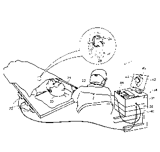
Fig. 1 is a schematic, pictorial illustration of a
cardiac mapping and ablation system 20, which operates in
accordance with an embodiment of the invention. System
20 may be based, for example, on the above-mentioned
CARTO system, with suitable additions to the system
software. System 20
comprises a probe, such as a
catheter 24, and a control console 34. In the embodiment
described hereinbelow, catheter 24 is used in ablating
sites of arrhythmias in one or more chambers of a heart
9
CA 02815755 2013-05-07
26 of a patient 30. Alternatively, catheter 24 or other
suitable probes may be used, mutatis mutandis, for other
therapeutic purposes in the heart or in other body
organs.
An operator 22, such as a cardiologist, inserts
catheter 24 through the vascular system of patient 30 so
that the distal end of the catheter enters a chamber of
heart 26. Operator 22 advances the catheter so that an
electrode 28 at the distal tip of the catheter engages
endocardial tissue at desired ablation sites. Catheter
24 is typically connected by a suitable connector at its
proximal end to console 34, and specifically to a radio
frequency (RE) generator 36, which generates RE energy
for transmission via catheter 24 to electrode 28.
Operator 22 actuates RE generator 36 to ablate tissue at
suspected sites of arrhythmia in the heart.
In this pictured embodiment, system 20 uses magnetic
position sensing to determine position coordinates of the
distal end of catheter 24 inside heart 26. For this
purpose, a driver circuit 38 in console 34 drives field
generators 32 to generate magnetic fields within the body
of patient 30. Typically,
field generators 32 comprise
coils, which are placed below the patient's torso at
fixed, known positions. These coils
generate magnetic
fields in a predefined working volume that contains heart
26. A magnetic
field sensor (not shown) within the
distal end of catheter 24 generates electrical signals in
response to these magnetic fields. A signal processor 40
processes these signals in order to determine the
position coordinates of the distal end of catheter 24,
typically including both location and orientation
coordinates. This
method of position sensing is
implemented in the above-mentioned CARTO system and is
well known in the art.
Alternatively or additionally,
system 20 may use other methods of position sensing that
are known in the art, such as ultrasonic or electrical
impedance-based methods.
In addition, catheter 24 may comprise a force sensor
(not shown) in its distal end, for measuring the contact
force between the catheter tip and the wall of heart 26.
The SmartTouchTm catheter developed by Biosense Webster
Inc. for the CARTO system offers this sort of capability.
A catheter of this sort is described, for example, in
U.S. Patent Application Publication 2011/0130648. The
force measurement is useful in ensuring that electrode 28
is in sufficiently firm contact with the heart wall to
effectively transfer RF energy and ablate the heart
tissue.
Processor 40 in console 34 typically comprises a
general-purpose computer processor, with suitable front
end and interface circuits for receiving signals from
catheter 24 and for controlling and receiving inputs from
the other components of console 34. Processor 40 may be
programmed in software to carry out the functions that
are described herein. The software may be downloaded to
processor 40 in electronic form, over a network, for
example, or it may be provided, alternatively or
additionally, on tangible, non-transitory media, such as
optical, magnetic or electronic memory media. Further
alternatively or additionally, some or all of the
11
CA 2815755 2019-09-04
CA 02815755 2013-05-07
functions of processor 40 may be carried out by dedicated
or programmable digital hardware components.
Based on the signals received from catheter 24 and
other components of system 20, processor 40 drives a
display 42 to present operator 22 with a three-
dimensional (3D) map 44 of heart 26. The map may
indicate cardiac electrophysiological activity measured
by catheter 24, as well as providing visual feedback
regarding the position of the catheter in the patient's
body and status information and guidance regarding the
procedure that is in progress. Other parameters that may
be measured by catheter 24 and by other elements of
system 20 and shown on display 42 may include, for
example, contact force between the catheter and heart
tissue, electrical impedance of the heart tissue, local
temperature, and RF power delivered through the catheter.
Processor 40 assesses the parameters that it
receives from system 20 as indicators of the adequacy of
ablation at each treated site in heart 26. When the
ablation parameters at a given site meet certain
predefined criteria, the processor automatically places a
mark 46 on map 44 to indicate the site. The
processor
may vary the appearance of marks 46 (such as their color)
in response to the parameters at each site. The criteria
for automatic marking of the ablation sites may be
preconfigured, or they may, alternatively or
additionally, be set by operator 22, typically using user
interface controls 48 and on-screen menus.
Although in the illustrated embodiment, catheter 24
is manipulated manually by operator 22, system 20 may
alternatively or additionally comprise an automated
12
CA 02815755 2013-05-07
mechanism (not shown) for maneuvering and operating the
catheter within the body of patient 30. In such
embodiments, processor 40 generates a control input for
controlling the motion of catheter 24 based on the
signals provided by the magnetic field sensor in the
catheter and other system parameters, such as those
mentioned above.
Fig. 2 is a schematic representation of map 44 as it
appears on display 42, in accordance with an embodiment
of the present invention. Map 44 is a 3D representation
of a chamber of the heart, which is colored to show local
electrical activity, as in the above-mentioned CARTO
system. (Colors are
represented by hatching in the
figure.) The map may
be generated based simply on
position measurements made using catheter 24, or
alternatively, these position measurements may be
registered with a pre-acquired image of the heart (such
as a CT, MRI, or ultrasound image) in order to create the
map.
Processor 40 has placed marks 50, 52 on map 44 to
indicate sites that have been ablated by catheter 24.
Typically, the processor automatically marks sites at
which the ablation parameters, such as dwell time and
force applied by the catheter against the tissue, meet
the predefined criteria. Marks 50 and 52 may be colored
differently to indicaLe different ranges of measured
parameters. Normally, as noted above, the criteria to be
applied by processor 40 in marking ablation sites may be
defined by operator 22. Additionally
or alternatively,
the operator may choose to mark sites manually by
manipulating appropriate system controls.
13
CA 02815755 2013-05-07
Although Figs. 1 and 2 show a particular system
configuration and application environment, the principles
of the present invention may similarly be applied in
other mapping and therapeutic applications using not only
catheters, but also probes of other types, both in the
heart and in other body organs and regions.
METHODS FOR AUTOMATIC MARKING
Fig. 3 is a flow chart that schematically
illustrates a method for tracking intracardiac ablation,
in accordance with an embodiment of the invention. The
method is described, for the sake of clarity and
convenience, with reference to system 20. As noted
above, however, the principles of the methods described
hereinbelow may similarly be applied in other systems and
application environments.
Processor 40 collects data continuously during the
operation of system 20 and saves the data in a cyclic
buffer in memory. The data are flushed from the buffer
on a first-in/first-out basis. Data
processing is
initiated when ablation starts, at an initiation step 60,
typically when operator 22 activates RF generator 36. At
this point, processor 40 is able to collect and process
data that accumulated in the buffer before ablation, at a
pre-ablation collection step 68. The
processor
continues collecting data during ablation, at a pen-
ablation collection step 64, until the RF generator is
deactivated, at an ablation termination step 62. The
processor may continue collecting data after step 62, at
a post-ablation collection step 70, for use in assessing
the results of the ablation at the current site.
14
Performing ablation at any given site typically
takes at least several seconds, and may take as long as a
minute. During
this period, patient 30 will typically
take one or more breaths, with the result that the
location of heart 26 shifts (along with other parts of
the patient's chest) relative to field generators 32.
These breaths are indicated in Fig. 3 by end-expirium
points 66. As a result of this respiratory motion of the
chest, position readings made by processor 40 with
respect to catheter 24 will shift cyclically in
synchronization with the respiration cycle, and the
catheter coordinates will change even when the catheter
is stably held in contact with a given ablation site.
In order to eliminate this confusing effect of
respiratory motion on the catheter position, processor 40
corrects the position coordinates to compensate for
respiration, at a respiration compensation step 70.
Typically, processor 40 uses end-expirium points 66 as a
baseline, determines the shift of heart 26 relative to
this baseline at each point during the respiratory cycle,
and then subtracts out this shift from the catheter
coordinates in order to project all position readings to
the equivalent end-expirium location. When the catheter
is actually moving relative to the heart, step 70 will
convert the sequence of catheter position reads to a
linear path between the start- and end-points of the
movement. A method
of compensation for respiratory
motion that may be used at this step is described, for
example, in U.S. Patent Application 13/017,469.
CA 2815755 2019-09-04
CA 02815755 2013-05-07
Processor 40 filters the collected data to identify
ablation sites that should be marked on map 44, at a
filtering step 72. At this step, the processor evaluates
whether a given site meets predefined criteria in terms
of stability of the contact between the probe and the
tissue and other factors, so as to qualify to be marked.
This step is described in greater detail below with
reference to Fig. 4. Sites that
satisfy the filter
criteria are selected to be marked on the map, while
sites that do not satisfy the criteria are discarded.
Figs. 5 and 6 show how stability criteria are applied in
this step, while Fig. 7 shows a user interface window
that can be used by operator 22 (or other personnel) to
set the filter criteria.
When a site satisfies the filter criteria, processor
40 assigns the site data to a specific position in the 3D
space corresponding to map 44, at a grid assignment step
74. In other
words, even if there is some residual
variation in measured catheter coordinates, following
respiration compensation, during the ablation, the
processor chooses a particular coordinate location to be
marked, such as the center of mass of the compensated
coordinate values. The position may be a specific voxel
in a 3D grid, for example, or it may correspond to a zone
of the map, such as zones in a Voronoi diagram
corresponding to the map shape.
Processor 40 then projects the volume points
assigned at step 74 to corresponding locations on the
surface of map 44, and creates marks 50, 52 at these
surface locations, at a projection step 76. This step is
16
CA 02815755 2013-05-07
described in greater detail with reference to Fig. 8
below.
Fig. 4 is a flow chart that schematically
illustrates a method for assessing intracardiac ablation
that is applied at step 72, in accordance with an
embodiment of the invention. Processor 40
applies
decision logic 80 (typically in software) based on the
date collected during ablation (step 64) at a given site,
and possibly the data .collected before and/or after the
ablation (steps 68,70). The following
filters may be
applied:
= A position stability filter 82: Processor 40 measures
changes in the location of catheter 24 during the
ablation, after compensating for respiratory movement
(step 70). Filter 82 typically
requires that the
variation of the location over a predefined minimum
ablation time be no greater than a predefined maximum
distance. The variation may be measured, for example,
in terms of a standard deviation about the mean
position during the ablation time.
= A velocity filter 84. This filter can be used to
detect loss of stability. For this purpose, processor
40 measures the displacement between successive
locations of the catheter and (implicitly or
explicitly) divides by the time increment between the
locations to find the velocity of motion of the
catheter. If the
velocity is above a specified
threshold, such as 10 mm/sec, the catheter may be
considered to be unstable.
= An impedance drop filter 86. Processor 40
typically
takes the impedance value of the first position in a
17
CA 02815755 2013-05-07
stable site as a base impedance value, and then tests
subsequent impedance values against a predefined
percentage threshold. For example, the impedance drop
in subsequent measurements may be defined as: (1¨
current position impedance value)
*100. If this drop is greater
base impedance value
than the percentage threshold, the site is considered
to have been ablated and is therefore marked on map 46.
Alternatively or additionally, the amount of impedance
drop may be taken into account in coloring the
corresponding mark 50, 52.
= An ECG drop filter 88. For each stable site, processor
40 may calculate, for example, the maximum peak-to-peak
ECG amplitude value in an ECG data span of two seconds
ending at the time at which a corresponding position
measurement was made. If the calculated
amplitude is
less than a predefined threshold, the site may be
marked as having been ablated and/or colored to
indicate the amount of ECG drop.
= A force
level filter 90. At each stable site,
processor 40 tests the average contact force between
catheter 24 and the heart tissue against a predefined
threshold. When the average contact force is above a
predefined threshold, the site is marked as having been
ablated and may be colored to indicate the average
force level. Additionally or alternatively, processor
40 may assess the force percentage, i.e., the fraction
of the time during which the catheter dwelled at a
given ablation site for which the force was above a
certain force threshold:
18
CA 02815755 2013-05-07
(number of postions exceeding the contact force threshold)
*100. If the
total number of positions in the stable site
percentage value is above a predefined time percentage
threshold, the site is marked, and/or the mark may be
colored according to the percentage value.
Additionally or alternatively, processor 40 may
apply other filters not shown in Fig. 4. For example, if
catheter 24 contains a temperature sensor in its distal
end, the processor may calculate the temperature increase
at each ablation site during the procedure. If the
temperature increase is above a predefined threshold, the
processor may place a mark at the site. Additionally or
alternatively, the mark may be colored according to the
temperature increase.
Other collected data and filters may apply to the
cumulative amount of RE energy delivered to each site, as
well as ultrasound reflectance data if available.
Processor 40 may also compute and filter integral
measures of the contact force of the catheter against the
tissue, which are indicative of the delivery of energy to
the tissue. Integral measures of this sort may include,
for example, the integral of force over time at a stable
location, or the integral of the product of force and RF
power.
Fig. 5 is a schematic representation of a part of
map 44, showing a series of catheter location data points
100. This figure
illustrates how processor identifies
and filters ablation sites 102, 104, 106 based on
catheter location stability, in accordance with an
embodiment of the invention.
19
CA 02815755 2013-05-07
Each data point 100 in Fig. 5 indicates a catheter
location measurement (after compensation for respiratory
movement). The data are
collected at predefined time
intervals, so that points 100 represent a time sequence
of successive catheter locations. A radius 108 defines
the spread of catheter locations that can be considered
to belong to a single site. Even when operator 22 holds
the catheter stably in place against the heart wall, the
actual measured position may appear to change due to
minor slippage, noise, and imperfect compensation for
respiratory motion, for example. Thus,
despite the
spread of the "clouds- of data points corresponding to
sites 102, 104, 106 in Fig. 5, processor 40 considers the
data points within each sphere to correspond to the
catheter dwelling stably at each of the sites during the
respective periods during which the data were collected.
A predefined time threshold determines the length of
time that the catheter must dwell at a given site (or
equivalently, the number of data points 100 within a
given sphere of radius 108) in order for the catheter to
be considered to have dwelled stably at the site. Thus,
when the number of successive data points 100 within
radius 108 around a given center point exceeds the
threshold, and RE' generator 36 is simultaneously
activated, processor 40 may consider the site to have
been ablated and may mark the site accordingly. Both
radius 108 and the threshold dwell time may be set by
operator 22. Processor 40 may decide whether and how to
mark a given site based not only on the dwell time,
however, but also on other parameters, as explained
above.
CA 02815755 2013-05-07
Fig. 6 shows a schematic plot 110 of catheter
contact force at an ablation site, in accordance with an
embodiment of the invention. The upper
trace in the
figure illustrates how processor 40 identifies that
catheter 24 is dwelling stably at a given site. Location
data are accumulated before and during ablation, at steps
68 and 64 (Fig. 3). The
processor accumulates and
processes the data over time until it determines, at a
time 112, that the location data points over an interval
that is equal to a time threshold 114 have all fallen
within radius 108. The
processor then retrospectively
marks a time 116 and the corresponding location as the
first stable location at the current ablation site. It
is assumed in this example that the catheter remains
stable within radius 108 of the current site after time
112, as well.
Concurrently, processor 40 monitors the contact
force between catheter 24 and heart 26 to generate and
store force plot 110 and computes a running average of
the contact force over a time window 120. At a time 118,
processor 40 determines that the average force over the
time window 120 ending at time 118 has exceeded the
contact force threshold that is shown in Fig. 6. (Both
the duration of window 120 and the level of the contact
force threshold may be set by operator 22.) Processor 40
then marks a time 122 at the beginning of window 120 and
the corresponding location of the catheter as the first
stable location at the current ablation site at which the
catheter exerted sufficient force to ablate the heart
tissue at the site.
21
CA 02815755 2013-05-07
Beginning from time 122, processor 40 accumulates
ablation data over a time interval 124 during which the
catheter location stability and average force level
continue to satisfy the threshold criteria described
above. A progress bar may be presented on display 42 to
show operator 22 the cumulative length of effective
ablation time at the current site. As explained
earlier, the accumulated data during interval 124 may
include ECG, tissue impedance, temperature, and/or RF
energy delivery, inter alia. The processor filters these
data and then marks map 44 accordingly.
Fig. 7 is a schematic representation of a window 130
that may be presented on display 42, in accordance with
an embodiment of the invention. The window contains on-
screen user controls 132 that operator 22 can use to set
the thresholds to be applied by processor 40 to the
ablation data values in choosing which ablation sites to
mark on map 44. The data
parameters and applicable
criteria have all been explained above. For each
criterion, a checkbox at the left side of the window
allows the operator to indicate whether or not processor
40 is to consider the criterion in filtering ablation
sites. A slider to
the right of each parameter name
enables the operator to set the threshold level, within
bounds that are set by system 20. For example,
the
duration (dwell Lime) for catheter stability may be set
to a value between 0 and 60 sec, while the location
stability (corresponding to radius 108) may be set to a
value between 0 and 8 mm. As another
example, the
threshold for average contact force may be set to a value
between 0 and 150 grams.
22
CA 02815755 2013-05-07
Display preference controls 134 in the lower part of
window 130 enable the operator to set other aspects of
how map 44 will be displayed.
Although window 130 as shown in Fig. 7 permits only
a single value of each threshold to be set, in an
alternative embodiment (not shown in the figures),
operator 22 may input different threshold values for
different parts of the heart. Additionally
or
alternatively, processor 40 may compute different
threshold values to apply based on operator inputs and on
physiological parameters, such as local heart wall
thickness.
Fig. 8 is a schematic representation of 3D map 44 of
a heart chamber showing how ablation marks 50 are applied
to the map, in accordance with an embodiment of the
present invention. In this example, ablation sites that
satisfied the threshold criteria at step 72 are assigned
to respective positions 140 in a 3D grid at step 74.
Processor 40 projects positions 140 onto the nearest
respective locations on the surface of map 44, and places
marks 50 at these locations.
It will be appreciated that the embodiments
described above are cited by way of example, and that the
present invention is not limited to what has been
particularly shown and described hereinabove. Rather,
the scope of the present invention includes both
combinations and subcombinations of the various features
described hereinabove, as well as variations and
modifications thereof which would occur to persons
skilled in the art upon leading the foregoing description
and which are not disclosed in the prior art.
23