Note : Les descriptions sont présentées dans la langue officielle dans laquelle elles ont été soumises.
CA 02817234 2013 05 06
WO 2012/064348
PCT/US2010/057814
ARTICULATING ORGAN SUPPORT
BACKGROUND OF THE INVENTION
[0001] This invention relates generally to organ support and bypass
processes, and
more particularly to methods and apparatus for physically supporting an organ
during
such processes.
[0002] Numerous medical procedures require circulation of a fluid through
an
internal organ, optionally with treatment of the fluid by processes such as
filtering,
oxygenation, and the like. For example, when an organ is harvested from a
donor for
transplantation, a neutral saline solution is used to flush out the donor's
blood from the
blood vessels of the organ.
[0003] It is also known that some devices use a similar process to
circulate an
aqueous organ preservation fluid, such as "Belzer's solution", through organs
which have
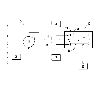
been harvested for transplantation. This action sustains the organ while it is
outside the
body by attempting to preserve functioning, and increases the limited "shelf
life" of
transplant organs compared to conventional chilled storage.
[0004] More advanced concepts provide methods and apparatus for supporting
an
organ (in vivo or in vitro) in a manner which closely mimics biological
processes by
providing carefully controlled fluid pressure and chemical profiles. One such
concept is
described in published U.S. patent application 2010/0028979 entitled "Methods
And
Apparatus For Organ Support".
[0005] All of these processes and devices require that an internal organ be
physically
supported outside the body, typically in a rigid or resilient static
container. Thus
supported, gravity forces on the organ tend to press it against whatever
support is used
underneath it, resulting in localized pressure on its lower and lateral
surfaces. This can
result in localized pressure ischemia, cellular damage and/or loss of organ
function and
vitality.
BRIEF SUMMARY OF THE INVENTION
- 1 -
SUBSTITUTE SHEET (RULE 26)
CA 02817234 2013 05 06
WO 2012/064348
PCT/US2010/057814
[0006] These and other shortcomings of the prior art are addressed by the
present
invention, which provides an articulating organ support.
[0007] According to one aspect of the invention, an organ support apparatus
includes: an enclosure having a floor, opposed side walls, opposed end walls,
and a lid; a
first support pad disposed on the floor of the enclosure, the first support
pad comprising a
plurality of inflatable and flexible chambers; and an inflation apparatus
coupled to the
chambers and operable to individually inflate or deflate each chamber.
[0008] According to another aspect of the invention, a method for
supporting an
organ, includes: providing an enclosure adapted to contain an organ; placing
the organ on
a first support pad disposed on a floor of the enclosure, the first support
pad comprising a
plurality of inflatable and flexible chambers; and selectively inflating and
deflating the
chambers to provide a time-varying contact pressure profile with the organ.
[0009] According to yet another aspect of the invention, a method for
supporting an
organ includes: providing an enclosure adapted to contain an organ, the
enclosure having
a floor opposite a lid; placing a first support pad against the floor, the
first support pad
comprising a plurality of inflatable and flexible chambers; placing a second
support pad
against the lid, the second support pad comprising a plurality of inflatable
chambers;
placing the organ between the first and second support pads; inflating the
chambers to
clamp the organ in position between the support pads; and selectively tilting
or rotating
the enclosure to provide a time-varying contact pressure profile between the
first and
second support pads and the organ.
BRIEF DESCRIPTION OF THE DRAWINGS
[0010] The invention may be best understood by reference to the following
description taken in conjunction with the accompanying drawing figures in
which:
[0011] Figure 1 is a schematic view of a organ support apparatus
constructed
according to an aspect of the present invention, coupled to a perfusion
system;
[0012] Figure 2 is top view of the organ support apparatus of Figure 1;
- 2 -
SUBSTITUTE SHEET (RULE 26)
CA 02817234 2013 05 06
WO 2012/064348
PCT/US2010/057814
[0013] Figure 3 is a partially-sectioned side view of the organ support of
Figure 2;
[0014] Figure 4 is a partially-sectioned end view of the organ support of
Figure 2;
[0015] Figure 5 is a top view of a support pad of the organ support
apparatus;
[0016] Figure 6 is a side view of the support pad of Figure 5;
[0017] Figure 7 is a front end view of the support pad of Figure 5;
[0018] Figure 8 is a rear end view of the support pad of Figure 5;
[0019] Figure 9 is a schematic diagram of a support pad coupled to an
inflation
apparatus;
[0020] Figure 10 is a side view of an organ support apparatus including a
rotation
apparatus;
[0021] Figure 11 is a side view of the organ support apparatus of Figure 10
in an
inverted position;
[0022] Figure 12 is a schematic end view of a support pad in a first
configuration;
and
[0023] Figure 13 is a schematic end view of a support pad in a second
configuration.
DETAILED DESCRIPTION OF THE INVENTION
[0024] Referring to the drawings wherein identical reference numerals
denote the
same elements throughout the various views, Figure 1 depicts diagrammatically
a
perfusion system 10 suitable for circulating a fluid through an organ, in
conjunction with
an organ support apparatus 12 which is constructed in accordance with the
present
invention. As used herein the term "perfusion system" broadly refers to any
apparatus
which functions to circulate fluid through an organ and could range from a
simple saline
flushing device to a highly sophisticated organ support apparatus such as the
one
described in U.S. published patent application 2010/0028979 entitled "Methods
And
- 3 -
SUBSTITUTE SHEET (RULE 26)
CA 02817234 2013 05 06
WO 2012/064348
PCT/US2010/057814
Apparatus For Organ Support". The perfusion system 10 comprises a fluid
circuit defined
by plastic tubing or another suitable type of conduit, connected to an organ,
depicted
generally at "K", by an inlet line 14 and an outlet line 16.
[0025] The perfusion system 10 includes some means for circulating fluid,
such as a
pump, along with appropriate fluid treatment equipment, such as one or more
filters, heat
exchangers, oxygenators, de-aerators, or chemical injectors. All of this
equipment is
depicted schematically at number 18. An electronic controller 20 may be used
to control
the operation of the perfusion system 10. The illustrated example is explained
in the
context of providing support for a kidney K which is contained in the organ
support
apparatus 12 and connected to a fluid collection container 22 which receives a
fluid flow
from the kidney K. However, it will be understood that the principles of the
present
invention are broadly applicable to support of many types of organs. The fluid
collection
container 22 may not be needed for other organs.
[0026] The basic components of the organ support apparatus 12 are an
enclosure 24,
a lower support pad 26, an optional upper support pad 28, and an inflation
apparatus 30.
[0027] Optionally, an imaging device 31 (such as a camera operating in the
visual,
UV, or IR frequency ranges) may be used to observe the condition of the organ
K
through the enclosure 24. One example of a suitable imaging device is a
confocal
microscope such as the VIVASCOPE device available from Lucid, Inc., Rochester,
NY
14623 USA. Positioning apparatus (not shown) capable of multi-axis positioning
may be
provided to point the imaging device 31 at a particular target area of the
organ K.
[0028] Figures 2-4 illustrate the organ support apparatus 12 in more
detail. The organ
enclosure 24 provides physical protection to the organ K and isolates it from
the external
environment. Preferably the material of the enclosure 24 is transparent to
visible light
and/or other select portions of the radio frequency ("RF") spectrum to
facilitate imaging
of the organ K. For example, it may be constructed from a material such as
sterilizable
transparent medical-grade polymer. As illustrated it is in the form of a
rectangular box
with a floor 32, side walls 34, front and rear walls 36 and 38, and a
removable lid 40. The
lid 40 may be secured with latches 42. The front wall 36 is provided with pass-
through
- 4 -
SUBSTITUTE SHEET (RULE 26)
CA 02817234 2013 05 06
WO 2012/064348
PCT/US2010/057814
openings for making connections between the artery "A" and vein "V" of the
kidney K
(for example) and the inlet and outlet lines 14 and 16 respectively of the
perfusion system
10. There is also a pass-through opening for making a connection to the ureter
U, to
allow urine to drain to the fluid collection container 22 (see Figure 1).
[0029] The lower support pad 26 rests on the floor 32 and the organ K rests
on top of
the lower support pad 26. The lower support pad 26 shown in more detail in
Figures 5-8.
It is constructed from a top sheet 44 and a bottom sheet 46 which are
selectively bonded
together along their mutual peripheral edges 48 and along dividing seams 50.
The
remaining unbonded portions define individual inflatable and flexible chambers
52A
through 52E. The sheets 44 and 46 may be made from any flexible, fluid-tight
material,
such as polymers, treated fabrics, or rubber. Preferably the material is
transparent to
visible light and/or other select portions of the radio frequency ("RF")
spectrum to
facilitate imaging of the organ K. The sheets 44 and 46 may be bonded together
by any
method which provides a leak-tight connection, such as by thermal or
ultrasonic bonding,
adhesives, or crimping.
[0030] In the illustrated example, there are five side-by-side, elongated,
generally
rectangular chambers 52A through 52E. As will be explained further below, the
shape,
number, and configuration of the chambers 52A through 52E is not critical and
could be
varied in a number of ways to suit a particular application. For example,
various patterns
of elongate shapes, grid patterns, and/or arcs or circles could be used to
define the
chambers. A fluid connection is provided to each of the individual chambers
52A-52E.
As shown, individual tube fittings 54 are employed.
[0031] If used, the upper support pad 28 would be identical in construction
to the
lower support pad 26. The upper support pad 28 would be placed between the
organ K
and the lid 40.
[0032] An inflation apparatus (shown schematically at 30 in Figure 1) is
provided for
selectively inflating and deflating each chamber 52A-52E. Figure 9 shows an
example of
the inflation apparatus 30 in a basic form comprising a controller 56 coupled
to an air
pump 58 which is in turn coupled to the individual chambers 52A-52E of the
lower
- 5 -
SUBSTITUTE SHEET (RULE 26)
CA 02817234 2013 05 06
WO 2012/064348
PCT/US2010/057814
support pad 26 through tubes 60. Water or another liquid could be used instead
of air.
The pump 56 may be a pressure pump only, or it may be a combination
pressure/vacuum
pump to provide for improved deflation. The controller 56 may be a general-
purpose
microcomputer of a known type, such as a PC-based computer, or it may be a
custom
processor, or may incorporate one or more programmable logic controllers
(PLC).
Depending on the type of perfusion system 10, the pump 58 may be controlled
through
software programming integrated into the perfusion system controller 20 (see
Figure 1).
As shown in Figure 9, the pump 58 is connected to the chambers 52A-52E through
a
multi-port remotely-controlled valve 62 whose position is commanded by the
controller
56. Alternatively, an independent pump could be provided for each chamber 52A-
52E. If
an upper support pad 28 is used, a separate inflation apparatus 30 (see Figure
1) may be
provided for it, or the valve 62 could be modified to accommodate additional
chambers
of the upper support pad 28.
[0033] Optionally, the capabilities of the support apparatus 12 may be
further
extended by providing apparatus for pivoting or rotating the enclosure 24.
Figure 10
illustrates an enclosure 24 with shafts 64 and 66 extending from the front and
rear walls
36 and 38, respectively, and mounted in pivot bearings 68 which are in turn
held by
stands 70. An electric motor 72 (for example a stepper motor), or other
suitable type of
rotary device, is coupled to one of the shafts 64 or 66. Rotation of the shaft
of the motor
72 pivots the enclosure 24 about the shafts 64 and 66. This function may be
used to tilt
the organ K (not seen in Figures 10 and 11) to specific angles or to
periodically invert it
during a perfusion procedure (the inverted position is shown in Figure 11). If
desired, a
multi-axis gimbal of a known type may be employed to mount the enclosure 24 so
that it
may be rotated about more than one axis.
[0034] The operation of the organ support apparatus 12 will be described
with
reference to figures 12 and 13. An organ K is placed on the lower support pad
26 within
the enclosure 24. The organ K is connected to the perfusion system 10 which is
placed in
operation circulating fluid through the organ K. While the organ K is resting
on the lower
support pad 26, the chambers 52A-52E are selectively inflated and deflated so
as to
provide a varying contact pressure profile with the organ K. For example,
Figure 12
- 6 -
SUBSTITUTE SHEET (RULE 26)
CA 02817234 2013 05 06
WO 2012/064348
PCT/US2010/057814
shows chambers 52A, 52C, and 52E as being fully inflated while chambers 52B
and 52D
are deflated. In this configuration the organ K is supported along three
spaced-apart lines,
and points of relatively higher pressure are present at the locations marked
with arrows
"Pl". At a subsequent time, chambers 52B and 52D may be fully inflated while
chambers
52A, 52C, and 52E are deflated. This configuration is shown in Figure 13. The
organ K
would thus be supported along two spaced-apart lines and points of relatively
higher
contact pressure are present at the locations marked with arrows "P2". The
locations P1
are relieved of pressure, allowing free flow of circulation and absence of
mechanical
stress. This cycle of alternating inflation and deflation may be repeated as
often as
necessary so that no one portion of the organ K is subjected to damaging
pressure for too
long, which could result in localized pressure ischemia. For example, the
pressure in any
one location may be relieved about 2 or 3 times per minute.
[0035] If the rotation apparatus described above are used, then the
enclosure 24 with
the organ K may be periodically tilted and/or inverted so that contact
pressures on the
organ K are shared between its opposite surfaces. For example, the organ K may
be tilted
and/or inverted with a frequency of about once per minute to about once every
30
minutes. The frequency is subject to the vascular resistance and condition of
the organ K
or tissue. The tilt and/or inversion may be in addition to or as an
alternative to the
selective inflation and deflation of the chambers 52A-52E. The imaging device
31, such
as a scanning high resolution infrared camera may be employed to take a series
of images
an build therefrom a mosaic image of the organ K for localized and global
comparison.
For example, the organ K may be imaged in small blocks, e.g. 20 mm x 20 mm
(0.8 in. x
0.8 in.). In the image, ischemic areas will exhibit relatively higher or lower
temperatures
than the surrounding tissue.
[0036] In response to the detection of such areas, the controller 56 may be
programmed to tilt and/or invert the organ enclosure 24, and/or to selectively
inflate or
deflate the chambers 52A-52E. To facilitate the imaging and control process,
the
temperature of the fluid circulating through the organ K may be altered (e.g.
using the
perfusion system 10) slightly up and down from a physiologically suitable
temperature
for organ characterization and preservation. For example, the fluid
temperature change
- 7 -
SUBSTITUTE SHEET (RULE 26)
CA 02817234 2014-02-17
W02012/064348 PCT/US2010/057814
may be plus or minus about 2 degrees C (plus or minus about 3.6 degrees F).
Any
ischemic areas will respond to the fluid temperature change at a substantially
slower rate
than the surrounding tissue, resulting in hot or cold spots which can be
detected by the
imaging device 3 I .
[0037] The upper support
pad 28 may be used to supplement the lower support pad
26. For example, if the enclosure 24 is inverted, then the organ K would rest
on the upper
support pad 28 and the alternate chamber inflation cycle described above would
be
carried out using the upper support pad 28. The upper support pad 28 may also
be used
simultaneously with the lower support pad 26 to provide a gentle clamping
action to the
organ K in order to support it during tilting and/or inversion, or during
movement or
transport of the enclosure 24.
- 8 -
SUBSTITUTE SHEET (RULE 26)