Note : Les descriptions sont présentées dans la langue officielle dans laquelle elles ont été soumises.
REAL TIME ASSESSMENT OF ABLATION FROM ELECTROCARDIOGRAM
SIGNALS
FIELD OF THE INVENTION
The present invention relates generally to cardiac
therapy, and particularly to methods and systems for
monitoring cardiac signals during ablation therapy.
BACKGROUND OF THE INVENTION
Various techniques are known in the art for cardiac
ablation therapy. U.S. Patent Publication No. 2010/0331658,
describes an open-irrigated catheter system comprising a tip
section, a distal insert, and mapping electrodes. The tip
section has an exterior wall that defines an open interior
region within the tip section. The exterior wall includes
mapping electrode openings and irrigation ports. The exterior
wall is conductive for delivering radio frequency (RE) energy
for an RE ablation procedure. The irrigation ports are in
fluid communication with the open interior region to allow
fluid to flow from the open interior region through the
irrigation ports. The distal insert is positioned within the
tip section to separate the open region into a distal fluid
reservoir and a proximal fluid reservoir. The mapping
electrodes are positioned in the mapping electrode openings in
the tip section.
1
CA 2820807 2018-09-19
U.S. Patent Publication No. 2007/0006254, describes a
method of controlling a remote navigation system that orients
the distal end of a medical device in response to user inputs,
including interrupting the operation of the remote navigation
system when the user inputs would navigate the medical device
to a location where the impedance exceeds a predetermined
value. A method of controlling ablation of cardiac tissue to
block an errant signal causing an arrhythmia includes ablating
tissue until there is a predetermined reduction in the
amplitude of the errant signal or a predetermined reduction in
local impedance.
U.S. Patent No. 7,959,630, describes assemblies, probes,
and methods for creating circumferential lesions in tissue,
e.g., the tissue within or around the ostium of a vessel. An
ablation probe with an ablative structure can be placed in
contact within or around the ostium of the vessel. A
diagnostic probe can be introduced through a lumen within the
ablation probe and inserted into the vessel. Energy can be
provided to the ablative structure to create a circumferential
lesion within or around the ostium of the vessel, and the
diagnostic structure can be used to diagnose the tissue to
determine whether the circumferential lesion can be properly
created.
U.S. Patent Publication No. 2006/0253115, describes
catheters, systems, and methods which are provided for
performing medical procedures, such as tissue ablation,
adjacent the ostia of anatomical vessels, such as pulmonary
veins. A catheter comprises an elongated flexible catheter
body including a proximal shaft portion and a distal shaft
2
CA 2820807 2018-06-26
CA 02820807 2013-06-25
portion, which has a proximal section pre-shaped to form a
curve having an apex sized to be inserted into an anatomical
vessel, such as a pulmonary vein, and a distal section
configured to contact an ostium of the vessel when the curve
apex is inserted within the vessel ostium. The catheter
further comprises a steering mechanism configured for
decreasing a radius of curvature of the curve.
SUMMARY OF THE INVENTION
An embodiment of the present invention provides an
apparatus including an intra-body probe and a processor. The
intra-body probe includes an electrode, which is configured to
contact tissue in a heart. The processor is configured to
receive an electrical signal from the electrode, to
distinguish a local component, due to the tissue with which
the electrode is in contact, in the electrical signal from a
remote-field contribution to the signal, and to control a
therapeutic procedure applied to the tissue responsively to
the distinguished local component.
In some embodiments, the therapeutic procedure includes
cardiac ablation therapy. In other embodiments, the intra-body
probe includes an additional electrode that is configured to
apply an ablation signal to the heart tissue. In other
embodiments, the electrode applies an ablation signal to the
tissue in the heart. Yet in other embodiments, the signal
includes a received electrocardiogram (ECG) signal. In some
embodiments, the local component is in response to cardiac
electrical activity generated in the heart within a target
ablation region, and the remote-field contribution is in
3
CA 02820807 2013-06-25
=
response to cardiac electrical activity generated in the heart
outside the target ablation region.
In some embodiments, the processor is configured to
distinguish the local component by identifying one or more
time intervals in the received signal in response to a change
therein. In other embodiments, the processor is configured to
distinguish the remote-field contribution by detecting no
change in the received signal in one or more additional time
intervals outside of the one or more identified time
intervals. In yet other embodiments, the processor is
configured to initiate a termination of an ablation procedure
by detecting that the signal within the one or more identified
time intervals no longer responds to successive ablation
cycles.
There is also provided, in accordance with embodiments of
the present invention, a method including receiving an
electrical signal from an intra-body probe, which includes an
electrode configured to contact tissue in a heart. A local
component, due to the tissue with which the electrode is in
contact, in the electrical signal is distinguished from a
remote-field contribution to the signal. A therapeutic
procedure applied to the tissue is controlled responsively to
the distinguished local component.
The present invention will be more fully understood from
the following detailed description of the embodiments thereof,
taken together with the drawings in which:
BRIEF DESCRIPTION OF THE DRAWINGS
4
CA 02820807 2013-06-25
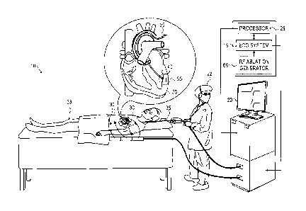
Fig. 1 is a schematic diagram showing an
electrocardiogram ablation monitoring system, in accordance
with an embodiment of the present invention;
Fig. 2 is a schematic diagram showing changes in the ECG
waveform with successive ablation cycles, in accordance with
an embodiment of the present invention; and
Fig. 3 is a flow chart that schematically illustrates a
method for monitoring an electrocardiogram signal after
successive ablation cycles, in accordance with an embodiment
of the present invention.
DETAILED DESCRIPTION OF EMBODIMENTS
OVERVIEW
Ablation is a known technique for the treatment of
various cardiac conditions. In an ablation procedure, an
intra-body probe, typically a catheter, is percutaneously
inserted into the cardiovascular system of a patient and
navigated into the heart to a region of tissue to be ablated.
Cardiac ablation can be performed with different modalities,
such as cryoablation and radio frequency (RF) ablation
therapies wherein the distal tip of the catheter can
respectively be used to locally freeze or heat the tissue. In
both cases, a lesion with a low conductance is formed. The
lesion typically blocks the faulty pathways of the heart's
electrical signals causing cardiac dysfunction, such as
tachyarrhythmias and atrial fibrillation.
Real time monitoring of electrocardiogram (ECG) signals
while performing the ablation therapy is beneficial for
assessing the lesion, e.g., to check if the procedure has
5
CA 02820807 2013-06-25
improved the heart function, or if more ablation needs to be
delivered to the patient. Such real time monitoring may
prevent terminating the procedure prematurely. However, if too
much ablation is applied to the heart tissue, irreparable
damage to the heart may result.
Embodiments of the present invention that are described
herein provide methods and systems for a real time assessment
of electrical signals received by an intra-body probe
contacting heart tissue during a therapeutic procedure, such
as cardiac ablation, so as to control the procedure in
response to monitoring the signals. The intra-body probe,
typically a catheter comprising an electrode, is navigated
into the heart cavity to a target ablation region and receives
a cardiac electrical signal from the electrode contacting the
target ablation region.
A monitoring system is configured to distinguish, in a
received electrical signal a remote-field contribution from a
local component of the signal. The signal is typically an
electrocardiogram (ECG) signal. The local component is due to
the electrical activity at the target ablation region, whereas
the remote-field contribution is due to electrical activity
outside the target region. The system then controls the
therapeutic procedure in response to the distinguished local
component by invoking actions, such as by causing the ablation
system to automatically terminate the ablation procedure, or
by notifying an operator of the system on a monitor of the
status of the ablation procedure. Such notification may avoid
damage to the heart by over-ablation.
6
CA 02820807 2013-06-25
SYSTEM DESCRIPTION
Fig. 1 is a schematic diagram showing an
electrocardiogram ablation monitoring system 10, in accordance
with an embodiment of the present invention. System 10
comprises an ECG system 15, a display monitor 20 for an
operator 22 to observe the ECG signal status, and a processor
25 for monitoring the received ECG data. In some embodiments,
the system may also comprise ECG body electrodes 30 which may
be placed at different positions along a body of a patient 33
and also utilized in monitoring ECG signals.
During a heart ablation procedure, an intra-body probe,
typically a catheter 35 comprising an electrode 40 formed at
the distal tip, is inserted into patient 33. Catheter 35 is
navigated through the patient's cardiovascular system and into
a heart 50 to contact a target ablation region 55 in the heart
tissue. Electrode 40 of catheter 35 is utilized by ECG system
15 to measure an ECG signal.
In some embodiments, ablation may be applied to region 55
through a separate catheter, or any other appropriate
therapeutic procedure, and the ECG signal monitored through
catheter 35. In other embodiments utilizing radio frequency
(RF) ablation therapy, the electrode at the distal tip of
catheter 35 may be used to both receive the ECG signal at
region 55 and also apply an RF ablation signal to region 55
from an RF ablation generator 60, as shown in the inset block
diagram of Fig. 1. In yet other embodiments, catheter 35 may
comprise an RF ablation electrode at the distal tip and a
separate electrode formed on the body of catheter 35 typically
7
CA 02820807 2013-06-25
near the distal tip.
Positioning electrode 40 to be in contact with ablation
region 55 enables the electrode to receive an ECG signal from
the region. As is explained in more detail below, the ECG
signal comprises a local component due to cardiac electrical
activity generated within target ablation region 55,
superimposed with remote-field cardiac electrical potentials
generated in heart 50 at positions outside target ablation
region 55. The blood, for example, can form a conductive path
for electrical cardiac activity occurring in any region of the
heart outside of target ablation region 55, and relay the
activity to catheter electrode 40 contacting the heart tissue
in region 55, resulting in a remote-field contribution of the
received ECG waveform.
Typically, faulty electrical pathways in region 55 of the
heart causing cardiac dysfunction as discussed previously are
incrementally removed with successive ablation cycles. As a
result, one or more time intervals in which the ECG waveform
decreases in amplitude, or changes shape, can be identified as
the local component of the electrical cardiac activity
detected from region 55, which is responding to the
therapeutic cardiac ablation therapy. Processor 25 is
configured to identify and track changes in the ECG waveform
with the application of successive ablation cycles to the
patient. The embodiment of the present invention shown in Fig.
1 is for conceptual clarity and not by way of limitation of
the present invention.
Processor 25 typically comprises a general-purpose
computer, with suitable front end and interface circuits for
receiving ECG waveform data from ECG system 15 from catheter
8
CA 02820807 2013-06-25
=
electrode 40 and body electrodes 30 contacting patient 33. The
processor may also comprise magnetic, optical, electronic or
any appropriate data storage device for storing the received
ECG waveform data. The processor may be programmed in software
to carry out the functions that are described herein. The
software may be downloaded to system 10 in electronic form,
over a network, for example, or it may be provided on non-
transitory tangible media, such as optical, magnetic or
electronic memory media. Alternatively, some or all of the
functions of processor 25 may be carried out by dedicated or
programmable digital hardware components. Based on signals
received from the catheter electrode and body electrodes,
processor 25 drives display monitor 20 to provide operator 22
with a visual display of the change of the ECG signal in the
defined time intervals with successive ablation cycles.
Display monitor 20 may also provide status information and
guidance regarding the procedure that is in progress.
Fig. 2 is a schematic diagram showing changes in the ECG
waveform with successive ablation cycles, in accordance with
an embodiment of the present invention. Before the application
of ablation therapy, an initial ECG waveform 100 is detected
by ECG system 15. After an ablation cycle is applied, a first
subsequent ECG waveform 110 changes in response to the
ablation relative to initial waveform 100. Processor 25
identifies one or more time intervals of the ECG waveform,
such as regions B and D as shown in Fig. 2 where the ECG
signal decreases. Processor 25 identifies one or more
additional time intervals of the ECG waveform, such as regions
A, C, and E where the ECG signal remains unchanged. Regions B
and D are assumed to be related to the local component of the
9
CA 02820807 2013-06-25
received ECG signal and regions A, C, and E are assumed to be
related to the remote-field contribution to the ECG signal
from electrical cardiac events outside target ablation region
55.
With another ablation cycle as shown in a second
subsequent waveform 120, the signal of the ECG waveform in
regions B and D continues to decrease as the ablation
decreases the local conductivity of the ablated heart tissue.
Note that initial ECG waveform 100 prior to ablation is shown
as a dotted line 115 on waveforms 110,120 merely for reference
and conceptual clarity. Ablation therapy is continued until
the system identifies no further changes in the ECG waveform
(e.g., in regions B and D) with successive ablation cycles, as
shown as a final ECG waveform 130. At this stage, system 10 is
configured to terminate the ablation therapy, or to notify
operator 22 that the ECG waveform no longer responds to
ablation as described previously, so as not to damage the
heart tissue.
Fig. 3 is a flow chart that schematically illustrates a
method for monitoring an electrocardiogram signal after
successive ablation cycles, in accordance with an embodiment
of the present invention. In a first receive step 200, a first
ECG signal is received from ECG catheter electrode 40. This
data is stored, as a first ECG signal data set, in a storage
medium within the processor. In an application step 210,
ablation is applied to heart 50 in region 55. In a subsequent
receive step 220, a subsequent ECG signal is received from ECG
catheter electrode 40. In an identification step 230,
processor 25 identifies changes between the subsequent and
previously acquired ECG signals in response to ablation,
CA 02820807 2013-06-25
typically after the ablation performed in step 210 has ceased.
The processor compares the subsequent ECG signal data set to
that of previously acquired ECG data sets to identify changes
in the ECG signal due to ablation cycles. From the comparison,
the processor may identify one or more time intervals in the
ECG waveform where the ECG signal has changed due to the
ablation. In a decision step 240, processor 25 assesses if, in
step 230, there has been a change in ECG signal levels between
the subsequent and previous sets of data. If the assessment is
positive, i.e., the ECG signals changed, then the flow chart
returns to ablation step 210, so that step 210 and step 220
repeat in an iterative process. If in step 240 there has been
no change, the ablation procedure is terminated in a
termination step 250, typically so as to prevent heart damage
due to over-ablation.
The changes occurring in the ECG signal typically occur
in particular time intervals as exemplified by time intervals
B and D (Fig. 2). Consideration of the waveforms of Fig. 2 and
of the flow chart of Fig. 3 demonstrates that waveform 130
corresponds to a remote-field contribution to the ECG signal
and that the differences between waveform 130 and waveforms
100, 110, or 120 correspond to a local component of the
signal.
Although the embodiment described in Fig. 2 and Fig. 3
refers to the one or more identified time intervals in which
the ECG signal responds to ablation, Fig. 2 and Fig. 3 are
merely for conceptual clarity and not by way of limitation
whatsoever of the embodiments of the present invention. Any
change in the ECG waveform shape in the one or more identified
time intervals after successive ablation cycles can be related
11
to the local component of the received ECG signal responding
to ablation as described previously.
Although the embodiments described herein typically
address the real-time monitoring of ECG signals during cardiac
ablation therapy, the methods and systems described herein can
also be used in other applications, such as in monitoring
neuro-electrical signals during RE ablation therapy of brain
tumors.
It will thus be appreciated that the embodiments
described above are cited by way of example, and that the
present invention is not limited to what has been particularly
shown and described hereinabove. Rather, the scope of the
present invention includes both combinations and sub-
combinations of the various features described hereinabove, as
well as variations and modifications thereof which would occur
to persons skilled in the art upon reading the foregoing
description and which are not disclosed in the prior art.
12
CA 2820807 2018-06-26
