Note : Les descriptions sont présentées dans la langue officielle dans laquelle elles ont été soumises.
CA 02824802 2013-07-15
WO 2012/107226
PCT/EP2012/000577
- 1 -
Implant system for bone fixation
Technical Field
The present disclosure generally relates to an implant system for use in
orthopaedic
surgery. Specifically, the disclosure relates to an intramedullary nail for
internal fixa-
tion of bone, such as a femur.
Background
Femur fractures commonly occur in the femoral neck and the trochanteric
regions.
Typically, trochanteric and sub-trochanteric femur fractures are currently
treated with
an intramedullary nail having a transverse bore to receive a bone fastener,
such as a
femoral neck screw usually provided in the form of a lag screw. The
intramedullary
nail is fitted in the intramedullary canal of the femur and the lag screw
passes
through the transverse bore of the intramedullary nail, through the neck of
the femur
and into the femoral head.
zo The lag screw is designed to transfer the load of the femoral head into
the nail shaft
by bridging the fracture line to allow fast and secure fracture healing.
Further, the
lag screw is allowed to slide in the intramedullary nail in accordance with
the sinter-
ing of the femoral fracture. Typically, a set screw is inserted into a bore of
the in-
tramedullary nail to prevent a rotation and an uncontrolled medial deviation
of the
lag screw.
The intramedullary nail may include a central cannulation along its
longitudinal axis
for receiving a surgical wire (guide wire), such as a Kirschner-wire. The
surgical wire
is inserted into the marrow cavity of the femur prior to the insertion of the
intrame-
dullary nail.
US 5,176,681 A relates to an intramedullary intertrochanteric fracture
fixation appli-
ance and fitting device. The intramedullary fracture fixation appliance
comprises an
intramedullary nail having a transverse bore for receiving a femoral neck
screw in the
form of a lag screw. The proximal end of the intramedullary nail is provided
with
another bore extending co-axially through the nail and opening into the
transverse
bore. A set screw is located within the co-axial bore of the nail. The lower
end of the
CA 02824802 2013-07-15
WO 2012/107226
PCT/EP2012/000577
- 2 -
set screw has a centrally arranged protrusion. When the set screw is in its
final posi-
tion, the central protrusion of the set screw engages in one of longitudinally
extend-
ing grooves arranged on the shaft of the lag screw.
US 6,835,197 relates to an intramedullary nail with a coupling mechanism. The
cou-
pling mechanism includes a body member and a flat prong laterally extending
from
the body member. Further, a short bolt for threadable engagement with a
partially
threaded channel that extends axially in the intramedullary nail is rotatably
coupled
to the body member. The body member further includes tabs, which are received
in
grooves of the channel, such that cooperation between the tabs and the grooves
prevents rotation of the body member within the channel. When the body member
is
urged toward a lag screw inserted through a transverse bore of the
intramedullary
nail, the flat prong contacts a side surface of the lag screw and fills a void
defined by
the flat portion of the lag screw, such that the prong fits tightly in the
space between
the channel wall and the lag screw.
US 6,648,889 62 relates to an intramedullary nail with a bifurcated lock.
Similar to
the body member described in US 6,835,197 62, a sleeve lock includes two
lateral
locking tabs in the form of flat prongs and an anti-rotation tab engaging
within a
groove of a channel of the intramedullary nail. The locking tabs of the sleeve
lock
engage within locking slots of a sleeve which is arranged on the lag screw.
US 6,406,477 61 relates to an intramedullary nail having a set hole in its
proximal
portion. The proximal portion of the nail further has two transverse bores in
which a
lag screw and an auxiliary connector are to be located. Since the auxiliary
connector
extends through the nail at a location between a set screw screwed into the
set hole
of the intramedullary nail and the lag screw, a spacer for transmitting a
clamping
force is interposed between the set screw and the lag screw. The spacer
includes a
body and two apart legs laterally extending from the body. When the set screw
is
placed on the spacer in the set hole and is screwed into the set hole, the set
screw
pushes the entire spacer down and the lower ends of the legs engage within
grooves
of the lag screw. The auxiliary connector is positioned between the two legs
of the
spacer and is securely held by a central boss formed at the forward end of the
set
screw and inserted through an opening formed in the body of the spacer.
US 2006/0200160 Al discloses a coupling arrangement between an intramedullary
nail and a lag screw. A coupling assembly includes an engagement member and an
CA 02824802 2013-07-15
WO 2012/107226
PCT/EP2012/000577
- 3 -
engagement driver. The coupling assembly is received in a proximal portion of
a bore
of the intramedullary nail for engaging a portion of the lag screw that is
located with-
in a transverse bore of the intramedullary nail. The engagement driver is
threadably
coupled with the intramedullary bore of the nail and operates to move the
engage-
ment member between a disengaged position and an engaged position. The en-
gagement member includes two engagement arms formed as flat prongs which can
engage the lag screw when the engagement member is in the engaged position.
Further technological background can be found in EP 1 175 872 A2 and EP 1 547
534
A2.
The conventional intramedullary nails have several drawbacks. A set screw
without a
through hole cannot be preassembled with the intramedullary nail and thus has
to be
inserted into the intramedullary nail intraoperatively after removal of a
guide wire. In
this case, the insertion of the relatively small set screw into the shaft of
the intrame-
dullary nail is cumbersome. Soft tissue overlapping the opening at the
proximal end
of the nail may hinder the insertion of the set screw and the mutual
engagement of
the threads. Thus, the set screw may get stuck within the intramedullary nail
and the
operation time increases due to additional operation steps. Moreover, a set
screw
having one or more prongs cannot prevent an uncontrolled medial deviation of
the
lag screw. Hence, the construct of intramedullary nail, coupling assembly and
lag
screw inserted through the transverse bore of the intramedullary nail and into
bone
can therefore not provide a high mechanical load stability within the body of
the
patient. Additionally, using set screws with prongs requires a modification of
the
current lag screw shaft design providing longitudinal extending grooves in
which a
pin of the set screw can engage to guarantee a defined sliding of the lag
screw with-
in the intramedullary nail.
Summary
Aspects of the present disclosure are directed to providing an implant system
simpli-
fying and facilitating the surgical procedure and implantation of an
intramedullary nail
and corresponding bone fasteners, as well as providing a sufficient mechanical
load
construct stability within the body of a patient.
According to a first aspect, there is provided an implant system for use in
orthopae-
dic surgery for fixation of bone. The implant system comprises an
intramedullary nail
CA 02824802 2013-07-15
WO 2012/107226
PCT/EP2012/000577
- 4 -
with a medial side, a lateral side and a proximal portion defining a
longitudinal axis.
The proximal portion includes a bore defining a first axis substantially
parallel to the
longitudinal axis of the proximal portion and at least one transverse bore
configured
to receive a bone fastener. Further, the implant system comprises a coupling
unit
configured to be movably arranged within the bore of the proximal portion and
in-
cluding one or more bone fastener engagement members at one or both of the lat-
eral side and the medial side of the intramedullary nail.
The proximal portion may be adapted to guide the coupling unit with the one or
more bone fastener engagement members in a direction substantially parallel to
the
longitudinal axis of the proximal portion. The guidance may be such that the
one or
more bone fastener engagement members can engage a bone fastener penetrating
the transverse bore of the proximal portion (at one or both of the lateral
side and the
medial side of the intramedullary nail). The transverse bore of the proximal
portion of
the intramedullary nail may be formed as an angulated or oblique bore having a
defined angle with respect to the longitudinal axis of the proximal portion.
In one
case, the one or more bone fastener engagement members may be located at oppo-
site sides with respect to a line which is substantially perpendicular to a
longitudinal
axis of the bone fastener. In other words, the one or more bone fastener
engage-
ment members may be located at opposite sides with respect to a line defined
by the
anterior side and the posterior side of the intramedullary nail.
In one implementation, the proximal portion may include one or more guiding
struc-
tures for the coupling unit, each defining a second axis substantially
parallel to the
longitudinal axis of the proximal portion. The one or more guiding structures
may be
configured to slidably receive the one or more bone fastener engagement
members
(or any other portion of the coupling unit) at one or both of the lateral side
and the
medial side of the intramedullary nail or at another side thereof. The second
axes of
the one or more guiding structures may be oriented eccentrically with respect
to the
longitudinal axis of the proximal portion.
The bore of the proximal portion and the one or more guiding structures may be
arranged adjacent to each other, for example, adjacent in transverse direction
(e.g.,
in lateral-medial-direction). The bore of the proximal portion of the
intramedullary
nail can be arranged co-axially. Further, the bore of the proximal portion of
the in-
tramedullary nail may be located at the medial side or at the lateral side of
the in-
tramedullary nail or is centrally located with respect to the longitudinal
axis of the
CA 02824802 2013-07-15
WO 2012/107226
PCT/EP2012/000577
- 5 -
proximal portion. The bore of the proximal portion of the intramedullary nail
and the
one or more guiding structures may thus be oriented eccentrically with respect
to the
longitudinal axis of the proximal portion of the intramedullary nail. The one
or more
guiding structures may be located at one or both of the lateral side and the
medial.
side of the intramedullary nail.
The one or more guiding structures can be formed as grooves or bores. The one
or
more guiding structures may, for example, have a V-, U- or C-shape or the like
in
cross-section. Alternatively, the one or more guiding structures may have a
round
(e.g., circular), square (e.g., quadrangular, trapezoidal, quadrat or
rectangle) or
triangular shape or the like in cross section.
The coupling unit may include at least a first bone fastener engagement member
located at the lateral side of the intramedullary nail and a second bone
fastener en-
gagement member located at the medial side. In such an implementation, the
first
and second bone fastener engagement members may have different lengths. For
example, the bone fastener engagement member on the lateral side may be longer
than the bone fastener engagement member on the opposite side. In one implemen-
tation, the first and second bone fastener engagement members are
interconnected
by a base member. The base member and the bone fastener engagement members
may constitute a one-piece structure.
In another realization, the coupling unit may include a first bone fastener
engage-
ment member and a second bone fastener engagement member which lie on a line
that extends perpendicularly to a plane including the longitudinal axis of the
proximal
portion and a longitudinal axis of the transverse bore. The line may be spaced
apart
from the longitudinal axis of the proximal portion in one of a lateral
direction and a
medial direction of the intramedullary nail (i.e., the two bone fastener
engagement
members may both be located at one of the lateral side and the medial side of
the
intramedullary nail). In certain other implementations, the line may cross the
longi-
tudinal axis of the proximal portion.
The coupling unit may be configured to urge, upon moving of the coupling unit
to-
ward a distal portion of the intramedullary nail, the one or more bone
fastener en-
gagement members in the direction of the longitudinal axis of the proximal
portion
towards the distal portion. In such a case the one or more bone fastener
engage-
ment members may engage within a groove or any other structure of the bone fas-
CA 02824802 2013-07-15
WO 2012/107226
PCT/EP2012/000577
- 6 -
tener to prevent rotation of the bone fastener about a longitudinal axis of
the bone
fastener.
In one implementation, the one or more bone fastener engagement members may
define a longitudinal axis intersecting a longitudinal axis of the bone
fastener. The
one or more bone fastener engagement members may each be formed as a blade, a
prong or a bolt having a shaft (and an optional tip with a spherical ball,
circular,
cone, flat, U, or V shape). The one or more bone fastener engagement members
may have a round (e.g., circular), square (e.g., quadrangular, trapezoidal,
quadrat or
rectangular) or triangle shape or the like in cross-section. Further, the one
or more
bone fastener engagement members can be eccentrically arranged on a drive mem-
ber of the coupling unit.
In one realization, the coupling unit may include a drive member for moving
the
coupling unit within the bore of the proximal portion. The drive member may or
may
not include a through hole for receiving a surgical wire. Further, the through
hole of
the drive member may be arranged centrally. The drive member may be movably
connected to the one or more bone fastener engagement members.
The intramedullary nail may include a channel substantially along a
longitudinal axis
of the intramedullary nail. The channel of the nail may have a circular or
angular
shape in cross-section. A cannulation can be defined through the
intramedullary nail
by the channel of the intramedullary nail, the through hole of the drive
member and
the bore of the proximal portion of the intramedullary nail, such that a
surgical wire
may be inserted through the cannulation. The surgical wire may be a guide
wire,
such as a Kirschner-wire or any other kind of wire.
In one implementation, the drive member may have an external thread for
threada-
ble engagement with the intramedullary nail, for example with the proximal
portion
of the intramedullary nail. The drive member can further include a ring (made
of, for
example, synthetic material) arranged in a circumferential groove of the drive
mem-
ber. Alternatively, the ring may be arranged on the external thread of the
drive
member (e.g., in a groove of the external thread). The material of the ring
may be
deformable. Thus, the ring can be a deformable plastic ring. The bore of the
proximal
portion of the intramedullary nail may include an internal thread, wherein the
exter-
nal thread of the drive member can mate with the internal thread of the
proximal
portion. Further, the drive member may be formed as a (short) bolt.
CA 02824802 2013-07-15
WO 2012/107226
PCT/EP2012/000577
- 7 -
The drive member may include a drive transmitting portion, and the one or more
bone fastener engagement members may include a groove substantially arranged
in
a direction transverse to the longitudinal direction of the one or more bone
fastener
engagement members. The drive transmitting portion can be configured to
movably
engage within the groove of the one or more bone fastener engagement members
(e.g., such that rotation of the drive member may cause movement of the one or
more bone fastener engagement members in the direction of the longitudinal
axis of
the proximal portion of the intramedullary nail). The drive transmitting
portion may
be rotatably supported in the groove of the one or more bone fastener
engagement
members.
In another implementation, the drive member may include a drive transmitting
por-
tion, and the one or more bone fastener engagement members may be arranged on
a base member, wherein the drive transmitting portion can movably engage the
base
member. In one realization, the base member may have a holding portion,
wherein
the drive transmitting portion can movably engage with the holding portion.
Rotation
of the drive member may cause movement of the one or more bone fastener en-
gagement members in the direction of the longitudinal axis of the proximal
portion of
the intramedullary nail.
The base member may include a through hole for receiving a surgical wire. The
base
member may have a circular shape and the through hole may be oriented
centrally
or eccentrically. Further, the channel of the intramedullary nail, the bore of
the prox-
imal portion of the intramedullary nail, the through hole of the base member,
the
through hole of the drive member and a central bore of the proximal portion
can
define a cannulation, such that a surgical wire may be inserted through the
cannula-
tion.
The implant system may further comprise a retainer arranged in the proximal
portion
of the intramedullary nail, wherein the range of motion of the coupling unit
in the
proximal direction can be limited by the retainer. The retainer may be formed
as a
snap ring or spring ring having a defined spring constant. The retainer can
further
have a circular shape.
The implant system may comprise the bone fastener. The bone fastener can be
formed as a sliding screw, a lag screw or femoral neck screw or any kind of
blade.
CA 02824802 2013-07-15
WO 2012/107226
PCT/EP2012/000577
- 8 -
The bone fastener may comprise one or more grooves or other structures, and
the
one or more bone fastener engagement members may be configured to engage
within the one or more grooves or other structures of the bone fastener to
prevent
rotation of the bone fastener about a longitudinal axis of the bone fastener.
The coupling unit may be captively held within the proximal portion of the
intrame-
dullary nail. Moreover, the drive member and the one or more bone fastener en-
gagement members may be preassembled within the proximal portion of the
intramedullary nail. The drive member may be movably connected to one or more
bone fastener engagement members.
Also provided is an implant system for use in orthopaedic surgery for fixation
of
bone, comprising an intramedullary nail with a proximal portion defining a
longitudi-
nal axis, wherein the proximal portion includes a bore defining a first axis
substantial-
ly parallel to the longitudinal axis of the proximal portion and at least one
transverse
bore, a bone fastener configured to penetrate the transverse bore and having
at
least one groove with one or more ramps, and a coupling unit configured to be
mov-
ably arranged within the bore of the proximal portion and including one or
more
bone fastener engagement members configured to engage the at least one groove
and to exert pressure on the bone fastener via the one or more ramps.
According to a further aspect there is provided a method of fracture fixation
of bone,
the method comprising the steps inserting an intramedullary nail with a medial
side
and a lateral side into a marrow cavity of bone, wherein the intramedullary
nail com-
prises a proximal portion defining a longitudinal axis, wherein the proximal
portion
includes a bore defining a first axis substantially parallel to the
longitudinal axis of
the proximal portion and a transverse bore configured to receive a bone
fastener,
and a coupling unit movably arranged within the bore of the proximal portion
and
including one or more bone fastener engagement members at one or both of the
lateral side and the medial side of the intramedullary nail; inserting a bone
fastener
through the transverse bore of the proximal portion of the intramedullary nail
into
bone for stabilization of the bone fracture; and driving the coupling unit for
produc-
ing an engagement of the one or more bone fastener engagement members with the
bone fastener penetrating the transverse bore of the intramedullary nail,
thereby
preventing rotation of the bone fastener.
CA 02824802 2013-07-15
WO 2012/107226
PCT/EP2012/000577
- 9 -
The method may further comprise an initial step of inserting a guide wire into
the
marrow cavity of bone, wherein the intramedullary nail is cannulated and
inserted
over the guide wire into the marrow cavity of bone. In a further step, the
guide wire
may be removed after insertion of the intramedullary nail.
When the bore and the one or more guiding structures of the proximal portion
of the
intramedullary nail are spaced apart from each other, and the coupling unit,
for ex-
ample, in form of a set screw, includes one or more bone fastener engagement
members and a drive member with a through hole, wherein the one or more
guiding
structures slidably receive the one or more bone fastener engagement members,
the
coupling unit (i.e., the one or more bone fastener engagement members and the
drive member) can be preassembled or preloaded within the intramedullary nail,
while allowing simultaneous passage of a surgical wire. In particular, the
surgical
procedure and the implantation of the intramedullary nail within an
intramedullary
canal of a femur is simplified and facilitated. Further, due to the fact that
the one or
more bone fastener engagement members are at one or both of the lateral side
and
the medial side of the intramedullary nail, the construct of intramedullary
nail, cou-
pling unit and bone fastener inserted through the transverse bore of the
intramedul-
lary nail and into bone provides a high mechanical load stability within the
body of
the patient. Moreover, modifications of the current bone fastener design are
not
necessarily required.
Brief Description of the Drawings
These and other features, aspects and advantages of the present disclosure
will
become more apparent from the following detailed description taken in
conjunction
with the accompanying drawings, wherein:
Fig. 1 is a cross-sectional view of an implant system embodiment;
Fig. 2 is a detailed view of a proximal portion of the implant
system embodi-
ment shown in Fig. 1;
Fig. 3 is a cross-sectional view of an alternative embodiment of
the proximal
portion of an intramedullary nail;
Fig. 4 shows top, side and bottom views of an alternative pin
embodiment;
CA 02824802 2013-07-15
WO 2012/107226
PCT/EP2012/000577
- 10 -
Fig. 5 is a side view of an alternative drive member embodiment;
Fig. 6 is a cross-sectional view of the alternative embodiment of
the proximal
portion shown in Fig. 3 including the pin embodiment shown in Fig. 4
and the drive member embodiment shown in Fig. 5;
Fig. 7 is a cross-sectional view of the assembly shown in Fig. 6
including a
guide wire
Fig. 8 is a cross-sectional view of an alternative embodiment of
the implant
system;
Fig. 9 is a cross-sectional view of a further alternative
embodiment of the
proximal portion of the implant system;
Fig. 10 is a cross-sectional view of an alternative embodiment of
the proximal
portion of the implant system;
Fig. 11 shows top, side and bottom views of the alternative pin embodiment
shown in Fig. 10;
Fig. 12 is a cross-sectional view of an alternative embodiment of
the proximal
portion of the implant system;
Fig. 13 is a cross-sectional view perpendicular to the cross-
sectional view of
Fig. 12; and
Fig. 14 is a perspective view of the embodiment of Fig. 12 wherein
the in-
tramedullary nail has been omitted.
CA 02824802 2013-07-15
WO 2012/107226
PCT/EP2012/000577
- 11 -
Detailed Description
In the following description of exemplary embodiments, the same or similar
compo-
nents will be denoted by identical reference numerals. It will be appreciated
that
certain components of different configurations may interchangeably be provided
in
different embodiments. It will further be appreciated that while the following
embod-
iments will primarily be described with respect to the treatment of a femur,
the im-
plant system presented herein can also be used for other treatments.
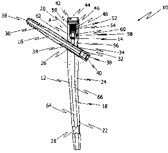
Referring to Fig. 1, there is shown a cross-sectional view of an embodiment of
an
implant system 10 for use in orthopaedic surgery for fixation of bone, such as
a
femur (not shown in Fig. 1). The implant system 10 comprises an intramedullary
nail
12, a coupling unit 14 and a bone fastener 16. The coupling unit 14 couples
the bone
fastener 16 to the intramedullary nail 12. The intramedullary nail 12 includes
a rod-
shaped body 18 insertable into the inner cavity (marrow cavity) of the femur,
i.e.,
into the intramedullary canal of the femur. The rod-shaped body 18 of the
intrame-
dullary nail 12 includes a proximal portion 20, a distal portion 22 which is
longer than
the proximal portion 20, and a bent portion 24 located between the proximal
portion
20 and the distal portion 22. In other words, the bent portion 24 connects the
proxi-
mal portion 20 and the distal portion 22.
As shown in Fig. 1, the intramedullary nail 12 includes a transverse bore 26
located
at the proximal portion 20. An axis of the transverse bore 26 has an angle
with re-
spect to a longitudinal axis of the intramedullary nail, such that a
longitudinal axis of
the transverse bore 26 has an oblique extension relative to an axial extension
of the
proximal portion 20. While in the present embodiment only a single transverse
bore
26 is utilized, in other embodiments multiple (e.g., two or more) transverse
bores
may be provided in the proximal portion 20.
The proximal portion 20 of the intramedullary nail 12 has a diameter
sufficient to
accommodate the transverse bore 26 therein, while the distal portion 22 of the
in-
tramedullary nail 12 has a smaller diameter with respect to the proximal
portion 20,
adapted to the shape of the marrow cavity of the femur in order to facilitate
the
insertion of the distal portion 22 into the intramedullary canal. Further, the
distal
portion 22 includes a through hole 28 extending orthogonally to a longitudinal
axis of
the distal portion 22. The through hole 28 is formed at an end of the distal
portion 22
CA 02824802 2013-07-15
WO 2012/107226
PCT/EP2012/000577
- 12 -
of the intramedullary nail 12 for receiving a bone fastener, such as a locking
screw,
in order to securely fix the intramedullary nail 12 to bone.
In the embodiment of the implant system 10 shown in Fig. 1, the bone fastener
16 is
a femoral neck screw in the form of a lag screw 16. The lag screw 16 is
adapted to
penetrate the transverse bore 26 of the intramedullary nail 12. The lag screw
16 has
a front portion 30 including a thread, for example a coarse thread, and a rear
portion
32. The rear portion 32 is provided with a plurality of longitudinally
extending
grooves 34 (two are shown in Fig. 1) arranged on the peripheral surface of the
rear
shaft portion 32 along the axis of the lag screw 16. Typically, four grooves
34 are
disposed on the peripheral surface of the lag screw 16 at intervals of 90
around the
longitudinal axis of the lag screw 16. Each groove 34 defines a ramp having a
shal-
low end and a deeper end. The rising ramp extends from the shallow end at a
rear
end of the rear portion 32 towards the threaded front portion 30 to the deeper
end.
The grooves 34 thus have an asymmetric depth profile. Further, the lag screw
16
includes a central cannulation 36 along the longitudinal axis of the lag screw
16. The
rear portion 32 of the lag screw 16 includes at the rear end a co-axial bore
38 and a
recess 40 (e.g., a hexalobular internal driving feature) for receiving a screw
driver or
a wrench (e.g., in the form of a entrainer driving feature).
As illustrated in Fig. 1, the proximal portion 20 of the intramedullary nail
12 includes
a recess 42 for receiving an end cap or a tool, such as a holding instrument
or tar-
geting instrument (not shown in Fig. 1) at the upper end of the proximal
portion 20.
The proximal portion 20 defines a longitudinal axis 44 and further includes a
bore 46
and a guiding structure 48. In the present embodiment, the bore 46 of the
proximal
portion 20 is co-axial with the longitudinal axis 44 of the proximal portion
20. As
further shown in Fig. 1, the bore 46 includes an internal thread 50 and a
recess por-
tion 52 for receiving a retainer 54 the exemplary in form of a snap ring.
The coupling unit 14 is preassembled and movably arranged within the proximal
portion 20 of the intramedullary nail 12. The coupling unit 14 includes one
bone
fastener engagement member 56 and a drive member 58 with a through hole 60.
The engagement member 56 is located at a lateral side of the intramedullary
nail 12
and realized in the exemplary form of a substantially cylindrical bolt or pin
56.
The terms medial and lateral are standard anatomical terms of direction and
denote
a direction toward the center or median plane of a body and the opposite
direction
CA 02824802 2013-07-15
WO 2012/107226
PCT/EP2012/000577
- 13 -
from the center to the side, respectively. With respect to the overall present
disclo-
sure and the exemplary embodiments, the medial and lateral directions may
general-
ly lie within a plane including the longitudinal axis 44 of the proximal
portion 20 and
a longitudinal axis of the transverse bore 26. In such a case, the medial side
of the
intramedullary nail 12 may be a side facing towards the outgoing side of the
trans-
verse bore 26 (e.g., towards a tip of the bone fastener 16 penetrating the
transverse
bore 26), whereas the lateral side may be a side facing towards ingoing side
of the
transverse bore 26 (e.g., towards a head of the bone fastener 16). In many
cases,
the intramedullary nail 12 will be anatomically adapted so that the nail 12
inherently
defines the medial and lateral sides, for example with respect to one or more
its
bending (e.g., as embodied by bent portion 24), an inclination of the
transverse bore
26, and so on.
Returning to Fig. 1, the drive member 58 is movably connected to the pin 56.
The
through hole 60 of the drive member 58 is a central through hole having an
axis
which coincides with the longitudinal axis 44 of the proximal portion 20. The
drive
member 58 further includes an external thread 62 for threadable engagement
with
the intramedullary nail 12 (e.g., with the proximal portion 20 as shown in
Fig. 1). The
internal thread 50 of the proximal portion 20 mates with the external thread
62 of
the drive member 58. In the present embodiment, the drive member 58 of the cou-
pling unit 14 is movably arranged within the bore 46 of the proximal portion
20 of
the intramedullary nail 12. Moreover, the coupling unit 14 is captively held
within the
proximal portion 20 of the intramedullary nail 12. As also illustrated in Fig.
1, the
guiding structure 48 slidably receives the pin 56 of the coupling unit 14,
such that
the pin 56 can engage within a groove 34 of the lag screw 16. Upon engagement
within the groove 34, the pin 56 can exert pressure on the lag screw 16 for
stabiliza-
tion purposes. The pressure is initially zero or low enough to still permit a
sliding
movement of the lag screw 16 relative to the intramedullary nail 12. The
pressure
will change (and typically increase) as the lag screw 16 slides due to the
depth pro-
file (i.e., the laterally and medially provided ramps) of the grooves 34.
As further shown in Fig. 1, the intramedullary nail 12 includes a channel 64
substan-
tially along the longitudinal axis of the intramedullary nail 12. Thus, a
cannulation 66
is defined through the intramedullary nail 12 by the channel 64 of the
intramedullary
nail 12, the through hole 60 of the drive member 58 and the bore 46 of the
proximal
portion 20, such that a surgical wire (not shown in Fig. 1) can be inserted
through
the cannulation 66.
CA 02824802 2013-07-15
WO 2012/107226
PCT/EP2012/000577
- 14 -
Fig. 2 illustrates a detailed view A of the proximal portion 20 of the
intramedullary
nail 12 shown in Fig. 1. As shown in Fig. 2, the bore 46 of the proximal
portion 20
defines a first axis 68 which, in the present embodiment, coincides with the
longitu-
dinal axis 44 of the proximal portion 20. In other embodiments, the first axis
68 of
the bore 46 may be spaced apart from and extend parallel to the longitudinal
axis 44
of the proximal portion 20. In certain cases, the first axis 68 of the bore 46
may be
slightly inclined (e.g., at an angle of up to 100 or 15 ) with respect to the
longitudinal
axis 44 of the proximal portion 20 and thus remain at least substantially
parallel
lo thereto.
Further, the guiding structure 48 defines a second axis 70. The first axis 68
of the
bore 46 and the second axis 70 of the guiding structure 48 are substantially
parallel
to the longitudinal axis 44 of the proximal portion 20 of the intramedullary
nail 12
and are spaced apart from each other. Moreover, the second axis 70 of the
guiding
structure 48 is oriented eccentrically with respect to the longitudinal axis
44 of the
proximal portion 20. The bore 46 of the proximal portion 20 and the guiding
struc-
ture 48 are thus arranged adjacent to each other. In the present embodiment
illus-
trated in Figs. 1 and 2, the bore 46 of the proximal portion 20 is located
centrally and
the guiding structure 48 of the proximal portion 20 is located at the lateral
side of the
intramedullary nail 12. The pin 56 of the coupling unit 14 guided within the
guiding
structure 48 is therefore arranged at the lateral side of the intramedullary
nail 12.
The bore 46 of the proximal portion 20 terminates at its lower end in the
channel 64
of the intramedullary nail 12. The guiding structure 48 terminates at its
lower end in
the transverse bore 26 of the proximal portion 20. In the present embodiment,
the
term "lower end" means that end which is nearer to the distal portion 22 of
the in-
tramedullary nail 12, and the term "upper end" is the opposite of the lower
end.
Further, the guiding structure 48 is formed as a groove having a circular
shape (e.g.,
C-shape) in cross-section.
As also illustrated in Fig. 2, the pin 56 of the coupling unit 14 is
eccentrically ar-
ranged on the drive member 58, i.e., arranged at an eccentric position (e.g.,
at a
lateral position as shown in Fig. 2). Further, the guiding structure 48 and
thus the pin
56 define a longitudinal axis intersecting the longitudinal axis of the lag
screw 16.
The pin 56 is formed as a bolt having a cylindrical shaft (here: with circular
cylindrical
cross-section) and a spherical tip at its lower end.
CA 02824802 2013-07-15
WO 2012/107226
PCT/EP2012/000577
- 15 -
The drive member 58 includes a drive transmitting portion 72 for transmitting
the
movement of the drive member 58 to the pin 56. The pin includes a groove 74 at
its
upper end. The groove 74 of the pin 56 is substantially arranged in a
direction trans-
verse to the longitudinal direction of the pin 56. The drive transmitting
portion 72 of
the drive member 58 movably engages within the groove 74 of the pin 56. For
this
purpose, the drive transmitting portion 72 is rotatably supported in the
groove 74 of
the pin 56. Thus, rotation of the drive member 58 causes movement of the pin
56 in
the direction of the longitudinal axis 44 of the proximal portion 20.
The drive member 58 of the coupling unit 14 has a receiving portion 76 in form
of a
cone having a recess (e.g., in the form of a hexalobular internal driving
feature) for
receiving a tool, screwdriver, wrench or the like. By driving the drive member
58
using such a tool, the entire coupling unit 14 moves along the longitudinal
axis 44 of
the proximal portion 20 of the intramedullary nail 12, since the external
thread 62 of
the drive member 58 mates with the internal thread 50 of the bore 46 of the
proxi-
mal portion 20. In other words, the position of the coupling unit 14, and
therewith
the position of its pin 56, within the proximal portion 20 of the
intramedullary nail 12
can be adjusted by screwing the drive member 54 of the coupling unit 14 along
the
longitudinal axis 44.
As shown in Fig. 2, the range of motion (i.e., the movement) of the coupling
unit 14
in the proximal direction is limited by the retainer 54. The retainer 54 in
form of a
snap ring engages within the recess portion 52. The recess portion 52 is
formed as a
circumferential groove within the proximal portion 20 of the intramedullary
nail 12 to
avoid an unintended disassembling of the coupling unit 14 and its drive member
58
and pin 56.
Upon moving of the coupling unit 14 towards the distal portion 22 of the
intramedul-
lary nail 12, the coupling unit 14 (particularly, the drive member 58 of the
coupling
unit 14) urges the pin 56 in the direction of the longitudinal axis 44 of the
proximal
portion 20 towards the distal portion 22 of the intramedullary nail 12. The
pin 56 of
the coupling unit 14 thus slides within the guiding structure 48 towards the
lag screw
16. In a final position (as shown in Fig. 2), the pin 56 engages within one of
the
grooves 34 of the lag screw 16 to prevent rotation of the lag screw 16 about
its Ion-
gitudinal axis.
CA 02824802 2013-07-15
WO 2012/107226
PCT/EP2012/000577
- 16 -
As illustrated in Figs. 1 and 2, the laterally arranged, eccentric pin 56
allows an en-
gagement within a groove 34 of the lag screw 16. The medial cannulation 66
formed
by the canal 64 of the intramedullary nail 12, the central through hole 60 of
the drive
member 58 and the bore 46 of the proximal portion 20 allows the simultaneous
in-
serting of a guide wire.
During a surgical procedure, the intramedullary nail 12 is positioned and
located in
the intramedullary canal of a bone, e.g., the femur. Then, a hole is bored
transver-
sally through the femur, the neck of the femur and into the head thereof for
receiv-
o ing the lag screw 16. Then, the lag screw 16 is screwed into position
through the
transverse bore 26 of the intramedullary nail 12 by operating a tool, e.g, a
screw
driver, such that one of the longitudinal grooves 34 of the lag screw 16 is
aligned in
the uppermost position. The drive member 58 of the coupling unit 14, which is
pre-
assembled within the proximal portion 20 of the intramedullary nail 12, is
then
is turned downwards (i.e., in the direction of the longitudinal axis 44 of
the proximal
portion 20 towards the distal portion 22 of the intramedullary nail 12) with a
screw
driver until the lower end of the pin 56 is engaged within one of the grooves
34 of
the lag screw 16.
20 Provided that the coupling unit 14 is not completely tightened (i.e.,
the drive member
58 of the coupling unit 14 is not completely tightened), the lag screw 16 has
the
facility to slide within the transverse bore 26 only in a lateral direction
(to the right in
Figs. 1 and 2) but is locked against rotation about its longitudinal axis. As
the lag
screw 16 is held against rotation by the coupling unit 14 (i.e., by the pin 56
of the
25 coupling unit 14), it merely slides through the transverse bore 26 and
draws the
head of the femur into close engagement with the rest of the bone. Due to the
rising
ramp of the groove 34 of the lag screw 16, an uncontrolled medial sliding (to
the left
in Figs. 1 and 2) of the lag screw 16 within the intramedullary nail 12 is
prevented.
30 Figs. 3 to 7 show another embodiment of a proximal portion with an
alternative cou-
pling unit embodiment, that can be adapted as needed (e.g., in terms of shape,
length, width, thickness, etc.) for use in the intramedullary nail 12 of the
implant
system 10 shown in Fig. 1.
35 Fig. 3 illustrates a cross-sectional view of the alternative embodiment
of the proximal
portion 78 of the intramedullary nail. The proximal portion 78 includes a
central bore
80 having an internal thread 82. The proximal portion 78 further includes the
recess
CA 02824802 2013-07-15
WO 2012/107226
PCT/EP2012/000577
- 17 -
portion 52 in form of the groove 52 for receiving the retainer 54 within the
central
bore 80. Moreover, the proximal portion 78 also includes the recess 42 for
receiving
an end cap or a tool, such as a holding instrument or targeting instrument
(not
shown in Fig. 3) at the upper end of the proximal portion 78.
As shown in Fig. 3, the guiding structure 48 is formed as a bore 48 located at
the
lateral side (right-hand side in Fig. 3) of the intramedullary nail. The
guiding struc-
ture 48 terminates at its upper end in the central bore 80 of the proximal
portion 78
and at its lower end in the transverse bore 26. Also in this present
embodiment, the
term "lower end" means that end which is nearer to the distal portion of the
in-
tramedullary nail, and the term "upper end" is the opposite of the lower end.
As
further illustrated in Fig. 3, the proximal portion 78 includes a bore 84
which is ar-
ranged adjacent to the guiding structure 48. The bore 84 of the proximal
portion 78
also terminates at its upper end in the central bore 80 and at its lower end
in the
transverse bore 26 of the intramedullary nail. Further, the bore 84 defines
the first
axis 68 and the guiding structure 48 defines the second axis 70, wherein the
first
axis 68 and the second axis 70 are substantially parallel to the longitudinal
axis 44 of
the proximal portion 78 and are spaced apart from each other (here: spaced
apart
from each other in the transverse direction), as shown in Fig. 3.
Fig. 4 illustrates a bottom view a), a side view b), and a top view c) of an
alternative
pin embodiment having a base member 86 in the form of a plate 86 on which the
pin
56 is arranged. In the present embodiment, the pin 56 is integrally formed
with the
plate 86 as a one-piece structure. The pin 56 is configured as generally
described
above with respect to Figs. 1 and 2. The plate 86 has a circular shape and a
through
hole 88 for receiving a surgical wire or a guiding wire. The pin 56 and the
through
hole 88 are eccentrically arranged on the plate 86.
The plate 86 further has a holding portion 90. The holding portion 90 is
arranged on
the upper surface opposite to the lower surface on which the pin 56 is
arranged. The
holding portion 90 extends from the plate 86 and has a L-shape in cross-
section as
shown in the side view b) of Fig. 4. Further, the holding portion 90 forms an
arc
along the outer peripheral side of the plate 86 as illustrated in the top view
c) of Fig.
4. For this purpose, the arc formed by the holding portion 90 may extend over
180
or less. Thus, the plate 86 and the holding portion 90 thereof form a circular
groove
92 for receiving a part of a drive member as described hereinafter.
CA 02824802 2013-07-15
WO 2012/107226
PCT/EP2012/000577
- 18 -
Referring to Fig. 5, there is shown a side view of another embodiment of a
drive
member 94 in form of a short bolt. The drive member 94 has an external thread
96
on its outer peripheral surface 98. The external thread 96 of the drive member
94 is
interrupted by a circumferential groove 97. The circumferential groove 97 may
re-
ceive a ring (not shown in Fig. 5) made of synthetic material. The drive
member 94
further includes a drive transmitting portion 100. The drive transmitting
portion 100
is formed as a flange arranged on the drive member 94, wherein the diameter of
the
drive transmitting portion 100 is slightly greater than the diameter of a
shaft portion
102 of the drive member 94. Thus, a circumferential step 104 is defined by the
drive
transmitting portion 100 and the shaft portion 102 of the drive member 94. The
drive
transmitting portion 100 can movably engage with the holding portion 90 of the
plate
86, wherein the step 104 of the drive member 94 engages within the circular
groove
92 of the holding portion 90. The drive member 94 further comprises a central
through hole 106 for receiving a guide wire and a recess 108 (e.g., in the
form of a
hexalobular internal driving feature or internal hexagon) for receiving a
tool, such as
a screwdriver, a wrench, or the like.
As illustrated in Figs. 6 and 7, a coupling unit 110 is formed by the drive
member 94
and the plate 86 having the pin 56, wherein the drive member 94 is movably con-
nected to the plate 86 as described above. Further, the coupling unit 110,
i.e, the
drive member 94 and the plate 86 with the pin 56, is preassembled within the
proxi-
mal portion 78 of the intramedullary nail. The guiding structure 48 of the
proximal
portion 78 of the intramedullary nail has a diameter which is slightly larger
than the
diameter of the pin 56, such that an optimal guiding and sliding respectively
of the
pin 56 within the bore 48 is guaranteed. As shown in Fig. 6, the pin 56 of the
cou-
pling unit 110 is located within the guiding structure 48 in the preassembled
configu-
ration. Moreover, the external thread 96 of the drive member 94 of the
coupling unit
110 mates with the internal thread 82 of the central bore 80 of the proximal
portion
78 of the intramedullary nail, such that the entire coupling unit 110 is
captively held
and movably arranged within the proximal portion 78 of the intramedullary
nail.
Thus, the height adjustment of the coupling unit 110, and therewith of the pin
56, is
driven by the drive member 94, as generally described above with respect to
Figs. 1
and 2 and hereinafter. To avoid an unintended loosening of the coupling unit
110,
the driving member 94 of the coupling unit 110 has a ring 112 made of
synthetic
material arranged in the circumferential groove 97 of the drive member 94 as
shown
in Figs. 6 and 7. Furthermore, the retainer 54 is positioned and engaged into
the
recess portion 52 formed as groove 52 within the central bore 80 of the
proximal
CA 02824802 2013-07-15
WO 2012/107226
PCT/EP2012/000577
- 19 -
portion 78 of the intramedullary nail to avoid an unintended disassembling of
the
coupling unit 110 or of its parts (drive member 94 and plate 86 with pin 56).
Thus,
the retainer 54 functions as a limiter which limits the range of motion of the
coupling
unit 110 in the proximal direction.
As shown in Figs. 6 and 7, the drive transmitting portion 100 of the drive
member 94
engages on the holding portion 90 of the plate 86. The plate 86 is centrally
inserted
within the proximal portion 78 of the intramedullary nail, providing
rotational stability
of the pin 56 of the coupling unit 110. Thus, rotation of the drive member 94
of the
coupling unit 110 causes movement of the pin 56, which is slidably received in
the
guiding structure 48, in the direction of the longitudinal axis 44 of the
proximal por-
tion 78 of the intramedullary nail. The rotation of the drive member 94 is
performed
by a tool such as a screw driver or the like which engages within the recess
108 of
the drive member 94. Upon moving of the coupling unit 110 along the
longitudinal
axis 44 of the proximal portion 78 of the intramedullary nail, the coupling
unit 110
(particularly, the drive member 94 of the coupling unit 110) urges the pin 56
through
the guiding structure 48 in the direction of the longitudinal axis 44 towards
the distal
portion of the intramedullary nail, such that the pin 56 engages within a
groove of
the lag screw to prevent rotation of the lag screw about its longitudinal
axis.
As further illustrated in Figs. 6 and 7, the channel of the intramedullary
nail, the bore
84 of the proximal portion 78 of the intramedullary nail, the through hole 88
of the
plate 86, the through hole 106 of the drive member 94, and the central bore 80
of
the proximal portion 78 define a cannulation. A guide wire 114 may be inserted
through the cannulation as shown in Fig. 7.
Fig. 8 illustrates a cross-sectional view of an alternative embodiment of a
proximal
portion 20 of the intramedullary nail 12 of the implant system 10 shown in
Figs. 1
and 2. The proximal portion 20 of the intramedullary nail 12 comprises the
bore 46
defining the first axis 68 and the guiding structure 48 defining the second
axis 70 as
shown in and generally described above with reference to Figs. 1 and 2. The
first
axis 68 of the bore 46 of the proximal portion 20 and the second axis 70 of
the guid-
ing structure 48 are substantially parallel to the longitudinal axis 44 of the
proximal
portion 20 of the intramedullary nail 12 and are spaced apart from each other.
As
described above with reference to Figs. 1 and 2, the second axis 70 of the
guiding
structure 48 is oriented eccentrically with respect to the longitudinal axis
44 of the
proximal portion 20. The intramedullary nail 12 further includes the coupling
unit 14
CA 02824802 2013-07-15
WO 2012/107226
PCT/EP2012/000577
- 20 -
having the pin 56 which is eccentrically arranged on the drive member 58 as
shown
in and generally described above with reference to Figs. 1 and 2.
As shown in Fig. 8, the first axis 68 of the bore 46 of the proximal portion
20 coin-
sides with the longitudinal axis 44 of the proximal portion 20. Thus, the bore
46 of
the proximal portion 20 is located, in this case, centrally with respect to
the longitu-
dinal axis 44 of the proximal portion 20 of the intramedullary nail 12. In the
present
embodiment illustrated in Fig. 8, the guiding structure 48 of the proximal
portion 20
is located at the medial side of the intramedullary nail 12. The guiding
structure 48 is
configured to slidably receive the cylindrical pin 56 of the coupling unit 14,
such that
the pin 56 can engage within a groove 34 of a bone fastener 16 configured to
pene-
trate the transverse bore 26 of the intramedullary nail 12. As shown in Fig.
8, the pin
56 of the coupling unit 14 is therefore arranged at the medial side of the
intramedul-
lary nail 12 (in Fig. 8, the medial side of the intramedullary nail 12 is on
the left side
of the drawing). Further, the pin 56 defines a longitudinal axis intersecting
the longi-
tudinal axis of the lag screw 16.
Upon moving of the coupling unit 14 towards the distal portion 22 of the
intramedul-
lary nail 12, the coupling unit 14, particularly the drive member 58 of the
coupling
unit 14, urges the pin 56 in the direction of the longitudinal axis 44 of the
proximal
portion 20 towards the distal portion 22 of the intramedullary nail 12. The
pin 56 of
the coupling unit 14 thus slides within the guiding structure 48 towards the
lag screw
16. In a final position (as shown in Fig. 8), the pin 56 engages at the medial
side of
the intramedullary nail 12 within a groove 34 of the lag screw 16 to prevent
rotation
of the lag screw 16 about its longitudinal axis and to provide a defined
sliding of the
lag screw 16 within the transverse bore 26 of the proximal portion 20.
Fig. 9 illustrates a cross-sectional view of an alternative embodiment of a
proximal
portion 20 of the intramedullary nail 12 of the implant system 10 as shown in
Figs. 1
and 2. The intramedullary nail 12 shown in Fig. 9 comprises the proximal
portion 20,
the coupling unit 14 and the bone fastener 16 as shown in and generally
described
above with reference to Figs. 1 and 2. Further, the intramedullary nail 12 has
the
transverse bore 26. The proximal portion 20 defines the longitudinal axis 44
as gen-
erally described above with reference to Figs. 1 and 2.
In the present embodiment the proximal portion 20 of the intramedullary nail
12
includes the bore 46 defining a bore axis 68 and two guiding structures 48,
wherein
CA 02824802 2013-07-15
WO 2012/107226
PCT/EP2012/000577
- 21 -
each guiding structure defines a guiding axis 70. As shown in Fig. 9, the bore
axis 68
of the bore 46 and the guiding axes 70 are substantially parallel to the
longitudinal
axis 44 of the proximal portion 20 of the intramedullary nail 12 and are
spaced apart
from each other. The coupling unit 14 is adapted to be movably arranged within
the
proximal portion 20 of the intramedullary nail 12 as shown in and generally
described
above with reference to Figs. 1 and 2. The coupling unit 14 includes, in this
embodi-
ment, two substantially cylindrical pins 56 and the drive member 58. As shown
in Fig.
9, one guiding structure 48 is located at the lateral side (right-hand side of
the draw-
ing in Fig. 9) and the other guiding structure 48 is located at the medial
side (left-
hand side in the drawing of Fig. 9) of the intramedullary nail 12. As
illustrated in Fig.
9, one pin 56 is arranged at both of the lateral side and the medial side of
the in-
tramedullary nail. In other words, one pin 56 is at the lateral side and one
further pin
56 is at the medial side of the intramedullary nail 12. Thus, the present
embodiment
is a combination of the embodiments shown in Figs. 1, 2 and 8. Further, each
guid-
ing structure is configured to slidably receive one of the substantially
cylindrical pins
56, such that the pins 56 can engage within a (e.g., one single) groove 34 of
the
bone fastener 16 which is configured to penetrate the transverse bore 26 of
the
intramedullary nail 12.
zo As shown in Fig. 9, the pins 56 are arranged substantially along a
direction of the
longitudinal axis 44 of the proximal portion 206. Alternatively, the pins 56
may be
shifted with respect to the longitudinal axis 44 and may be located in a
region at the
lateral and medial side of the intramedullary mail 12 respectively. Upon
moving of
the coupling unit 14 towards the distal portion 22 of the intramedullary nail
12, the
coupling unit (particularly, the drive member 58 of the coupling unit 14)
urges the
two pins 56 in the direction of the longitudinal axis 44 of the proximal
portion 20
towards the distal portion 22 of the intramedullary nail 12. The pins 56 of
the cou-
pling unit 14 thus slide within the guiding structures 48 toward the lag screw
16. In a
final position (as shown in Fig. 9) the pins 56 engage within a (e.g., one
single)
groove 34 of the lag screw 16 to prevent rotation of the lag screw 16 about
its longi-
tudinal axis and to provide a high mechanical load stability of the construct
of in-
tramedullary nail 12, coupling unit 14 and bone fastener 16 within the body of
the
patient.
Figs. 10 and 11 show another embodiment of a proximal portion within an
alternative
coupling unit embodiment, that can be adapted as needed (e.g., in terms of
shape,
CA 02824802 2013-07-15
WO 2012/107226
PCT/EP2012/000577
- 22 -
length, width, thickness, etc.) for use in the intramedullary nail 12 of the
implant
system 10 shown in Fig. 1.
Fig. 10 illustrates a cross-sectional view of the alternative proximal portion
20 of the
intramedullary nail 12. The proximal portion 20 of the intramedullary nail 12,
the
drive member 58 and the bone fastener 16 are configured as shown in and
generally
described above with reference to Figs. 1, 2, 8 and 9. The proximal portion 20
of the
intramedullary nail 12 defines the longitudinal axis 44. Further, the proximal
portion
20 includes the bore 46 defining a bore axis 68 which is coaxial with the
longitudinal
axis 44 of the proximal portion 20, i.e., the bore 46 is centrally arranged in
the prox-
imal portion 20 of the nail 12 with respect to the longitudinal axis 44 of the
proximal
portion 20.
In the present embodiment, the proximal portion 20 again includes two guiding
structures 48, wherein each guiding structure 48 defines a guiding axis 70.
The bore
axis 68 and the guiding axes 70 are substantially parallel to the longitudinal
axis 44
of the proximal portion 20 of the intramedullary nail 12 and are spaced apart
from
each other as shown in Fig. 10.
Further, the implant system includes a coupling unit 116 having the drive
member 58
as shown in and generally described above with reference to Figs. 1, 2, 8 and
9. The
coupling unit 116 comprises an alternative pin embodiment 118 including two
sub-
stantially cylindrically pins 56 as generally described above with reference
to Figs. 1,
2, 8 and 9. The pin embodiment 118 of the coupling unit 116 is described with
refer-
to Fig. 11 in more detail below.
As shown in Fig. 10, the guiding structures 48 are formed as grooves. One
guiding
structure 48 is located at the lateral side (right-hand side in Fig. 10) and
the other
guiding structure 48 is located at the medial side (left-hand side in Fig. 10)
of the
intramedullary nail 12. Each guiding structure 48 is configured to slidably
receive one
of the pins 56 of the pin embodiment 118 of the coupling unit 116, such that
the pins
56 can engage within the groove 34 of the bone fastener 16 which is configured
to
penetrate the transverse bore 26 of the intramedullary nail 12. As illustrated
in Fig.
10, one pin 56 is arranged at both of the lateral side and the medial side of
the in-
tramedullary nail. In other words, one pin 56 is at the lateral side and one
pin is at
the medial side of the intramedullary nail.
CA 02824802 2013-07-15
WO 2012/107226
PCT/EP2012/000577
- 23 -
=
Fig. 11 illustrates a bottom view a), a side view b), and a top view c) of the
alterna-
tive pin embodiment 118 used with the drive member 58, which both form the cou-
pling unit 116 as inserted in the proximal portion 20 of the intramedullary
nail 10 as
shown in the embodiment of Fig. 10. The pin embodiment 118 has a base member
120 in the form of a plate 120 on which two pins 56 are arranged. In the
present
embodiment, each pin 56 is integrally formed with the plate 120. Each pin 56
is con-
figured as generally described above with reference to Figs. 1 and 2. The
plate 120
has two plate segments 122 with a substantially circular shape. The two plate
seg-
ments 122 of plate 120 are connected with each other by two curved arms 124,
such
that the two arms 124 form a through hole 126 for receiving a surgical wire or
a
guiding wire. Further, the through hole 126 is centrally arranged on the base
mem-
ber 120. As shown in Fig. 11, each pin 56 is arranged on a corresponding plate
seg-
ment 122 of the base member 120 respectively. Thus, the pins 56 are arranged
opposite to each other and extend in the same direction from the base member
120.
As further shown in Fig. 11, the pins 56 have a different length in its
longitudinal
direction. The length of the pins 56 can be adapted as needed for use in the
proximal
portion 20 of the intramedullary nail 12 of the implant system shown in Fig.
10.
With reference to Fig. 10, the pin embodiment 118 is inserted in the proximal
portion
20 of the intramedullary nail 12, such that each pin 56 is received by a
guiding struc-
ture 48 of the proximal portion 20. In the present embodiment, the pin 56 with
the
shorter length is received by the guiding structure 48 which is located at the
medial
side (left-hand side in Fig. 10) of the intramedullary nail 12. Further, the
pin 56 hav-
ing the longer length is received by the guiding structure 48 which is located
at the
lateral side (right-hand side in Fig. 10) of the intramedullary nail 12. The
drive mem-
ber 58 of the coupling unit 116 includes a drive transmitting portion 128
which en-
gages on the top surface of the base plate 120 of the pin embodiment 118.
Thus, as
illustrated in Fig. 10, the coupling unit 116 is formed by the drive member 58
and the
base member 120 having the pins 56, wherein the drive member 58 is movably con-
nected to the base member 120. Further, the coupling unit 116, i.e., the drive
mem-
ber 58 and the base member 120 with the pins 56, is preassembled within the
proximal portion 20 of the intramedullary nail 12. The guiding structures 48
of the
proximal portion 20 of the intramedullary nail 12 are formed as open bores
(e.g., c-
shaped grooves) and have a diameter which is slightly larger than the diameter
of
each pin 56, such that an optimal guiding and sliding respectively of the pins
56
within the guiding structures 58 is guaranteed.
CA 02824802 2013-07-15
WO 2012/107226
PCT/EP2012/000577
- 24 -
As shown in Fig. 10, the drive transmitting portion 128 of the drive member
58,
which is, in this case, the bottom surface of the drive member 58, engages on
the
base member 120 of the pin embodiment 118. The base member 120 is centrally
inserted within the proximal portion 20 of the intramedullary nail 12,
providing rota-
s tional stability of the pins 56 of the coupling unit 116. Thus, rotation
of the drive
member 58 of the coupling unit 116 causes movement of the pins 56, which are
slidably received in the guiding structures 48, in the direction of the
longitudinal axis
44 of the proximal portion 20 of the intramedullary nail 12. The rotation of
the drive
member 58 is performed by a tool such as a screw driver or the like which
engages
within a recess of the drive member as generally described above with
reference to
Figs. 1 and 2. Upon moving of the coupling unit 116 along the longitudinal
axis 44 of
the proximal portion 20 of the intramedullary nail 12, the coupling unit 116
(particu-
larly, the driving member 58 of the coupling unit 116) urges the two pins 56
through
the corresponding guiding structure 48 in the direction of the longitudinal
axis 44
towards the distal portion 22 of the intramedullary nail 12, such that the
pins 56
engage within a (e.g., one) groove of the bone fastener 16. Thus, rotation of
the
bone fastener about its longitudinal axis is prevented and a high mechanical
load
stability of the construct of intramedullary nail, coupling unit and bone
fastener in-
serted through the transverse bore 26 of the intramedullary nail 12 and into
bone
within the body of the patient is provided.
As further shown in Fig. 10, the channel 64 of the intramedullary nail 12, the
bore 46
of the proximal portion 20 of the intramedullary nail 12, the through hole 126
of the
base member 120 of the pin embodiment 118, the through hole 60 of the drive
member 58 define a cannulation 66. A surgical wire or a guide wire (not shown
in
Fig. 10) may be inserted through the cannulation 66.
Figs. 12 to 14 show a still further embodiment of an implant system. As
illustrated in
the cross-sectional view of Fig. 12, the proximal portion 20 of the
intramedullary nail
12, the drive member 58 and the bone fastener 16 are configured as shown in
and
generally described above with reference to Figs. 1, 2, 8, 9 and 10. Thus, the
proxi-
mal portion 20 of the intramedullary nail 12 defines the longitudinal axis 44
and
includes the bore 46 defining the bore axis 68 which is, in this case, coaxial
with the
longitudinal axis 44 of the proximal portion 20, i.e., the bore 46 is
centrally arranged
in the proximal portion 20 with respect to the longitudinal axis 44 of the
proximal
portion 20.
CA 02824802 2013-07-15
WO 2012/107226
PCT/EP2012/000577
- 25 -
In the present embodiment, the proximal portion also includes the two guiding
struc-
tures 48, wherein each guiding structure 48 defines a guiding axis 70. The
bore axis
68 and the guiding axes 70 are substantially parallel to the longitudinal axis
44 of the
proximal portion 20 of the intramedullary nail 12 and are spaced apart from
each
other as shown in Fig. 12.
In the present embodiment, the implant system includes an alternative coupling
unit
embodiment that can be adapted as needed (e.g., in terms of shape, length,
width,
thickness, etc.) for use in the proximal portion 20 of the intramedullary nail
12 shown
in Fig. 12. The alternative coupling unit 130 includes the drive member 58 as
shown
in and generally described above with reference to Figs. 1, 2, 8, 9 and 10.
The cou-
pling unit 130 further comprises an alternative pin embodiment 132 including
two
substantially cylindrical pins 56 as generally described above with reference
to Figs.
1, 2, 8, 9 and 10.
The pin embodiment 132 of the coupling unit 130 has a base member 134 in form
of
a plate 134 on which the two pins 56 are arranged. In the present embodiment,
each
pin 56 is integrally formed with the plate 134. Each pin 56 is configured as
generally
described above with reference to Figs. 1 and 2. The plate 134 has a circular
shape
and through hole 136 for receiving a surgical wire or a guiding wire (not
shown in
Fig. 12). As shown in Fig. 12, the through hole 136 is centrally arranged on
the base
member 134. The pins 56 are eccentrically arranged on the plate 134. Further,
the
pins 56 are arranged opposite to each other and extend in the same direction
from
the base member 134. In the present embodiment, the pins 56 are positioned on
the
base member 134 along a direction of a diameter of the circular plate 134 that
per-
pendicularly crosses the longitudinal axis 44. Alternatively, the pins 56 can
be shifted
for example in a parallel manner with respect to a line defined by that
diameter of
the circular plate 134, e.g., shifted in a direction which is substantially
perpendicular
to the diameter of the circular plate 134. In this case, the pins 56 may lie
on a line
which is parallel to the diameter of the circular plate 134. As further shown
in Fig.
12, each pin 56 is arranged on the plate 134 near the outer periphery of the
base
member 134.
As illustrated in Fig. 12, the guiding structures 48 are located at opposite
sides with
respect to the longitudinal axis of the bone fastener 16 which is configured
to pene-
trate the transverse bore 26 of the intramedullary nail 12. Thus, the guiding
struc-
tures 48 can be located in a central position with respect to the proximal
portion 20
CA 02824802 2013-07-15
WO 2012/107226
PCT/EP2012/000577
- 26 -
(as shown in Fig. 12), or alternatively, at the medial side or at the lateral
side of the
intramedullary nail 12. Thus, the two pins 56 can alternatively be arranged
closer to
one of the lateral side and the medial side of the intramedullary nail 12. In
other
words, the pins 56 can be at the lateral side or at the medial side of the
intramedul-
lary nail 12.
The pin embodiment 132 is inserted in the proximal portion 20 of the
intramedullary
nail 12, such that each pin 56 is received by a guiding structure 48 of the
proximal
portion 20. The drive member 58 of the coupling unit 130 includes a drive
transmit-
ting portion 138 which engages on the top surface of the plate 134 of the pin
em-
bodiment 132. In this case, the drive transmitting portion 138 is the bottom
surface
of the drive member 58. Thus, as illustrated in Fig. 12, the coupling unit 130
is
formed by the drive member 58 and the base member 134 having the pins 56,
wherein the drive member 58 is movably connected to the base member 134. Fur-
ther, the coupling unit 130, i.e., the drive member 58 and the base member 134
with
the pins 56, is preassembled within the proximal portion 20 of the
intramedullary nail
12.
As shown in Fig. 12, the drive transmitting portion 138 of the drive member 58
en-
gages on the base member 134 of the pin embodiment 132. The base member 134
is centrally inserted within the proximal portion 20 of the intramedullary
nail 12, i.e.,
centrally inserted within the bore 46 of the proximal portion 20, providing
rotational
stability of the pins 56 of the coupling unit 130. Thus, rotation of the drive
member
58 of the coupling unit 130 causes movement of the pins 56, which are slidably
re-
ceived in the guiding structures 48, in the direction of the longitudinal axis
44 of the
proximal portion 20 of the intramedullary nail 12. The rotation of the drive
member
58 is performed by a tool as generally described above with reference to Figs.
1, 2,
8, 9 and 10. Upon moving of the coupling unit 130 along the longitudinal axis
44 of
the proximal portion 20 of the intramedullary nail 12, the coupling unit 130
(particu-
larly, the drive member 58 of the coupling unit 130) urges the two pins 56
through
the corresponding guiding structure 48 in the direction of the longitudinal
axis 44
towards the distal portion of the intramedullary nail 12, such that each pin
56 engag-
es within a dedicated groove 34 of the bone fastener 16. Hence, as shown in
Figs. 12
and 14, the pins 56 of the coupling unit 130 engage within two different
grooves 34
of the bone fastener 16. Moreover, upon engagement within the grooves 34, the
pins
56 can exert pressure on the lag screw 16 for stabilization purposes. Thus, a
rotation
of the bone fastener 16 about its longitudinal axis is prevented and a high
mechani-
CA 02824802 2013-07-15
WO 2012/107226
PCT/EP2012/000577
- 27 -
cal load stability of the construct of intramedullary nail, coupling unit and
bone fas-
tener inserted through the transverse bore 26 of the intramedullary nail 12
and into
bone within the body of the patient is provided.
As stated above, in other embodiments the pins 56 may be shifted out of the
central
nail plane illustrated in Fig. 12 towards one of the lateral side and the
medial side of
the intramedullary nail 12. The pins 56 will then lie on a line (the above
"diameter")
that extends perpendicularly to a plane including the longitudinal axis 44 of
the prox-
imal portion 20 and a longitudinal axis of the transverse bore 26, wherein the
line is
spaced apart from that plane in one of a medial direction and a lateral
direction of
the intramedullary nail 12. In such case the engagement of two grooves 34 by
the
two pins 56 may be maintained, although in certain cases the grooves 34 may
need
to be modified (e.g., as regards their width or number).
As further shown in Fig. 12, the channel 64 of the intramedullary nail 12, the
bore 46
of the proximal portion 20 of the intramedullary nail 12, the through hole 136
of the
base member 134 of the pin embodiment 132, the through hole 60 of the drive
member 58 define a cannulation 66. A surgical wire or a guide wire (not shown
in
Fig. 12) may be inserted through the cannulation 66.
In an exemplary method for fracture fixation of bone using any of the above or
other
implant system embodiments, the guide wire 114 (see Fig. 7) is firstly
inserted into a
marrow cavity of bone. Then, the cannulated intramedullary nail 12 of any of
the
above or other embodiments is inserted over the guide wire 114 into the marrow
cavity of bone. The intramedullary nail 12 comprises a medial side, a lateral
side and
the proximal portion 20 or 78, the transverse bore 26 and the coupling unit
14, 110,
116 or 130 as generally described above. The guide wire 114 is then removed
and a
bone fastener 16 is inserted through the transverse bore 26 of the
intramedullary
nail 12 into bone for stabilization of the bone fracture. Finally, the
coupling unit of
the intramedullary nail 12 is driven for producing an engagement of the pin 56
with
the bone fastener 16 penetrating the transverse bore 26 of the intramedullary
nail
12, thereby preventing rotation of the bone fastener 16.
Since the proximal portion of the intramedullary nail and the coupling unit
having the
drive member and the pin are configured as described above, the coupling unit
can
be preassembled or preloaded within the intramedullary nail, while allowing a
simul-
taneous inserting/passage of a guide wire. The channel of the intramedullary
nail,
CA 02824802 2013-07-15
WO 2012/107226
PCT/EP2012/000577
- 28 -
the bore(s) of the proximal portion of the intramedullary nail and the through
hole(s)
of the coupling unit (which together define a cannulation) may be
substantially
aligned to permit insertion of a guide wire completely through the
preassembled unit
and the intramedullary nail. Thus, a guide wire can be used to guide the
intramedul-
s lary nail, including the preassembled coupling unit, into the
intramedullary canal of,
e.g., the femur. Therefore, the coupling unit has not to be assembled
intraoperative-
ly. Consequently, the operation steps that need to be performed by a surgeon
are
reduced, whereby the surgical procedure and the implantation of the
intramedullary
nail within an intramedullary canal of a femur is facilitated and simplified.
Due to this
fact, the operation time is reduced. Since the intramedullary nail is provided
with the
coupling unit (including the pin and the drive member movably connected
thereto)
that is preassembled into the hollow portion (bore) of the proximal portion of
the
intramedullary nail, the amount of time associated with implanting the
intramedullary
nail as well as the number of parts which have to be handled by a surgeon is
re-
duced.
While the one or more guiding structures and/or the one or more pins (i.e.,
the one
or more engagement members) as described herein are substantially located at
the
lateral or medial side of the intramedullary nail, the one or more guiding
structures of
the proximal portion of the intramedullary nail and the one or more pins of
the cou-
pling unit can be adapted to different applications as needed. Thus, the
guiding
structures of the proximal portion of the intramedullary nail and the one or
more pins
of the coupling unit may, for example, be located in an area at the lateral or
medial
side of the intramedullary nail. Moreover, more, e.g. two or more, pins may be
locat-
ed at one or both of the lateral side and the medial side of the
intramedullary nail.
All parts of the implant system described above are easily and cheaply
produceable
with the current state of machine tools. Moreover, since the pin can engage
within a
groove of the bone fastener, any modifications or changes of the current bone
fas-
teners are not necessary. Since the guide wires deviate to an eccentric
position (e.g.,
to the medial side) within the intramedullary nail due to the bending of the
intrame-
dullary nail, the eccentric arrangement of the pin of the coupling unit and in
particu-
lar of the bore of the proximal portion of the intramedullary nail facilitates
the fence
of the guide wire inside the intramedullary nail.
While the rod-shaped body of the intramedullary nail includes a distal portion
and a
bent portion in the embodiment illustrated in the drawings, the nail body can
be
CA 02824802 2013-07-15
WO 2012/107226
PCT/EP2012/000577
- 29 -
adapted as needed (e.g., in terms of shape, length, width, thickness, etc.)
for use in
orthopaedic surgery for fixation of bone and for insertion into an
intramedullary canal
of, e.g., a femur. Thus, the intramedullary nail can be adapted to different
applica-
tions and may thus have a different shape. Moreover, while the threads as
shown
herein are one start threads, they could also be multiple start threads (e.g.,
a two-
start thread).
While the bone fastener as described herein is formed as a lag screw, the bone
fas-
tener can be of any type of, e.g., a femoral neck screw or any kind of blade,
and can
io be adapted to different applications as needed. The bone fasteners may
thus have
different diameters, lengths, shapes or threads. Further, the bone fastener
and the
implant described above can generally be made of stainless steel, titanium or
any
other biocompatible material.
While the above embodiments have exemplarily been described in relation to a
bone
screw and an intramedullary nail, it will be readily apparent that the
techniques pre-
sented herein can also be implemented in combination with other types of bone
fasteners (such as bone pegs having a rod-like or pin-like shafts, wire-like
bone fas-
teners such as Kirschner wires, etc.) as well as other types of implants (such
as bone
plates, bone distractors, etc). Accordingly, the present disclosure is not
limited to any
type of bone fastener or any type of implant.
The features described in the above description taken in conjunction with the
accom-
panying drawings can be readily combined to result in different embodiments.
It will
thus be apparent that the disclosure described above may be varied in many
ways.
Such variations are not to be regarded as a departure from the scope of the
inven-
tion, and all modifications are intended to be included within the scope of
the follow-
ing claims.