Note : Les descriptions sont présentées dans la langue officielle dans laquelle elles ont été soumises.
CA 02829881 2013-09-11
TITLE: SCAFFOLD SYSTEM TO REPAIR CARDIOVASCULAR CONDITIONS
BACKGROUND OF THE INVENTION
I. Field of the Invention
The invention generally relates to devices for the treatment of cardiovascular
conditions.
More specifically, the invention relates to tissue engineering for the
treatment of aneurysms or
other damaged cardiovascular tissue.
2. Description of the Relevant Art
Abdominal aortic aneurysms, commonly referred to as AAA, consist of a 50%
enlargement of the abdominal aorta which is believed to be caused by the
breakdown of the
tunica media, a vessel wall layer primarily composed of smooth muscle cells.
While the exact
cause of AAA is not well understood, it is believed to be a complex process
involving
hemodynamic forces as well as local extracellular matrix remodeling,
infiltration of macrophages
and lymphocytes and increase in matrix metalloproteinase enzymes which all
play a role in the
destruction of elastin fibers and smooth muscle cells. Overtime, a gradual
reduction of medial
elastin fibers, thinning collagen within the media and thickening of the
intima heighten the
aneurismal tendency. Loss of elasticity and strength of the tunica media along
with
compensatory collagen production lead to arterial expansion, forming an
aneurysm.
Histologically, the aneurysm elastin fragmentation, chronic transmural
inflammation, and
depletion of smooth muscle cells are observed. Aneurysm progression is
characterized by
molecular mediators and extracellular matrix-degrading proteinases including
matrix
metalloproteinases 2 and 9. Increased collagen turnover has been targeted as a
potential cause of
aneurysm growth and rupture.
Studies show that 3% of all individuals aged 50 and over, predominately males,
have
AAA. In addition, 2.1% of men over 65 years of age will die of ruptured aortic
aneurysms. The
average aorta at the renal level is approximately 2 cm in diameter; therefore,
an aneurysm is
technically a 3 cm dilation. By the age of 65, 5% of men and 1.7% of women
have an aortic
diameter of at least 3 cm. The prevalence of AAA greater than or equal to 3 cm
increases 6%
with each decade beyond 65 years of age. However, most aneurysms are not
considered
.. clinically relevant until they reach 4 cm, and surgery is generally not
prescribed until they are
approximately 5 cm. The risk of rupture is known to increase with the diameter
of the aneurysm.
Only 25% of patients with ruptured aneurysms reach the hospital and only 10%
make it to the
operating room. Because of such high mortality rates, it is important to treat
the aneurysm before
it ruptures.
Substitute Specification
1
CA 02829881 2013-09-11
Current treatment of the AAA includes either open surgery or endovascular
aneurysm
repair, depending on the patient physiology and pathology. Open surgical
treatment of
aneurysms was first performed by Dubost and colleagues in 1951 but was
reintroduced by
Charles Rob in 1963 using the current retroperitoneal approach. With the
retroperitoneal
approach, the aneurysm is accessed no higher than the 11th rib when the
patient is prone. An
alternative open surgical method is the transperitoneal technique in which the
aneurysm is
accessed through an incision along the midline. In 1991, an alternative
approach to the open
surgical method was introduced by Juan Parodi in which iliofemoral access was
used to insert an
endovascular graft to cover the aneurysm: endovascular aneurysm repair (EVAR).
EVAR utilizes stent technology to place the graft over the aneurysm and into
the
iliofemoral arteries, splitting at the bifurcation. The graft serves to block
off the aneurismal
segment of the aorta without extensive damage to the arteries. Currently FDA
approved stent-
grafts contain either a woven polyester (PET) or ePTFE graft on a stainless
steel, a Cobalt-
Chromium alloy, or Nitinol stent. The grafts are fixated using either self-
expansion, stents,
barbs, or a combination of these. However, because the graft is meant to
separate the unhealthy
portion from the blood flow, inherent problems exist in the implementation.
Tortuosity of the
aorta and iliac bifurcation, particularly an angulation of 90 or greater, may
lead to an endoleak
after implantation in which blood seeps between the graft and the lumen of the
aorta, reaching
the aneurysm. Calcification and thrombotic events also play a role in limiting
EVAR
effectiveness, particularly when calcification is greater than 50% or
thrombosis is 25%-50%.
Success of an EVAR graft is usually defined by the absence of any of the four
types of
endoleaks. Type I endoleak occurs when blood flows between the graft and the
vessel wall at
either the proximal or distal ends of the graft. When blood flows into the
aneurysm sac from
branch vessels, it is considered a Type II endoleak. Type III endoleaks are
the result of poor
anastomsoes between different sections of the graft. If leakage occurs through
the graft material,
it is considered a Type IV endoleak. Types II and IV generally resolve
spontaneously while
Types I and Ill pose a greater danger and must be repaired during a subsequent
procedure.
Testing endovascular grafts for treatment of AAA require first, appropriate
cell culture
evaluation in vitro and structural mechanical properties tests, then an
appropriate AAA animal
model in order to be properly assessed, particularly in terms of coagulation
and fibrinolytic
systems. Both canine and swine models are considered appropriate for testing
current EVAR
devices.
A popular technique used to induce an aneurysm in an animal is the patch model
which
involves suturing an elliptical patch made of materials such as jejunum, iliac
vein, rectus fascia
Substitute Specification
2
or DacronTM into a longitudinal incision made in the aorta. This technique
allows gradual
enlargement and rupture similar to what is observed in humans. The greatest
drawback of
this technique is its inconsistency with physiological aneurysm formation and
anatomy. More
physiologically relevant techniques include mural-stripping, a model in which
adventitia and
60-70% of the media are cut away allowing the vessel wall to expand, and the
elastase model
which uses temporary exposure to an elastolytic agent to break down layers of
the vessel wall.
However, this technique is difficult to control.
SUMMARY OF THE INVENTION
A device for treating a cardiovascular condition includes an expandable
scaffold
positionable in a portion of a vasculature of a mammal, wherein the scaffold
is comprised of
electrospun fibers composed of a biodegradable compound. The cardiovascular
condition, in
some embodiments, is an aneurysm. The biodegradable compound may be formed
from
poly(a-hydroxy esters), for example, Polycaprolactone or other expandable
biomaterials. A
cardiovascular condition may be treated by inserting the device endovascularly
and expanding
it to provide a template for and to encourage regrowth of the damaged tissue.
It is secured
using stent technology.
In an embodiment, a device for treating a cardiovascular condition includes an
expandable scaffold supported by stent technology, wherein the scaffold is
comprised of
nonwoven fibers electrospun from a biomaterial compound, and wherein the
scaffold is
substantially tubular and comprises a concave surface having a higher
concentration of fibers
than the convex surface. When the scaffold is positioned in the vasculature of
a mammal to
direct tissue development and control blood flow into the vasculature, the
concave surface of
the scaffold may inhibit blood flow while the less concentrated convex surface
facilitates the
ingress of cells.
The vascular condition may be an aneurysm, a void, or a semi-void space.
Biomaterials that may be used include poly(a-hydroxy esters). Other
biomaterials include
natural polymers, such as Elastin, Collagen, DNA, RNA, Glucosaminoglycans, or
mixtures
thereof.
In one embodiment, the scaffold is composed of an expandable nonwoven textile.
The stent may be an expandable stent.
In an embodiment, a method of treating a vascular condition comprising
inserting a
device as described above into the vasculature of a mammal; and securing the
device in the
vasculature.
BRIEF DESCRIPTION OF THE DRAWINGS
Advantages of the present invention will become apparent to those skilled in
the art with
3
CA 2829881 2017-08-24
the benefit of the following detailed description of embodiments and upon
reference
to the accompanying drawings in which:
FIG. 1 depicts a schematic diagram of an electrospinner;
FIGS. 2A-2C depict graphs comparing the effect of solution concentration,
extrusion rate and voltage on ultimate tensile stress of electrospun tubular
scaffolds;
FIGS. 3A-3C depict graphs comparing the effect of solution concentration,
extrusion rate and voltage on strain at failure of electrospun tubular
scaffolds;
FIG. 4 depicts a graph of the average porosity of scaffolds fabricated using
varying parameters;
FIG. 5A-C depict SEM images of the contrast between the concave and
convex surfaces of a single tubular scaffold representing the gradient of
morphological changes throughout the scaffold;
FIGS. 6A-6C depict graphs of the degradation of tubular electrospun scaffolds
over 90 days in PBS at 37 C agitated at 50 RPM (n=6);
FIG. 7 depicts an SEM image of human aortic endothelial cells spread on
electrospun tubular scaffold;
FIG. 8 depicts the metabolic activity of human aortic smooth muscle cells in
static culture over 14 days on tubular electrospun PCL scaffolds;
FIG. 9 depicts the metabolic activity of human aortic smooth muscle cells in a
bioreactor on tubular electrospun scaffolds;
FIG. 10 depicts a graph comparing human aortic smooth muscle cells using
different sterilization and seeding techniques;
FIGS. 11A-B depicts SEM images of electrospun scaffolds A (nano) and B
(micro) at 2000X;
FIGS. 12A-B depict graphs of change in metabolic activity of hAoEC and
hAoSMC in response to scaffolds of different fiber morphology (normalized to
day 0
values for each sample);
FIGS. 13A-B depict graphs of cell proliferation over time of hAoEC and
hAoSMC on scaffolds composed of either nanofibers (A), microfibers (B) or
films
(C). Determined using Picogreen to measure dsDNA content, n=6;
FIGS. 14A-D depicts SEM images of electrospun microfibers with human
aortic endothelial cells on days 1, 3, 7 and 10;
FIGS. 15A-D depicts SEM images of electrospun microfibers with human aortic
smooth muscle cells on days 1, 3, 7 and 10;
4
CA 2829881 2017-08-24
FIGS. 16A-L depicts images of electrospun microfibers with human aortic
smooth muscle cells on days 1, 3, 7 and 10.
While the invention may be susceptible to various modifications and
alternative forms, specific embodiments thereof are shown by way of example in
the
drawings and will herein be described in detail. The drawings may not be to
scale. It
should be understood, however, that the drawings and detailed description
thereto are
not intended to limit the invention to the particular form disclosed, but to
the contrary,
the intention is to cover all modifications, equivalents, and alternatives
falling within
the scope of the present invention as defined by the appended claims.
DETAILED DESCRIPTION
It is to be understood the present invention is not limited to particular
devices
or biological systems, which may, of course, vary. It is also to be understood
that the
terminology used herein is for the purpose of describing particular
embodiments only,
and is not intended to
5
CA 2829881 2017-08-24
CA 02829881 2013-09-11
be limiting. As used in this specification and the appended claims, the
singular forms "a", "an",
and "the" include singular and plural referents unless the content clearly
dictates otherwise.
EVAR utilizes stent technology to place a graft over the aneurysm from within
the blood
vessel, essentially blocking off the aneuyrsmal sac from blood flow. Many of
the risks
associated with EVAR are due to the permanent introduction of a material that
is not bioactive.
Such risks may be circumvented using a tissue engineering approach to treat
AAA. Tissue
engineering is a means of rebuilding a tissue by introducing a biodegradable
scaffold which is
seeded with cells into a defect area. The scaffold provides a three
dimensional structure on
which the cells can proliferate and organize into a new tissue. Changing the
scaffold properties
alters the way the cells grow and organize. Taking a tissue engineering
approach to treating
abdominal aortic aneurysms would allow native cells to infiltrate the scaffold
and remodel into
an aortic wall of proper diameter.
Applying concepts of tissue engineering, our system uses a highly porous,
tubular
. scaffold placed over the aneurysm endovascularly and seeded naturally by
infiltrating cells. This
allows for the aneurysm to be "repaved" as the cells secrete extracellular
matrix components and
organize in response to the scaffold morphology. Infiltrating cells will come
from both the blood
flowing through the scaffold as well as the surrounding tissue. Initially, the
cells act according to
the wound healing response. Then the initially adhered cells signal for other
more appropriate
cells to adhere and migrate through the scaffold.
As different cells adhere, migrate and proliferate a remodeling process takes
place in
which extracellular matrix components and scaffold fibers are broken down in
some areas and
bolstered in others. Therefore, as time progresses the scaffold is slowly
replaced by functional
tissue organized in response to physiological conditions. Eventually the
scaffold will be
completely degraded leaving tissue in its place of the correct shape and
containing vital
components such as collagen, elastin and vasa vasorum. At this point the
aneurysm will be
minimized or no longer present.
By placing the scaffold endovascularly, it is able to reduce the effect of
mechanical
stimuli while concomitantly providing a structure with high porosity on which
appropriate cells
can adhere, migrate, proliferate and organize into a new vessel wall. In
addition, the infiltration
.. of cells increases the scaffold strength, compliance and integration into
the existing tissue. This
reduces the chances of endoleaks present by current EVAR stent-grafts. As the
vessel wall
remodels, the scaffold degrades allowing the new tissue to take over both form
and function.
Unlike current EVAR treatments which try to present an impermeable barrier,
the
scaffold disclosed herein will initially be permeable to allow cell
infiltration. Once appropriate
Substitute Specification
6
CA 02829881 2013-09-11
cells adhere, put down extracellular matrix components and proliferate, the
scaffold will become
substantially impermeable. Furthermore, the scaffold is biodegradable, so that
as new tissue is
formed, our scaffold will slowly be broken down by natural metabolic pathways.
Unlike current tissue engineered blood vessels, the described device may be
positioned
within the damaged cardiovascular tissue with minimum excision or damage to
surrounding
tissue.
In an embodiment, scaffolds intended for use in an engineered blood vessel
have: a
porosity and surface area conducive to cell migration, proliferation and
differentiation; stiffness
and mechanical strength congruent to native vessels; and a biodegradation rate
coinciding with
tissue formation. In an embodiment, a scaffold is intended for the aorta and
is configured to be
implanted endovascularly. A stent for deployment in an aorta is inserted using
a catheter in the
femoral artery and expanded to the nominal size of the aorta at the aneurysm
site. In an
embodiment, the scaffold includes a material that can withstand the 5-6x
expansion of the stent
in the aorta which is necessary for an EVAR procedure. Furthermore the
scaffold includes a
material that degrades and losses mechanical properties as the tissue is
developed allowing the
mechanical stresses to gradually be transferred to the new tissue.
In an embodiment, a scaffold includes a biodegradable material and/or a
bioresorbable
material. Polymers may be chosen based on water permeability, crystallinity,
glass transition
temperature, and degradation time.
In one embodiment, the scaffold consists of nonwoven polycaprolactone (PCL)
fibers.
PCL is a biodegradable material commonly used in FDA approved clinical
applications based on
its strength, elastic properties, and extended degradation time. Other
polymers, copolymers or
polymer blends which may be used as a scaffold include, but are not limited
to, Poly(a-hydroxy
esters) such as polylactic acid (PLA), polyglycolic acid (PGA) poly (D,L-
lactide-co-glycolide)
(PLGA), polydioxanone (PDO).
PGA is a widely used bioresorbable aliphatic polyester commonly used in FDA
approved
sutures. PGA may have average biocompatibility and consistent mechanical
properties, which
promoted makes PGA acceptable for tissue engineering applications. The in vivo
degradation
rate of PGA is reported to be 2-4 weeks. PGA has a crystallinity of 46-52%, a
melting point
(Tm) of 225 C and has a low solubility in organic solvents. Due to its high
crystallinity, PGA is
soluble in highly fluorinated organic solvents. The hydrophilic polymer is
especially susceptible
to hydrolytic degradation, which accounts for 60% loss in strength in 2 weeks
as well as a
marked decrease in local pH and crystallinity. The glass transition
temperature (Tg) of PGA is
near physiologic temperature, which contributes to the water diffusion and the
resulting
Substitute Specification
7
CA 02829881 2013-09-11
hydrolysis in vivo. PGA is a good choice for applications requiring high
initial toughness and
fast degradation.
PLA is also a bioresorbable aliphatic polyester synthesized as either the D(-
), L(+) or
D,L isomers based on the position of a methyl group in the monomer. PLA is
more hydrophobic
than PGA due to the methyl group, which increases its solubility in organic
solvents and
decreases its rate of hydrolysis (30-50 weeks). The crystallinity of PLA is
approximately 37%
and the Tm is 96 C. Like PGA, PLA is also commonly used in medical
applications.
Polycaprolactone (PCL) is a semicrystalline, hydrophobic, bioresorbable,
aliphatic
polyester and demonstrates high elasticity with slow degradation (1-4 years).
The Tm of PCL is
60 C and the Tg is -60 C but the decomposition temperature is 350 C.
Hydrolytic degradation
of PCL occurs in the amorphous regions of the bulk material by random chain
scission of ester
groups as a result of loose structural packing in these regions. The result of
the cleaved ester
bonds is capronic acid, which can be a catalyst for further degradation if not
removed. The
cleaved chains, however, can rearrange and lead to ordered packing that
maintains or increases
the crystallinity. The degradation rate of PCL can also be affected by the
structural and
morphological forms as well as the surface area to volume ratio. Fibrous PCL
has been reported
to have a relatively low Young's modulus but a higher yield stress due to its
increased yield
strain. When comparing PDLLA, PLLA and PCL, it was determined that PDLLA and
PLLA
exhibited higher tensile modulus but PCL exhibited higher percentage
elongation at break.
Copolymers and polymer blends allow for properties to be tailored to a
specific
application, with the percentage of each dependent on the desired properties
of the copolymer.
For example, poly(lactic-co-glycolic acid) (PLGA) which is an amorphous
polymer because the
PGA and PLA chains are not tightly packed.
Polydioxanone (PDO) is a biodegradable polymer with high crystallinity (55%
crystalline fraction) and a degradation rate between PLA and PGA. A unique
property of PDO is
its shape memory. The bulk material properties of PDO are similar to
structural components of
native ECM.
These polymers degrade through hydrolysis of their ester bond into acidic
monomers,
which can be removed from the body through normal metabolic pathways and, thus
making them
suitable to biodegradation and/or bioresorbable applications. The synthetic
nature of PCL makes
it more easily tailored for a particular application due to its consistency.
Natural polymers such
as collagen, elastin or DNA may also be used for this application.
In addition to choosing a feasible material, the scaffold manufacturing
process must be
appropriate for the given application. Electrospinning is a fiber
manufacturing process using
Substitute Specification
8
CA 02829881 2013-09-11
electrostatic forces to form nonwoven fibers. A high voltage of one polarity
is applied to a
polymeric solution or melt, which causes coulombic repulsion as the
concentration of positive
ions exceeds negative ions. As the solution or melt is expelled and the
voltage is applied, the
similar charges within the expelled droplet repel each other. The combination
of the repulsion
within the expelled droplet and the attraction to the collector allows the
molecules within the
droplet to overcome the surface tension that maintains the droplet form. A jet
of solution then
accelerates towards the collector, allowing the volatile solvent to evaporate
in the distance
between the tip of the spinneret and the collector plate. When a fluid is
expelled at a sufficient
rate and a potential greater than the threshold is applied, the jet is
continuous and forms
continuous nonwoven fibers ranging from a few nanometers to a few micrometers
on the
collecting unit. Electrospinning polycaprolactone yields a compliant nonwoven
textile well
suited for use in aorta scaffolds due to the potential for high porosity and
fiber sizes comparable
to extracellular matrix components as well as its degradation and mechanical
properties. By
changing the processing parameters or collecting unit, a myriad of different
scaffolds may be
formed.
Electrospinning process parameters have a significant effect on the resultant
fiber
diameter and consistency. In order to prepare a scaffold for use in aneurysm
repair, it is
desirable to understand how those parameters affect properties of the
resultant scaffolds that will
play a role in cell proliferation and the success of the scaffold in general.
Electrospinning relies
on appropriate combinations of a number of parameters including solution
concentration,
extrusion rate, applied voltage, tip to collector distance, temperature,
humidity, volatility of
solvent, and polymer characteristics. The effects of these parameters on the
properties of
electrospun polycaprolactone were studies. To limit the number of variables
simultaneously
affecting the outcome, the tip to collector distance of the electrospinning
device was maintained
at 10 cm, and the mandrel rotation was fixed at 587.5 RPM based on preliminary
studies.
Additionally, polycaprolactone dissolved in chloroform was used as the polymer
and
environmental conditions within the electrospinning equipment were maintained
in the range:
23-24 C temperature and 45-55% humidity.
In one embodiment, once the scaffold is produced, it is gas plasma treated in
order to
introduce moieties on the surface that are conducive to cell infiltration and
proliferation. Gas-
plasma treatment of a scaffold may include subjecting the scaffold to a plasma
formed by a
reactive gas. A reactive gas may include oxygen, nitrogen, argon, ammonia or
combinations
thereof.
Substitute Specification
9
CA 02829881 2013-09-11
In one embodiment, the scaffold is treated with chemical stimuli including but
not
limited to Platelet Derived Growth Facor (PDGF), Vascular Endothelial Growth
Factor (VEGF),
Angiotensin II (Ang II), Collagen VIII, Collagen I or Collagen V.
A stent system is then used to deploy the scaffold. The scaffold may be
attached to a
stainless steel, cobalt-chromium alloy, Nitinol, or polymeric stent. The
scaffold may be sutured,
mechanically adhered, chemically adhered, directly to a stent or structural
system. In some
embodiments, the scaffold is directly or indirectly electrospun onto the
stent. In an embodiment,
a structural system may be incorporated into the electrospun scaffold. The
stent scaffold system
is implanted using normal EVAR procedures in which a femoral artery is
accessed to introduce
the system endovascularly then deployed using a balloon catheter. Alternative
setups may
include spinning the fibers directly onto the stent; altering the polymer
used; using a different
solvent; or using barbs instead of a stent. Each of these setups would
essentially be designed
using the same embodiment as the original but would implicate minor changes to
the deployment
or degradation characteristics of the scaffold system.
After the scaffold system is expanded in the aneurismal aorta, cells from the
blood as
well as from the native vessel will infiltrate the scaffold as a result of the
normal wound healing
response. Because the tube is in an expanded form, the fibers will be aligned
somewhat
concentrically allowing the smooth muscle cells to orient along the same
direction, similar to
native tunica media while the blood flow will instigate endothelialization
with cells oriented in
the direction of the flow. Over time, the biomaterial scaffold is
hydrolytically degraded and
disposed of through natural metabolic pathways leaving new tissue in its
place. Because the cells
will infiltrate the scaffold, the resulting graft will be directly connected
to native tissue thus
reducing or eliminating the occurrence of endoleaks unlike current stent-graft
systems. In
addition, the reinforcement provided by collagen and other extracellular
matrix components may
contribute to increased stiffness and strength of electrospun scaffolds
observed when cells are
present. As an added benefit, tissue remodeling may allow collateral
vasculature to attach to the
new vessel wall, unlike currently used stent grafts.
Investigating interaction of various cells on electrospun fibers, it has been
observed that
scaffolds made of polymers more resistant to degradation and containing
sufficient porosity
promote cell integration and proliferation purportedly due to the 3-
dimensional structure. This
supports the widely held assumption that three-dimensional as opposed to two-
dimensional
surfaces are preferred by cells over a period of time.
In one embodiment fibers within the scaffold may range in diameter (<200 nm to
>10
pim) and may be arranged to display different porosities (70-85% porous) to
accommodate
Substitute Specification
CA 02829881 2013-09-11
different cell types and attachment tendencies. In addition, the fiber
orientation has been noted
to play a role in cell adhesion, migration and proliferation. Cells located
within arranged fibers
frequently display a similar orientation - a characteristic which may be
utilized for growing
aligned tissues. Investigating cell response to aligned verses nonaligned
fibrous scaffolds shows
that when fibroblasts were cultured on aligned as opposed to non-aligned
polyurethane (PU)
fibers, there was an increased amount of collagen produced on the aligned
scaffolds, although no
increase in cell number was detected. The fiber concentration per area and
fiber curviness may
alter the cell attachment, proliferation and remodeling. Therefore, in one
embodiment, scaffolds
may be designed to include a morphological gradient from the concave to the
convex side. The
concave side, for example, may include fibers with a more looped appearance,
while the convex
side includes fibers that are more linear. This morphological difference may
aid in organization
of different cell types throughout the scaffold without the need of an
additional structure. In
addition, the change may aid in reducing blood flow across the scaffold,
therefore reducing
mechanical force on the aneurysm and reducing the chance of rupture.
Current technology uses more bioinert materials, which may result in a fibrous
capsule
as a result of the immune response. The described embodiments encourage the
graft to
endothelialize so that it is not rejected (encapsulated). In one embodiment, a
scaffold graft,
formed as described herein, may utilize the immune response by providing a
means for the cells
to attach, migrate and proliferate in an organized manner. The gradient comes
into play with the
cells when the endothelial cells attach to the looped concave surface-- they
have more potential
points of contact without compromising the porosity. The endothelial cells
prefer to grow in a
single layer so the concentration of fibers may aid in their attachment and
communication.
Meanwhile, the convex, more linear, less concentrated side is designed for
smooth muscle cells
which prefer to organize in striations and follow the length of the fiber. The
linearity of the
fibers may aid in their organization into circumferential striations. By
providing a scaffold
designed for cells as opposed to an inert surface, the complications may be
decreased. The
scaffold grafts described herein may allow for the blood vessels, which supply
blood to the aorta,
to develop out of necessity. This is, generally, not possible with the current
technology which
simply blocks off these vessels and potentially leads to burst sacs if one of
these is supplying
blood to the sac.
To tailor the scaffolds for a particular application, the effects of solution
concentration,
applied voltage, and extrusion rate on tensile stress and strain, porosity and
fiber morphology
were examined by changing one of these parameters at a time. After these
results were
compiled, parameters that yielded scaffolds with unacceptable stress or strain
were eliminated
Substitute Specification
11
CA 02829881 2013-09-11
and three parameter sets yielding a high, a medium and a low porosity scaffold
continued in a
degradation study over a 90 day period.
In all our work, the electrospinner used was a custom built model consisting
of a 0-30kV
voltage source (Information Unlimited) attached to a 22Gs, 2" blunt needle
(Hamilton) on a
2.5mL gas tight syringe (Hamilton). An image and schematic of the
electrospinner are shown in
FIG. 1. The syringe was depressed with a noncaptive bipolar linear actuator
(Haydon Switch and
Instruments) controlled with a bistep controller (Peter Norberg Consulting,
Inc.) using serial
commands input through the Hyperlink terminal feature of the PC. In the
preliminary work,
serial commands of 50r, 125r and 200r were used to define the run rate in
microsteps/s/s in order
to slew the motor at rates of 16.575 mm/hr, 42.188 mm/hr and 67.5 mm/hr
respectively. This
produced polymer solution flow rates of 0.012 mL/min, 0.029 mL/min and 0.047
mL/min which
are comparable to parameters found in other studies. The positive terminal of
the high voltage
source was connected via a small alligator clip approximately 3 mm from the
tip of the needle.
For flat scaffolds, a collecting plate consisting of replaceable aluminum foil
over an aluminum
screen was connected to the negative terminal of the voltage source and is
positioned from the tip
of the needle using a screw sensitive to under I mm. When tubular scaffolds
were made, the
aluminum foil and screen were replaced by an aluminum mandrel system. The
mandrel was
composed of a 0.5 diameter aluminum rod attached to the negative terminal
through a bushing.
It was turned using a 12 VDC permanent magnet motor (Grainger) which was
operated using
only 3 VDC to give 587.5 RPM. The spinning area was enclosed by an acrylic
case to reduce
external interference. Scaffolds were stored in individual vials at room
temperature under
vacuum at 634.92 mmHg (25 inHg). Both the flat and tubular scaffolds were
classified by their
manufacturing parameters to determine how these parameters affect mechanical
properties. In
addition, the effect of the manufacturing parameters on porosity and
degradation for the tubular
scaffolds was explored.
Electrospinning parameters were optimized to determine which setup provides
the best
tensile strength and expansion characteristics. After initial testing of a
wider range of tip to
collector plate distances, solution concentrations and applied voltages, an
experiment was setup
to examine parameters with the most potential. Samples were made using
polycaprolactone (Mn
80000 kDa, Aldrich) dissolved in chloroform (>= 99.8% HPLC grade; Sigma-
Aldrich).
Concentrations of 8 wt%, 10 wt% and 12 wt% concentrations were used for flat
scaffolds while
10 wt%, 12 wt% and 14 wt% solutions were used for tubular scaffolds. Each
solution was used
within 24 hours and stored in sealed amber bottles between uses. PCL in DCM
was examined in
early trials with poor results and was thus eliminated from further studies.
Substitute Specification
12
CA 02829881 2013-09-11
For the flat scaffolds, 8kV, lIkV, 14kV and 17kV voltages were applied to each
concentration and the syringe was depressed with the 50r serial command
corresponding with a
0.012 mL/min flow rate. In addition to a 50r input, the 12 wt% solution was
also spun using
125r and 200r commands for the same voltages. This allowed for analyses of the
effect of
concentration on the resulting scaffolds as well as the effects of voltage and
flow rate.
Each flat sample was approximately 0.3 mm thick and cut for mechanical testing
using a
straight razor blade. The exact thickness and width of each sample was
measured by placing the
samples between two glasses slides and using calipers to determine the
thickness then subtracting
the thickness of the slides. This information was used when determining the
stress values during
mechanical testing. The average fiber diameter, distribution of fiber sizes
and sample
morphology was analyzed using SEM. For the tubular scaffolds, transverse
strips were cut so
that the extension axis when tested corresponded with the circumferential
stress associated with
uniformly expanding the tubular scaffolds. Two straight razor blades were
affixed parallel, 0.5
cm apart, allowing consistent strips to be cut without dragging the blade
across the samples. Prior
to testing, the width and thickness of each strip were measured using an
inverted microscope at
40x magnification with Bioquant software. Ten measurements of each dimension
were taken
and the average was used to determine an average cross sectional area of each
sample. The
overall average strip measured 1.1 cm x 0.538 cm x 0.080 cm.
For tensile and elongation testing, ASTM D 5035, Standard Test Method for
Breaking
Force and Elongation of Textile Fabrics (Strip Method), was followed with some
modification
due to limitations of scaffold size. Electrospun scaffolds were cut into 20 mm
x 10 mm strips
and placed in clamps spaced 10 mm apart for a constant rate of extension (CRE)
test using an
Insight 5 (MTS) system with a 200 lbf load cell. Stress, strain, force,
displacement and time
were recorded for each strip but only stress and strain were used in analysis
due to the variation
in sample thickness. The test method was set up to apply a 0.5 N preload to
adjust for slack in the
samples then the actuator was moved at 1.000 mm/s up to 150 mm. The length of
extension was
set to exceed the circumference change that would occur when a graft is
inserted using a 22F
catheter then expanded to 40 mm in diameter.
Data collected from tensile testing of the flat scaffolds showed a clear
distinction
between lower and higher concentrations in terms of strain. With both 8 and 10
wt% solutions,
the scaffolds failed at relatively low strain. However, all 12wt% solutions
exceeded tensile stress
and strain properties of their lower concentration counterparts. Within the
12wt% group, there
was less distinction when comparing expulsion rates and voltages but mid range
on both seemed
more favorable.
Substitute Specification
13
CA 02829881 2013-09-11
Six flat scaffolds of each parameter were cut into octagons 1.5cm in diameter
and three
of each were sterilized with oxygen gas plasma while the other three were
sterilized with Et0
gas. Treatment occurred directly prior to seeding and samples were wetted with
Smooth Muscle
Growth Supplemented cell media (Medium 231 + SMGS, Cascade Biologics) then
incubated for
30 min. Scaffolds were seeded with Human Aortic Smooth Muscle cells (Cascade
Biologics, P4)
at a density of 2 x 10⁴ cells/cm' using a drop seeding technique. Seeded
scaffolds were placed in
an incubator and media was changed every other day for 7 days. At day 7,
scaffolds were fixed
with 4% Formalin then stained with FITC and DAPI. Samples were analyzed using
a Leica
Fluorescent confocal microscope.
Based on a qualitative assessment of the number of cells per scaffold, gas
plasma treated
samples made with PCL dissolved in chloroform showed the most promising
results. While the
sample size was not large enough for statistical significance, the overarching
pattern of cell
spreading and proliferation on gas plasma treated scaffolds as opposed to Et0
scaffolds as well
as on scaffolds with larger rather than smaller fibers gives some direction
for future studies.
By adjusting electrospinning parameters for flat scaffolds, we observed that
while each
parameter has an effect on the resulting fibers, the concentration of a
solution has a greater
impact on fiber morphology than the expulsion rate or the voltage. As the
concentration of a
solution increased, the diameter of the fiber also increased. However,
concentrations higher than
12 wt% displayed two distinct fiber sizes, presumably where a smaller fiber
spun off of a larger
one due to charge repulsion within the jet as the spinning occurred. Perhaps
the most distinct
effect of concentration dependence can be observed with the small change in
concentration that
occurred as solvent evaporated while solution was in the syringe, waiting to
be used which
created fibers consistent with higher concentrations.
Based on the results from these preliminary studies on flat scaffolds, it was
postulated
that scaffolds made with a 12 wt% concentration extruded at 0.012 mL/min with
14 kV as well
as scaffolds made with 14 wt% concentration extruded at 0.029 mL/min with 12
kV, at a distance
of 10 cm will provide sufficient expansion and porosity at the highest tensile
strength. In
addition, our studies using hASMC support the feasibility of cells prospering
on scaffolds made
using these conditions. From this data, a more robust study featuring tubular
scaffolds was
designed and implemented.
As described previously, tubular scaffolds were electrospun from PCL and
mechanically
tested using a constant rate of extension (CRE) test following ASTM D-5035
"Standard Test
Method for Breaking Force and Elongation of Textile Fabrics" as a guideline,
although some
deviations from the method were necessary due to inherent limitations of the
scaffolds. The strip
Substitute Specification
14
CA 02829881 2013-09-11
method was used because it is prescribed for nonwoven textiles under the
standard although it
differs from some currently reported methods which use a dogbone shape.
Failure was defined
as the point at which the tensile strength became less than or equal to 50% of
the ultimate tensile
strength. A 889.64N (200 lbf) load cell sending data to Test Works 4 (MTS
Systems) was used
to calculate stress. Both stress and strain were recorded and graphed from the
raw data recorded
by Test Works 4. Nine samples of each electrospinning parameter combination
were tested
(n=9). However, in some cases there was slippage between the specimens and the
clamps during
testing and these were not included in the analysis.
A pycnometer with a 1.0 cm3 chamber and Helium gas (AccuPyc 1340,
Micromeritics)
was used to determine the true volume of each tubular scaffold, taking 10
measurements per
sample. Bioquant software was used to measure the nominal volume at 40x
magnification on
an inverted microscope. For the nominal measurement, samples were sandwiched
between two
glass slides and an area measurement was taken. Then the samples were stood on
end and ten
measurements of thickness were taken and averaged. The area was multiplied by
the average
thickness to determine an average nominal volume. The nominal volume and true
volume were
used to determine the porosity of the samples. Six samples from each parameter
set (n=6) were
measured then averaged to determine average porosity for each parameter set.
Using scanning
electron microscopy (SEM), images were acquired for the various parameters and
evaluated for
the overall morphology of both the interior and exterior of each sample.
For the degradation study, a high, medium and low porosity scaffold were
chosen for
analysis from scaffolds considered feasible for aortic aneurysm applications.
Aorta scaffolds
used with the EVA R technique are introduced into the femoral artery using a
catheter. In
general, smaller catheter sizes are preferred to reduce damage to the
arteries. If a 22F catheter is
used, the scaffold circumference will have to expand 5-6 times when it is
deployed in the aorta.
Because of this demanding high strain capacity during deployment, scaffolds
with average strain
values less than 550% were considered irrelevant for the degradation study.
The scaffold
considered to be highly porous has a porosity of 85.4 1.8% (12wt% solution,
0.012 mL/min,
10kV); the medium porosity scaffold is 80.9 1.5% porous (14wt% solution,
0.029 mL/min,
10kV); and the low porosity scaffold is 76.8 5.6% porous (12 wt% solution,
0.029 mL/min,
10kv applied).
A total of 72 scaffolds were made from these three parameter sets (24
scaffolds per set)
and were weighed on a microbalance then submerged in 2.0 mL Phosphate Buffered
Saline
(PBS) in a water bath at a temperature of 37 C shaking at 50 RPM. After time
periods of 1
hour, 30, 60 and 90 days, scaffolds (n=6) corresponding to each parameter set
were removed and
Substitute Specification
CA 02829881 2013-09-11
rinsed three times in deionized water. The scaffolds were then allowed to dry
under vacuum for
48 hours at room temperature before being weighed a second time then subjected
to mechanical
testing as previously described. Results were compared to those of the I hour
time point which
served as control samples to determine trends in mechanical data and changes
in weight loss.
Care was taken to insure that samples from each time point were tested as
quickly as possible and
stored under vacuum with desiccant and protected from light between tests.
Parameter sets were compared using one-way ANOVA (a=0.05) to determine
significant
effects of parameters on stress, strain and porosity as well as degradation. Z-
test (a=0.05) and
box plots were used to determine outliers within a sample data population.
The ultimate tensile strength results from the constant rate of extension test
are presented
in Figure 2A-2C. While the experimental design called for three separate
extrusion rates to be
used, all of the samples produced at the 0.047mL/min rate were wet upon
reaching the mandrel.
This prohibited fiber formation and resulted in a hard, twisted sample
incapable of being
mechanically tested and inappropriate for use as a tissue scaffold. Therefore,
samples spun at the
0.047mL/min rate were eliminated from the study. Some samples spun at 0.029
mL/min
exhibited non-uniform collection, occasionally to the extreme of forming a
single disc on the
mandrel. Although non-uniform, these scaffolds contained well formed fibers
and could be
tested. Because this was a recurring trend that appears inherent to the set of
manufacturing
parameters, these samples remained in the study. The 0.012 mL/min rate did not
appear to
present problems in uniformity on a macroscopic scale. The greatest ultimate
tensile strength
(UTS) was 1.893 0.458 MPa which was associated with the 14vvt% solution spun
at 0.029
mL/min with 12 kV applied. Out of the nine samples of this configuration
tested, 5 slipped out
of the clamps and were thus unable to be measured. Comparing UTS based on
extrusion rates,
all samples showed a significant difference between the slower and faster
rate, regardless of the
concentration of the solution or the voltage applied. When the UTS was
compared based on
concentrations, the only significant difference occurred between 12wt% and
14wt%
concentrations at 0.029 mL/min when 12kV was applied. Similarly, when
comparing UTS based
on applied voltage, the only significant difference occurred by applying
either 10kV or 12 kV
while using the lOwt% concentration at 0.012 mL/min.
The practical requirements for a device which is inserted in a small vessel
then expanded
to a large vessel include the strain which can be achieved before failure.
Figure 3A-3C
demonstrates the average recorded values for strain at failure from the
constant rate of extension
test. The greatest average strain at failure was recorded at 951.87 + 272.90%
for the sample
fabricated using the 12wt% solution extruded at 0.029 mL/min with 10kV
applied. When
Substitute Specification
16
CA 02829881 2013-09-11
comparing the effect of applied voltage on strain, the strain with 14 kV is
significantly less than
strain with either 10 kV or 12 kV applied for both 10 wt% and 12 wt% solutions
extruded at
0.029 mL/min. However, when the extrusion rate is 0.012 mL/min, the only
significant
difference occurs with the 10 wt% solution where the samples with 12 kV
applied exhibit greater
strain at failure than those with 10 kV applied. Examining the effect of
concentration on strain at
failure, the only significant differences occur between the 10 wt% and 14 wt%
as well as
between the 12 wt% and 14 wt% when the solution is extruded at 0.029 mL/min
with 14 kV
applied. Interestingly, unlike the effect extrusion rate has on stress, its
effect on strain is
minimal. The only significant difference occurs between the 0.012 mL/min and
0.029 mL/min
when the 10 wt% solution has either 10 kV or 12 kV applied.
In addition to mechanical requirements, the scaffolds are designed to be
favorable for
cells. This includes sufficient porosity for cell attachment, migration and
proliferation. One of
the touted properties of electrospun scaffolds is their fibers resembling
extracellular matrix and
its porous nature. The average porosity within each sample group remained, for
the most part,
very similar and with small standard deviations as shown in FIG. 4. The
extrusion rate had the
greatest effect on porosity as samples made with lOwt% with all applied
voltages; 12wt% with
10kV applied; and 14wt% with 10 kV or 12 kV applied showed a significant
decrease in porosity
when the rate was increased from 0.012 mL/min to 0.029 mL/min. When comparing
the effect of
solution concentration on porosity, the 10 wt% solution spun at 0.012 mL/min
with 12 kV
applied was significantly greater than both the 12 wt% and 14 wt% solutions
spun with the same
configuration. When the extrusion rate was 0.029 mL/min and 14 kV was applied,
the scaffolds
made with 12wt% solution had significantly greater porosity than those made
with the 14wt%
solution which were significantly more porous than the 10 wt% solution
scaffolds. There were,
however, no significant differences between scaffold porosity when comparing
applied voltage.
SEM images revealed mild changes in morphology from the interior of the sample
to the
exterior as shown in FIG. 5. The fibers on the concave side occasionally
presented a more
curved alignment whereas the fibers on the convex side appeared more linear.
While this was
not the case for all samples, the 12 wt% concentration appeared to
consistently present a greater
contrast in linearity between the concave and convex faces while the lOwt%
concentration
displayed the least contrast.
As a bioresorbable polymer, it is expected that PCL will undergo degradation.
However,
it is important for the scaffolds to maintain their integrity until viable
tissue is formed. It may be
expected that scaffolds of higher porosity may lose integrity before scaffolds
of lower porosity
due to increased surface area.
Substitute Specification
17
CA 02829881 2013-09-11
Comparing scaffolds with different porosities over a 90 day time period, there
was no
significant difference between the ultimate tensile stress from one time point
to the initial
strength for any of the scaffolds. Results for the tensile stress over time
are shown in FIG. 6A
and are comparable with values obtained in other areas of this study.
Similarly to results for UTS, there was no significant difference in strain at
failure over
the 90 day period for any of the scaffolds. FIG. 6B shows a graph of these
results.
While weight loss over the 90 day time period was observed for all samples as
shown in
FIG. 6C, it appears to plateau after the initial loss and is minute.
The most definitive effect in this study was the relationship between
extrusion rate and
ultimate tensile strength. Extrusion rate may have a greater influence on
ultimate tensile strength
because like the conventional drawing process, as the polymer is extruded, it
is drawn and the
individual polymer units are aligned to provide greater strength. However,
because the voltage
component is involved in electrospinning, it provides the mechanism for
drawing instead of a
mechanical stimulus. The higher extrusion rate appears to result in residual
charge buildup as
evidenced by the formation of rings on the mandrel at higher rates. This
residual charge may be
related to increased alignment of polymer units and thus increased ultimate
tensile strength.
As noted, the scaffolds made with increased extrusion rates are more likely to
form
thicker scaffolds on a narrower portion of the mandrel - occasionally leading
to a ring formation -
whereas the lower extrusion rates tend to form scaffolds that spread out along
the mandrel more
evenly. The ring phenomenon observed may be related to an extension phenomenon
described in
polyaniline, an electrically conductive polymer, which allows free movement of
electrons.
Instead of polyaniline collecting in a flat mat like insulative polymers, the
nanofiber network has
a tendency to expand in the direction of the applied electric field. This
extension is explained as
a shortened electron redistribution time causing an accumulation of electric
charge at portions of
.. the fiber network which are oriented or bending in a favorable direction.
While the uniform collection is preferred for consistency of the scaffolds and
ease of
manufacturing, it may be detrimental to the scaffold expansion properties. The
scaffolds
manufactured at a lower extrusion rate and thus more consistent, presented
lower ultimate tensile
strength and failed at lower strain values than those made at faster rates.
However, the scaffolds
that were made at increased extrusion rates presented lower porosity, in
general. This may
dictate an important compromise to balance the mechanical properties with a
preferred porosity
for better cell migration and proliferation for an AAA scaffold.
The concave side having more curvy fibers and the convex side having more
straight
fibers within the same scaffold may contribute to the mechanical properties of
the overall
Substitute Specification
18
CA 02829881 2013-09-11
scaffold. We also noted that some scaffolds displayed significant necking
which led to increased
strain. With these scaffolds, they generally broke either by the introduction
of elliptical
vacancies or by delamination, insinuating that some fibers are breaking before
others causing a
transfer of tensile forces onto the remaining fibers. On the other hand, some
samples had very
little necking and broke more abruptly. Samples made at the lower extrusion
rate were more
likely to break abruptly, occurring in about half of the samples from the
parameter set. This may
account for the increased deviation within these groups in terms of strain. It
was noted, however,
that sample sets with large deviations in strain did not show large deviations
in stress insinuating
that some type of fiber rearrangement is occurring to allow for the expansion
and necking but
that the fibers themselves have a breaking threshold. This may be related to a
gradient of fiber
configuration and entanglement throughout the scaffold.
When comparing these properties to the overall trends in porosity, the
scaffolds with
lower porosity tended to correspond with more consistent concave and convex
sides.
Based on the results from the current study, electrospun scaffolds can be
classified not
only by their manufacturing parameters but also by the morphological
characteristics as a whole.
For example, the most prominent effect of a manufacturing parameter on
mechanical properties
is that of extrusion rate on ultimate tensile strength. However, while there
is not an equally
prominent parameter affecting strain at failure, there are several
combinations of parameters
which have a significant effect. Ultimately, the entanglement of the scaffold
and other
morphological properties dictate how the tensile force is distributed and thus
influence the strain
of the individual scaffolds at failure.
The manufacturing processing parameters - extrusion rate, applied voltage, and
solution
concentration - can significantly impact the mechanical properties, and
morphology of
electrospun PCL scaffolds which in turn affect their efficacy as aneurysm
repair scaffolds.
However, the parameters have less of an effect on the degradation rate of the
scaffolds and the
corresponding mechanical properties over time. The extrusion rate has the
greatest effect on
both the ultimate tensile stress and the porosity while playing a lesser role
in increasing the strain
at failure. Strain at failure appears to rely more on the applied voltage and
morphology of the
scaffold in general.
Additional studies were performed to assess cell proliferation on the
scaffolds. Tubular
scaffolds were placed in both static and dynamic cultures and either human
aortic endothelial
cells (Cascade Biologics) or human aortic smooth muscle cells (Lonza) were
placed on the
scaffolds to observe their respective proliferation in vitro. FIG.7 shows
human aortic endothelial
cells spreading on a scaffold when cultured under dynamic flow. While studies
with endothelial
Substitute Specification
19
CA 02829881 2013-09-11
cells are preliminary, this spreading suggests that the endothelial cells will
adhere to the scaffolds
and proliferate under dynamic flow. In another study, tubular scaffolds were
sterilized with
either Ethylene Oxide gas (Et0) (n=3) or Oxygen Gas Plasma (GP) (n=3) then
placed in
individual well plates and smooth muscle cells were drop-seeded onto the
scaffolds. The cells
were allowed to proliferate for 14 days, with media changes every other day.
The metabolic
assay AlamarBlue (Invitrogen) was used to extrapolate cell number at days 0,
3, 5, 7, and 14 as
shown in FIG. 8. An increase in cell number indicates that the scaffolds were
conducive to cell
growth and proliferation. Next, tubular scaffolds were placed in a bioreactor
and exposed to a
dynamic flow for 5 days with media changes every other day. Scaffolds were
once again
sterilized with either Et0 (n=3) or GP (n=3), seeded with human aortic smooth
muscle cells and
AlamarBlue was used to measure metabolic activity on days 0, 3 and 5. Results
from this study
are shown in FIG. 9. The increase in cell number indicates that the cells can
proliferate under
dynamic flow. While these results are positive it is also important to note
whether cells in the
fluid passing by the scaffolds will attach. A study was performed in which
tubular scaffolds
were sterilized with either Et0 (n=1) or GP (n=3) and placed in the
bioreactor. However, instead
of pre-seeding the scaffolds, the cells were placed in suspension in the media
that would be
perfusing through the system. At day 3, the scaffolds were removed and
AlamarBlue was used to
determine cell number. The results indicate that the cells are able to adhere
to the scaffolds
without pre-seeding. FIG. 10 compares the results of the suspension test to
the static and
dynamic tests in which the cells were pre-seeded. This is an important
indication that scaffolds
placed in a flow system such as the cardiovascular system will be able to
retain cells in the flow
thus reducing the need to pre-seed the scaffolds and in turn reducing the time
a patient must wait
to receive the scaffold.
Studies were conducted to compare different scaffold morphologies. PCL was
prepared
in three configurations. The first, A, consisted of electrospinning a 9 wt%
(e.g., about 8-10 wt%)
solution of PCL in 75:25 Chloroform:Methanol (e.g., halogenated organic
solvent and alcohol
mixture) at 0.035 mL/min with a tip to collector distance of 15 cm and 15 kV
applied to the
needle of the syringe. The second, B, used electrospinning with a 14 wt%
(e.g., about 13-15
wt%) solution of PCL in Chloroform (e.g., a halogenated organic solvent) at
0.029 mL/min
extrusion rate, a 10 cm tip to collector distance and 12.0 kV applied. The
third set, C, was made
from casting 12 wt% (e.g., about 11-13 wt%) PCL solution in Chloroform (e.g.,
a halogenated
organic solvent) on a piece of glass, under a Styrofoam box. After the
chloroform evaporated, a
film was left which was consistently the same thickness as the B setup,
approximately 0.5 mm.
The A setup produced thinner scaffolds, approximately 0.3 mm. "C" samples
serve as a control
Substitute Specification
CA 02829881 2013-09-11
to compare the theoretical three-dimensional structure of A and B with a two-
dimensional
structure. The collector, as mentioned before, consisted of a piece of
aluminum foil, shiny side
up, which covered an aluminum screen with the negative terminal of the high
voltage source
applied. After making the scaffolds, they were cut into 5 mm x 5 mm squares
using a straight
razor blade. SEM was used to image the scaffolds to determine average fiber
diameter. FIGS.
11A-B depicts SEM images of electrospun scaffolds A (nano) and B (micro) at
2000X.
In some embodiments, scaffolds were sterilized in open glass scintillation
vials by
exposing them to high RF oxygen gas plasma for 3 minutes. Scaffolds were
grouped for
sterilization so that all time points for a group for both cell types were
sterilized together to
reduce the error that may result in different sterilization within a group.
After sterilization,
samples may be exposed to sterile cell culture media for their respective cell
types in individual
wells of ultra-low adhesion well plates.
Human aortic endothelial cells (EC) and human aortic smooth muscle cells (SMC)
were
purchased from Lifeline cell technologies. The SMC donor was a 49 year old
African American
male, non-smoker, with hypertension and cardiac disease who died from
intracerebral
hemorrhage. The EC donor was a 61 year old Caucasian male, non-smoker, with
hypertension
and cardiac disease who died of intracerebral hemorrhage. SMC were cultured in
Invitrogen's
basal media, M231, with smooth muscle cell growth supplement and EC were
cultured in
Lifeline's basal media with Endothelial growth supplement. Both cell types
were brought up
through P5. Cells were trypsinized, centrifuged, resuspended and counted using
a
hemacytometer. SMC were introduced to wells with SMC media at a concentration
of 4 x 10⁴
cells/scaffold. EC were introduced to wells with EC media at a concentration
of 4 x 10⁴
cells/scaffold. Standard curves were also made by seeding a range of volumes
of each cell type
into a regular well plate. Three scaffolds for each time point were seeded and
three replications
for the standard curve were seeded. Cells were allowed to attach for 2.5 hours
before initial
analysis. For metabolic data, this study was replicated 4 times, for
proliferation data, the study
was replicated twice and for microscopy the study was replicated twice. An n=3
was used for
each replication.
To measure metabolic activity, media was withdrawn from the scaffolds and a
10%
alamarBlue (AB) solution in media was added to each well, including the
standard curves. The
AB solution used the respective media for each cell type. Scaffolds were
incubated for 2.5 hours
with the AB then the AB was aliquoted in 100 luL volumes into black opaque 96
well plates and
read with a fluorescent plate reader at EX:530 EM:590. After AB solution was
removed from the
wells, scaffolds were rinsed with PBS then plates with day 0 time point
scaffolds were wrapped
Substitute Specification
21
CA 02829881 2013-09-11
in parafilm and placed in the -80C freezer. Media was replaced in the
remaining scaffolds and
the plates were placed back in the incubator. This AB process was repeated for
days 1, 3, 7 and
10. FIGS. 12A-B depict graphs of change in metabolic activity of hAoEC and
hAoSMC in
response to scaffolds of different fiber morphology (normalized to day 0
values for each sample).
After all time points were completed and frozen, a dsDNA quantification study
was
performed using Picogreen (PG). Scaffolds were removed from -80 C and allowed
to thaw for
30 min at RT. Proteinase K was diluted in EC media to I mg/mL and 100 !IL was
added to each
sample and standard curves. The plates were placed in the incubator which was
ramped up to 42
C for 30 min. Plates were removed and placed on a plate shaker for 2 min at #3
intensity. The
plates were then placed back in -80 C and left overnight. The next morning,
the plates were
removed from the -80 C and allowed to thaw at room temperature for 30 min.
They were once
again placed on a plate shaker for 2 min at #3 intensity before being frozen a
third time at -80 C
for another 30 min then thawed at room temperature for 30 min. 500 [IL of TE
buffer was added
to all of the Plate 1 samples. Then 5 replicates of 100 p,L each was removed
to a DNAse and
RNAse-free 96 well plate. Plates 2 and 3 were placed in -20 C freezer. The PG
assay solution
was mixed and consisted of 100 L PG with 21 mL of TE buffer. 100 at of PG
solution was
added to the well plates so that the total volume per well was 200 L. The
plates were allowed to
incubate a few minutes in the dark then read with a fluorescence plate reader
at EX:485 EM:528.
The same technique was repeated for plates 2 and 3. FIGS. 13A-B depict graphs
of cell
proliferation over time of hAoEC and hAoSMC on scaffolds composed of either
nanofibers (A),
microfibers (B) or films (C). Determined using Picogreen to measure dsDNA
content, n=6.
Scanning electron microscopy was used to image both fibrous scaffolds before
the
introduction of cells as well as at each time point. When cells were present,
the samples were
fixed in 4% Paraformaldehyde, then dehydrated using an ethanol gradient before
being placed in
a vacuum oven at room temperature.
Samples for each time point were fixed in 4% paraformaldehyde then stained
with either
a-actin conjugated FITC or anti-CD-31 with a fluoraphor and DAPI to stain the
nuclei. The
samples were mounted in Slowfade then observed with a confocal fluorescence
microscope using
their respective wavelengths. FIGS. 14A-D depicts SEM images of electrospun
microfibers with
human aortic endothelial cells on days 1, 3, 7 and 10. FIGS. 15A-D depicts SEM
images of
electrospun microfibers with human aortic smooth muscle cells on days 1, 3, 7
and 10.
One-way ANOVA was used to determine a significant increase in cell number and
metabolic activity. Tukey test was used Post hoc. A z test was used to
determine outliers.
Substitute Specification
22
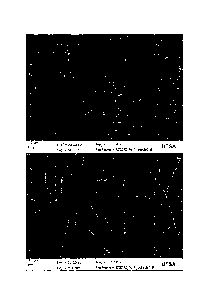
FIGS. 16A-L depicts images of electrospun micro fibers with human aortic
smooth muscle cells on days 1, 3, 7 and 10 (FIGS. 16A-D depict SMC on scaffold
A,
FIGS. 16E-H depict SMC on scaffold B, and FIGS. 16I-L depict SMC on scaffold C
each set for respective days 1, 3, 7 and 10). The scaffolds are shown to be
significantly different although they are manufactured from the same material
using
similar techniques. The "A" scaffolds are measured to be 0.245 Kit 0.158
whereas
"B" scaffolds are 6.744 gni 0.265. Based on both the metabolic and
proliferation
data, it can be determined that endothelial cells respond more positively to
microfibers than either films or nanofiber scaffolds made of the same
material. More
specifically, it should be noted that on the nanofibers, the endothelial cells
show
increased metabolism but not increased proliferation suggesting that the cells
may be
distressed. A similar trend is observed on the film controls but not on the
microfiber
scaffolds. The contrast of metabolic activity as well as proliferation with
visual
images for microfiber scaffolds suggests that the cells have infiltrated the
scaffolds,
unlike the other samples.
Further modifications and alternative embodiments of various aspects of the
invention will be apparent to those skilled in the art in view of this
description.
Accordingly, this description is to be construed as illustrative only and is
for the
purpose of teaching those skilled in the art the general manner of carrying
out the
invention. It is to be understood that the forms of the invention shown and
described
herein are to be taken as examples of embodiments. Elements and materials may
be
substituted for those illustrated and described herein, parts and processes
may be
reversed, and certain features of the invention may be utilized independently,
all as
, would be apparent to one skilled in the art after having the benefit of this
description
of the invention. Changes may be made in the elements described herein without
departing from the scope of the invention as described in the following
claims.
23
CA 2829881 2017-08-24