Note : Les descriptions sont présentées dans la langue officielle dans laquelle elles ont été soumises.
CA 02832484 2013-10-04
WO 2012/139030
PCT/US2012/032573
[0001] POLYNIALIC ACID BASED NANOCONJUGATES FOR IMAGING
[0002] This application claims the benefit of U.S. Provisional
Application
No. 61/472,362, filed April 6, 2011, which is incorporated by reference as if
fully
set forth.
[0003] GOVERNMENT RIGHTS
[0004] The invention was made in part with support from grants
R01CA123495 and U01CA151815 from the National Institutes of Health. The
government has certain rights in the invention.
[0005] FIELD OF INVENTION
[0006] The disclosure relates to nanoconjugates containing imaging
moieties and targeting modules attached to a polymalic acid-based molecular
scaffold. The disclosure also relates to methods of synthesizing
nanoconjugates
and targeting the diseased cells or tissues in by administering nanoconjugates
to
a subject.
[0007] BACKGROUND
[0008] Diagnostic imaging allows avoidance of unnecessary invasive
surgical interventions by confirmation of the nature of various pathological
conditions including differentiating between edema and a tumor, detection of
multiple metastases, or detection of mental illness or dementia. Non-invasive
imaging may be especially useful for diagnostics of diseases or pathological
conditions of the human brain, which is not easily accessible by many
conventional probing methods such as biopsy and light imaging. Non-invasive
imaging is also needed for diagnosis of Alzheimer disease (AD), the most
common
form of dementia observed in people over 65 years of age.
[0009] The oldest approach to diagnose the AD was demonstration of
Alzheimer's plaques in human tissue post mortem by employing small chemical
compounds that attached specifically to the plaques and that could be
visualized
CA 02832484 2013-10-04
WO 2012/139030
PCT/US2012/032573
2
by staining ex vivo or by radioactive scintigram in vivo (Newberg A B et al.
2006
J Nuc Med 47:748).
[0010] After mouse models became available for AD and cancers, such as
triple negative breast cancer, HER2-positive breast cancer, and glioblastoma,
in
vivo imaging methods could be developed. In vivo imaging approaches utilized
fluorescent agents or tagged antibodies binding specifically to components of
the
diseased cells or tissues, or employed positron emission tomography (PET;
Raymond SB et al. 2008 Plos One 3:e2175, 1; Klunk WE et al. 2004 Annals
Neurol 55:306).
[0011] Although some of these approaches could demonstrate the existence
of the diseased tissues, applications required long exposure times and were of
insufficient resolution for clearly distinguishing details, or small
Alzheimer's
plaques. Breakthrough imaging techniques made use of magnetic resonance
imaging (MRI). MRI is one of the most advanced non-invasive imaging systems
due to application of high resolution contrast agents that include gadolinium
(Gd). However, MRI fails to differentiate pathological conditions occurring
within a brain. For example, MRI cannot distinguish cancer types, or even
cancer
from other malignancies. An inefficiency of many in vivo imaging approaches,
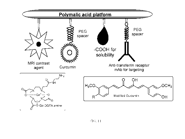
including MRI, stems from the inability of the contrasting agents, such as
gadolinium, to cross the blood-brain barrier (BBB) in combination with rapid
elimination of the contrast agent through the kidneys.
[0012] SUMMARY
[0013] In an aspect, the invention relates to a nanoconjugate that
includes
a polymalic acid-based molecular scaffold, at least one imaging moiety and at
least one targeting module. One or more of the at least one imaging moiety and
one or more of the at least one targeting module is conjugated to the
polymalic-
acid based molecular scaffold.
[0014] In an aspect, the invention relates to a kit for facilitating
imaging of
a cell or a tissue in a subject. The kit contains a nanoconjugate that
includes a
polymalic acid-based molecular scaffold, at least one imaging moiety and at
least
CA 02832484 2013-10-04
WO 2012/139030
PCT/US2012/032573
3
one targeting module. One or more of the at least one imaging moiety and one
or
more of the at least one targeting module is conjugated to the polymalic-acid
based molecular scaffold.
[0015] In an aspect, the invention relates to a method of targeting a
cell or
a tissue in a subject. The method includes administering to the subject a
composition that includes a polymalic acid-based molecular scaffold, at least
one
imaging moiety and at least one targeting module. One or more of the at least
one
imaging moiety and one or more of the at least one targeting module is
conjugated to the polymalic-acid based molecular scaffold.
[0016] In an aspect, the invention relates to a method of synthesizing a
nanoconjugate. The method involves providing a polymalic acid having a
plurality of pendant carboxyl groups. The method further involves reacting a
compound containing sulfhydryl groups and amino acid groups through the
pendant carboxyl groups to add sulfhydryl groups to the polymalic acid to form
an activated polymalic acid. The method involves reacting at least one imaging
moiety containing a sulfhydryl binding group to the activated polymalic acid
to
form a preconjugate. The method also involves reacting at least one targeting
module containing a sulfhydryl binding group to the preconjugate.
[0017] BRIEF DESCRIPTION OF THE DRAWINGS
[0018] The following detailed description of the preferred embodiments
will
be better understood when read in conjunction with the appended drawings. For
the purpose of illustration, there are shown in the drawings embodiments which
are presently preferred. It is understood, however, that the invention is not
limited to the precise arrangements and instrumentalities shown. In the
drawings:
[0019] FIG. 1 is a schematic drawing illustrating a nanoconjugate
designed
to facilitate imaging of triple negative breast cancer (TNBC) metastasized to
brain.
[0020] FIG. 2 is a diagram illustrating synthesis of gadolinium (Gd) -
1, 4, 7,10-tetraazocyclododecane- 1, 4, 7,10-tetraacetic acid (DOTA) amine.
CA 02832484 2013-10-04
WO 2012/139030
PCT/US2012/032573
4
[0021] FIG. 3 is a diagram illustrating synthesis of the Gd-DOTA-
Polycefin
nanoconjugate.
[0022] FIG.4 illustrates the HPLC elution profile of the Gd-DOTA-
Polycefin
nanoconjugate containing Cetuximab.
[0023] FIG.5 is a set of line graphs illustrating calculation of Tl-
relaxivity
for a Polycefin nanoconjugate that includes polymalic acid (P), 12% Gd-DOTA
and 15% 2-mercapto-ethane- 1-amine (MEA).
[0024] FIG.6 is a set of line graphs illustrating affinity determination
of
monoclonal antibody specific to mouse transferrin receptor (anti-MsTfR mAb) by
saturation ELISA. Solid line indicates free anti-MsTfR mAb. Broken line
indicates MsTfR mAb attached to the Gd-DOTA-Polycefin nanoconjugate that
also contains Cetuximab and AlexaFluor 680.
[0025] FIG.7 is a set of Fluorescence Activated Cell Sorting (FACS)
histograms illustrating binding of a Rhodamine-labelled Gd-DOTA-Polycefin
nanoconjugate containing Cetuximab to an epidermal growth factor receptor
(EGFR) expressed in MDA-MB-468 cells in comparison to free Cetuximab and
phosphate buffered saline (PBS).
[0026] FIG. 8 is a set of MRI images showing brain sections of mice
having
TNBC metastatic tumors. Images on the left were obtained without a contrast
agent administered to animals. Images on the right were obtained after animals
received a Polycefin-Gd nanoconjugate intravenously. Scale bar = 50 gm.
[0027] FIG. 9 is a set of MRI images showing tumors in brain sections of
mice having TNBC metastatic tumors. Top images were taken 15 minutes (left)
and 1 hour 45 minutes (right) after administering commercially available
Gd(III)
enhancer reagent to animals. Bottom images were taken 15 minutes (left) and 3
hours 15 minutes (right) after administering to animals a Polycefin
nanoconjugate containing polymalic acid, Gd-DOTA, MsTfR, Cetuximab and
Alexa Fluor 680 dye. Scale bar = 50 gm.
[0028] FIGS. 10A and 10B illustrate Xenogen fluorescence imaging of
animals injected with a Polycefin-Gd-DOTA nanoconjugate containing Gd-DOTA,
MsTfR, Cetuximab Alexa Fluor 680 dye.
CA 02832484 2013-10-04
WO 2012/139030
PCT/US2012/032573
[0029] FIG. 11 is a set of line graphs illustrating MRI kinetics for
tumors
after injecting to animals clinically used Gd (III) (open circles) and a
Polycefin
nanoconjugate containing Gd-DOTA, MsTfR, Cetuximab and Alexa Fluor 680
(closed circles).
[0030] FIGS. 12A and 12B illustrate MRI kinetics for parts of the brain
having tumor (solid line) in comparison with healthy parts of the brain
(broken
line) after injecting to the subjects clinically used Gd(III) (FIG.12A) and a
Gd-
DOTA-Polycefin nanoconjugate containing Gd-DOTA, MsTfR, Cetuximab and
Alexa Fluor 680 (FIG.12B).
[0031] FIGS.13A to 13D are a set of schematic drawings illustrating
nanoconjugates designed to target primary brain and TNBC metastasized to
brain (FIG.13A), and HER2 positive breast cancer metastasized to brain
(FIG.13B) glioblastoma (FIG.13C), in comparison to a control molecule lacking
specific targeting modules (FIG.13D).
[0032] FIG.14 is a schematic drawing illustrating a nanoconjugate
designed to facilitate imaging of Alzheimer's plaques.
[0033] FIG.15 is a diagram illustrating synthesis of a curcumin-PEGi000-
amine.
[0034] FIG.16 is a diagram illustrating attachment of curcumin and Gd-
DOTA modules to polymalic acid.
[0035] FIG.17 is a set of photographs of fluorescent microscopy of slices
of
human brain having AD (top images) and normal (bottom images) stained with
20 pM of free curcumin (right) and 20 pM of a Polycefin-curcumin nanoconjugate
(left).
[0036] DETAILED DESCRIPTION OF THE PREFERRED
EMBODIMENTS
[0037] Certain terminology is used in the following description for
convenience only and is not limiting. The words "right," "left," "top," and
"bottom" designate directions in the drawings to which reference is made.
CA 02832484 2013-10-04
WO 2012/139030
PCT/US2012/032573
6
[0038] The words "a" and "one," as used in the claims and in the
corresponding portions of the specification, are defined as including one or
more
of the referenced item unless specifically stated otherwise. This terminology
includes the words above specifically mentioned, derivatives thereof, and
words
of similar import. The phrase "at least one" followed by a list of two or more
items, such as "A, B, or C," means any individual one of A, B or C as well as
any
combination thereof.
[0039] An embodiment provides a nanoconjugate that may include a
polymalic acid-based molecular scaffold, one or more imaging moieties and one
or
more targeting modules. At least one of the imaging moieties and at least one
of
the targeting modules may be conjugated to the polymalic acid-based molecular
scaffold. All of the imaging moieties may be conjugated to the polymalic acid-
based molecular scaffold. All of the targeting modules may be conjugated to
the
polymalic acid-based molecular scaffold.
[0040] Conjugated means covalently bound.
[0041] In an embodiment, the nanoconjugate may be Polycefin. As used
herein, the term "Polycefin" refers to a family of compounds based on a
polymalic
acid as the platform for attachment of various specific residues for
therapeutic
targeting. Polycefin may include polymalic acid derived from a slime mold.
Polycefin may be 20 to 30 nm in size and may act as a drug. Polycefin may be
engineered to transport other therapeutic molecules. The polymalic acid (PMLA)
may include a homopolymer that contains a main chain ester linkage. The
polymalic acid may be obtained from cultures of Physarum polycefallum. The
polymalic acid may be of any length and of any molecular mass. The polymalic
acid may have a molecular mass of 10, 20, 30, 40, 50, 60, 70, 80, 90, 95, or
100
kDa, or more. The polymalic acid may have a molecular mass in a range between
any two of the following molecular masses: 10, 20, 30, 40, 50, 60, 70, 80, 90,
95, or
100 kDa. The polymalic acid may be at least one of biodegradable and of a high
molecular flexibility, soluble in water (when ionized) and organic solvents
(in its
acid form), non-toxic, or non-immunogenic (Lee Bs et al., Water-soluable
aliphatic
polyesters: poly(malic acid)s, in: Biopolymers, vol.3a (Doi Y, Steinbuchel A
eds.,
CA 02832484 2013-10-04
WO 2012/139030
PCT/US2012/032573
7
pp 75-103, Wiley-VCH, New York 2002, which is incorporated herein by reference
as if fully set forth).
[0042] In an embodiment, a polymalic acid may be used as a molecular
scaffold carrying target modules. In an embodiment, targeting modules may
have functions in addition to targeting. Polymalic acid-based molecular
scaffolds
that may be in embodiments herein were described in PCT Appl. Nos.
PCT/US04/40660, filed December 3, 2004, PCT/US09/40252, filed April 10, 2009,
and PCT/US10/59919, filed December 10, 2010, PCT/US10/62515, filed
December 30, 2010; and U.S. Appl. Nos. 10/580,999, filed March 12, 2007,
issued
as U.S. Pat. 7,935,677, and 12/935,110, filed Sept 28, 2010. All of the
foregoing
PCT and U.S. applications are incorporated herein by reference as if fully set
forth.
[0043] A polymalic acid-based molecular scaffold may be a molecule having
at least two or more targeting modules attached to the polymalic acid-based
molecular scaffold. The targeting modules may also transport a drug, or other
therapeutic entity to a targeted tissue.
[0044] In an embodiment, the polymalic acid-based molecular scaffold may
be based on po1y(I3-L-ma1ic acid). The po1y(I3 -L-malic acid) may be
chemically
conjugated at its carboxylic groups at defined ratios with a variety of
modules.
[0045] In an embodiment, the nanoconjugate having a polymalic acid-based
molecular scaffold may target cells or tissues with high specificity. The high
specificity of nanoconjugates as drug vehicles may result from enhanced
permeability and retention in target tissues that originates from high
molecular
mass, which may be greater than 20000 (Duncan R. 1999 Research Focus 2:441;
Seymour LW et al., 1995 Eur J Cancer Res 31A:766).
[0046] In an embodiment, the one or more imaging moieties may include a
compound suitable to facilitate an imaging procedure. The compound may be a
contrast agent. An imaging may be any imaging procedure used as a clinical
diagnostic tool. An imaging may be an MRI procedure that allows non-invasive
imaging of optically opaque subjects and may provide contrast among soft
tissues
at high spatial resolution. An imaging moiety in the one or more imaging
CA 02832484 2013-10-04
WO 2012/139030
PCT/US2012/032573
8
moieties may be a chelating molecule used for MRI. The chelating molecule may
be but is not limited to 1,4,7,10-tetraazocyclododecane-1,4,7,10-tetraacetic
acid
(DOTA), dibenzo-DOTA, diethylenetriaminepentaacetic acid (DTPA), 1,4,7,10-
tetraazacyclododecane-1, 4, 7.10-tetrakis(2-propionic acid) (DOTMA), 1, 4,
8,11-
tetrazacyclotetradecane-1,4,8,11-tetraacetic acid (TETA), 1,4,7,-
tricarboxymethyl
1,4,7,10 teraazacyclododecane triacetic acid (DO3A), 1,4,7,10-tetraazacyclo-
dodecan- 1-(2-hydroxypropy1)-4,7,10-triacetic acid (HP-DO3A), ethylenediamine-
tetraacetic acid (EDTA), bis-2 (hydroxybenzy1)-ethylene-diaminediacetic acid
(HBED), or 1,4,7-triazacyclo-nonane N,N',N"-triacetic acid (NOTA).
[0047] In an
embodiment, the chelating molecule may form a complex with
a paramagnetic ion. A paramagnetic ion may be a metal ion which may
magnetize parallel or antiparalell to a magnetic field. The paramagnetic ion
may
be a multivalent ion of paramagnetic metal. The paramagnetic metal may be
selected from but is not limited to lanthanides and transition metals. The
transition metals may include but are not limited to manganese, iron,
chromium,
nickel and cobalt. The lanthanides may include but are not limited to
praseodymium, neodymium, samarium, gadolinium, terbium, dysprosium,
holmium, erbium, europium and ytterbium.
[0048] In an
embodiment, the contrast agent may be gadolinium, a highly
paramagnetic ion. This embodiment may be utilized in an MRI procedure.
Gadolinium may be combined with a chelating molecule. Gadolinium (Gd) may be
combined with
(2,2',2"- (2- (2- (2- mercaptoethylamino)-2- oxoethyl)- 1, 4, 7-
tetraazacyclododecane-1,4,7-triy1)triacetic acid)(DOTA) and may form a Gd-DOTA
complex. Gd-DOTA may form a stable contrast agent. Gd-DOTA may be used in
humans.
[0049] A
nanoconjugate herein having a high molecular weight and
including a Gd-DOTA molecule may improve both the efficacy of BBB permeation
and prolong the circulation time. This may improve the accumulation of the
contrast agent inside brain tumor regions or in other areas with pathological
conditions due to the high molecular weight of the nanoconjugate.
CA 02832484 2013-10-04
WO 2012/139030
PCT/US2012/032573
9
[0050] The
one or more targeting modules attached to the polymalic acid-
based molecular scaffold may include biological activities other than
targeting.
The one or more targeting modules may be configured to perform delivery of a
pro-drug. The one or more targeting modules may include a releasable
functional
module that may become effective in the cytoplasm. The one or more targeting
modules may be configured to direct the nanoconjugate towards a specific
tissue
by being capable of binding to the surfaces of cells. The one or more
targeting
modules may be configured to facilitate internalization of the nanoconjugate
into
the targeted cell through endosomes. The one or more targeting modules may be
configured to promote escape of the nanoconjugate from endosomes into the
cytoplasm by virtue of hydrophobic functional units that integrate into and
disrupt endosomal membranes. The one or more targeting modules may be
configured to increase effectiveness during acidification of endosomes en
route to
lysosomes. The one or more targeting modules may be configured to protect
against degradative enzyme activities, for example, peptidases and proteases.
[0051] In an
embodiment, a targeting module may be but is not limited to
an antibody, a polypeptide, an oligonucleotide, a therapeutic chemical, or a
phage. The one or more targeting modules may be capable of targeting a
component of a diseased cell or a tissue.
[0052] In an
embodiment, a targeting module may be an antibody. The
antibody may have an ability to recognize and specifically bind to a target.
The
target may be but is not limited to a protein, a polypeptide, a peptide, a
carbohydrate, a polynucleotide, a lipid, or combinations of at least two of
the
foregoing through at least one antigen recognition site within the variable
region
of the antibody.
[0053] In an
embodiment, a targeting module may be an antibody of a
class described as antagonist antibodies, which specifically bind to a cancer
stem
cell marker protein and interfere with, for example, ligand binding, receptor
dimerization, expression of a cancer stem cell marker protein, and/or
downstream
signaling of a cancer stem cell marker protein.
CA 02832484 2013-10-04
WO 2012/139030
PCT/US2012/032573
[0054] In an
embodiment, a targeting module may be an antibody of a
class described as agonist antibodies which specifically bind to a cancer stem
cell
marker protein and promote, for example, ligand binding, receptor
dimerization,
and/or signaling by a cancer stem cell marker protein. In an embodiment, a
targeting module may be an antibody that does not interfere with or promote
the
biological activity of a cancer stem cell marker protein and may instead
function
to inhibit tumor growth by, for example, antibody internalization and/or
recognition by the immune system.
[0055] A
targeting module may be selected from any type of antibody. The
antibody may be a polyclonal antibody, an intact monoclonal antibody, an
antibody fragment, which may be but is not limited to Fab, Fab', F(ab')2, an
Fv
fragment, a single chain Fv (scFv) mutant, a chimeric antibody or a
multispecific
antibody. A multispecific antibody may be a bispecific antibody generated from
at
least two intact antibodies. A targeting module may be a humanized antibody or
a human antibody. A targeting module may be a fusion protein comprising an
antigen determination portion of an antibody. A targeting module may be
fragment of an antibody comprising an antigen recognition site. Antibodies
selected from may include any of the five major classes of immunoglobulins:
IgA,
IgD, IgE, IgG, and IgM, or subclasses (isotypes) thereof (e.g. IgG 1, IgG2,
IgG3,
IgG4, IgAl and IgA2), based on the identity of their heavy-chain constant
domains referred to as alpha, delta, epsilon, gamma, and mu. A targeting
module
may be a naked antibody or an antibody conjugated to other molecules. A
targeting module may be an antibody conjugated to, for example, toxins or
radioisotopes.
[0056] In an
embodiment, a targeting module may be a monoclonal
antibody. In an embodiment, a targeting module may be a polyclonal antibody.
In
an embodiment, a targeting module may be an antibody specific to at least one
vasculature protein in a cell. The vasculature protein may be a transferrin
receptor protein. The transferrin receptor protein as used herein refers to
the
receptor expressed on endothelium cell surfaces, and at elevated levels on
certain
tumors (Lee JH et al. 2001 Eur J Biochem 268:2004; Kovar MK et al., 2003 J
CA 02832484 2013-10-04
WO 2012/139030
PCT/US2012/032573
11
Drug Targeting 10:23). A monoclonal antibody targeting module (TfR-mAb) may
bind the transferrin receptor protein and thereby achieve transcytosis through
endothelium associated with BBB. An embodiment includes Tfr-mAb attached to
a Gd-containing nanoconjugate that may bind specifically to transferrin
receptor
residing at the endothelial surface on the luminal side of brain capillaries
thus
binding the nanoconjugate thereto. Once bound, the nanoconjugate may
efficiently cross the BBB endothelium by transcytosis. A Tfr mAb-containing
nanoconjugate may be of the size of 20-30 nm (molecular weight 100,000), which
is known to be well above the renal exclusion limit.
[0057] A TfR mAb targeting module may be a humanized (hu-Tfr-mA) or a
chimeric antibody. To study in vivo imaging in mouse and rat models of
Alzheimer's disease (AD models), or TNBC metastasized to brain, hu-TfR mAb of
the nanoconjugate could be replaced by mouse- or rat-TfR mAb. The
nanoconjugate may contain other polypeptides used for similar purposes.
[0058] A targeting module may include a lectin or another ligand specific
to
the transferrin receptor. A targeting module may be a ligand to one of any
number of cell surface receptors or antigens.
[0059] A targeting module may be a small drug molecule or a chromophore
molecule, or a protein molecule, or a lectin that are covalently joined to
polymalic
acid in constructing the nanoconjugate.
[0060] A targeting module may be an antibody configured to specifically
bind a protein selected from but not limited to EGFR, human epidermal growth
factor (HER), laminin 411, insulin-like growth factor (IGF) and tumor necrosis
factor-alpha (TNF-a). The antibody binding EGFR may be Cetuximab. The
antibody binding HER may be Herceptin . The antibody binding laminin 411
may bind either laminin 131 subunit, or laminin a4 subunit, or both.
[0061] A targeting module may be an oligonucleotide. The oligonucleotide
may be an antisense oligonucleotide inhibiting expression of a target nucleic
acid
molecule. The oligonucleotide may be one of the antisense oligonucleotides
inhibiting expression of lamin 411 that were described in PCT Appl.
PCT/US04/29956, filed September 13, 2004; and U.S. Appl. Nos. 10/570,747,
filed
CA 02832484 2013-10-04
WO 2012/139030
PCT/US2012/032573
12
January 30, 2007, issued as U.S. Pat. 7,547,511, and 12/473,992, filed May 28,
2009, which are incorporated by reference as if fully set forth.
[0062] A targeting module may include an endosomal escape unit as
described in PCT application PCT/US09/40252, filed April 10, 2009, which is
incorporated by reference as if fully set forth. An endosomal escape may be a
carrier module attached to the polymalic acid-based scaffold that becomes
active
by acidification during maturation of the endosomal vesicles towards
lysosomes.
The carrier module may include a plurality of leucine residues in a
polypeptide
linked to the polymalic acid-based molecular scaffold by amide bonds. The
carrier
module may include a plurality of valine residues in a polypeptide linked to
the
polymalic acid-based molecular scaffold by amide bonds. The carrier module may
include a leucine ethylester linked to the polymalic acid-based molecular
scaffold
by amide bonds. During acidification of the endosomes en route to lysosomes,
these stretches of the carrier module may become charge-neutralized and
hydrophobic, and capable of disrupting membranes. Other molecules that become
charge neutralized at lysomal pH's may be used in place of leucine or valine
residues, or a leucine ethylester in construction of the compositions
containing
polymalic acid and an endosomal escape unit module.
[0063] A targeting module may be a module for protection against
resorption by the reticuloendothelial system (RES) and/or enzyme degradation.
For example, the module for protection against resorption may be but is not
limited to a polyethylene glycol (PEG) molecule. PEG may be used to increase
the
in vivo half-life of conjugated proteins, to prolong the circulation time, and
enhance extravasation into targeted solid tumors (Arpicco S et al. 2002
Bioconjugate Chem 13:757; Maruyama K et al., 1997 FEBS Letters 413:1771,
which is incorporated by reference as it fully set forth). Other molecules
known to
increase half-life of the nanoconjugate may be used in design of
nanoconjugates
herein.
[0064] FIG. 1 depicts an exemplary nanoconjugate including Gd-DOTA
complex attached to the polymalic acid platform. The nanoconjugate may be for
tumor-type specific MRI in mouse model for human TNBC metastasized to brain.
CA 02832484 2013-10-04
WO 2012/139030
PCT/US2012/032573
13
The modules attached to the polymalic acid may include an MRI contrast agent
(Gd-DOTA), targeting modules (chimeric mouse-human monoclonal antibodies
Cetuximab (Erbitux ) specific to EGFR displayed by tumor cells and MsTfR for
penetration through BBB) and a carboxyl group for improving solubility. For
use
in humans, the anti-mouse TfR mAb may be replaced by anti-human TfR mAb.
[0065] Polymalic acid of any molecular weight (Mw) may be used as the
platform to carry one or more targeting modules and one or more imaging
moieties. Polymalic acid used herein may have a Mw of 10,000; 15,000; 20,000;
30,000; 40,000; 50, 000; 60,000; 70,000; 80,000; 90,000; 100,000; 110,000;
120,000;
130,000; 140,000; or 150,000, or more, or any value in a range between any two
of
the foregoing (endpoints inclusive). The polymalic acid of Mw 80,000 may be
platform for a nanoconjugate that caries covalently bound MsTfR mAb and a
tumor specific mAb together with multiple covalently bound Gd-DOTA. The
platform may contain any number of derivatisable carboxyl group. In
embodiments, the platform may contain 700 or more derivatisable carboxyl
groups and a large number of Gd-DOTA units can be loaded for generating a
strong MRI signal.
[0066] In an embodiment, one or more targeting modules may be capable of
targeting a component of a diseased cell or tissue. The component may be, but
not
limited to, beta amyloid plaques thought to contribute to the degradation of
the
neurons in the brain and the subsequent symptoms of Alzheimer's disease. The
one one or more targeting modules may include curcumin (5-hydroxy-1,7-bis(4-
hydroxy-3- methoxypheny1)-1,4,6-heptatrien-3-on) for specific binding to
Alzheimer's amyloid plaques. Curcumin may bind specifically and tightly to the
beta amyloid plaques and thereby may allow accumulation of the nanoconjugate
within the brain and a high staining intensity. The nanoconjugate may contain
one or more curcumin molecules. The presence of multiple curcumin molecules on
the nanoconjugate results in firm attachment of the nanoconjugates around to a
beta-amyloid plaque contributing to sharp contours with high contrast.
[0067] The nanoconjugate molecule containing curcumin may carry any
number of gadolinium ions. The nanoconjugate may carry a single gadolinium
CA 02832484 2013-10-04
WO 2012/139030
PCT/US2012/032573
14
ion. The nanoconjugate may carry a plurality of gadolinium ions. The
nanoconjugate may carry 1, 5, 10, 20, 30, 40, 50, 60, or more Gd ions per
molecule
of nanoconjugate. The nanoconjugate may carry a number of Gd ions per
molecule of nanoconjugate in a range between any two of the following numbers:
1, 5, 10, 20, 30, 40, 50, or 60. A high concentration of Gd on a target
tissue, for
example amyloid plaques, may allow imaging by MRI at high contrast and
resolution quality.
[0068] The one or more targeting module may include therapeutic
polypeptides.
In embodiments, the one or more targeting modules may include additional
therapeutic agents. In embodiments, the additional therapeutic agent or agents
is selected from the group consisting of growth factors, anti-inflammatory
agents,
vasopressor agents, collagenase inhibitors, topical steroids, matrix
metalloproteinase inhibitors, ascorbates, angiotensin II, angiotensin III,
calreticulin, tetracyclines, fibronectin, collagen, thrombospondin,
transforming
growth factors (TGF), keratinocyte growth factor (KGF), fibroblast growth
factor
(FGF), insulin-like growth factors (IGF), epidermal growth factor (EGF),
platelet
derived growth factor (PDGF), neu differentiation factor (NDF), hepatocyte
growth factor (HGF), and hyaluronic acid.
[0069] In an embodiment, the nanoconjugate may include a tracking
fluorescent dye to follow in vivo distribution of the nanoconjugate in a
subject.
The tracking dye may facilitate gross in vivo monitoring of the nanoconjugate
distribution by imaging systems other than by using MRI. In the absence of Gd,
the tracking dye may allow the validation of curcumin-polymalic acid conjugate
entrance into the brain in the first phase of the investigation of a disease
or
condition in a subject. A tracking dye may also validate whether curcumin is
attached to polymalic acid within the brain. Thus, the tracking dye may be
useful
in optimization experiments. Tracking may be performed, for example, by using
Xenogen fluorescence imaging system.
[0070] In an embodiment, a kit for facilitating imaging of a cell or
tissue is
provided. The cell may be a diseased cell. The tissue may be a diseased
tissue.
CA 02832484 2013-10-04
WO 2012/139030
PCT/US2012/032573
The kit may be implemented in a method for visualizing pathological
conditions.
The kit may include a nanoconjugate comprising a polymalic acid-based
molecular scaffold, one or more imaging moiety and one or more targeting
module. The kit may include any one or more nanoconjugates described herein.
At least one of the imaging moieties and at least one of the targeting modules
may be conjugated to the polymalic acid-based molecular scaffold. All of the
imaging moieties may be conjugated to the polymalic acid-based molecular
scaffold. All of the targeting modules may be conjugated to the polymalic acid-
based molecular scaffold.
[0071] The exact nature of the modules and moieties configured in the kit
may depend on its intended purpose. In embodiments, the kit may be configured
for the purpose of visualizing, treating or monitoring Alzheimer's disease or
other
conditions involving abnormal brain function, activity or pathology. For this
purpose, the kit may include a nanoconjugate comprising a module for binding
amyloid beta plaque and MRI imaging. In embodiments, the kit may be
configured for the purpose of visualizing, treating, or monitoring cancer.
[0072] In an embodiment, the kit may be configured particularly for the
purpose of treating mammalian subjects. The kit may be configured particularly
for the purpose of treating human subjects. The kit may be configured for
veterinary applications. The kit may be configured to, but is not limited to,
treating farm animals, domestic animals, or laboratory animals. Instructions
for
use may be included in the kit. Instructions for use may include a tangible
expression describing the technique to be employed in using the components of
the kit to effect a desired outcome. For example, instructions may describe
the
technique to visualize amyloid beta plaques or tumor cells or cell types. The
kit
may also contain other useful components. For example, the kit may contain
diluents, buffers, pharmaceutically acceptable carriers, syringes, catheters,
applicators, pipetting or measuring tools, bandaging materials or other useful
paraphernalia as will be readily recognized by those of skill in the art.
[0073] The materials or components assembled in the kit may be provided
to the practitioner stored in any convenient and suitable ways that preserve
their
CA 02832484 2013-10-04
WO 2012/139030
PCT/US2012/032573
16
operability and utility. For example, the components may be provided be in
dissolved, dehydrated, or lyophilized form. The components may be provided at
room, refrigerated or frozen temperatures. The components may be contained in
suitable packaging material(s). As used herein, the phrase "packaging
material"
refers to one or more physical structures used to house the contents of the
kit,
such as inventive compositions and the like. The packaging material may be
constructed by well known methods, preferably to provide a sterile,
contaminant-
free environment. As used herein, the term "package" refers to a suitable
solid
matrix or material such as glass, plastic, paper, foil, and the like, capable
of
holding the individual kit components. The packaging material may have an
external label which indicates the contents and/or purpose of the kit and/or
its
components.
[0074] In an embodiment, a method of targeting a cell or a tissue in a
subject is provided. The cell may be a diseased cell. The tissue may be a
diseased
tissue. The method may involve administering to the subject a nanoconjugate
that includes a polymalic acid-based molecular scaffold, at least one imaging
moiety, and at least one targeting module. At least one of the imaging
moieties
and the at least one of the targeting modules may be conjugated to the
polymalic
acid-based molecular scaffold. All imaging moieties may be conjugated to the
polymalic acid-based molecular scaffold. All targeting modules may be
conjugated
to the polymalic acid-based molecular scaffold. The nanoconjugate may be any
nanoconjugate described herein. The method may also include providing
conditions permitting interaction of the nanoconjugate with a component of the
diseased cell or a diseased tissue.
[0075] The subject may be a patient. As used herein, the term "patient"
refers to a human. The patient may be a human with a symptom or symptoms of
a disease or condition. The patient may need treatment for the disease or
condition in a clinical setting. The symptoms of the disease or condition may
change as a result of a treatment, or spontaneous remission, or development of
further symptoms with the progression of the disease. The term "patient" may
also refer to non-human organism. The patient may be a laboratory animal, a
CA 02832484 2013-10-04
WO 2012/139030
PCT/US2012/032573
17
farm animal or a zoo animal. The patient may be a mouse, a rat, a guinea pig,
a
hamster, a horse, a rabbit, a goat, or a cow.
[0076] In an
embodiment of the method of targeting a cell or a tissue, a
nanoconjugate may be administered to a subject by any suitable route. The
nanoconjugated may be administered by intravenous injections. The
nanoconjugate may be delivered by a technique selected from the group
consisting of: intramuscular injection, subcutaneous injection, intravenous
injection, intradermal injection, intranasal injection, inhalation, oral
administration, sublingual administration, buccal administration, or topical
administration.
[0077] In an
embodiment of the method of targeting a cell or a tissue, the at
least one imaging moiety may be a molecule facilitating an imaging technique.
An imaging may be performed by any technique including but not limited to X-
ray imaging, computer tomography (CT) scans, and MRI. The imaging moiety
may include an imaging contrast agent. The imaging contrast agent may be a Gd-
DOTA. The method may involve visualizing the imaging contrast agent in the
subject. Visualizing may be performed by the imaging technique; e.g., by X-
ray,
CT, or MRI.
[0078] In an
embodiment, the method of targeting a cell or a tissue may
also include diagnosing a disease or other condition in the subject.
Diagnosing
may be based on an image of the diseased cell or the diseased tissue.
Diagnosing
may include comparing the image with a control image of a normal cell or
tissue
in a healthy individual. The image may be obtained by any non-invasive
clinical
diagnostic imaging procedure. For example, the image may be obtained by MRI.
The MRI apparatus utilizes the nuclear magnetic resonance phenomenon and
may produce images of cross sections of the cells or tissues being imaged. The
MRI may measure signal derived from protons of the water molecules present in
cells or tissues in a subject positioned for imaging. The intensity of MRI
images
may depend on physical properties of specific tissues. The intensity of MRI
signal
may depend on proton density, spin lattice relaxation time (T1), and the spin-
spin
relaxation time (T2).
CA 02832484 2013-10-04
WO 2012/139030
PCT/US2012/032573
18
[0079] An "abnormal condition" refers to a function in the cells and
tissues
in a body of a patient that deviates from the normal function in the body. An
abnormal condition may refer to a disease. Abnormal condition may include
brain
disorders. Brain disorders may be but are not limited to Alzheimer's disease,
Multiple sclerosis, Parkinson's disease, Huntington's disease, schizophrenia,
anxiety, dementia, mental retardation, and anxiety. Abnormal condition may
include proliferative disorders. The terms "proliferative disorder" and
"proliferative disease" refer to disorders associated with abnormal cell
proliferation. Proliferative disorders may be, but are not limited to, cancer,
vasculogenesis, psoriasis, and fibrotic disorders. Cancer is a physiological
condition in mammals in which a population of cells is characterized by
unregulated cell growth. Examples of cancers include, but are not limited to,
carcinoma, lymphoma, blastoma, sarcoma, and leukemia. More particular
examples of such cancers include squamous cell cancer, small-cell lung cancer,
non-small cell lung cancer, adenocarcinoma of the lung, squamous carcinoma of
the lung, cancer of the peritoneum, hepatocellular cancer, gastrointestinal
cancer, pancreatic cancer, glioblastoma, cervical cancer, ovarian cancer,
liver
cancer, bladder cancer, hepatoma, breast cancer, colon cancer, colorectal
cancer,
endometrial or uterine carcinoma, salivary gland carcinoma, kidney cancer,
liver
cancer, prostate cancer, vulval cancer, thyroid cancer, hepatic carcinoma and
various types of head and neck cancers. Breast cancer may include TNBC and
HER2-positive breast cancer.
[0080] Cancer may be a primary cancer or a metastatic cancer. The term
"primary cancer" refers to the original site at which a cancer originates. For
example, a cancer originating in the breast is called a primary breast cancer.
If it
metastasizes; i.e., spreads to the brain, the cancer is referred to as a
primary
breast cancer metastatic to the brain.
[0081] The term "metastasis" refers to the process by which a cancer
spreads or transfers from the site of origin to other regions of the body with
the
development of a similar cancerous lesion; i.e., having the same or
substantially
the same biochemical markers at the new location. A "metastatic" or
CA 02832484 2013-10-04
WO 2012/139030
PCT/US2012/032573
19
"metastasizing" cell is one that has a reduced activity for adhesive contacts
with
neighboring cells and migrates by the bloodstream or within lymph from the
primary site of disease to additional distal sites, for example, to invade
neighboring body structures or distal structures.
[0082] An abnormal condition may also include diabetes, rheumatoid
arthritis, asthma, psoriasis, atherosclerosis, cardiovascular disorders,
glaucoma,
and rethinopathy. The term "disease" refers to all abnormal conditions.
Diagnosing may include diagnosing of another condition in addition to an
abnormal condition. The other condition may be associated with an abnormal
condition. The other condition may not be associated with an abnormal
condition.
For example, diagnosing of schizophrenia may be made in addition to diagnosing
Alzheimer's disease.
[0083] The term "tumor" refers to any mass of tissue that result from
excessive cell growth or proliferation, either benign (noncancerous) or
malignant
(cancerous) including pre-cancerous lesions. Tumor cell may derive from a
tumor
or a pre-cancerous lesion including both a non-tumorigenic cell and a
tumorigenic
cell; i.e., cancer stem cell.
[0084] An embodiment includes a tumor-specific nanoconjugate, which
may be implemented for enhancement of MRI and facilitating diagnostic
imaging. An enhancement includes such a method. In particular, a tumor-
specific nanocomjugate may be used to distinguish between tumor and non-tumor
lesions of the brain which are indistinguishable by a common MRI procedure. A
nanoconjugate may be used to distinguish between different types of tumors
occurring side-by side in the same individual. A nanoconjugate may be used for
MRI enhancement in the brain of cancer patient with a history of primary
breast
cancer, metastatic brain tumor from primary breast cancer, metastatic tumors
from a different type of cancer, a primary brain tumor, and/or infection
resulting
from impairment of the immune system as a complication of chemotherapy.
[0085] A nanoconjugate herein may be designed to enhance MRI-based
diagnostics of specific conditions. In an embodiment, a nanoconjugate (MRI
enhancer) may include antibodies specific for tumor markers at the surface of
CA 02832484 2013-10-04
WO 2012/139030
PCT/US2012/032573
tumor cells. The antibodies may be specific to overexpressed cell-surface
antigens
such as EGFR, HER2, B lymphocyte antigen CD 20 or laminin. The antibodies
may facilitate access to the tumor tissue across the BBB into tumor
interstitial
using transcytosis through targeting of transferrin receptor on the
endothelium
of tumor capillaries. Once attached, the enhancer could be retained over a
time
scale that exceeds by far the clearance of unbound free MRI enhancer through
the kidneys. On basis of the prolonged retention in the brain or other tumors,
MRI could recognize the labeled tumor by a signal sent as a shortened
relaxation
time T1 of the reagent surrounding water molecules after given pulses of a
spin
orientating external magnetic field of the MRI apparatus. The shortening of
the
reciprocal of T1 is proportional to the concentration of the MRI enhancer, and
thus the enhancement of the signal may be the result of an accumulation due to
tumor specific binding. The tumor nonspecific MRI signal may be accounted for
by measurement of T1 measured for healthy portions of the brain. The
difference
of T1 values between tumor and healthy brain may be measured as a function of
time reflecting specific tumor retention of the enhancer reagent, while the
reagent in the healthy brain and elsewhere may be already cleared through the
kidneys. Tumor-type specific MRI scanning may be performed when T1 for the
healthy brain has approached zero value.
[0086] A number of contrast agents may be included in a nanoconjugate
herein to improve resolution of MR images. A contrast agent may be a molecule
suitable to generate a contrasting effect in vivo. A contrast agent may form
metalloprotein complex. A contrast agent may form a complex that affects the
relaxation times T1, or T2, or both. A contrast agent that affects T1 may be a
lanthanide metal ion. A contrast agent may be Gd that is chelated to a low
molecular-weight molecule in order to limit toxicity. A contrast agent that
affects
T2 may consist of small particles of magnetite (FeO--Fe203). Contrast agents
may
interact with mobile water in tissue to produce contrast.
[0087] In an embodiment, diagnosing the disease or condition may involve
a patient with abnormal brain function, activity or pathology. Diagnosing the
CA 02832484 2013-10-04
WO 2012/139030
PCT/US2012/032573
21
Alzheimer disease may be based on the presence of amyloid beta plaques in the
patient's brain.
[0088] Diagnosing may be performed by administering a composition that
includes a polymalic-acid based nanoconjugate containing a targeting module
for
binding amyloid beta plaques and an imaging moiety for MRI imaging to the
patient and acquiring images of localization of the nanoconjugate in a
particular
type of tissue in the patient's body.
[0089] The nanoconjugate may be able to pass the BBB and then target
plaques, a hallmark of Alzheimer's disease, by simultaneously having attached
plaque-binding curcumin and TfR mAb. Access to beta-amyloid plaque imaging
may allow determining the status of Alzheimer's disease and to follow patients
during the treating the disease. Similar Polycefin nanoconjugates containing
curcumin and/or other active compounds could be used to treat Alzheimer's
disease.
[0090] In an embodiment, application of a nanoconjugate may improve both
the efficacy of BBB permeation and may prolong circulation of the Gd-
containing
contrast agent. It may also improve the accumulation inside brain regions that
contain plaques due to tight binding to Alzheimer's amyloid plaques to
curcumin.
[0091] In an embodiment, targeting the diseased cell or tissue may result
in reduction or elimination of at least one symptom of the disease or
condition,
and thereby treatment of the disease or condition in the subject. Targeting
the
diseased cell or tissue may be a therapeutic measure to promote regression of
a
cancer or prevent further development or metastasis, or as a prophylactic
measure to minimize complications associated with development of a tumor or
cancer.
[0092] In an embodiment, the condition and/or disease monitored or
treated may be Alzheimer's disease. In an embodiment, a method of treating a
condition in a patient is provided. The method may include administering a
composition comprising a nanoconjugate comprising a targeting module for
binding amyloid beta plaques and an imaging moiety for MRI imaging. The
method may also include treating the patient with the composition.
CA 02832484 2013-10-04
WO 2012/139030
PCT/US2012/032573
22
[0093] To
achieve the desired effect; i.e., inhibit the expression of at least
one ligand of the target receptor, a composition may be administered at a
therapeutically effective amount. A "therapeutically effective amount" of the
composition may be the amount effective for preventing further development of
a
cancer or transformed growth, and even to effect regression of the cancer.
[0094] The
exact dosage may be chosen by the individual physician with
regard to the need of the patient to be treated. Dosage and administration may
be adjusted to provide sufficient levels of the active agent(s) or to maintain
the
desired effect. Additional factors which may be taken into account include the
severity of the disease state; e.g., cancer size and location; age, weight and
gender
of the patient; diet, time and frequency of administration; drug combinations;
reaction sensitivities; and tolerance/response to therapy. Long
acting
compositions might be administered every 3 to 4 days, every week, or once
every
two weeks depending on half-life and clearance rate of the particular
composition.
[0095] In an
embodiment, the one or more targeting modules may include
active agents for treating a disease or condition in a patient. The active
agents
may be formulated in dosage unit form for ease of administration and
uniformity
of dosage. The expression "dosage unit form" as used herein refers to a
physically
discrete unit of active agent appropriate for the patient to be treated.
[0096] For
any active agent, the therapeutically effective dose may be
estimated initially either in cell culture assays or in animal models, usually
mice,
rabbits, dogs, or pigs as shown in Examples herein. The animal model may be
also used to achieve a desirable concentration range and route of
administration.
Such information may then be used to determine useful doses and routes for
administration in humans. A therapeutically effective dose refers to that
amount
of active agent which ameliorates the symptoms or condition. Therapeutic
efficacy and toxicity of active agents can be determined by standard
pharmaceutical procedures in cell cultures or experimental animals, e.g., ED50
(the dose is therapeutically effective in 50% of the population) and LD50 (the
dose is lethal to 50% of the population). The dose ratio of toxic to
therapeutic
CA 02832484 2013-10-04
WO 2012/139030
PCT/US2012/032573
23
effects is the therapeutic index, and it can be expressed as the ratio,
LD50/ED50.
Compositions herein may exhibit large therapeutic indices. The data obtained
from the animal studies may be used in formulating a range of dosage for human
use.
[0097] As discussed above and described in greater detail in the
Examples,
a nanoconjugate herein may be administered in a method to prevent development
or metastasis of a cancer condition. In particular, a nanoconjugate may be
useful in preventing further growth of a particular cancer type, including but
not
limited to breast cancer; skin cancer; ovarian cancer; cervical cancer;
retinoblastoma; colon cancer and other conditions including those arising from
the lining of the gastrointestinal tract; lung cancer and cancers of the
respiratory
tract; renal carcinoma and other tumors arising from the inner surface of
kidney
tubules; leukemias and lymphomas and such disorder of blood; and other types
of
genital cancer including those associated with various strains of papilloma
virus;
brain tumors; and cancers of the uterus, of the vagina, and of the urethra.
[0098] In embodiments, diagnostic, prognostic and therapeutic methods
may not be limited to treating conditions in humans, but may involve similar
conditions in any mammal including but not limited to bovine, canine, feline,
caprine, ovine, porcine, murine, and equine species.
[0099] In an embodiment, a method of monitoring an efficiency of
treatment of a disease or condition in a subject is provided. Monitoring may
include obtaining a first image of a diseased cell or a diseased tissue in the
subject after treatment, and, after a period of time, a second image of the
diseased cell or tissue. Comparison can be made between the first and the
second
images to determine a clinically significant difference in cells and tissues
after
the treatment. For example, two or more images may be compared to determine
whether the treatment reduced the number of cancer cells in a tumor, or the
size
of a particular tumor.
[0100] A subject may be a patient in need of MRI procedure. A composition
that includes a polymalic acid-based molecular scaffold, at least one imaging
moiety, and at least one targeting module may be administered at any time
CA 02832484 2013-10-04
WO 2012/139030
PCT/US2012/032573
24
before or after placing a patient in an MRI apparatus. The composition may
target cells or tissues at different locations of the patient's body before
images
may be produced. In this case, the composition may be accumulated in the
specific location before imaging. The images may be also produced during the
period of accumulation of the composition in target cells or tissues. Any
disease
cells or tissues targeted by the composition may be identified by examining
the
image or images. The composition may be re-administered to the subject after a
period of time depending on the scheme of a particular therapeutic treatment.
For example, the composition may be administered every week, every two weeks,
every three weeks, or every month. Image(s) may be produced during or after
subsequent administration of the composition and comparison may be made
between images taken during different phases of therapeutic treatment to
assess
the efficacy of treatment.
[0101] Methods herein may include providing a period of time sufficient
for
accumulation of a nanoconjugate in targeted cells or tissues.
[0102] In another embodiment, a method of prognosing a condition and/or
disease is provided for an individual having abnormal brain function, activity
or
pathology. The method may include administering a composition comprising a
nanoconjugate comprising a targeting module for binding amyloid beta plaques
and a module for MRI imaging to the individual, and prognosing a severe form
of
the condition and/or disease based on the presence of an extensive level of
amyloid beta plaques in the individual relative to a normal subject.
[0103] In an embodiment, a composition including a polymalic acid-based
molecular scaffold, at least one imaging moiety, and at least one targeting
module may further include a pharmaceutically acceptable carrier. As used
herein, the term "pharmaceutically acceptable carrier" includes any and all
solvents, diluents, or other liquid vehicle, dispersion or suspension aids,
surface
active agents, isotonic agents, thickening or emulsifying agents,
preservatives,
solid binders, and lubricants as suited to the particular dosage form desired.
A
pharmaceuitically acceptable carrier may be one described in Remington's
Pharmaceutical Sciences Ed. by Gennaro, Mack Publishing, Easton, PA, 1995,
CA 02832484 2013-10-04
WO 2012/139030
PCT/US2012/032573
which is incorporated herein by reference as it fully set forth, and discloses
various carriers used in formulating pharmaceutical compositions and known
techniques for the preparation thereof. Some examples of materials which can
serve as pharmaceutically acceptable carriers include but are not limited to
sugars, lactose, glucose, and sucrose; starches, corn starch and potato
starch;
cellulose and its derivatives, sodium carboxymethyl cellulose, ethyl
cellulose, and
cellulose acetate; powdered tragacanth; malt; gelatin; talc; excipients, cocoa
butter and suppository waxes; oils, peanut oil, cottonseed oil, safflower oil,
sesame oil, olive oil, corn oil, and soybean oil; glycols, propylene glycol;
esters,ethyl oleate and ethyl laurate; agar; buffering agents, magnesium
hydroxide, and aluminum hydroxide; alginic acid; pyrogen-free water; isotonic
saline; Ringer's solution; ethyl alcohol, and phosphate buffer solutions, as
well as
other non-toxic compatible lubricants, sodium lauryl sulfate and magnesium
stearate. Coloring agents, releasing agents, coating agents, sweetening,
flavoring
and perfuming agents, preservatives and antioxidants may also be present in
the
composition.
[0104] In an embodiment, a method of synthesizing a nanoconjugate is
provided. The method may include providing a polymalic acid having a plurality
of pendant carboxyl groups. The method may include reacting a compound
containing sulfhydryl groups and amino groups through the pendant carboxyl
group to add sulfhydryl groups to the polymalic acid to form an activated
polymalic acid. The method may also include reacting at least one imaging
moiety containing a sulfhydryl binding group to the activated polymalic acid
to
form a preconjugate. The method may further include reacting at least one
targeting module containing a sulfhydryl binding group to the activated
polymalic acid.
[0105] The method of synthesizing may include activating pendant carboxyl
carboxyl groups on polymalic acid by adding N-hydroxysuccinimide (NHS) to the
polymalic acid to form an NHS-ester. The method may also include reacting the
activated carboxyl groups with the amino group of 2-mercapto-ethane- 1-amine.
The method may also include reacting at least one imaging moiety containing an
CA 02832484 2013-10-04
WO 2012/139030
PCT/US2012/032573
26
amino group with the NHS-activated pendant carboxyl group. The method also
may involve reacting at least one targeting module containing a sulfhydryl
group
to the preconjugate. The at least one imaging moiety may include an activated
molecule of a contrast agent. The activated molecule of the contrast agent may
include gadolinium (Gd)-1, 4, 7,10-tetraazocyclododecane-1,4, 7,10-tetraacetic
acid
(DOTA)-amine. The at least one targeting module may include an activated
antibody. The activated antibody may include an antibody-polyethylene glycol-
maleimide. The antibody-polyethylene glycol-maleimide may further react with
the preconjugate to form the nanoconjugate.
[0106] The at least one targeting module may include an activated
curcumin-polyethylene-glycol amine. The at least one targeting module may
specifically bind to a component of a diseased cell or tissue in a subject
selected
from the group consisting of: an epidermal growth factor receptor (EGFR),
human receptor growth factor (HER), laminin 411, insulin-like growth factor
(IGF), transferrin receptor protein, curcumin and tumor necrosis factor-alpha
(TNF- ct).
[0107] A polymalic acid having one or more targeting modules may be
synthesized by any known method. For example, a polymalic having attached one
or more target modules may be synthesized by ring-opening polymerization of
derivatized malic acid lactones. Doxorubicin-poly-malic acid may be
synthesized
from synthetic poly- 13 -D, L-malic acid.
[0108] Further embodiments herein may be formed by supplementing an
embodiment with one or more element from any one or more other embodiment
herein, and/or substituting one or more element from one embodiment with one
or more element from one or more other embodiment herein.
[0109] Examples
[0110] The following non-limiting examples are provided to illustrate
particular embodiments. The embodiments throughout may be supplemented
with one or more detail from one or more example below, and/or one or more
CA 02832484 2013-10-04
WO 2012/139030
PCT/US2012/032573
27
element from an embodiment may be substituted with one or more detail from
one or more example below.
[0111] Example 1. Chemical synthesis of a tissue specific
nanoconjugate for MRI enhancement
[0112] Materials. High purity polymalic acid (PMLA; Mw 800,000,
polydispersity factor P=1.2 by Sec-HPLC) isolated from the culture medium of
Physarum polycephalum was used as Polycefin platform (Ljubimova JY et al.
2007 Chem-Biol Interactions 171:195). Mouse anti-human TfR mAb RVS10 was
purchased from Southern Biotech (Birmingham, AL, USA) and ERBITUX
(Cetuximab) from Bristol-Myers Squibb (New York, NY, USA). mPEG5000-Amine
and maleimide-PEG3400-maleimide were obtained from Laysan Bio, Inc. (Arab,
AL, USA). 3-(2-Pyridyldithio)-propionate (PDP; Carlsson J et al. 1978 Biochem
J
173:723. Alexa Fluor 680 C2 maleimide (A1ex680) was purchased from
Invitrogen Corporation (Carlsbad, CA, USA), 2-Aminoethyl-mono-amide-DOTA-
tris(t-Bu ester) from (Macrocyclics, Inc. TX, USA). Unless indicated,
chemicals
and solvents of highest purity were obtained from Sigma-Aldrich (St. Louis,
MO)
USA.
[0113] Analytical methods for chemical synthesis. The conjugation
reaction
of Gd-DOTA-amine and 2-MEA with PMLA was followed by thin layer
chromatography (TLC) on precoated silica gel 60 F254 aluminum sheets (Merck,
Darmstadt, Germany) under UV light and/or by ninhydrin staining. Size
exclusion chromatography was performed on an Elite LaChrom analytical system
with Diode Array Detector L 2455 (Hitachi, Pleasanton, CA, USA), and Mu, was
measured using either BioSep-SEC-S 3000 or PolySep-GFC P4000 (300 x 7.80
mm; Phenomenex, Torrance, CA, USA) using 50 mM sodium phosphate buffer pH
6.8 as mobile phase (0.75 mL/min) and polystyrene sulfonates as molecular
weight standards. Thiol residues were assayed by the method of Ellman (Ellman
GL 1959 Arch Biochem Biophys 82:70). TfR binding activity of anti-mouse TfR
mAb conjugated to polymalic acid was assayed using Protein DetectorTM ELISA
Kit (KPL, Inc., Gaithersburg, MA, USA). The mouse TfR ectodomain used as the
CA 02832484 2013-10-04
WO 2012/139030
PCT/US2012/032573
28
antigen was obtained from Protein Expression Center, California Institute of
Technology (Pasadena, CA USA). Binding of polymalic acid conjugated
Cetuximab to EGFR expressed on triple-negative breast cancer cells was
demonstrated by fluorescence activated cell sorting (FACS) analysis.
Gadolinium
was measured by ICP-MS at UCLA, Los Angeles, CA, USA). In the absence of
protein, the reaction of DOTA-Gd was followed by its intrinsic fluorescence at
280
nm excitation wavelength and 316 nm emission wave length (Hagan JJ et al.
1988 Anal Biochem. 60:514).
[0114] Example 2. Synthesis - an overview
[0115] Synthesis of the tumor-type specific MRI enhancer reagent was
accomplished in two parts: first the synthesis of Gd-DOTA-amine (FIG. 2) and
second the conjugation of Gd-DOTA-amine to NHS-activated PMLA (FIG. 3). The
alternative route includes first conjugating DOTA-amine with PMLA and then
loading with Gd3+ . The first part of the synthesis started with deprotection
of
the commercially available DOTA amino derivative (FIG. 2). The conjugation of
Gd-DOTA amino with activated polymalic acid shown in FIG.3 may be subject to
variation for further increase in number of Gadolinium per polymer chain and
for
increase in reaction yields.
[0116] Example 3. General procedure for Boc deprotection
[0117] Referring to FIG. 2, (1) 2-Aminoethyl-mono-amide-DOTA-tris(t-Bu
ester) (1.23 g, 1.77 mmol) was dissolved in trifluoroacetic acid (TFA) (25 mL)
and
Triisopropylsilane (TIS) (1.12 g, 7.1 mmol) was added. The reaction mixture
was
stirred at 50 C for 3hours and cooled to room temperature. Evaporation of the
solvent under reduced pressure yielded viscous brown oil. An ice-cold diethyl
ether (25 mL) was added and the white precipitate was filtered and washed with
diethyl ether. The dried precipitate was dissolved in pure water and freeze
dried.
Reaction yield was 97%.
[0118] Example 4. General procedure for preparation of metal
complex
[0119] Referring to FIG. 2, an equivalent of DOTA amine (2) (295 mg, 0.56
mmol) dissolved in 4 mL of water, received dropwise a slight stoichiometric
CA 02832484 2013-10-04
WO 2012/139030
PCT/US2012/032573
29
excess of a Gadolinium (III) acetate (250 mg, 0.61 mmol) in 4 mL of water. The
solution was stirred at room temperature (RT) while the pH was continuously
maintained at pH 5.5 by adding 1M KOH. After stirring for 48 hours, EDTA (0.2
equivalent) was added and the mixture stirred at room temperature for lourh
and then freeze dried. Reaction yield was 95%.
[0120] Example 5. Synthesis of preconjugate [P/Gd-
DOTA(15%)/MEA(5%)]
[0121] N-Hydroxysuccinimide (NHS) (0.62 mmol) and N,N'-
dicyclohexylcarbodiimide (DCC; 1 mmol) dissolved in 2 mL of dimethylformamide
(DMF) were added consecutively to 72 mg of PMLA (0.62 mmol with regard to
malyl units) in 1.5 mL of anhydrous acetone. After stirring at RT for 3 hours
to
complete the activation the mixture was filtered and acetone removed by rotary
evaporation. A solution of Gd-DOTA in DMF 15 Mol-% (with regard to malyl
units) was added drop-wise at RT under stirring followed by addition of 0.15
mmol of triethylamine (TEA). The reaction was complete after 2 h according to
TLC (ninhydrin, Rf = 0 for the polymer conjugate, Rf = 0.2 for Gd-DOTA; n-
butanol:acetic acid:water 1:1:1). After addition of 2-mercapto-ethane- 1-amine
(MEA) 0.5 mmol of in DMF (100 L, 5 Mol-% with regard to malyl units) and
stirring at RT for lhour, buffer (100 mM sodium phosphate and 150 NaC1, pH
6.8) was added and stirring continued at RT for 30 min. After centrifugation
at
1500 x g for 10 minutes the clear supernatant was passed over a Sephadex
column (PD-10, GE Healthcare, Piscataway, NJ, USA) equilibrated with
deionized (DI) water. The product containing fractions containing the
conjugate
polymalic acid (P), Gd-DOTA(15%) and 2-mercapto-ethane- 1-amine (MEA; 5%)
were lyophilized (white powder). Reaction yield was 34.4%.
[0122] Referring to FIG. 3, the PMLA based preconjugate contains 25% of
Gd-DOTA, 70% of derivatisable carboxyl groups and 5% of sulfhydryl groups.
[0123] Example 6. General procedure for synthesis of antibody-
PEG3400-Maleimide
[0124] Referring to FIG. 3, antibodies (each of anti-MsTfR mAb and
Cetuximab; 5 mg ¨ 33 nmol, Mr ¨ 150 kD) were dissolved in 2 mL of 100 mM
CA 02832484 2013-10-04
WO 2012/139030
PCT/US2012/032573
sodium phosphate buffer containing 150 mM NaC1 pH 5.5. Tris(2-carboxy ethyl)
phosphine hydrochloride (TCEP, 50 mM in water) was added at a final
concentration of 5 mM. After 30 minutes at room temperature. TCEP was
removed over Sephadex PD10 and the reduced antibody was immediately added
dropwise to maleimide (MAL)-PEG3400-MAL (10 mmol) dissolved in 5 mL sterile
sodium phosphate buffer, 100 mM, 150 mM NaC1 ( pH 5.5) (always freshly
prepared before use). After overnight stirring at 4 C, the mixture was
concentrated over centrifuge membrane filter (Vivascience, cut off 30 kD, 20
mL,
100 mM sodium phosphate buffer containing 150 mM NaC1, ¨pH 5.5) and
purified over Sephadex G75 equilibrated with 100 mM sodium phosphate buffer,
150 mM NaC1, pH 6.2. Reaction yield was 75-85%.
[0125] Example 7. General procedure for synthesis a Gd-DOTA-
Polycefin nanoconjugate
[0126] A total of 6 mg (2 mg/mL) of anti-mouse transferrin receptor mAb
(anti-MsTfR mAb) and Cetuximab (each conjugated with PEG3500/maleimide) in
100 mM sodium phosphate buffer/150 mM NaC1 (pH 6.2) was added to 10 mg (2-3
mg/mL) of a preconjugate P/Gd-DOTA(15%)/MEA(5%) in the same buffer. After 1
hour at room temperature, the extend of the reaction was analysed by SEC-
HPLC. Alexa Fluor 680 C2-maleimide (Alx 680) 1 mg in ml DMF was added and
stirred for lh at RT. Remaining ¨SH-groups were blocked by adding excess of
pyridyl(dithio)propionate (PDP) for 30 min at room temperature. After
concentration over a centrifuge membrane filter Vivaspin 20, cutoff 30 kDa, 20
mL at 1500 x g (Sartorius Stedim Biotech, Concord, CA, USA), the final volume
was adjusted to 2 ml before purification over Sephadex G-75 equilibrated with
PBS, pH 7.4. Product containing fractions were isolated, combined and
concentrated via membrane filtration. Reaction yield was 80-90%. FIG. 3
illustrates a synthesised Gd-DOTA-Polycefin nanoconjugate containing 15% Gd-
DOTA, 0.25% Cetuxumab, 0.25% anti-MsTfR mAb, 1% Alexa Fluor 680 (Alx
680), 3.5% PDP and 70% pendant carboxyl groups. Results of Gd-analysis
indicated 12% loading with regard to polymalic acid carboxyls. 12 % loading
CA 02832484 2013-10-04
WO 2012/139030
PCT/US2012/032573
31
corresponds to an average of 82 molecules of Gd loaded on each enhancer
molecule.
[0127] Example 8. Characterization of Gd-DOT-Polycefin with
covalently bound Cetuximab
[0128] Purity of the synthesized nanoconjugate was assessed by HPLC
profiling. FIG. 4 depicts the elution profile of Gd-DOTA-Polycefin molecule
carrying covalently bound Cetuximab. The detection was performed at 220 nm
wavelength. Referring to FIG. 4, the position of the peak eluted as an early
fraction (8 min) indicates a high purity and high molecular weight (Mw
470,000)
of the nanoconjugate.
[0129] FIG.5 shows calculation of T1-relaxivity of Polycefin-Gd-
DOTA(12%)-MEA(5%). Relaxivity refers to a measure of the ability of magnetic
compounds to increase the relaxation rates of the surrounding water proton
spins
in nuclear magnetic resonance applications. Referring to FIG.5, the T1
relaxivity
value was calculated to be equal to 7 s-1mM-1. The calculated value is smaller
than that of clinical MRI systems using a static magnetic field strength of
1.4
Tesla. A static magnetic field strength of the Siemens Microscan used was 9.4
Tesla. Relaxivity was calculated by measuring the slope of 1/T1 versus Gd
concentration (pM). The equation Y = 7E-.6x+0.0004 allowed to translate
absorbance at OD 450 directly to pM concentrations. The R2 value equal to
0,9989 shows high accuracy of the calculation (with R2 equal to 1 being
perfect).
[0130] Affinity of ani-mouse TfR mAb to a target antigen (mouse-TfR) was
determined by saturation ELISA (FIG. 6). The data shows that binding of a Gd-
DOTA-Polycefin nanoconjugate containing Cetuximab, MsTfR and Alexa Fluor
680 was comparable to that o free anti-mouse TfR mAb. Reffering to FIG.6, it
was observed that the values of the dissociation constants of the antigen-
antibody
complexes were similar and in the range of 0.03 to 0.08 pg/mL corresponding to
0.2 nM to 0.5 nM. These values are close to published values and indicate that
the antigen binding of anti-Mouse TfR mAb was not affected by its attachment
to
the Gd-DOTA-Polycefin nanoconjugate.
CA 02832484 2013-10-04
WO 2012/139030
PCT/US2012/032573
32
[0131] Specificity of Cetuximab to EGFR receptor was determined by
Fluorescent Activated Cell Sorting (FACS) based on binding of Rhodamine-
labelled Gd-DOTA-Polycefin-Cetuximab (2.5 iug/mL to EGFR expressed in MDA-
MB-468 cells (amount 30,000) in comparison to that of phosphate buffered
saline
(PBS) (negative control) and free Cetuximab (positive control) (FIG. 7). This
figure shows that the peak to the right corresponds to Rhodamine-labelled Gd-
DOTA-Polycefin-Cetuximab bound to EGFR. In comparison, the peak the in the
middle of histogram was found to correspond to free unlabeled Cetuximab at
25 ,g/mL which did not bind EGFR. The positions of the peak corresponding to
free unlabeled Cetuximab and the peak corresponding to that of the negative
control PBS were very close.
[0132] Analysis data indicated that both anti-mouse TfR mAb and
Cetuximab conjugated to Polycefin-Gd-DOTA preserved their functional
activities
and may be active during in vivo MRI.
[0133] Example 9. Materials and methods for tumor-type specific
MRI
[0134] Cell lines and culture conditions. Human breast cancer cell line
MDA-MB-468 (TNBC, EGFR positive) and human lung cancer cell line A549
(EGFR positive) were obtained from American Type Culture Collection
(Manassas, VA). Cells were cultured in L-15 and F-12K medium, respectively,
supplemented with 10% FBS and antibiotics/antimycotics.
[0135] Tumor xenografts in nude mice. All experiments with animals were
performed in accordance with the protocols approved by the Cedars-Sinai
Medical
Center Institutional Animal Care and Use Committee (IACUC). Athymic NCr-
nu/nu mice were obtained from NCI-Frederick. MDA-MB-468 cells were
stereotactically implanted at either 1.5 x 10⁶ or 2.5 x 10⁶ into the right
basal
ganglia field of mice. A549 cells were stereotactically implanted at 5 x 10⁵.
[0136] Xenogen fluorescent imaging. For the MRI and near infrared studies
of contrast agent accumulation in the healthy brain and tumor tissue, the mice
were anesthetized by inhalation of Isoflurane (2-4% to effect) inside an
induction
chamber. Once anesthetized, the mice were removed from the chamber; their tail
CA 02832484 2013-10-04
WO 2012/139030
PCT/US2012/032573
33
was dipped in warm water to allow the vain to dilate and placed in a Mouse
Tail
Illuminator (Braintree Scientific Inc., Braintree, MA) to avoid failure of
injection
due to unexpected fast recovery from anesthesia. Contrast agent in PBS at a
dose
0.1 mmol Gd/kg was administered via the tail vein of desired via the tail vein
using a 30-gauge needle 1 ml syringe, at a rate of 100 ial within 5 seconds.
(Single
injection per mouse). Then, mice were anesthetized again by inhalation of
Isoflurane (2-4% to effect) inside of an induction chamber before image
detection.
A nose cone was placed to maintain anesthesia during MRI measurements.
During measurements, 1.8% isoflurane was maintained. The mouse bed was
heated to prevent cooling of the mice during anesthesia.
[0137] MRI measurement. The MRI sessions were performed on a Siemens
Microscan System 9.14 Tesla, 45 after (for A549) and 27, 48 and 52 days after
(for
MDAMB 468) cell inoculation when tumors were ¨4 mm in diameter. Spin echo
and T1 images of the entire brain were acquired. Axial slices were positioned
over
the entire brain. A multisided echo sequence was used with TR = 900 ms. 50
Slices with a 0.5 mm thickness were acquired for a 1.8 x 1.8 cm field of view
with
a 196 x 196 matrix size. The in-plane resolution was 92 x 92 pm/pixel. T1
values
of the samples were measured from regions of interest using a single
exponential
fitting of the intensity for different repetition times scans. In this case,
the in-
plane resolution was 234 x 234 pm/pixel.
[0138] Xenogen IVIS 200 imaging. For the assessment of drug distribution
and localization in nude mice, animals were studied in a Xenogen IVIS 200
imager under isoflurane anesthesia at different time points (before drug
administration and 24 h after the injection of the drug). Twenty-four hours
after
drug administration, mice were euthanized. Intra-arterial PBS perfusion was
done in order to wash out the circulating drugs in blood vessels. The tumor
and
major organs were harvested to detect the fluorescent signal. The fluorescent
signal intensities in the tumor and different organs were analyzed by Xenogen
Living ImageH software, Version 2.50 (WaveMetrix, USA).
[0139] Example 10. MRI-ennhancement by Gd-DOTA-Polycefin
CA 02832484 2013-10-04
WO 2012/139030
PCT/US2012/032573
34
[0140] Initial experiments with TNBC tumor A549 were negative due to
insufficient Gd-DOTA bound to polymalic acid in a Polycefin nanoconjugate
(less
than 5%), % refers to the fraction of total carboxyls of the polymalic acid
platform
covalently bound to Gd-DOTA. Subsequent experiments were conducted with Gd-
DOTA-Polycefin loaded with 12-13% Gd. FIG. 8 shows the result of imaging of
two animals representing mouse model of TNBC injected with the human TNBC-
specific MRI enhancer nanoconjugate. MRI imaging of human TNBC on mouse
was performed 27 days after tumor inoculation. Referring to FIG, 8, it was
observed that animals injected with a Polycefin-Gd nanoconjugate displayed
considerable accumulation of Polycefin-Gd in tumors which made tumors visible.
In contrast, no tumors were visible on images of animals which were not
injected
with the contrast agent. The data shows feasibility of MR imaging using a
Polycefin-Gd nanoconjugate.
[0141] FIG.9 shows MR imaging of the animals having the same type of
tumors as shown in FIG. 8 using a Polycefin-Gd nanoconjugate and a
commercially available Gd(III) enhancer reagent. However the time of injection
of Gd(III) enhancer reagents were 49-52 days after tumor inoculation. This
tumor MRI was used for to time dependent evaluation. Top images show
administration of Gd(III) for clinical use. Top image on the left was made 15
minutes after reagent injection and shows visible tumor. Top image on the
right
was made 1 hour 40 minutes after injection of Gd (III) and does not show tumor
image, because Gd(III) enhancer reagent was already cleared through the
kidneys. Bottom images show administration of a Gd-DOTA-Polycefin
nanoconjugate specific for EGFR expression on TNCB cells. Lower image on the
left was made 15 minutes after injection of P/Gd-DOTA/MsTfR/Cetux/A1x680
nanoconjugate and shows visible tumor. Lower image on the right was made 3
hours 15 minutes after injection of the nanoconjugate. The data indicates that
the enhancement effect of Gd-DOTA-Polycefin is retained for much longer time
than that of the Gd(III) reagent routinely used in clinics. This prolongation
may
be explained by an effect of slower clearance through the kidneys as it takes
longer to clear the nanoconjugate because of its high molecular weight above
CA 02832484 2013-10-04
WO 2012/139030
PCT/US2012/032573
clearance cut-off, and retention of the polymer bound Gd(III) because of tumor
specific binding.
[0142] To evaluate specific localization of accumulated enhancement
reagents within a body of an experimental animal, Alexa Fluor 680 was attached
to a Gd-DOTA-Polycefin nanoconjugate for Xenogen imaging using fluorescence.
Referring to FIGS. 10A-10B, the image on FIG.10A demonstrates high amounts
of imaging agent accumulated in kidneys and liver of an animal. The image on
FIG.10B shows tumor in the middle identifiable by blue fluorescence and
accumulation of Polycefin-Gd-Alexa Fluor 680.
[0143] Example 11. Evaluation of specificity of MRI enhancement
reagents
[0144] To separate the retention effect based on binding to target from
the
prolonged natural clearance effect through the kidneys the kinetics of the T1-
values were evaluated.
[0145] FIG. 11 shows kinetics of MR imaging after injecting the subject
with clinically used formulation of Gd(III) and formulation of a Gd-DOTA-
Polycefin nanoconjugate carrying covalently bound Cetuximab. The kinetics of
imaging was not deconvoluted and contain effects of different blood clearance
times due to different molecular weights and, in the case of Gd-DOTA-Polycefin
retention by interaction of covalently bound Cetuximab with EGFR on tumor cell
surface. Referring to FIG.11, it was observed that the high values of 1/T1
were
maintained for several hours for Polycefin-Gd-DOTA while the curve for
clinical
Gd(III) rapidly decayed after reaching a maximum value. The differences in
kinetics profiles may be explained by the fact that the 1/T1 value depends on
the
amount of clinical Gd(III) or Polycefin bound Gd(III) in the circulating
blood; and
on the retention of Polycefin-Gd-DOTA by the tumor. Clinical Gd(III) cannot
penetrate BBB and is not retain by the tumor, and may only circulate in the
tumor blood capillaries. The levels of both a clinically used Gd(III) and
Polycefin-
Gd-DOTA decrease because of clearance through kidneys. However, the clearance
of Polycefin-Gd-DOTA is slower than that of clinically used Gd(III) because
large
molecules, such as Polycefin-Gd-DOTA are less rapidly cleared.
CA 02832484 2013-10-04
WO 2012/139030
PCT/US2012/032573
36
[0146] FIGS. 12A and 12B compares kinetics of T1 relaxation of MRI for
healthy and tumor areas of brain after injection of clinically used Gd(III)
enhancer reagent and a Gd-DOTA-Polycefin nanoconjugate containing
Cetuximab. FIG.12A shows that after application of Gd(III) 1/T1 values
obtained
for a healthy and tumor areas of brain are not significantly different for 50
minutes following the injection of the contrast agent. The data may be
explained
by the fact that the Gd(III) formulation does not recognize the tumor. FIG.12B
shows that 1/T1 values obtained for a healthy and tumor areas of brain after
injection of the Gd-DOTA-Polycefin nanoconjugate containing Cetuximab are
significantly different. Half-life of the nanoconjugate in the healthy area of
brain
is 20-30 minutes and that in the tumor area is 130 minutes. The higher half-
time
value obtained for the noconjugate in the tumor area may be explained by
retention of the nanoconjugate by tumor due to specific binding of the
nanoconjugate to EGFR.
[0147] Example 12. MRI-enhancing reagents targeting different
types of tumors
[0148] Imaging of different types of tumors involves formulation of
nanoconjugates having an ability to target tumors specifically and
differentiate
between different types of tumors. FIGS. 13A - 13D show schematic drawings of
molecules designed to target primary brain and TNBC metastasized to brain
(FIG. 13A), HER2-positive brain cancer metastasized to brain (FIG.13B),
glioblastoma (FIG. 3C) and a control molecule lacking specific targeting
modules
(FIG.13D). All nanoconjugates of FIGS. 13A ¨ 13D were designed for targeting
specific tumors and a control molecule include Gd-DOTA, as an MRI contrast
agent for MRI, and having a carboxyl group COOH for improving solubility,
where each of these moieties attached to polymalic acid platform. Referring to
FIG.13A, a nanoconjugate designed for targeting and imaging primary brain and
metastatic brain tumor of triple negative breast cancer includes mAbs for
targeting: mAb specific to laminin 131, MsTfR mAb and Cetuximab specific to
EGFR. Referring to FIG.13B, a nanoconjugate designed to target and facilitate
imaging of HER2 positive breast cancer metastasized to brain includes mAbs for
CA 02832484 2013-10-04
WO 2012/139030
PCT/US2012/032573
37
targeting: mAb specific to laminin J31, Herceptin specific to HER2 and TfR
mAb.
Referring to FIG.13C nanoconjugate designed to target and facilitate imaging
of
glioblasoma includes mAbs for targeting: mAb specific to laminin 01, mAb
specific to laminin a4 and MsTfR mAb.
[0149] Referring to FIG.13D, a nanoconjugate designed as a control for
other agents includes mouse mAbs for targeting: two IgG1 monoclonal antibodies
that do not bind specific targets in tumors.
[0150] Validation of specific effect of the nanoconjugates on MRI is
performed on mouse models of TNBC, the HER2-positive breast cancer
metastasized to brain and glioblastoma. These models may also be used to
differentiate specific and non-specific effects of the nanonoconjugates on
MRI. For
example, although the nanoconjugates are designed for binding to specific
targets, unspecific penetration of the nanoconjugates may occur through the
permissive (while damaged) endothelia of BBB called a typical tumor effect due
to an "enhanced permeation and retention" (EPR). Images obtained after
application of nanoconjugates designed for specific targets will be compared
with
control images. Together the results will indicate the strength of specificity
and
the "background" effect of injection of the control molecule, in which
specific
targeting modules are replaced with IgG1 mAbs. The background effect is of
interest for translation into the human system since in human tumors,
transferrin receptors are typically present in capillary endothelia and on
tumor
cell surface. A tumor specificity may be improved by eliminating the anti-
human
Tfr mAb and relying only on the EPR effect for penetration of BTB and
targeting
cancerous tissues.
[0151] Example 13. MRI enhancing reagent targeting Alzheimer
plaques
[0152] An MRI enahancing nanoconjugate was designed to image
Alzheimer plaques. Previously it was shown that curcumin can bind beta amyloid
plaque (Ryu EK et al. 2006 J Med Chem 49: 6111).
[0153] A nanoconjugate based on polymalic acid contains simultaneously
attached curcumin (5-hydroxy-1, 7-bis(4-hydroxy- 3- methoxypheny1)-1, 4,
6-
CA 02832484 2013-10-04
WO 2012/139030
PCT/US2012/032573
38
heptatrien-3-on) and Gd-DOTA (2,2',2"-(2-(2-(2mercaptoethylamino)-2-oxoethyl)-
1,4,7- tetraazacyclododecane-1,4,7-triy1)triacetic acid),and is designed to
target
and image Alzheimer's disease beta-amyloid plaques in vivo (FIG. 14). The
nanoconjugate is a composite molecule containing features of Polycefin and the
following chemically functional modules: an MRI contrast agent Gd-DOTA,
curcumin for binding amyloid plaques, a carboxy group and TfR specific mAb
attached to polymalic acid. Each of curcumin and TfR mAb modules is linked to
polymalic acid by the PEG spacer.
[0154] To
study in vivo imaging in mouse and rat models of Alzheimer's
disease (AD models), mouse or ratTfR mAb could be used. Mouse or rat TfR
could be replaced with human TfR for imaging in human patients. A
nanoconjugate can carry multiple curcumin molecules which may result in firm
attachment of a nanoconjugate around beta-amyloid plaque contributing to
sharp contours with high contrast. Nanoconjugate molecules containing curcumin
can also carry a large number of covalently attached Gd-DOTA, typically 40-60
or
more Gd per molecule of nanoconjugate. This high concentration of Gd on
amyloid plaques may allow imaging by MRI at high contrast and resolution
quality. The optional covalent attachment of a tracking dye may facilitate
gross
in vivo monitoring of the nanoconjugate distribution by Xenogen imaging
systems
other than by using MRI and may allow the validation of curcumin-Polycefin
(Gadolinium absent) entrance into brain in the first phase of the
synthesis/investigation. A tracking dye may also validate whether curcumin is
attached to Polycefin within the brain. Thus, it can be useful in optimization
experiments with curcumin-Polycefin in the absence of Gd.
[0155] If
curcumin binding is not sufficiently strong, an antibody that
specifically recognizes human Alzheimer plaques may be used. Penetration of
the
enhancement reagent may be accomplished by the attached anti-transferrin
antibody (anti-TfR mAb), which carries the enhancer through the BBB by
transcytosis. Because BBB transcytosis is reversible, the enhancer reagent
could
be very firmly attached to the plaques. If curcumin does not bind sufficiently
strong, the plaque-specific mAb may be attached to the platform instead of
CA 02832484 2013-10-04
WO 2012/139030
PCT/US2012/032573
39
curcumin. Multiple curcumin residues attached to the platform may enhance the
strength of plaque binding through multiple binding. If further strength
enhancement is designed an alternative of using the antibody may be employed.
[0156] Example 14. General procedure for N-alkylation
[0157] FIG. 15 shows synthesis of a curcumin-PEGthoo-amine. A solution of
Boc-PEGi000-NH2 (0.2 mmol) in 2 ml of acetonitrile was added to a suspension
of
K2CO3 (1.2 mmol) in 2 ml acetonitrile, and the reaction mixture was stirred at
room temperature for 10 min. A solution of modified curcumin (0.2 mmol) in 2m1
acetonitrile was added to the reaction mixture, and the reaction was allowed
to
proceed at RT for 72 hours. The reaction mixture was filtered to remove
undissolved solids and washed with acetonitrile. The filtrate was concentrated
and the residue was passed over sephadex LH 20 in methanol. Product
containing fractions were collected, methanol was removed. Product was used
for
next step without further purification. Reaction yield was 73%.
[0158] Example 15. General procedure for Boc deprotection
[0159] 3M methanolic HCL 9m1 was added to Boc-NH-PEGthoo-curcumin
and reaction mixture was stirred at room temperature for 16 hour. Solvent was
evaporated to dryness with rotary evaporator. Thick solid was dissolved in
water
and freeze dried to obtain a desired product as dark yellow solid. Reaction
yield
96%.
[0160] The curcumin derivatives will be covalently attached to NHS-
activated carboxyls of polymalic acid together with 2-mercapto- 1-ethylamin
and
Gd-DOTA to receive the MRI-enahcer as shown in FIG. 16. In this figure,
curcumin and Gd-DOTA are shown to be attached to polymalic acid (PMLA,
30KDa). Each of curcumin and Gd-DOTA is attached to 5% of polymalic acid
pendant carboxylates. The percentage of attached modules may be increased up
to 30% or more of pendant carboxylates to improve MRI enhancement of the
reagent.
[0161] Example 15. Binding of polymalic acid-bound curcumin(5%)
[0162] Polymalic acid-bound curcumin can be used to stain plaques in
human brain tissue ex vivo (FIG. 17). Slices of human brains having AD (top
CA 02832484 2013-10-04
WO 2012/139030
PCT/US2012/032573
images) and normal hunan brains (lower images) were analyzed by fluorescent
imaging after staining with 20 pM of Polycefin-curcumin (images to the right)
and 20 pM of free curcumin (images to the left). Referring to FIG. 17, the
higher
number of bright light spots observed on the top left image compared to that
on
the top right image indicates stronger binding of the polymalic acid-curcumin
conjugate than of free curcumin to human Alzheimer plaques in slices of brain
obtained from a patient having Alzheimer disease (AD). No binding occurred in
control that included slices of brain obtained from a healthy individual as
visible
on the lower images. Concentration of a polymalic acid curcumin conjugate may
be reduced to 2 pM. The use of polymalic acid-curcumin is advantageous
compared to the used of free curcumin because it does not show staining
background even at high concentrations, such as higher than 200 M. This
demonstrates that binding to polymalic acid greatly enhances the solubility of
curcumin.
[0163] Example 16. Diagnosing and monitoring Alzheimer's disease
[0164] The nanoconjugate Gd-DOTA/polymalic acid/Curcumin(5%)/anti-
mouse TfR mAb may be used as MRI enhancer for imaging the plaques. The
strategy for imaging may also include replacing of Gd-DOTA by the highly
fluorescent dye AlexaFluor 680 and finding conditions that allow detection of
fluorescence in the brain of Alzheimer-mouse using Xenogen Imaging System.
Curcumin may also be replaced by anti-plaque mAb. For detection by
fluorescence, the MRI system using Gd-DOTA (highest possible %)/polymalic
acid/Curcumin or anti-plaque antibody/anti-TfR mAb for imaging.
[0165] The references cited throughout this application, are incorporated
for all purposes apparent herein and in the references themselves as if each
reference was fully set forth. For the sake of presentation, specific ones of
these
references are cited at particular locations herein. A citation of a reference
at a
particular location indicates a manner(s) in which the teachings of the
reference
are incorporated. However, a citation of a reference at a particular location
does
not limit the manner in which all of the teachings of the cited reference are
incorporated for all purposes.
CA 02832484 2013-10-04
WO 2012/139030
PCT/US2012/032573
41
[0166] It is understood, therefore, that this invention is not limited to
the
particular embodiments disclosed, but is intended to cover all modifications
which are within the spirit and scope of the invention as defined by the
appended
claims; the above description; and/or shown in the attached drawings.
* * *