Note : Les descriptions sont présentées dans la langue officielle dans laquelle elles ont été soumises.
CA 02832749 2013-10-08
WO 2012/142111
PCT/US2012/033053
METHOD FOR PREPARING QUANTITATIVE VIDEO-MICROSCOPY AND
ASSOCIATED SYSTEM
FIELD OF THE INVENTION
The present invention relates to image analysis and, more particularly, to a
method
for calibrating or otherwise preparing a video-microscopy system for
quantitative video-
microscopy in cellular biology and pathology applications and an associated
system and
computer software program product therefore.
BACKGROUND OF THE INVENTION
Effective analysis of microscopic images is essential in cellular biology and
pathology, particularly for detection and quantification of genetic materials
such as, for
example, genes or messenger RNA, or the expression of this genetic information
in the
form of proteins such as through, for example, gene amplification, gene
deletion, gene
mutation, messenger RNA molecule quantification, or protein expression
analyses. Gene
amplification is the presence of too many copies of the same gene in one cell,
wherein a
cell usually contains two copies, otherwise known as alleles, of the same
gene. Gene
deletion indicates that less than two copies of a gene can be found in a cell.
Gene
mutation indicates the presence of incomplete or non-functional genes.
Messenger RNAs
(mRNA) are molecules of genetic information, synthesized from a gene reading
process,
that serve as templates for protein synthesis. Protein expression is the
production of a
given protein by a cell. If the gene coding for the given protein, determined
from a protein
expression process, is enhanced or excess copies of the gene or mRNA are
present, the
protein may be over-expressed. Conversely, if the gene coding is suppressed or
absent,
the protein may be under-expressed or absent.
Normal cellular behaviors are precisely controlled by molecular mechanisms
involving a large number of proteins, mRNAs, and genes. Gene amplification,
gene
deletion, and gene mutation are known to have a prominent role in abnormal
cellular
behaviors through abnormal protein expression. The range of cellular behaviors
of
concern includes behaviors as diverse as, for example, proliferation or
differentiation
regulation. Therefore, effective detection and quantification in gene
amplification,
deletion and mutation, mRNA quantification, or protein expression analyses is
necessary
in order to facilitate useful research, diagnostic and prognostic tools.
CA 02832749 2013-10-08
WO 2012/142111
PCT/US2012/033053
There are numerous laboratory techniques directed to detection and
quantification
in gene amplification, deletion and mutation, mRNA quantification, or protein
expression
analyses. For example, such techniques include Western, Northern and Southern
blots,
polymerase chain reaction ("PCR"), enzyme-linked immunoseparation assay
("ELISA"),
and comparative genomic hybridization ("CGH") techniques. However, microscopy
is
routinely utilized because it is an informative technique, allowing rapid
investigations at
the cellular and sub-cellular levels while capable of being expeditiously
implemented at a
relatively low cost.
When microscopy is the chosen laboratory technique, the biological samples
must
first undergo specific detection and revelation preparations. Once the samples
are
prepared, a human expert typically analyzes the samples with a microscope
alone in a
qualitative study, or with a microscope coupled to a camera and a computer in
a
quantitative and generally standardized study. In some instances, the
microscope may be
configured for fully automatic analysis, wherein the microscope is automated
with a
motorized stage and focus, motorized objective changers, automatic light
intensity
controls and the like.
The preparation of the samples for detection may involve different types of
preparation techniques that are suited to microscopic imaging analysis, such
as, for
example, hybridization-based and immunolabeling-based preparation techniques.
Such
detection techniques may be coupled with appropriate revelation techniques,
such as, for
example, fluorescence-based and visible color reaction-based techniques.
In Situ Hybridization ("ISH") and Fluorescent In Situ Hybridization ("FISH")
are
detection and revelation techniques used, for example, for detection and
quantification in
genetic information amplification and mutation analyses. Both ISH and FISH can
be
applied to histological or cytological samples. These techniques use specific
complementary probes for recognizing corresponding precise sequences.
Depending on
the technique used, the specific probe may include a chemical (ISH) marker or
a
fluorescent (FISH) marker, wherein the samples are then analyzed using a
transmission
microscope or a fluorescence microscope, respectively. The use of a chemical
marker or a
fluorescent marker depends on the goal of the user, each type of marker having
corresponding advantages over the other in particular instances.
In protein expression analyses, immunohistochemistry ("IHC") and
immunocytochemistry ("ICC") techniques, for example, may be used. IHC is the
application of immunochemistry to tissue sections, whereas ICC is the
application of
2
CA 02832749 2013-10-08
WO 2012/142111
PCT/US2012/033053
immunochemistry to cultured cells or tissue imprints after they have undergone
specific
cytological preparations such as, for example, liquid-based preparations.
Immunochemistry is a family of techniques based on the use of a specific
antibody,
wherein antibodies are used to specifically target molecules inside or on the
surface of
cells. The antibody typically contains a marker that will undergo a
biochemical reaction,
and thereby experience a change color, upon encountering the targeted
molecules. In
some instances, signal amplification may be integrated into the particular
protocol,
wherein a secondary antibody, that includes the marker stain, follows the
application of a
primary specific antibody.
In both hybridization and immunolabeling studies, chromogens of different
colors
are used to distinguish among the different markers. However, the maximum
number of
markers that may be used in a study is restricted by several factors. For
example, the
spectral overlapping of the colors used to reveal the respective markers may
be a limiting
factor because dyes may absorb throughout a large portion of the visible
spectrum.
Accordingly, the higher the number of dyes involved in a study, the higher the
risk of
spectral overlapping. Further, the spectral resolution of the acquisition
device may be a
limiting factor and the minimal color shift that the device is able to detect
must be
considered.
In addition, immunochemistry, as well as chemistry in ISH, are generally
considered to exhibit poor sensitivity when quantification of a marker must be
achieved.
However, the quantification accuracy of these techniques may be dependent upon
several
factors. For instance, the type of reaction used may play a role in the
accuracy of the
technique since the linearity of the relationship between ligand concentration
and the
degree of the immunochemical staining reaction may strongly depend on the
reaction type.
More particularly, for example, a peroxidase / anti-peroxidase method may be
more linear
than a biotin-avidin method. The cellular localization of the markers may also
affect
accuracy where, for example, if membrane and nuclear markers spatially
overlap, the
resulting color is a mixture of the respective colors. Accordingly, since the
corresponding
quantification is subjective, the accuracy of the determination may be
affected. In
addition, a calibration standard such as, for example, cells with known
features, gels with
given concentrations of the marker, or the like, may be required where a
developed
analysis model is applied to a new and different case. Staining kits are
generally available
which incorporate calibration standards. However, the calibration standard is
usually only
applicable to a particular specimen, such as a specific cell or a structure of
a specific type
3
CA 02832749 2013-10-08
WO 2012/142111
PCT/US2012/033053
which is known to exhibit constant features with respect to the standard, and
may be of
limited utility when applied to a sample of a different nature.
Overall, the described "colorimetric" studies present sample analysis
information
in color and facilitate processing and quantification of the information to
thereby help to
provide a diagnosis or to form a prognosis of the particular case. For
illustration, the
detection and quantification of the HER2 protein expression and/or gene
amplification
may be assessed by different approaches used in quantitative microscopy. HER2
is a
membrane protein that has been shown to have a diagnostic and prognostic
significance in
metastatic breast cancer. Because HER2 positive patients were shown to be more
sensitive to treatments including Herceptin0 (a target treatment developed by
Genentech),
the definition of the HER2 status of metastatic breast cancers has been proven
to be of first
importance in the choice of the appropriate treatment protocol. This
definition of the
HER2 status was based on a study of samples treated with either hybridization
(FISH,
ISH) or immunolabeling (IHC) techniques.
In such studies, using FISH with, for example, an FDA approved kit such as
PathVysion0 produced by Vysis, requires an image analysis protocol for
counting the
number of copies of the HER2 gene present in every cell. In a normal case, two
copies of
the gene are found in each cell, whereas more than three copies of the gene in
a cell
indicate that the gene is amplified. Alternatively, using IHC with, for
example, an FDA
approved kit such as HerceptestO produced by Dako, requires an image analysis
protocol
that classified the cases into four categories depending on the intensity and
localization of
the HER2 specific membrane staining. Current studies tend to show that these
two
investigation techniques (hybridization and immunolabeling) may be
complementary and
may help pathologists in tumor sub-type diagnosis when combined.
However, such colorimetry studies require extensive sample preparation and
procedure control. Thus, when disposing of adapted staining protocols, it is
critical to be
able to verify that the staining for each sample matches the particular model
used in the
image acquisition and processing device such that useful and accurate results
are obtained
from the gathered information. Otherwise, the analysis may have to be
repeated, starting
again from the sample preparation stage, thereby possibly resulting in a
costly and time-
consuming process.
In a typical microscopy device based on image acquisition and processing, the
magnified image of the sample must first be captured and digitized with a
camera.
Generally, charge coupled device (CCD) digital cameras are used in either
light or
4
CA 02832749 2013-10-08
WO 2012/142111
PCT/US2012/033053
fluorescence quantitative microscopy. Excluding spectrophotometers, one
technique used
to perform colorimetric microscopy studies includes the use of a black and
white (BW)
CCD camera. In such an instance, a gray level image of the sample is obtained,
corresponding to a monochromatic light having a wavelength specific to the
staining of the
sample to be analyzed. The specific wavelength of light is obtained either by
filtering a
white source light via a specific narrow bandwidth filter, or by directly
controlling the
wavelength of the light source, using either manual or electronic controls.
Images of the
sample, showing the spectral response of the sample at different wavelengths,
are
individually captured in sequential order to facilitate the analysis. When
multiple scenes
or fields of view are analyzed, the typical protocol is to automate the
sequence in a batch
mode to conserve processing time. However, when multiple scenes, fields of
view, or
regions of interest are analyzed, the scene, field of view, or region of
interest examined in
one image acquired under one particular wavelength must correspond to a scene,
field of
view, or region of interested examined in a separate image acquired under a
different
wavelength to ensure accurate analysis of the scene, field of view, or region
of interest.
Furthermore, images acquired under different wavelengths must be corrected for
different
magnification factors produced by chromatic aberrations.
Accordingly, techniques used in colorimetric analyses of prepared samples are
of
limited use in the detection and quantification of species of interest due to
several factors
such as, for example, spectral overlapping, mixing of colors due to spatially
overlapping
of membrane, cytoplasmic, and nuclear markers, chromatic aberrations in the
optical path,
limited spectral resolution of the acquisition device, calibration
particularities, subjectivity
of the detection and quantification process, and inconsistencies between human
operators.
The image processing portion of colorimetric analysis techniques has
historically been
directed to the subjective detection of contrast within the prepared sample or
to a complex
and voluminous analysis of the sample at various specific wavelengths of light
using a
combination of light sources and filters. Therefore, there exists a need for
preparing
imaging systems to provide accurate comparisons between multiple images,
scenes, fields
of view and/or regions of interest in order to generate high quality data,
comprising the
necessary analysis information about the sample, while reducing subjectivity
and
inconsistency in the sample analysis.
5
CA 02832749 2013-10-08
WO 2012/142111
PCT/US2012/033053
SUMMARY OF EMBODIMENTS OF THE INVENTION
The above and other needs are met by the present invention which, in one
embodiment, provides a method for calibrating an imaging system for analyzing
a
plurality of molecular species in a sample. The method generally includes
acquiring a
plurality of images of the sample with an image acquisition device, such as a
camera in a
video-microscopy system, at a plurality of different wavelengths. The method
includes
comparing a region of interest associated with at least one of the images
acquired at one
respective wavelength to a region of interest association with at least one of
the images
acquired at a different wavelength. Further, the method includes aligning the
plurality of
images such that the region of interest associated with at least one of the
images acquired
at one respective wavelength corresponds to the region of interest associated
with the at
least one of the images acquired at the different wavelength.
According to one embodiment of the invention, the method may further include
determining a magnification factor for each of a plurality of wavelengths from
a reference
image, wherein the magnification factor characterizes the difference in
magnification
between one image taken with respect to one wavelength and another image taken
with
respect to a different wavelength. Determining the magnification factor may
comprise
capturing a defocused image of a calibration slide in each of the plurality of
wavelengths.
Such a calibration slide may include a lattice of a plurality of cells
arranged in an
alternating pattern, each cell further comprising a plurality of pixels.
According to one
technique, the defocused image is captured by applying a low pass filter to a
focused
image of the calibration slide. In another embodiment, the defocused image may
be
captured by adjusting the focus plane in the positive or negative z-axis.
In one aspect, the method may further include determining a shading of each of
the
plurality of pixels so as to form an image mask that discriminates between a
percentage of
pixels having the lightest shading and a percentage of pixels having the
darkest shading.
In one embodiment, the percentage of pixels having the darkest and lightest
shading is
equivalent, and may each be about 25%. In addition, the method may include
determining
an area and the center for each of the plurality of cells, measuring the
distance between the
centers of each of the plurality of cells, and refining the measurements of
the areas for
each of the plurality of the cells and the distances between the centers of
each of the
plurality of cells.
6
CA 02832749 2013-10-08
WO 2012/142111
PCT/US2012/033053
The distance between the centers of each of the plurality of cells may be
measured
by averaging the distances measured between the centers of each of the
plurality of cells
and the centers of each of the plurality of cells' cardinal neighboring cells
in the north,
east, south, and west directions. Furthermore, the distance between the
centers of each of
the plurality of cells may be refined by excluding distances from the mean
calculation that
fall outside a confidence interval from the mean calculation. Likewise, the
measurements
for the areas of the plurality of cells may be refined by excluding the areas
from the mean
calculation that fall outside a confidence interval from the mean calculation.
Furthermore, the magnification factor measurements may be refined by
displacing
the calibration slide in a random or otherwise not established in advance
direction a
plurality of times, capturing a defocused image for each of the times the
calibration slide is
displaced, determining a shading of each of the plurality of pixels in each of
the displaced
calibration slides so as to form a mask for discriminating between a
percentage of the
pixels having the lightest shading and an equal percentage of the pixels
having the darkest
shading, determining an area for each of the plurality of cells in each of the
calibration
slides, determining a center for each of the plurality of cells in the
displaced calibration
slides, measuring the distances between the centers of each of the plurality
of cells in each
of the displaced calibration slides, and refining the measurements for the
areas of each of
the plurality of cells in each of the displaced calibration slides and the
measurements of
distances between the centers of each of the plurality of cells in each of the
displaced
calibration slides.
In one embodiment of the present invention, acquiring the plurality of images
of
the sample includes scanning the image at a plurality of different
wavelengths. Each scan
produces a displacement factor in a first direction and a second direction.
The
displacement factor defines the difference in displacement between a region of
interest of
an image taken with respect to one wavelength and a region of interest of an
image taken
with respect to a second wavelength.
According to one embodiment, comparing a region of interest associated with at
least one of the images acquired at one respective wavelength to a region of
interest
associated with at least one of the images acquired at a different wavelength
further
comprises applying a low-pass filter to each of the plurality of images,
determining a
plurality of histograms of an optical density in each of the images,
binarizing the plurality
of images according to a threshold from each of the respective histograms so
as to form an
image mask for discriminating between negative and positive regions in each of
the
7
CA 02832749 2013-10-08
WO 2012/142111
PCT/US2012/033053
images, determining a plurality of profile areas for each of the images from
each
respective binarized image mask, the plurality of profile areas configured to
at least
represent the region of interest selected for comparison, rescaling the
coordinates of the
plurality of profile areas according to a spline function, and determining a
shift between
the plurality of images with respect to a reference image.
In one embodiment, applying the low pass filter may comprise applying a low
pass
filter on the image with a kernel comprising a square matrix (i.e., X = X
matrix) having an
equal number of rows and a number of columns, such as a 3 x 3 matrix, with
elements
having a value equal to about the inverse of the product of the total number
of rows and
the total number of columns (i.e., 1/X2), such as a value of 1/9 for a 3 x 3
matrix.
Furthermore, the threshold formed from each of the respective histograms may
comprise a
value defined by the mode and standard deviations of the optical densities of
each of the
plurality of pixels of the respective histograms. Furthermore, the profiles
areas may be
oriented in a horizontal and vertical fashion with respect to each of the
plurality of images,
wherein each image further comprises a region of interest. The horizontal
profile area
width may be defined in part by a horizontal displacement factor, and the
number of
horizontal profiles may be defined in part by a vertical displacement factor.
The vertical
profile area height may be defined in part by the horizontal displacement
factor and the
number of vertical profile areas may be defined in part by the horizontal
displacement
factor.
Another embodiment of the present invention includes rescaling the horizontal
coordinates of the horizontal and vertical profile areas from coordinates
measured in
pixels to coordinates measured by the distance between a center reference
pixel position
and each of the corresponding horizontal pixel positions. In one embodiment,
the distance
may be measured in micrometers. The vertical coordinates of the horizontal and
vertical
profile areas may be rescaled to relate to an average optical intensity at a
particular
horizontal coordinate position and the related wavelength of the image.
Further,
embodiments of the present invention may further comprise determining a shift
between
the plurality of images by determining the difference in displacement between
the rescaled
horizontal and vertical profile areas of a reference image to the rescaled
horizontal and
vertical profile areas of a target image. In addition, aligning the plurality
of images may
further comprise rescaling the plurality of images by a respective
magnification factor and
shifting the images in a horizontal and vertical direction so as to align the
rescaled
8
CA 02832749 2013-10-08
WO 2012/142111
PCT/US2012/033053
horizontal and vertical profile areas of each of the images with the rescaled
horizontal and
vertical profile areas of the reference image.
Another advantageous aspect of the present invention comprises determining an
amount of molecular specie, as indicated by a respective dye, for each pixel
at each
corresponding pixel location in the plurality of images, the plurality of
images being
aligned with respect to one another. Additionally, the present invention may
comprise an
imaging system for analyzing an amount of a plurality of molecular species in
a sample,
the system comprising an image acquisition device configured to acquire a
plurality of
images of the sample at different wavelengths, and a processor device in
communication
with the image acquisition device. The processor device may be configured to
compare a
region of interest associated with at least one of the images acquired at one
respective
wavelength to a region of interest associated with at least one of the images
acquired at a
different wavelength and to align the plurality of images captured by the
imaging system
such that the region of interest associated with at least one of the images
acquired at one
respective wavelength corresponds to the region of interest associated with
the at least one
of the images acquired at the different wavelength.
In one embodiment, the image acquisition device may be further configured to
acquire the plurality of images by scanning the images, each scan producing a
displacement factor in a first and a second direction, the displacement factor
defining the
difference in displacement between a region of interest of an image taken with
respect to
one wavelength and a region of interest of an image taken with respect to a
different
wavelength. The processor device may be configured to determine an amount of
molecular species, as indicated by a respective dye, for each pixel in the
plurality of
images. Further, the processor device may be configured to determine a
magnification
factor for each of the plurality of images taken with wavelengths which are
different from
a reference image wavelength. In another embodiment, the image acquisition
device may
comprise a black and white camera and may comprise a plurality of filters,
each filter
corresponding to a different wavelength representative of a respective dye in
the sample.
Still another advantageous aspect of the present invention comprises a
computer
software program product configured to be executable on a computer device for
calibrating an imaging system for determining an amount of a plurality of
molecular
species in a sample. The computer program product comprises an executable
portion for
acquiring a plurality of images of the sample with an image acquisition device
at a
plurality of different wavelengths, an executable portion for comparing a
region of interest
9
CA 02832749 2016-01-14
67044-106
associated with at least one of the images acquired at one respective
wavelength to a region of
interest associated with at least one of the images acquired at a different
wavelength, and an
executable portion for aligning the plurality of images such that the region
of interest
associated with at least one of the images acquired at one respective
wavelength corresponds
to a region of interest associated with at least one of the images acquired at
the different
wavelength.
Another embodiment of the present invention includes a method of calibrating
an imaging system for analyzing a plurality of molecular species in a sample,
said method
comprising: acquiring a plurality of images of the sample with an image
acquisition device at
a plurality of different wavelengths: scanning the acquired images obtained at
a plurality of
different wavelengths, each scan producing a displacement factor in a first
and second
direction, the displacement factor defining the difference in displacement
between a region of
interest of an image taken with respect to one wavelength and a region of
interest of an image
taken with respect to a different wavelength; comparing a region of interest
associated with at
least one of the images acquired at one respective wavelength to a region of
interest associated
with at least one of the images acquired at a different wavelength to
determine the
displacement factors among the images; and aligning the plurality of images
using the
displacement factors such that the region of interest associated with at least
one of the images
acquired at one respective wavelength corresponds to the region of interest
associated with the
at least one of the images acquired at the different wavelength.
Another embodiment of the present invention includes an imaging system for
analyzing an amount of a plurality of molecular species in a sample, said
system comprising:
an image acquisition device configured to acquire a plurality of images of the
sample at
differing wavelengths by scanning the images, each scan producing a
displacement factor in a
first direction and in a second direction, the displacement factor defining
the difference in
displacement between a region of interest of an image taken with respect to
one wavelength
and a region of interest of an image taken with respect to a different
wavelength; and a
processor device in communication with the image acquisition device and
configured to:
compare a region of interest associated with at least one of the images
acquired at one
respective wavelength to a region of interest associated with at least one of
the images
CA 02832749 2016-01-14
67044-106
acquired at a different wavelength to determine the displacement factor
between the images;
and align the plurality of images captured by the imaging system using the
displacement
factor such that the region of interest associated with at least one of the
images acquired at one
respective wavelength corresponds to the region of interest associated with
the at least one of
the images acquired at the different wavelength.
Another embodiment of the present invention includes a computer-readable
medium encoded with a computer program for calibrating an imaging system for
determining
an amount of a plurality of molecular species in a sample, said computer-
readable medium
encoded with a computer program being executable on a computer device and
comprising: an
executable portion for acquiring a plurality of images of the sample with an
image acquisition
device at a plurality of different wavelengths: scanning the images, each scan
producing a
displacement factor in a first direction and a second direction, the
displacement factor
defining the difference in displacement between a region of interest of an
image taken with
respect to one wavelength and a region of interest of an image taken with
respect to a different
wavelength for aligning the plurality of images; an executable portion for
comparing a region
of interest associated with at least one of the images acquired at one
respective wavelength to
a region of interest associated with at least one of the images acquired at a
different
wavelength and determining the displacement factor between the images; and an
executable
portion for aligning the plurality of images using the displacement factor
such that the region
of interest associated with at least one of the images acquired at one
respective wavelength
corresponds to a region of interest associated with the at least one of the
images acquired at
the different wavelength.
Thus, embodiments of the present invention comprise a calibration technique
for preparing an imaging system to provide accurate comparisons between
multiple images,
scenes, fields of view, and/or regions of interest in order to generate high
quality data,
comprising the necessary information about the sample, while reducing the
subjectivity and
inconsistency in the sample analysis. Embodiments of the present invention may
further
provide an apparatus and a computer program product for preparing an imaging
system to
provide accurate comparisons between multiple images, scenes, fields of view,
and/or regions
of interest in order to generate high quality data for sample analysis.
10a
CA 02832749 2016-01-14
67044-106
BRIEF DESCRIPTION OF THE DRAWINGS
Having thus described the invention in general terms, reference will now be
made to the accompanying drawings, which are not necessarily drawn to scale,
and wherein:
FIG. 1 is a general schematic representation of a quantitative video-
microscopy
system according to one embodiment of the present invention;
FIG. 2 is a general schematic representation of a quantitative video-
microscopy
system according to one embodiment of the present invention;
FIG. 3 is a flowchart for calibrating a video-microscopy system according to
one embodiment of the present invention;
FIG. 4 is a flowchart for calibrating a video-microscopy system according to
one embodiment of the present invention;
FIG. 5 is a scatter plot illustrating a distortion evaluation of an image
taken
from a video-microscopy system according to one embodiment of the present
invention;
FIG. 6 is a histogram illustrating optical density statistics of a plurality
of
pixels in a image of a sample taken from a video-microscopy system according
to one
embodiment of the present invention; and
FIG. 7 is an illustration of a region of interest of a slide comprising
vertical and
horizontal profile areas.
10b
CA 02832749 2013-10-08
WO 2012/142111
PCT/US2012/033053
DETAILED DESCRIPTION OF THE INVENTION
The present invention now will be described more fully hereinafter with
reference
to the accompanying drawings, in which preferred embodiments of the invention
are
shown. This invention may, however, be embodied in many different forms and
should
not be construed as limited to the embodiments set forth herein; rather, these
embodiments
are provided so that this disclosure will be thorough and complete, and will
fully convey
the scope of the invention to those skilled in the art. Like numbers refer to
like elements
throughout.
Embodiments of the present invention are generally directed to systems and
methods for calibrating or otherwise preparing a video-microscopy system,
wherein the
system may be configured to determine an amount (e.g., concentration) of a
plurality of
molecular specie in a sample, the molecular specie being indicated by a dye.
The amount
of the molecular specie is determined by analyzing images of the sample that
are captured
using an image acquisition device, such as a camera or a scanner, in a video-
microscopy
system. According to one embodiment of the invention, the system may be
configured so
as to be capable of detecting one or more particular dyes, each dye
corresponding to a
particular spectral signature, so as to determine the amount of a molecular
specie in each
pixel at each pixel location in the images. In one embodiment, the video-
microscopy
system is prepared prior to determining the amount of a molecular specie in
each pixel at
each pixel location in the images. Thus, embodiments of the present invention
may
provide advantages over the prior art, such as reducing errors in the
estimations of the dye
concentrations estimations due to improper calibration or inaccurate
comparisons of each
pixel at each pixel location in the plurality of images.
According to one embodiment of the present invention, the analysis of the
sample
may be used to quantify melastatin staining in both normal melanocyte nuclei
(melanocytes from the basal layer of epithelial cells), considered as
reference nuclei, and
abnormal melanocyte nuclei (melanocytes from tumor foci). The results of such
a
quantitative analysis indicate whether the gene is either downregulated or
normally
expressed in the abnormal nuclei. However, the efficiency of the quantitative
analysis
heavily depends upon the image analysis methodology, which must consider and
perform
segmentation of the melanocyte nuclei, as well as colorimetric analysis of the
specific
dyes used in the protocol.
A plurality of chromogens may be present in a histological or cytological
sample
such as, for example, one or more markers (e.g., Brown DAB or BCIP-NBT), one
or more
11
CA 02832749 2013-10-08
WO 2012/142111
PCT/US2012/033053
morphological counterstains (e.g., Nuclear Fast Red-NFR, Haematoxylin, Eosin,
Light
Green SF, Orange G), and one or more natural pigments (e.g., melanin). All of
the
chromogens are typically taken into account for analyzing the sample, and
embodiments
of the present invention provide techniques for analyzing the sample given
each of the
chromogens, on a per pixel basis, to quantify the amount of one or more
molecular specie
in the sample. For example, the cytological test based on Papanicolaou stain
is a
multichromatic staining procedure that would contain 4 different dyes:
haematoxylin,
Orange G, Eosin Y and Light Green SF.
The platform for the evaluation of biological samples via image analysis is
increasingly shifting from a general-purpose image analyzer to a more, and
often highly,
specialized dedicated "pathology workstation." Such workstations are typically
designed
to facilitate routine work, often combining many of the tools needed to
provide a
pathologist with the necessary information to determine the best possible
results. One
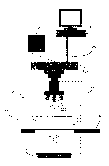
example of such a workstation is illustrated in FIG. 1 as a quantitative video-
microscopy
system, indicated by the numeral 100, according to one embodiment of the
present
invention. The system 100 generally comprises a microscope 150 haying a light
source
200 and a magnifying objective 250, a plurality of filters 600, a camera 300,
a computer
device 350, and a data transmission link 400 between the camera 300 and the
computer
device 350. The microscope 150 may comprise, for example, an Axioplan (or
Axioyert)
microscope produced by ZEISS of Germany or a similar microscope haying a
bright field
light source. The camera 300 operably engages the microscope 150 and, in one
embodiment, comprises a black-and-white camera, such as, for instance the
prosilica
GE1910 from Allied Vision Technologies. Typically, such a camera 300 also
includes an
associated frame grabber (not shown) to facilitate image capture, both the
camera 300 and
associated frame grabber being referred to herein as the "camera 300" for
convenience. In
some instances, both camera 300 and microscope 150 may be replaced by, for
example, a
black-and-white linear flat scanner and a controlled illumination source. Note
that, though
different configurations of the necessary system 100 are contemplated by the
present
invention, the present invention will be described herein in terms of a camera
300 and
associated microscope 150. Accordingly, one skilled in the art will understand
and
appreciate the capabilities and methodologies associated with these different
configurations for accomplishing the present invention as detailed herein.
Further,
although the present embodiment is disclosed as a camera, it is understood
that the camera
may be any image acquisition device, such as a camera, scanner, or any device
configured
12
CA 02832749 2013-10-08
WO 2012/142111
PCT/US2012/033053
to capture a plurality of images. The image acquisition system is capable of
capturing low
and/or high resolution images at any desired magnification, various regions of
interest, and
within various fields of view that may correspond to all or a portion of the
sample or the
slide.
The camera 300 is generally configured to capture a plurality of images 450 of
a
sample 500 through the magnifying objective 250 (where a flat scanner is used,
the image
450 is captured through internal lenses), wherein the images 450 may further
comprise a
digital image having corresponding image data (collectively referred to herein
as "the
image 450"). According to one embodiment, a calibration image 451 of a
calibration slide
550 as shown in FIG. 2, captured by image acquisition system, may be used for
preparing
the microscopy system prior to analysis of samples placed on slides. The
filters 600 filter
light from light source 200, and during operation of the system 100, multiple
images of the
sample 500 are taken using different filters, the differing filters provided
for by a filter
wheel or other filtering device as known to those skilled in the art.
According to one
embodiment, each wavelength corresponds to a respective dye of interest that
may be
present in the images. In one embodiment, the filters employed may correspond
to the
wavelengths of 460 nm, 490 nm, 520 nm, 570 nm, and 630nm. Accordingly, the
images
450 are generally captured individually, wherein each image corresponds to an
individual
wavelength filtered image of the field of view. According to one embodiment of
the
present invention, the camera 300 is configured to capture a plurality of
calibration images
455 of a calibration slide 550 corresponding to each of the plurality of
filters, the filters
corresponding to different spectral signatures. The data transmission link 400
is
configured so as to be capable of transmitting the calibration image 455 to
the computer
device 350, wherein the computer device 350 is further configured to be
capable of
analyzing the calibration image 455 with respect to each of the wavelengths.
One skilled
in the art will appreciate the computer device 350 may be any sort of
processor device or
processing element configured to communicate with the image acquisition system
and is
further configured to analyze a plurality of images as described herein.
According to a particularly advantageous aspect of the present invention, the
system 100 is configured to analyze the calibration images for preparing a
video-
microscopy system for quantitative video-microscopy in cellular biology and
pathology
applications in accordance with the Lambert-Beer law. The Lambert-Beer law
generally
describes a proportionality that can be observed between the concentration of
molecules in
13
CA 02832749 2013-10-08
WO 2012/142111
PCT/US2012/033053
a solution (the concentration of the "molecular specie" or the "sample") and
the light
intensity measured through the solution. The Lambert-Beer law is typically
expressed as:
a. OD = c = 1. C
(1)
where OD is the optical density of the solution, is a proportionality constant
called the molar extinction or absorption coefficient, 1 is the thickness of
the
sample, and C is the concentration of the molecular specie. The absorption
coefficient is specific to the molecular specie and is typically expressed in
units
of L=mol-l=cm-1.
This proportionality relationship defined by the Lambert-Beer law has been
verified under the several conditions including, for example, monochromatic
light
illuminating the sample, low molecular concentration within the sample,
generally
no fluorescence or light response heterogeneity (negligible fluorescence and
diffusion) of the sample, and lack of chemical photosensitivity of the sample.
Further, another requirement for an analysis according to the Lambert-Beer law
includes, for instance, correct Kohler illumination of the sample under the
microscope. Kohler illumination is available with many modern microscopes,
providing an even illumination of the sample in the image plane and allowing
for
effective contrast control. Kohler illumination is critical for certain
processes such
as, for example, densitometry analysis. Correct Kohler illumination is
typically
provided by, for example, a two-stage illuminating system for the microscope
in
which the source is imaged in the aperture of the sub-stage condenser by an
auxiliary condenser. The sub-stage condenser, in turn, forms an image of the
auxiliary condenser on the object. An iris diaphragm may also be placed at
each
condenser, wherein the first iris controls the area of the object to be
illuminated,
and the second iris varies the numerical aperture of the illuminating beam.
In order to accurately measure the concentration of given species imaged under
a
microscope, the measurements of the optical densities performed at different
wavelengths
must specifically correspond to the observed portion of the sample.
Accordingly, one
advantageous aspect of the present invention includes a method for determining
a relative
magnification of a given calibration slide, scene, or pattern imaged under
different
wavelengths in order to specifically correspond a pixel at a given pixel
location of one
14
CA 02832749 2013-10-08
WO 2012/142111
PCT/US2012/033053
observed portion of a sample under one wavelength to a pixel at the
corresponding pixel
location in the observed portion of the sample in one or more different
wavelengths. The
coordinates of the center of the magnifying objective 250 are determined with
respect to
the center of the electronic device or chip comprising the image-producing
component of
the camera 300. An observed magnification factor is then determined for each
wavelength
and compared to the magnification factor for an arbitrarily chosen wavelength.
For
example, a central wavelength would comprise the chosen wavelength to which
the
magnification factor for each of the other wavelengths would be compared. The
image for
each wavelength is then adjusted according to the determined relative
magnification factor
and the relative coordinates of the center of the magnifying objective 250.
According to one embodiment and with reference to FIG. 3, a calibration slide
comprising a chessboard pattern image, such as a FocalPoint Calibration Plate,
is captured
(Block 10) by the camera 300 at different wavelengths. Specifically, the
chessboard
pattern may be captured as a defocused image by applying a low pass filter to
a focused
image of the chessboard pattern. In another embodiment, a defocused image of
the
chessboard pattern may be captured by directly capturing a defocused image of
the
chessboard pattern by adjusting the focus plane in a vertical direction,
either above or
below the best z-focus plane. After the defocused image is obtained, a
histogram of the
defocused image is obtained detailing, for example, the 25% darkest and 25%
lightest
pixels in the image.
Accordingly, the defocused image may be modified to display the 25% darkest
pixels and the 25% lightest pixels, and the middle 50% pixels are discarded
from the
image to create an image mask (Block 11) of the defocused image comprising a
lattice of
cells, the cells containing, for example, the 25% darkest and 25% lightest
pixels. Cells
containing the darkest and lightest pixels that touch the border of the
defocused image
may also discarded and excluded from the subsequent calibration steps.
Furthermore, the
area of the cells of the image mask may be measured (Block 12), and the mean
area size
and the standard deviations may be computed. The cells having area sizes that
fall outside
of, for example, a 95% confidence interval are also discarded from the
calibration process.
The coordinates for the center of gravity for each of the remaining cells
comprising
the lattice of the defocused image may then be calculated. The distance
between the
center coordinates of each cell and the center coordinates of each of the
neighboring cells
in different directions, such as the north, east, south, and west directions,
may then be
measured (Block 12). An initial average distance may then be computed, which
averages
CA 02832749 2013-10-08
WO 2012/142111
PCT/US2012/033053
the measured distances between each of the center coordinates of each of the
cells and the
center coordinates of each of the cells' neighboring cells. For a given cell,
the average
distance between the center coordinate of the cell and the center coordinates
of cell's
cardinal neighbors can be computed by the following equation, wherein ktv is
the cell's
lc,th cardinal neighbor.
=1 Ed(i,kNdi) (2)
4 CardinalNeighbors
In addition to the mean distance, the calibration process may also include
determining the
standard deviation such that those distances that fall outside of, for
example, the 95%
confidence interval are then discarded from lattice cell mean distance
characterization
(Block 13). Accordingly, any cell within the image mask not having, for
instance, 4 valid
distances to 4 valid cardinal neighbors in the north, east, south, and west
directions are
excluded from the computation of the mean and standard deviation of the cell
lattice
distances. From the remaining valid distances, the average cell lattice
distance then
provides a relative magnification factor for the observed scene (Block 14).
Another advantageous aspect of the present invention includes refining the
magnification factor taken from a calibration slide, image, or pattern under
each of the
differing wavelengths. Once the average cell lattice distance is computed for
a given
calibration slide, image, or pattern under a specific wavelength, the
calibration slide,
image, or pattern may be displaced in a plurality of random directions or
directions
otherwise not established in advance, such as directions in both the x-axis
and y-axis. The
calibration slide may then be automatically focused in the z-axis focus plane,
and the
calibration process is repeated. According to one embodiment, repeating the
calibration
process may include obtaining a new defocused image, creating a new image
mask, and
calculating new average cell lattice distances for each of the newly displaced
images. In
one embodiment of the present invention, this magnification refining process
is repeated a
plurality of times, such as at least thirty times. Although the current
embodiment includes
a magnification refining process that is repeated at least thirty times, one
skilled in the art
will appreciate the invention may be repeated any number of times.
According to a further advantageous aspect of the present invention, the
system
also limits distortion, an aberration that can cause straight lines to curve
near the edges of
a captured image. This aberration causes the image to either curve in an
outwardly
fashion like a barrel or curve in an inwardly fashion like a pincushion.
Distortion
16
CA 02832749 2013-10-08
WO 2012/142111
PCT/US2012/033053
aberrations are problematic in video-microscopy systems as a pixel location in
an image
taken under one specific wavelength should correspond to the respective pixel
location in
each of the multiple images taken under differing wavelengths. As such,
distortion
aberrations may create inaccuracies when comparing images taken under
differing
wavelengths. Distortion aberrations can be determined by plotting the
distances between
the center coordinates of each cell and the center coordinates of each of the
neighboring
cells in a plurality of directions, such as the north, east, south, and west
directions, with
respect to the cell's distance from the center of the field of view of the
image. Once these
points are plotted, a linear regression line may be used to model the linear
function
between the cell's cardinal distances versus the cell's distance to the center
of the field of
view. An image having distortion aberrations will create a linear regression
line that has a
slope that is either substantially positive or negative. A barrel or
pincushion like distortion
would generate a negative and positive slope, respectively. In addition, the
system may
capture images with the same optical path such that if any significant
distortion aberration
was present, the distortion would be present in each of the images, and if
necessary,
precise image unwaming techniques, as is well-known in the art, could be
applied to
correct for this distortion.
According to one exemplary system, high end Plan Apo Achromat objectives, a
Allied Vision Technology GE1910 camera, FocalPoint's Chessboard 100 pattern of
the
calibration plate, and images with region of interests that are smaller than a
field of view
may be used to limit the distortion. Distortion is mainly expected on edges of
the field of
view. Further, according to one embodiment of the present invention, the
analyzed
regions of interest may represent a central part of the field of view image
and may roughly
represent only one-third to one-half of the whole field of view image. A
scatter plot and
regression line using such a system, as shown in FIG. 5, illustrates a
regression line having
a slope of approximately 0.0001. Accordingly, this embodiment of the present
invention
limits distortion aberrations to an insignificant amount.
As previously mentioned, in order to solve chromogen separation equations
derived from the Lambert-Beer law, a basic premise is that the same part of
the object in
the field of view should be examined. Thus, one advantageous aspect of the
present
invention is the correction of lateral chromatic aberration, which when
observed, will
provide a difference in magnification for light of different wavelengths due
to the different
focal lengths thereof For instance, an image viewed under relatively short
blue light
wavelengths will appear larger than the same image viewed under relatively
longer red
17
CA 02832749 2013-10-08
WO 2012/142111
PCT/US2012/033053
length wavelengths. However, even after the plurality of images are corrected
with the
appropriate magnification factor from the process previously described, a
pixel location in
one image taken under one wavelength may not correspond to a pixel location in
a second
image taken under a different wavelength if the images are not aligned.
Referring again to exemplary embodiment of FIG. 3, images of the sample are
captured by the system (Block 30), which scans the area or region of interest
of the sample
on the slide. Each image is transformed from transmittances into optical
densities
according to the Lambert-Beer law, as is well known in the art. In one
embodiment, a
calibration step may be used to capture a black reference image (B) and a
white reference
image (Io) for each of a plurality of wavelengths (X). Each shading-corrected
optical
density image may be computed by transforming the transmittances of each pixel
(x,y)
captured at a given wavelength into the optical density of each pixel OD,yA.
captured at the
specific wavelength with the following formula:
(I, ¨B
OD = N xlogio ___ .17 B (3)
/ ¨
xyA. 2g,
N is a multiplication factor depending upon the pixel depth of the images,
which may be
about 10,000 for a 16bits per pixel image.
The system scans the sample fully with respect to one particular wavelength
before
scanning the sample again with respect to a differing wavelength. Therefore,
images
obtained for separate wavelengths of light may be adjusted to provide
correlation with
respect to the regions of the field of view where chromogen separation
equations are
subsequently employed to determine the amount of molecular species in a
sample.
Further, the plurality of images acquired from each of the scans of the object
taken with
respect to a specific wavelength may be aligned such that the pixel locations
of an image
captured at one wavelength correspond to pixel locations of an image captured
at a
different wavelength. According to one embodiment of the present invention,
when
scanning the area or region of interest of the sample on the slide, the system
may produce
displacement factors as each scan of the sample with respect to one wavelength
is
completed before scanning the system with respect to a different wavelength.
As such, a
scan completed under one wavelength may produce an image having a region of
interest
that is displaced from the corresponding region of interest of an image taken
with respect
to a different wavelength, the displacement being characterized by the
displacement factor.
18
CA 02832749 2013-10-08
WO 2012/142111
PCT/US2012/033053
Accordingly, embodiments of the present invention provide a method of aligning
each of the images scanned such that each pixel location of one image taken
under one
wavelength corresponds to a respective pixel location of an image taken under
a differing
wavelength, as shown in FIG. 3. Another embodiment of the invention provides a
method
of aligning each of the images scanned such that a region of interest of at
least one of the
images taken under one wavelength corresponds to a region of interest of at
least one of
the images taken under a different wavelength. After each image has been
corrected by
the appropriate magnification factor with respect to the central wavelength,
the images
may be aligned such that the pixel locations of each of the images correspond
to the pixel
locations of each of the images captured. One embodiment of the present
invention
provides for extracting a number of profiles in a plurality of different
directions (e.g.,
horizontal and vertical profiles) from each of the plurality of images based
on the
background and object optical density to align the plurality of the images.
Specifically,
the horizontal and vertical profiles may be extracted from a first image to
align a region of
interest within the first image taken with respect to a reference or central
wavelength with
a region of interest within a second image taken with respect to a different
wavelength.
Furthermore, horizontal and vertical profiles may be extracted from each of
the images to
align the same region of interest from the reference image with the
corresponding region
of interest in the other images.
In one aspect of the present invention, a low-pass filter is applied (Block
31) to
each of the images to reduce high frequency noise artifacts. According to one
exemplary
embodiment, the low-pass filter comprises a kernel, such as a square matrix
having an
equal number of rows and number of columns (e.g., a 3x3 kernel), with each
element
having a particular value equal to about the inverse of the product of the
total number of
rows and the total number of columns (e.g., +1/9 for a 3x3 kernel).
Accordingly, when a
3x3 kernel is applied to a particular pixel, the center pixel and each of the
eight
neighboring pixels that surround the center pixel are added together and then
divided by 9.
The resulting value then replaces the value for the center pixel. This process
may be
repeated for each pixel within the image to create a filtered optical density
image.
To extract the horizontal and vertical profiles from the image, a binarized
image
mask may be created from the shade-corrected image. A histogram detailing the
optical
densities of the pixels may be created (Block 32) based upon the shade-
corrected optical
density image, the histogram being constructed to binarize the shade corrected
image to
form a binarized image mask. Thus, according to one embodiment, the histogram
is
19
CA 02832749 2013-10-08
WO 2012/142111
PCT/US2012/033053
created based on the background peak statistics of each of the pixels. Where,
for example,
a 16-bit black-and-white camera 300 is used in the system 100, the light
intensity
transmitted through each of the pixels in each wavelength filter may be
expressed as 216
(=65536) values between 0 and 65535. Further, the optical density for each of
the pixels
may be calculated according to Lambert-Beer law and may be stored in a
computer device
350 using a dynamic range of 16bits. Accordingly, the histogram detailing the
optical
densities of each of the pixels will have a number of bins between 0 and
65535, as shown
in FIG. 6. For example, the initial intensity Io of the light source 200,
which corresponds
to 100% transmittance, will preferably be expressed in each of plurality of
wavelengths as
a value approaching 65535, representing the brightest possible value in each
wavelength,
and a value of 0 after being converted from transmittance to optical density.
Conversely,
in the absence of light, generally corresponding to transmittance approaching
0%, a "black
image" will have an intensity value approaching 0 in each of the wavelengths
or the high
end of the histogram after being converted to optical density.
Once the histogram detailing the background peak statistics of each of the
pixels
has been constructed, a threshold utilized for binarizing the image to create
a binary image
mask (Block 33) may be calculated based on the background mode and standard
deviations. According to one embodiment, the mode is determined by the optical
density
background peak max found between the minimum bin and the minimum bin plus
4096.
The standard deviation may be calculated based on the full width at half
maximum.
Specifically, in one embodiment, the standard deviation of the optical density
histogram is
calculated as the full width at half maximum (FWHM) divided by, for example,
2.35, as
shown in the equation below.
FWHM
StDev ¨ (4)
2.35
The full width at half maximum is calculated by adding the two distances
between the
mode (i.e., the background peak between the minimum bin and the minimum bin
plus
4096) and the bins equaling, for example, 50% of the mode, as shown by the
equation
below and illustrated in FIG. 6.
FWHM = D1+ D2 (5)
If one of the distances between the mode and the bin equaling 50% of the mode
is, for
instance, 1.2 times greater than the other distance between the mode and the
other bin
equaling 50% of the mode, the full width at half maximum value may be
calculated as, for
CA 02832749 2013-10-08
WO 2012/142111
PCT/US2012/033053
example, 2 times the smaller of the two distances, as shown by the equation
below and
illustrated in FIG. 6.
If (D2 >1.2 FWHM = 2 .4 (6)
Accordingly, as mentioned before, the standard deviation may then be
calculated as the
full width at half maximum divided by 2.35, and the threshold used to create
the binarized
image mask (Block 33) is calculated as the mode plus, for example, 6 times the
standard
deviation, as shown by the equation below.
Threshold = Mode+ 6 StDev (7)
In another embodiment, the threshold may be calculated as the mode plus at
least 3 times
the standard deviation.
The value of each of the pixels in the image may be then compared to the
threshold
to create the binary image mask. Those pixels having a value less than or
equal to the
threshold may be assigned a value of zero, while the pixels having a value
greater than the
threshold may be assigned a value of one. Accordingly, the image may then be
converted
into a binarized mask, each pixel within the image mask having a value or zero
or one.
According to one embodiment of the invention, the percentage of pixels having
a value
greater than the threshold in the reference image can be used to refine the
threshold used
to generate the binarized mask of the image to be realigned. Once the binary
image mask
is applied to each of the images, the horizontal and vertical profiles can be
extracted for
each of the images, the images corresponding to a differing wavelength. The
horizontal
and vertical profiles are extracted (Block 34) to align the region of interest
from a portion
of each of the images that are captured.
From the binarized image, the horizontal profiles extracted represent the
vertical
projection of the region of interest on the horizontal axis, as illustrated by
FIG. 7.
Likewise, the vertical profiles extracted represent the horizontal projection
of the region of
interest on the vertical axis. The number of horizontal profiles correlates to
the number of
pixels necessary to cover the tolerance in the displacement in the vertical
direction, while
the number of vertical profiles correlates to the number of pixels necessary
to cover the
tolerance in displacement in the horizontal direction. In one embodiment of
the invention,
the region of interest examined has a length and width of equal value.
Accordingly, the
horizontal profiles have a horizontal length equal to the size of the region
plus two times a
horizontal displacement factor. The horizontal displacement factor is equal to
the mean
horizontal displacement plus, for example, twelve standard deviations.
Likewise, a
21
CA 02832749 2013-10-08
WO 2012/142111
PCT/US2012/033053
vertical displacement factor is equal to the mean vertical displacement plus,
for example,
twelve standard deviations. Furthermore, the vertical profiles have a vertical
length equal
to the size of the region plus two times the vertical displacement factor.
Once the horizontal and vertical profiles are extracted for each of the
plurality of
images, the horizontal and vertical profiles for each of the images may be
rescaled using
spline functions. According to one embodiment of the invention, the horizontal
coordinates of each of the horizontal and vertical profiles for each of the
plurality of
images are converted from coordinates defined by the number of pixels to
coordinates defined in micrometers, as shown in the equation below.
X(um)=[X(px)¨ X .õ(px)]= pixelSize(um)+X.,(px) (8)
The new horizontal coordinate value rescaled in micrometers, X(i.im), is equal
to the
original horizontal coordinate value in pixels, X(pm), minus the horizontal
coordinates of
the reference point, such as the center of the image, in pixels, Xcenter(PM),
multiplied by the
size of a pixel in micrometers, pixelSize( m) plus the horizontal coordinates
of the
reference point. According to one embodiment, the horizontal coordinate value
of the
reference point may equal zero.
The vertical coordinates of each of the horizontal and vertical profiles for
each of
the plurality of images may be rescaled by the spline functions (Block 35) to
a new value
so as to be related at least in part by the profile intensity. Specifically,
the vertical
coordinates of each of the horizontal and vertical profiles are rescaled
according to the
equation below.
Yvalue = profileValue = pixelSize(i1) pixelSize(ilref) (9)
The new vertical coordinate value, Yvahie, is equal to the average intensity
of the pixel at
the corresponding horizontal position, profileValue, multiplied by the size of
the pixel at
the corresponding wavelength, pixelSize(X), divided by the size of the pixel
of the
reference wavelength, pixelSize(kref). In one embodiment of the invention, the
reference
wavelength may be equal to 570nm. The number of rescaled spline profiles may
be equal
to the number of profiles.
22
CA 02832749 2013-10-08
WO 2012/142111
PCT/US2012/033053
Once each of the horizontal and vertical profiles extracted from each of the
plurality of images are rescaled according to the spline functions,
embodiments of the
present invention provide for evaluating the shift between spline profiles
from the
reference image with spline profiles for each of the plurality of images
(Block 36). First,
the spline profiles from a target image may be evaluated against the spline
profiles from
the reference image by calculating an error factor according to the equation
below.
Error = E [P A(dx, dy) ¨ PAref (0,0)]2 (10)
The spline profile with the minimum amount of error best matches the reference
profile.
The error factor is equal to the sum of the square of the differences between
the
coordinates of the spline profile and the coordinates of the reference
profile. The shift is
evaluated for both the horizontal spline profiles and the vertical spline
profiles. According
to one embodiment of the invention, averaging the shifts between the spline
profiles and
the reference spline profiles provides a preferred precision to evaluate the
shift between
profiles. Furthermore, another embodiment may comprise extracting profiles
from a
plurality of regions of interests from the two images that are to be aligned
and compared.
Once the spline profile with the least amount of error is determined, the
target
image may be aligned to the reference image (Block 37) by first rescaling the
image with
the previously determined magnification factor. The image may then be aligned
to the
reference image by shifting the image in the horizontal and vertical
directions by the shift
factors that were determined to provide the smallest amount of error. The
magnification
rescaling and corrective shifting in the horizontal and vertical direction may
be applied to
each of the plurality of spectral images with respect to the reference
wavelength image.
Once each of the plurality of spectral images have been rescaled and aligned
with respect
to the reference wavelength image, according to one embodiment of the
invention, the
system may then proceed with chromogen separation analysis of the sample
(Block 40).
Particularly, the system may then proceed with determining an amount of a
plurality of molecular species in a sample, each molecular specie being
indicated by a dye.
In one embodiment, the system may determine an amount of a plurality of
molecular
species by using chromogen separation techniques. Such a technique includes
determining an optical density of the sample in each pixel at each
corresponding pixel
location in the plurality of images. In one embodiment, a corresponding
optical density
23
CA 02832749 2016-01-14
67044-106
matrix is formed for that pixel and multipled by the inverse of a relative
absorption
coefficient matrix so as to form a resultant matrix for the pixel. The
relative absorption
coefficient matrix comprises a relative absorption coefficient for each dye,
independently
of the sample, in each of the plurality of wavelengths. In one embodiment, the
method
may include refining the amount of a plurality of molecular species at one or
more pixel
locations in the plurality of images. Further, the system may comprise a video-
microscopy
system comprising an image acquisition device configured to capture a
plurality of
magnified digital images of the sample and a processor device configured to
determine an
amount of each molecular specie. For further exemplary discussion regarding
techniques
for determining the amount of a molecular species in a sample, see U.S. Patent
Application No. 61/474,250, entitled METHOD FOR OPTIMIZATION OF
QUANTITATIVE VIDEO-MICROSCOPY AND ASSOCIATED SYSTEM, which was
filed on April 12, 2012.
According to the methodology described herein, the determined dye
concentrations
may then be used to reconstruct an artificial image of the sample. The
artificial images
may be generated as a substantially real time or live image, or as a still
image, using
combinations of the dyes comprising a marker and/or a counterstain used to
prepare the
sample. More particularly, an artificial image of the field of view may be
produced which
shows the sample as affected by all of the dyes, the sample as affected by one
or more
marker dyes, or the sample as affected by the counterstain. Consequently,
since the dyes
used to prepare the sample are characterized by the system, the capabilities
of the system
may be extended such that, for instance, the sample or field of view may be
automatically
scanned to detect a specific region of interest as identified by the
characteristics of a
particular dye or to affect or facilitate a task to be performed on that
specific region of
interest.
Still further, the artificial image of the field of view may also be used to
facilitate
the identification and extraction of selected features of the treated sample.
For example,
marked point processes, contextual analysis, and/or geo-statistics may be used
to identify
and extract features from the image based on, for instance, a spatial
distribution analysis of
a particular dye. Such a feature extraction capability would also allow, for
example, fields
of view or objects of interest to be sorted, flagged, or otherwise identified
or grouped
based on, for instance, the overall content of a given marker dye or a
selected ratio of
particular marker. Where, for example, a threshold criteria can be
established, such a
capability would be the detection of rare, worsening, or other serious events.
Proceeding
24
CA 02832749 2013-10-08
WO 2012/142111
PCT/US2012/033053
further, classifiers based specifically on the image processing resulting from
the
counterstain and/or marker dye specific images may then be established and
used to
evaluate the presence of certain cell types or to perform a diagnosis based
upon the field of
view. For example, HER2 may be evaluated in this manner by comparison to a
continuous
diagnosis scale established according to the system and methods described
herein. Such
classifiers may usually also encompass other informative features such as, for
example,
detail based upon the morphology or the texture of the cells.
It will be understood that the methodology and procedures detailed herein in
conjunction with the system 100 specify a method of calibrating or otherwise
preparing a
video-microscopy system for acquiring and analyzing images of a sample. One
skilled in
the art will also appreciate that such a method may be automated so as to
provide a
computer software program product, executable on a computer device, having
executable
portions capable of determining a magnification factor and aligning the
plurality of
images. Accordingly, embodiments of the present invention describe the
implementation
of a method and/or corresponding computer software program product which may
be
accomplished in appropriately configured hardware, software, or a combination
of
hardware and software in accordance with the scope of the present invention.
Thus,
embodiments of the present invention comprise a preparation technique for a
video-
microscopy system for analyzing prepared samples that may provide effective
detection
and quantification of species of interest that overcomes limiting factors of
prior art
technologies, such as errors in dye concentration estimations due to noise
fluctuations,
improper calibration, and/or chromatic aberrations.
Many modifications and other embodiments of the invention will come to mind to
one skilled in the art to which this invention pertains having the benefit of
the teachings
presented in the foregoing descriptions and the associated drawings.
Therefore, it is to be
understood that the invention is not to be limited to the specific embodiments
disclosed
and that modifications and other embodiments are intended to be included
within the
scope of the appended claims. Although specific terms are employed herein,
they are used
in a generic and descriptive sense only and not for purposes of limitation.
25