Note : Les descriptions sont présentées dans la langue officielle dans laquelle elles ont été soumises.
CA 02833518 2013-11-15
USING LOCATION AND FORCE MEASUREMENTS
TO ESTIMATE TISSUE THICKNESS
FIELD OF THE INVENTION
[0001]
The present invention relates generally to invasive probes,
and specifically to estimating tissue thickness based on
location and contact force measurements received from an
invasive probe.
BACKGROUND OF THE INVENTION
[0002]
A wide range of medical procedures involve placing objects,
such as sensors, tubes, catheters, dispensing devices and
implants, within a patient's body.
Position sensing systems
have been developed for tracking such objects.
Magnetic
position sensing is one of the methods known in the art.
In
magnetic position sensing, magnetic field generators are
typically placed at known positions external to the patient. A
magnetic field sensor within the distal end of a probe generates
electrical signals in response to these magnetic fields, which
are processed in order to determine the position coordinates of
the distal end of the probe.
These methods and systems are
described in U.S. Patents 5,391,199, 6,690,963, 6,484,118,
6,239,724, 6,618,612 and 6,332,089, in PCT International
Publication WO 1996/005768, and in U.S. Patent Application
Publications 2002/0065455 Al, 2003/0120150 Al and 2004/0068178
Al, whose disclosures are all incorporated herein by reference.
[0003]
When placing a probe within the body, it may be desirable
to have the distal tip of the probe in direct contact with body
tissue. The contact can be verified, for example, by measuring
1
CA 02833518 2013-11-15
,
,
the contact pressure between the distal tip and the body tissue.
U.S. Patent Application Publications 2007/0100332, 2009/0093806
and 2009/0138007, whose disclosures are incorporated herein by
reference, describe methods of sensing contact pressure between
the distal tip of a catheter and tissue in a body cavity using a
force sensor embedded in the catheter.
[0004] Some probes include both a force sensor and a position
sensor. U.S. Patent Application 13/152,423, whose disclosure is
also incorporated herein by reference, describes a method for
detecting tenting in tissue (due to a force exerted by the
distal tip of the probe on the tissue) using location and force
measurements received from a probe that includes a position
sensor and a force sensor.
[0005] Documents incorporated by reference in the present patent
application are to be considered an integral part of the
application except that to the extent any terms are defined in
these incorporated documents in a manner that conflicts with the
definitions made explicitly or implicitly in the present
specification, only the definitions in the present specification
should be considered.
SUMMARY OF THE INVENTION
[0 0 0 6] An embodiment of the present invention provides a method,
including:
pressing a distal end of a medical probe against a wall of
a body cavity;
receiving from the probe first measurements of a force
exerted by the distal end on the wall;
receiving from the probe second measurements indicating a
displacement of the wall in response to the force; and
2
CA 02833518 2013-11-15
estimating a thickness of the wall based on the first and
the second measurements.
[0007] Typically the probe includes a catheter.
[0008] In a disclosed embodiment the method includes, prior to
pressing the distal end of the probe against the wall,
initializing one or more calibration matrices, each of the
calibration matrices associated with a type of tissue.
Typically, the type of tissue is selected from a list comprising
artery tissue and endocardial tissue.
[0009] Initializing a given calibration matrix may include storing
a force value, a displacement value, and an associated thickness
value to each element of the calibration matrix.
[0010] In a further disclosed embodiment estimating the thickness
of the wall includes identifying, in a given calibration matrix,
a given element of the calibration matrix having a given force
value corresponding to the first measurements and a given
displacement value corresponding to the second measurements, and
retrieving the thickness value from the identified matrix
element.
[0011] Estimating the thickness of the wall may include
interpolating between the thickness values stored in two
calibration matrix elements. In one embodiment the method
includes, subsequent to initializing the one or more calibration
matrices and prior to estimating the thickness of the wall,
selecting a given calibration matrix associated with the type of
tissue corresponding to the wall of the body cavity. In another
embodiment, the method includes, prior to selecting the given
calibration matrix, identifying the type of tissue based on a
location of the distal end.
[0012] In an alternative embodiment receiving the second
3
. CA 02833518 2013-11-15
measurements indicating the displacement includes receiving
first position measurements from the probe indicating a first
location of the probe upon the probe engaging the wall,
receiving second position measurements indicating a second
location of the probe upon the distal end exerting the force on
the wall, and calculating a distance between the first and the
second locations.
[0013] There is also provided, according to an embodiment of the
present invention, medical apparatus, including:
a probe having a distal end configured for insertion into a
body cavity having a wall, the probe including:
a force sensor in the distal end, configured to generate a
first signal indicative of a force exerted by the distal end on
the wall; and
a position sensor in the distal end, configured to generate
a second signal indicative of a location of the distal end
within the body cavity; and
a processor, which is coupled to receive and process the
first and second signals from the probe so as to estimate a
thickness of the wall.
[0014] There is also provided, according to an embodiment of the
present invention, a computer software product, operated in
conjunction with a probe that is configured for insertion into a
body cavity of a patient and that includes a position sensor for
measuring a position of a distal end of the probe inside the
body cavity and a force sensor for measuring a force between the
distal end and a wall of the body cavity, the product including
a non-transitory computer-readable medium, in which program
instructions are stored, which instructions, when read by a
computer, cause the computer to receive from the probe, while
4
CA 02833518 2013-11-15
pressing the distal end against the wall, first measurements of
a force exerted by the distal end on the wall, to receive from
the probe second measurements indicating a displacement of the
wall in response to the force, and to estimate a thickness of
the wall based on the first and the second measurements.
BRIEF DESCRIPTION OF THE DRAWINGS
[0015] The disclosure is herein described, by way of example only,
with reference to the accompanying drawings, wherein:
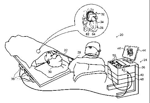
[0016] Figure 1 is a schematic pictorial illustration of a medical
system that is configured to estimate tissue thickness, in
accordance with an embodiment of the present invention;
[0017] Figure 2 is a schematic side view showing details of the
distal portion of a pressure-sensitive catheter, in accordance
with an embodiment of the present invention;
[0018] Figure 3 is a flow diagram that schematically illustrates a
method of calibrating the catheter, in accordance with an
embodiment of the present invention;
[0019] Figures 4A and 4B are schematic detail illustrations of
tissue displacements due to a force exerted by the distal
portion of the catheter on the tissue, in accordance with an
embodiment of the present invention; and
[0020] Figure 5 is a flow diagram that schematically illustrates a
method of estimating tissue thickness based on location and
force measurements received from the catheter, in accordance
with an embodiment of the present invention.
, CA 02833518 2013-11-15
DETAILED DESCRIPTION OF EMBODIMENTS
OVERVIEW
[0021]
Various diagnostic and therapeutic procedures, such as
cardiac ablation and intracardiac electrical mapping, use an
invasive probe, such as a catheter, whose distal tip is fitted
with at least one electrode.
The electrode is typically
operated when the probe is pressed against a wall (also referred
to herein as tissue) of a body cavity. In these procedures, it
is usually important to ascertain both the precise location of
the probe in the body cavity, and the force that the distal tip
is exerting on the body cavity wall. Therefore, some catheters
comprise position sensors for ascertaining the location of the
distal tip and force sensors for measuring the force exerted by
the probe on intra-body tissue, such as the endocardium.
[0022]
During an ablation procedure, in embodiments of the present
invention the thickness of the tissue being ablated is
monitored. Applying (by the distal tip) too much force to thin
tissue may cause perforation, and on the other hand, applying
too little force to thicker tissue may be inefficient in
isolating the tissue area electrically.
[0023]
As an operator presses the distal tip of a probe against a
body cavity wall, embodiments of the present invention provide
methods and systems for estimating a thickness of the body
cavity wall, based on location and force measurements received
from sensors within the probe. The received force measurements
indicate a force applied by the distal tip against the body
cavity wall, and the position measurements indicate a
displacement of the wall in response to the applied force. As
explained in detail hereinbelow, the tissue thickness can be
6
' CA 02833518 2013-11-15
estimated by locating an entry in a calibration matrix with
force and displacement values that correspond to the force and
the displacement measurements received from the probe. Tissue
thickness measurements incorporating embodiments of the present
invention may be used by medical systems to replace or
complement other known methods of tissue thickness measurement,
such as magnetic resonance imaging (MRI) or computerized
tomography (CT).
SYSTEM DESCRIPTION
[0024]
Figure 1 is a schematic pictorial illustration of a medical
system 20 that is configured to estimate tissue thickness, in
accordance with an embodiment of the present invention. System
20 may be based, for example, on the CARTOTm system, produced by
Biosense Webster Inc. (Diamond Bar, California).
System 20
comprises a probe 22, such as a catheter, and a control console
24. In the embodiment described hereinbelow, it is assumed that
probe 22 is used for diagnostic or therapeutic treatment, such
as for mapping electrical potentials in a heart 26 or performing
ablation of heart tissue. Alternatively, probe 22 may be used,
mutatis mutandis, for other therapeutic and/or diagnostic
purposes in the heart or in other body organs.
[0025]
An operator 28, such as a cardiologist, inserts probe 22
through the vascular system of a patient 30 so that a distal end
32 of probe 22 enters a chamber of heart 26.
Operator 28
advances probe 22 so that a distal tip 34 of probe 22 engages
endocardial tissue at a desired location or locations. Probe 22
is typically connected by a suitable connector at its proximal
end to console 24.
[0026]
Console 24 typically uses magnetic position sensing to
7
, CA 02833518 2013-11-15
determine position coordinates of distal end 32 inside heart 26.
To determine the position coordinates, a driver circuit 36 in
console 24 drives field generators 38 to generate magnetic
fields within the body of patient 30.
Typically, field
generators 38 comprise coils, which are placed below the
patient's torso at known positions external to patient 30.
These coils generate magnetic fields in a predefined working
volume that contains heart 26.
A magnetic field sensor 62
within distal end 32 of probe 22 (sensor 62 is shown in more
detail in Figure 2) generates electrical signals in response to
these magnetic fields.
A signal processor 40 processes these
signals in order to determine the position coordinates of distal
end 32, typically including both location and orientation
coordinates.
The method of position sensing described
hereinabove is implemented in the above-mentioned CARTOTm system
and is described in detail in the patents and patent
applications cited above.
[0027]
Signal processor 40 typically comprises a general-purpose
computer, with suitable front end and interface circuits for
receiving signals from probe 22 and controlling the other
components of console 24.
Processor 40 may be programmed in
software to carry out the functions that are described herein.
The software may be downloaded to console 24 in electronic form,
over a network, for example, or it may be provided on non-
transitory tangible media, such as optical, magnetic or
electronic memory media.
Alternatively, some or all of the
functions of processor 40 may be carried out by dedicated or
programmable digital hardware components.
[0028]
An input/output (I/0) interface 42 enables console 24 to
interact with probe 22.
Based on the signals received from
8
CA 02833518 2013-11-15
probe 22 (via interface 42 and other components of system 20),
processor 40 drives a display 44 to present operator 30 with an
image 46 showing the position of distal end 32 in the patient's
body, as well as status information and guidance regarding the
procedure that is in progress.
[0029] In the present embodiment, processor 40 monitors
measurements received from position sensor 62 and a force sensor
64 within distal end 32 (force sensor 64 is shown in more detail
in Figure 2) during periods in which the catheter is believed to
be pressing against endocardial tissue of heart 26.
As
explained hereinbelow, when distal tip 34 is pressing against
the endocardial tissue, processor 40 can determine the thickness
of the tissue based on measurements received from the probe's
position and force sensors.
[0030]
Processor 40 stores data representing image 46 in a memory
48.
In some embodiments, operator 28 can manipulate image 46
using one or more input devices 50.
[0031]
Alternatively or additionally, system 20 may comprise an
automated mechanism (not shown) for maneuvering and operating
probe 22 within the body of patient 30.
Such mechanisms are
typically capable of controlling both the longitudinal motion
(advance/retract) of probe 22 and transverse motion
(deflection/steering) of distal end 32 of the probe.
In such
embodiments, processor 40 generates a control input for
controlling the motion of probe 22 based on the signals provided
by the magnetic field sensor in the probe.
[0032]
Although Figure 1 shows a particular system configuration,
other system configurations can also be employed to implement
embodiments of the present invention, and are thus considered to
be within the spirit and scope of this invention. For example,
9
CA 02833518 2013-11-15
the methods described hereinbelow may be applied using position
transducers of types other than the magnetic field sensor
described above, such as impedance-based or ultrasonic position
sensors. The term "position transducer" as used herein refers
to an element mounted on probe 22 which causes console 24 to
receive signals indicative of the coordinates of the element.
The position transducer may thus comprise a receiver on the
probe, which generates a position signal to the control unit
based on energy received by the transducer; or it may comprise a
transmitter, emitting energy that is sensed by a receiver
external to the probe.
Furthermore, the methods described
hereinbelow may similarly be applied in therapeutic and
diagnostic applications using not only catheters, but also
probes of other types, both in the heart and in other body
organs and regions.
[0033]
Figure 2 is a schematic sectional view of distal end 32 of
probe 22, in accordance with an embodiment of the present
invention. Specifically, Figure 2 shows functional elements of
distal end 32 used for therapeutic and/or diagnostic activity.
An electrode 60 (e.g., an ablation electrode) at distal tip 34
of the probe is typically made of a metallic material, such as a
platinum/iridium alloy or another suitable material.
Alternatively, multiple electrodes (not shown) along the length
of the probe may be used for this purpose.
[0034]
Position sensor 62 transmits a signal to console 24 that is
indicative of the location coordinates of distal end 32.
Position sensor 62 may comprise one or more miniature coils, and
typically comprises multiple coils oriented along different
axes.
Alternatively, position sensor 62 may comprise either
another type of magnetic sensor, an electrode which serves as a
= CA 02833518 2013-11-15
position transducer, or position transducers of other types,
such as impedance-based or ultrasonic position sensors.
Although Figure 2 shows a probe with a single position sensor,
embodiments of the present invention may utilize probes with
more than one position sensor.
[0035]
In an alternative embodiment, the roles of position sensor
62 and magnetic field generators 38 may be reversed. In other
words, driver circuit 36 may drive a magnetic field generator in
distal end 32 to generate one or more magnetic fields.
The
coils in generator 38 may be configured to sense the fields and
generate signals indicative of the amplitudes of the components
of these magnetic fields.
Processor 40 receives and processes
these signals in order to determine the position coordinates of
distal end 32 within heart 26.
[0036]
Force sensor 64 measures a force applied by distal tip 34
to the endocardial tissue of heart 26 by conveying a signal to
the console that is indicative of the force exerted by the
distal tip on the intra-body tissue.
In one embodiment, the
force sensor may comprise a magnetic field transmitter and
receiver connected by a spring in distal end 32, and may
generate an indication of the force based on measuring the
deflection of the spring. Further details of this sort of probe
and force sensor are described in U.S. Patent Application
Publications 2009/0093806 and 2009/0138007, whose disclosures
are incorporated herein by reference. Alternatively, distal end
32 may comprise another type of force sensor.
TISSUE THICKNESS ESTIMATION
[0037]
Prior to performing a medical procedure such as cardiac
ablation, probe 22 is typically calibrated using embodiments
11
CA 02833518 2013-11-15
described hereinbelow. During a medical procedure, processor 40
can utilize the calibration data in order to estimate tissue
thickness based on force and displacement measurements received
from probe 22 (i.e., when the probe is pressing against a wall
of a body cavity).
[0038]
Figure 3 is a flow diagram that schematically illustrates a
method of calibrating probe 22, and Figures 4A and 4B are
schematic detail views of displacements 92 in body cavity walls
90 in response to a force exerted by distal tip 34, in
accordance with an embodiment of the present invention. In the
description herein, different body cavity walls 90 and different
displacements 92 may be separately identified by appending a
letter to the identifying numeral, so that body cavity walls 90
comprise a body cavity wall 90A and a body cavity wall 90B, and
displacements 92 comprise a displacement 92A, also indicated by
Lx/ in Figure 4A, and a displacement 92B, also indicated by Lx2
in Figure 43.
Calculating Lx/ and Lx2 is described in detail
hereinbelow.
[0039]
In an initial step 70, operator 28 selects a first body
cavity wall 90 having a first known thickness.
In a force
application step 72, the operator first positions probe 22 so
that distal tip 34 engages the selected body cavity wall, and
then presses the distal tip against the wall.
Pressing distal
tip 34 against body cavity wall 90 causes displacement 92 of
wall 90 in response to the force exerted by the distal tip on
the wall.
[0040]
As operator 28 positions probe 22, position sensor 62
outputs a signal indicative of locations of distal tip 34.
Additionally, as the operator presses distal tip 34 against the
12
CA 02833518 2013-11-15
selected body cavity wall, force sensor 64 outputs a signal
indicative of the force exerted by the distal tip on the wall.
Both the position and the force signals, providing respective
location and force measurements, are conveyed to medical system
20.
[0041]
When operator 28 presses distal tip 34 against the selected
body cavity wall, processor 40 collects, in a first collection
step 74, a first signal from sensor 64 indicating a force
exerted by the distal tip against the wall. Processor 40 also
collects, in a second collection step 76, a second signal from
sensor 62 indicating locations of distal tip 34. The locations
indicated by the signal comprise a first location comprising
where distal tip 34 initially engages the selected body cavity
wall and a second location comprising a location of the distal
tip after the operator presses the distal tip against the wall.
Displacement 92 comprises a distance between the first location
and the second location.
[0042] In a calibration step 78, processor 42 creates a
calibration matrix entry based on the collected position and
force measurements.
To create the calibration matrix element,
processor 42 maps the known thickness of body cavity wall 90
against the location measurements received from position sensor
62 and the force measurements received from force sensor 64.
Therefore, each calibration matrix element typically comprises a
force value, a displacement value, and an associated thickness
value.
Alternatively, the thickness, force and displacement
values may be stored as a range of values. For example, for a
range between 1.8 and 2.0, the range of values can be stored in
the calibration matrix as a lower and an upper threshold (e.g.,
1.8, 2.2) of the range, or as the midpoint of the range and the
13
CA 02833518 2013-11-15
,
value to be added to and subtracted from the midpoint (2.0,
0.2).
[0043]
In a first comparison step 80, if additional calibration
for the selected body cavity wall is needed to calibrate the
selected body cavity wall, then in a prompting step 82, console
24 prompts operator 28 to change the force applied by distal tip
34 against the selected body cavity wall (i.e., apply lower or
greater force), and the method continues with step 72.
For
example, to accurately calibrate a given body cavity wall,
processor 40 may need to collect at least a defined number of
force (and displacement) values, within a range typically used
during a given medical procedure. If no additional calibration
for the selected body cavity wall is needed, then in a second
comparison step 84, console 24 prompts operator 28 to determine
if there is an additional body cavity wall to be calibrated.
[0044]
If an additional body cavity wall is needed to calibrate
probe 22, then in a selection step 86, console 24 prompts
operator 28 to select a different body cavity wall 90 having a
different known thickness, and the method continues with step
72. The method ends when there are no additional body cavity
walls needed for calibrating probe 22.
[0045]
In some embodiments, operator 28 can decide if additional
calibration is desired in the comparison steps described supra
(i.e., in steps 80 and 84).
In alternative embodiments, a
software application executing on processor 40 can determine if
further calibration is desired.
[0046]
During calibration, operator 28 may select a variety of
different types of body cavity walls 90, since different types
of tissue may generate different calibration tables.
For
example, a specific part of the endocardium may generate a
14
CA 02833518 2013-11-15
calibration matrix that differs from a calibration matrix for an
artery, typically because of different elasticities of the
different tissues.
Sets of calibration matrices for different
types of tissue can be created using the steps described
hereinabove, wherein a given calibration matrix is associated
with a given tissue type.
In some embodiments, the set of
calibration matrices can be stored to memory 48. Alternatively,
the calibration matrices can be stored to a memory coupled to
probe 22 (not shown).
[0047]
In the examples shown in Figures 4A and 4B, operator 28
applies the same force vector F, as measured by force sensor 64,
orthogonally to walls 90A and 90B having different thicknesses
(T/ and T2 respectively). As described supra, processor 40 can
measure the displacement in the tissue by identifying a first
location of distal tip 34 when the distal tip first engages the
given tissue, and identifying a second location when the force
applied by the distal tip on the given tissue is F.
The
difference between the first location and the second location
(i.e., the displacement) is Axl in Figure 4A and Lx2 in Figure
4B. As illustrated in the examples shown in the Figures, there
is a relation between tissue thickness and tissue displacement.
In other words, given the same force vector F applied by distal
tip 34, the resulting displacement Axi in thin body cavity wall
90A is typically greater than the displacement Lx2 in thick body
cavity wall 90B.
[0048]
Figure 5 is a schematic flow diagram that schematically
illustrates a method of estimating tissue thickness based on
position and force measurements conveyed by probe 22, in
accordance with an embodiment of the present invention. In an
= CA 02833518 2013-11-15
initial step 100, operator 28 positions distal end 32 within a
given body cavity (e.g., heart 26) and presses distal tip 34
against a given body cavity wall 90. As explained supra, there
may be multiple of calibration matrices defined for different
types of tissue that can be encountered during a medical
procedure. Therefore, prior to pressing distal tip 34 against a
given body cavity wall 90, operator 28 may identify, using input
devices 50, the type of tissue in the body cavity. In response
to the operator identifying the type of tissue, processor 40 can
select a given calibration matrix that is associated with the
identified tissue. In an alternative embodiment, processor 40
can identify the type of tissue based on the location of distal
tip 34.
[0049]
While operator 28 presses distal tip 34 against the given
body cavity wall, processor 40 collects, in a first collection
step 102, a first signal from sensor 64 indicating a force
exerted by the distal tip against the wall. Processor 40 also
collects, in a second collection step 104, a second signal from
sensor 62 indicating locations of distal tip 34. The locations
indicated by the signal comprise a first location where distal
tip 34 initially engages the given body cavity wall, and a
second location comprising a location of the distal tip after
the operator presses the distal tip against the wall.
As
explained supra, displacement 92 (in response to the applied
force) comprises the distance between the first location and the
second location.
[0050]
In an estimation step 106, processor 40 identifies an
element in the calibration matrix that has force and
displacement values corresponding to the collected force and the
displacement measurements, and retrieves a thickness value from
16
CA 02833518 2013-11-15
the identified calibration matrix element, and the method ends.
In instances where corresponding values for the collected force
and displacement measurements are not explicitly found in the
calibration matrix, processor 40 can estimate the thickness by
calculating a thickness based on an interpolation between two
force and/or displacement values found in the calibration
matrix.
[0051]
It will be appreciated that the embodiments described above
are cited by way of example, and that the present invention is
not limited to what has been particularly shown and described
hereinabove.
Rather, the scope of the present invention
includes both combinations and subcombinations of the various
features described hereinabove, as well as variations and
modifications thereof which would occur to persons skilled in
the art upon reading the foregoing description and which are not
disclosed in the prior art.
17