Note : Les descriptions sont présentées dans la langue officielle dans laquelle elles ont été soumises.
CA 02833654 2013-10-18
WO 2012/143709
PCT/GB2012/050846
IMMUNOASSAY
The invention relates to an immunoassay method of quantifying IgE levels in a
sample,
methods of calibrating a device suitable for carrying out such quantification
and a system
that enables such quantification.
Immunoassays are methods that utilise the binding capacity of antibodies.
Often,
immunoassays are used to assay for the presence of a particular antigen-
specific
antibody in a sample. This is done by washing the sample over the particular
antigen
o immobilised on a solid support, and subsequently visualising any bound
antibody using
various techniques.
Generally, immunoassays require the use of calibrators to assign values or
concentrations to unknown samples. In a classical immunoassay, a set of
calibrators is
run, a calibration curve of signal versus concentration is plotted and the
concentration of
the unknown samples determined by interpolation.
Allergic conditions are characterised by inappropriate and exaggerated immune
responses to innocuous environmental antigens. These antigens are collectively
called
allergens. Immune responses to allergens include a first phase of
sensitization consisting
of (i) processing of the allergens by antigen presenting cells (APCs), (ii)
presentation of
the processed allergens by the APCs to T helper 0 (Th0) naïve cells, (iii)
differentiation of
the Th0 naïve cells to Th2 cells, and (iv) stimulation of B cells by the Th2
cells, leading to
the production and secretion of allergen-specific IgE by the B cells.
Each specific allergen will stimulate the production of an IgE specific to
that allergen. IgE
antibodies can interact with two different cell types; mast cells and
basophils, which
contain histamine-containing granules.
When the same allergen that has elicited the sensitization phase enters the
body a
second time and is recognised by the appropriate mast-cells and basophils, it
stimulates
a second phase of the allergy mechanism known as the "challenge phase". The
allergen
binds to its specific IgE presented on the surface of mast-cells and basophils
triggering a
mechanism which eventually leads to degranulation of the mast cells and
basophils and
secretion of histamine, which is responsible for the inflammatory reaction
typical of an
allergic reaction. The severity of the ensuing allergic reaction corresponds
with the level
of allergen-specific IgE in that individual. Therefore, it is important to
detect and quantify
1
CA 02833654 2013-10-18
WO 2012/143709
PCT/GB2012/050846
the concentration of IgEs raised against particular allergens in an individual
to enable
identification of allergic (or atopic) individuals and characterisation of
their allergies.
Quantitative immunoassays for the diagnosis of allergy normally use or refer
to the World
Health Organization (WHO) International Reference Preparation 75/502 to build
their
calibration systems. This is a freeze-dried human serum sample with an
assigned IgE
reactivity (Kontis K, et al (2006) Correlation of the Turbo-MP RIA with
ImmunoCAP FEIA
for Determination of Food Allergen-Specific Immunoglobulin-E. Ann Clin Lab
Sci. 36(1):
79-87; Bousquet J et al (1990) Comparison between RAST and Pharmacia CAP
system:
A new automated specific IgE assay. J Allergy Clin Immunot 86(6):1 039-43;
Reference
for the product: http://www.nibsc.ac.uk/documents/ifu/75-502.odf).
Measured response values for allergen-specific IgE antibodies are typically
evaluated
against a total IgE calibration curve (WHO International Reference
Preparation) and
expressed as concentration of Allergen specific Units per litre (kUA/I). The
IgE reference
curve is used to describe the dose-response curve for all the allergens
tested. This
requires that the concentrations of allergenic components that are immobilised
on a solid
support for the immunoassay are optimised such that the dose-response curves
for the
IgE and the allergens show the same trend. To optimise the concentrations of
the
allergenic concentrations for the dose response curves, different
concentrations of the
allergens are tested against a panel of samples at known reactivity to
identify the
concentration that gives a dose-response curve similar in shape and slope to
the one
generated using the WHO International Reference total IgE curve.
Immunoassays presently used for the diagnosis of allergic disease include:
Radioallergosorbent Test (RAST), Enzyme-Linked Immunosorbent Assay (ELISA),
and
ImmunoCAP.
RAST involves covalently coupling an allergen (or other antigen) to a paper
disk solid
phase. The paper disk is then incubated with serum from a patient whose
allergenic
status is to be investigated. If antibodies against the allergen are present
in the serum,
they react with the conjugated allergen and binding is revealed with radio-
labelled anti-
IgE antibodies. In its original form, the results of the test were reported in
classes or
arbitrary units by interpolating from a heterologous IgE anti-birch pollen
reference curve.
Birch allergen coupled to the paper disk is incubated with known reactivity
serum
samples and the reference curve is generated by plotting the signal obtained
against the
2
CA 02833654 2013-10-18
WO 2012/143709
PCT/GB2012/050846
known IgE concentration. WHO/NIBSC International Reference IgE Preparation, as
above, is generally used for calibration of the birch reference system.
Similarly, ELISA involves allergens (or other antigens) adsorbed to a solid
phase
(typically a plastic multi-welled plate) incubated with serum of a patient to
be
investigated. Binding of IgE in the sample to the adsorbed allergens is
revealed by
incubating the IgE bound to the allergens adsorbed onto the solid phase with
an enzyme-
linked anti-19E antibody and then adding an appropriate substrate for the
enzyme.
Catalysis of the substrate leads to a colour change on the plate. Measurement
of the
io colour intensity allows quantification of the serum IgE by interpolation
of the colour signal
to a reference curve. The reference curve is generated by coating ELISA wells
with
capture anti-human IgE antibodies and incubating them first with WHO/NIBSC
International Reference IgE Preparation standard, followed by the enzyme-
linked anti-
IgE antibodies with an appropriate substrate. The concentration of the capture
anti-IgE
remains constant across the reference wells and the reference IgE preparation
is titrated
to obtain the reference curves.
As an example ELISA RV-5 kit produced by ALLERGOPHARMA consists of a single
concentration of allergens adsorbed to papers disks placed on the bottom of
flat 96-well
plates along with disks coated with a single concentration of capture anti-
human IgE.
While allergen-coated disks are incubated with patient serum, capture anti-IgE-
coated
disks are hybridized with human IgE Preparation derived from the WHO/NIBSC
International Reference. The reference curve is then generated by plotting the
signal
intensity obtained from the capture anti-IgE-coated wells against the known
WHO/NIBSC
IgE concentrations. The fully automated Enzyme Immunoassay (EIA) utilized by
HYCOR for Allergy testing is based on the same principle.
The CAP system-PHADIA (reference method) essentially differs from the above
methods
in the nature of the solid phase ¨ ImmunoCAP. The solid phase of ImmunoCAP is
a
CNBr-activated cellulose derivative which has higher binding capacity compared
to other
substrates (David W. (2006), The immunoassay handbook, published by Elsevier
Ltd).
Allergens of interest are covalently coupled to a hydrophilic carrier polymer
encased
within a capsule. The carrier consists of the cellulose derivative with high
protein binding
properties. The ImmunoCAP can react with specific IgE in patient serum and
after
washing away the unbound IgE, enzyme-labelled anti-IgE antibodies are added to
form a
complex which is then incubated with a fluorogenic substrate. As with the
ELISA
method, the colour intensity provides an indication of the level of allergen-
specific IgEs in
3
CA 02833654 2013-10-18
WO 2012/143709
PCT/GB2012/050846
the serum by interpolation to a heterologous total serum-IgE dose-response
curve used
for calibration. The assay is calibrated against the WHO standard for IgE and
includes
two sets of calibrators: 0.35-100 kUA/I (for specific IgE Ab and low range
total IgE) and
2-2000 kW! (for wide range total IgE). The anti-IgE is designed to permit a
wider
The ImmunoCAP ISAC Kit produced by PHADIA is a microarray-based test for the
Deinhofer et al (2004) Methods: 32: 249-254 describes the application of
microarray
EP 1 322 960 B1 describes a microarray-based allergen test system.
The listing or discussion of an apparently prior-published document in this
specification
The invention seeks to address problems with the above immunoassays. The
invention
provides a more accurate immunoassay for quantifying IgE levels in test
samples.
In a first aspect the invention provides a method of quantifying multiple
antigen-specific
immunoglobulins in a test sample, the method comprising the steps of;
(i) assaying binding of a series of samples, for example serum samples,
containing immunoglobulin of known antigen reactivity, the immunoglobulin
being of the
4
CA 02833654 2013-10-18
WO 2012/143709
PCT/GB2012/050846
(ii) comparing the level of binding in step (i) with the known reactivity to
produce a
dose response curve for each antigen component or fragment thereof,
(iii) assaying binding of a serial dilution of a reference immunoglobulin
sample of
the same immunoglobulin subtype as that used in part (i) with a known total
amount of
immunoglobulin to a serial dilution of anti-immunoglobulin antibodies,
fragments or
derivatives thereof, immobilised on the first, or a second, solid support,
(iv) comparing the level of binding in step (iii) with the known total amount
of
reference immunoglobulin to produce a binding capacity curve for each anti-
immunoglobulin antibody, fragment or derivative dilution,
(v) comparing the dose response curves produced in step (ii) with the binding
capacity curves produced in step (iv), identifying the binding capacity curve
that most
closely matches the dose-response curve for each antigen or fragment thereof,
and
assigning a binding capacity curve to each antigen or fragment thereof on this
basis,
(vi) assaying binding of antigen-specific immunoglobulin in the test sample to
the
recombinant or purified antigen components or fragments thereof immobilised on
the
first, second, or a third solid support, and
(vii) comparing the level of binding in step (vi), with respect to each
individual
antigen or fragment thereof, to the binding capacity curve assigned to that
antigen or
fragment thereof in step (v) and quantifying the level of antigen-specific
immunoglobulin
present in the test sample.
The samples containing known immunoglobulin reactivity may be any sample
containing
known reactivity of immunoglobulin to the specific antigens. It is preferred
that the
samples contain known lgE reactivity. The reactivity of the sample will have
been
determined prior to their use in the methods of the present invention through
the use of
an appropriate immunoassay, as would be appreciated by a skilled person.
Examples of
appropriate immunoassays are provided above, for example ELISA. It is
preferred that
the samples are serum samples, for example human serum samples. The reactivity
may
be expressed in International Units per millilitre (IU/m1). Dose-response
curves are
produced, for example, by plotting fiuorophore signal intensity obtained in an
immunoassay against IgE reactivity expressed in International Unit/ml.
It is intended that the series of samples used in step (i) comprise a panel of
samples;
each sample may be reactive with one or more of the immobilised, or other,
antigens.
This step utilises the differing properties of different samples, which
samples can each
have different levels of reactivity towards the same antigens to provide a
wide range of
different binding levels to each antigen. For example, a Sample 1 may have
reactivity x
5
CA 02833654 2013-10-18
WO 2012/143709
PCT/GB2012/050846
for Antigen A, a Sample 2 may have reactivity y for Antigen A, and a Sample 3
may have
reactivity z for Antigen A. When the reactivity of Samples 1 to 3 to Antigen A
are plotted
on a curve, a dose response curve is generated. Thus, these samples with
differing
reactivity to the same antigens, and to different antigens, are used to build
a dose-
response curve for each immobilised antigen to be used in later steps on the
method.
It is envisaged that the known reactivity sample and/or the test sample are
samples
obtained from a patient, for example a human. The sample may be a serum
sample,
whole blood sample, plasma sample, lymph sample, cerebrospinal fluid sample,
bone
io marrow sample, lung aspirate sample, urine sample, stool sample, saliva
sample,
sputum sample, tissue sample or any other sample that may contain
immunoglobulin. It
is preferred that the samples are serum samples containing known IgE
reactivity.
The reference immunoglobulin sample contains an appropriate class of
immunoglobulins
according to the class of immunoglobulins that are intended to be detected in
the test
sample. For example, if the immunoassay is for the detection of IgE in the
test sample,
then the reference immunoglobulin sample will contain known total IgE. The
reference
immunoglobulin sample may be any sample of immunoglobulin whose total
immunoglobulin concentration is known. For example, when the immunoglobulin is
IgE
the reference IgE sample may be the WHO/NIBSC International Reference. The
total IgE
may be expressed in International Unit/ml.
Alternative arrangements are envisaged where the immunoglobulin is IgG, IgA,
and/or
IV, or any other immunoglobulin subclass that may be used in the methods of
the
invention. Appropriate anti-immunoglobulins would be provided for generating
binding
capacity curves to represent specific antigen binding capacity with each of
these different
classes of immunoglobulin. In other words, when the immunoglobulin subclass to
be
detected is IgA, the reference immunoglobulin and known reactivity
immunoglobulin
sample would contain appropriate IgA and the anti-immunoglobulin antibody
would be
anti-IgA.
The anti-immunoglobulin antibodies provided in step (iii) of the first aspect
immobilised
on the first, or a further, solid support are directed to the antibody class
that is intended
to be detected in the test sample. For example, if allergen-specific IgE
antibodies are
intended to be detected in the test sample, then the serum samples and the
reference
sample will contain IgE of known reactivity and known total IgE respectively.
Thus, the
anti-immunoglobulin antibodies will be anti-IgE antibodies. The antibody
subclass of the
6
CA 02833654 2013-10-18
WO 2012/143709
PCT/GB2012/050846
anti-immunoglobulin antibodies would generally be IgG. Thus, it is envisaged
that the
anti-immunoglobulin antibodies may be anti-IgE, IgG antibodies.
By "antigen" we include the meaning of any compound that contains an epitope
that is
specifically recognised by an immunoglobulin. Thus, the antigen may be derived
from
natural extracts, it may be a recombinant protein, or another protein or other
molecule
(such as a polysaccharide) purified from natural extracts, or any other
source. It is
preferred that the antigen is an allergen, i.e. an antigen that is recognised
as being
capable of causing an allergic reaction in an individual upon contact with
that individual.
The antigen may be a characterised allergen, or a yet to be characterised
allergen. It is
envisaged that the antigen may be any compound that is specifically recognised
by IgE
molecules.
Examples of allergen components that may be included on the solid support
include: Der
p1 and Der p2 - major allergenic molecules in the Dust Mite, Dermatophagoides
pteronyssinus; Bet v1 and Bet v2 - major allergenic molecules of Birch pollen;
Phi p1, Phl
p5, Phl p2 and Phi p6 - major allergenic molecules of Timothy Grass pollen.
Step (ii) of the first aspect above provides a dose response curve for each
antigen
contained on the solid support. The antigen concentrations on the solid
support may be
optimised such that an appropriate response is obtained. Antigens (for example
allergens) may be optimized in terms of concentration of protein and by way of
the most
appropriate buffer. Optimisation of the antigen concentration and buffer is
performed
before the methods of the present invention are carried out. Optimisation is
carried out
by identifying, for each antigen, the most appropriate concentration of
protein and most
appropriate buffer in which to dilute the protein that gives the highest
concordance in
terms of reactivity when compared with samples at known reactivity for that
given
antigen. Examples of appropriate buffers for use in solubilising the antigens
to be
immobilised on the solid support and optimising the antigen concentration
include:
Phosphate buffer saline pH 7.4; Phosphate buffer saline pH 7.4 with 0.1 g/I
Tween 20;
and/or Phosphate buffer saline pH 7.4 with 10 % Glycerol.
Following completion of steps (i) to (iv) of the first aspect, the skilled
person will be in
possession of a dose response curve for each immobilised antigen and a binding
capacity curve for each concentration of anti-immunoglobulin antibody present
on the
first, or the further solid support. Thus, two graphs will be produced: the
first comparing
binding intensity for each antigen with the reactivity of antigen-specific
immunoglobulin
7
CA 02833654 2013-10-18
WO 2012/143709
PCT/GB2012/050846
present in a known sample (see Figure 1 for an example of such a curve with
IgE
containing samples); the second comparing binding intensity for each dilution
of anti-
immunoglobulin antibody with increasing concentrations of total immunoglobulin
present
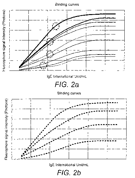
in a reference sample (see Figure 2A for an example of such a curve with lgE
containing
samples).
Step (v) of the first aspect provides for a comparison of these two graphs to
match the
curves produced for each antigen with the curves produced for each anti-
immunoglobulin antibody concentration. Such comparison may be carried out
visually or
io by some other means, such as with the use of computer software.
Once each antigen sample is matched with a particular anti-immunoglobulin
concentration that immunoglobulin concentration is assigned to that antigen
and is later
used as a more accurate reference, or calibration, curve for that antigen,
which more
accurately describes that particular antigen's binding capacity. Thus, the
concentration
of antibodies specific to that antigen in a test sample may be more accurately
elucidated
through the use of the newly assigned binding capacity calibration curve for
that antigen.
The inventors have identified that the use of a single reference curve for
calibrating
immunoassays for multiple antigens, particularly multiple allergens, as is
standard
practice in the art, is inadequate to describe the different binding
capacities that different
allergens exhibit. The inventors found that when immunoassays are performed on
serum samples with known IgE reactivity, incubated with different allergen
extracts,
different dose-response curves are obtained with the data generated for each
allergen.
This is exemplified in Figure 1. Thus, in the example of allergen testing, the
calibration
systems used in previous allergen immunoassays were inadequate for providing
accurate quantitative information for IgE reactivity of serum samples when
multiple
allergens are tested in a multiplex assay.
The inventors sought to provide a calibration system that would address the
disadvantages of the available immunoassays and provide a more accurate
quantitative
immunoassay. The step of assigning a different reference curve to each antigen
as in
the present invention, based on their binding capacity, enables more accurate
quantification of specific immunoglobulin in a test sample. In the example of
allergen
testing, the present invention describes allergen dose-response behaviour in a
more
accurate way than previous assays.
8
CA 02833654 2013-10-18
WO 2012/143709
PCT/GB2012/050846
The binding capacity curves may be loaded into software that controls
immunoassay
analysing instruments to be used as standards for future assays. Internal
controls on
each assay may be used to compensate for minor environmental variations in
each
assay. Nevertheless, it is envisaged that when new batches of allergen are
prepared for
loading onto a solid support, the binding capacity curves may be adjusted
appropriately
or new binding capacity curves produced, as taught herein, to ensure accuracy
of the
assay. Such quality control activities will be readily understood by the
skilled person.
Steps (vi) and (vii) of the first aspect utilise the binding capacity curves
generated in the
io earlier steps to quantify antigen-specific immunoglobulin levels in the
test sample. It is
envisaged that all of the immobilised components described in the first aspect
may be
immobilised on the same, or different solid supports, as required by the assay
equipment
that is utilised to carry out the method. Thus, all the appropriate
immobilised
components may be immobilised on the same solid support, for example chip,
including
antigens, capture immunoglobulins and positive and negative controls.
Nevertheless, it
is envisaged that each sample will be incubated with a different solid
support, for
example chip, to prevent cross-contamination. Thus, a number of solid
supports, for
example chips, will be used in each assay. For example, if 50 serum samples
are to be
incubated on the solid support, 50 separate solid supports with the
appropriate
immobilised antigens/immunoglobulins/controls may be used. Equally, each
reference
sample may be incubated on a different solid support to prevent cross
contamination.
The assay equipment utilised will influence the exact mechanics of incubation
of the
samples, as would be understood by a person of skill in the art.
In a preferred embodiment, the reference immunoglobulin sample (for example
the WHO
international standard IgE) is used at final immunoglobulin concentrations
ranging from
0.1 to 100 IU/ml. Preferably, the anti-immunoglobulin antibody to be
immobilised on the
solid support (for example anti-IgE antibody) is used at concentrations
ranging from 30 to
0.1 pg/ml.
The term "immunoglobulin(s)" is used herein interchangeably with the term
"antibody" or
"antibodies".
The first aspect may include a further step wherein the binding capacity
curves produced
in step (iv) are clustered into representative binding capacity curves to
represent different
levels of binding capacity, for example, very high binding, high binding,
medium binding
and low binding, and wherein the comparing in step (v) is carried out with
respect to the
9
CA 02833654 2013-10-18
WO 2012/143709
PCT/GB2012/050846
dose-response curves produced in step (ii) and the representative binding
capacity
curves, rather than the binding capacity curves produced in step (iv).
It is envisaged that including this further step of providing fewer
consolidated binding
capacity curves may simplify data management, thus simplifying the calibration
process.
These binding capacity curves, as exemplified in Figure 2B may be stored in
the
software of immunoassay analyser instruments for future analysis of samples.
By "anti-immunoglobulin antibodies, fragments or derivatives thereof' we
include the
io meaning that the antibodies comprise an antibody or antigen binding
fragment thereof
such a Fab-like molecules; Fv molecules; single-chain Fv (ScFv) molecules
where the VH
and VI_ partner domains are linked via a flexible oligopeptide and single
domain
antibodies (dAbs) comprising isolated V domains, but it may also be any other
ligand
which exhibits the preferential binding characteristic mentioned above.
In a second aspect, the invention provides a method of calibrating a device
suitable for
assaying binding of multiple antigen-specific immunoglobulins to multiple
antigens or
fragments thereof immobilised on a solid support, the method comprising the
steps of;
(i) assaying binding of a series of samples, for example serum samples,
containing immunoglobulin of known antigen reactivity to multiple recombinant
or purified
antigen components or fragments thereof immobilised on a first solid support,
(ii) comparing the level of binding in step (i) with the known reactivity to
produce a
dose response curve for each antigen component or fragment thereof,
(iii) assaying binding of a serial dilution of a reference immunoglobulin
sample of
the same subtype as that used in part (i) with a known total amount of
immunoglobulin to
a serial dilution of anti-immunoglobulin antibodies, fragments or derivatives
thereof,
immobilised on the first, or a second, solid support,
(iv) comparing the level of binding in step (iii) with the known total amount
of
reference immunoglobulin to produce a binding capacity curve for each anti-
immunoglobulin antibody, fragment or derivative dilution,
(v) comparing the dose response curves produced in step (ii) with the binding
capacity curves produced in step (iv), identifying the binding capacity curve
that most
closely matches the dose-response curve for each antigen or fragment thereof,
and
assigning a binding capacity curve to each antigen or fragment thereof on this
basis, and
(Vi) inputting the binding capacity curves generated in step (v) into the
device
such that the binding capacity curves for each antigen can be interpolated
with signals
CA 02833654 2013-10-18
WO 2012/143709
PCT/GB2012/050846
produced from samples containing unknown amounts of immunoglobulin that
specifically
binds that antigen.
The second aspect may include a further step wherein the binding capacity
curves
produced in step (iv) are clustered into representative binding capacity
curves to
represent different levels of binding capacity, for example, very high
binding, high
binding, medium binding and low binding, and wherein the comparing in step (v)
is
carried out with respect to the dose-response curves produced in step (ii) and
the
representative binding capacity curves, rather than the binding capacity
curves produced
io in step (iv).
Once such a device has been calibrated using the method of the second aspect,
it may
be utilised to assay and quantify antigen-specific immunoglobulin in test
samples.
For example, a microarray slide (solid support) following the appropriate
treatment to
visualise the antigens bound to antibody can be read using an ADAM instrument
(Microtest Matrices Ltd). Raw data are collected and used to calculate the
dose-
response curve. The output of the instrument is a textual file where all the
dots of the
microarray are listed; they are described by the coordinates and a numeric
value that is
the photons count emitted by each dot on the microarray. Using a numerical
computing
environment similar to Matlab, all the data obtained from the reader are
computed and a
set of factors that describe the dose-response curves are generated. The ADAM
instrument uses these factors to build the internal Master Calibration Curve,
inserted in a
configuration file.
In preferred embodiments of the first and second aspects, the antigens are
allergens, the
immunoglobulin is IgE and the anti-immunoglobulin antibodies are anti-IgE
antibodies.
Thus, in such embodiments, the methods may be used in the detection of
allergies in
patients to certain allergens by assaying samples obtained from the patient
for IgE
reactivity to the allergens components or fragments thereof immobilised on the
solid
support. Such information may aid in the diagnosis of allergy to particular
allergens.
Examples of allergens that may be immobilised on the solid support include
those listed
in Table 1.
Table 1: List of allergens
11
CA 0 2 8 3 3 65 4 2 0 1 3 ¨ 1 0 ¨ 1 8
WO 2012/143709
PCT/GB2012/050846
List of allergens
Drags: 2. F44 (Strawbeny) M2 (Cladorporium erbanon)
Cl (Penicillin G) F45 (Baker's yeast) M3
(Aspergillusfurnigatus)
C2 (Penicillin V) F46 (Pepper) M4 (Mucor racemosus)
C214 (Arnoxicillin F49 (Apple) M5 (Candida albicans)
NI* M6 (Alternaria semis)
:
DI (Dermatophagoides pteronyssinias) F74 (Hen's egg) M7
(Botrytis cinerea)
D2 (Dermatophagoides farinae) F76 (Alpha-Lactalbumin) M9 (Fusarium
monitiforme)
D3 (Dermatophagoides microceras) F77 (13-Lactoglobulin) MI3
(Phonics betae)
1)70 (Acarus siro) M20 (Mucor rnucedo)
D71 (Lepidog(yfus destructor) F83 (Chicken meat) Tree pellet*:
,
D72 (Tyrophagus putrescentiae) F84 (Kiwi) T2 (Alder)
D73 (Gbsciphagus domesticus) F85 (Celery) T3 (Birch pollen)
Animal epithelia F92 (Banana) 14 (Hazel)
. = ' es4k..
El (Cat hair) F95 (Peach) T5 (European beech)
E2 (Dog hair) =Grass pelvis T6 (Mountain cedar)
,
E3 (Horse hair) GI (Sweet vernal grass) T7 (Oak)
G2 (Bermuda grass/squitch) 1'9 (Olive)
E78 (Budgerigar feathers) G3 (Orchard grass) TI I (Plane)
E81 (Sheep epithelium) G4 (Meadow fescue) T14 (Poplar)
E82 (Rabbit epithelium) G5 (Ryegrass perennial) T901 (Ash)
Food slicriens ' ' = G6 (Timothy grass) T904 (Sallow)
Fl (Egg white) G8 (Bluegrass, June¨ 'Yfe.ert Pontik''
Kentucky)
F2 (Cow's milk) GI2 (Rye cultivated) WI (Ragweed common)
F3 (Cod) G14 (Oats cultivated) W6 (Mugwort)
F4 (Wheat flour) G15 (Wheat) W8 (Dandelion)
F7 (Oat flour) G18 (Barley) W9 (English plantain)
F8 (Corn flour) ==hisiets' ==. ' W20 (Stinging nettle)
F13 (Peanuts) II (Honeybee venom) W2I (Parietaria)
FI4 (Soybean) 13 (Wasp venom) W32 (Rape)
F16 (Walnut) Turifted pilot*
F17 (Hazelnut) 171 (Midge/Mosquito/Gnat) Bet v I
F23 (Shrimp) OcculiatIona!=alkrgens Phi p5 (66-V)
K81 (Ficus benjamina) Phl p I
F26 (Pork) K82 (Latex) Der p I (1)1-1)
F27 (Beef) K87 (Alpha amylase) Der p2 (1)1-11)
F31 (Carrot) K905 (HSA) Bet v 2
12
CA 02833654 2013-10-18
WO 2012/143709
PCT/GB2012/050846
F33 (Orange) Moulds Phi p 2
F35 (Potato) M1 (Penicillium notatun) Phi p6
In a third aspect, the invention provides a multi-allergen test system
comprising a serial
dilution of anti-IgE antibodies, fragments or derivatives thereof immobilised
on a solid
support. Such multi-allergen test system may be used in the methods of the
earlier
aspects of the invention.
In an embodiment of the third aspect, the system further comprises recombinant
or
purified allergen components or fragments thereof immobilised on the, or a
second, solid
support.
In a fourth aspect, the invention provides a kit of parts comprising the mufti-
allergen test
systems of the third aspect, and one or more of the following:
i) a reference IgE sample;
ii) a first antibody preparation comprising first antibodies that bind IgE;
iii) a second antibody preparation comprising second antibodies that
specifically
bind the first antibodies;
iv) a third antibody preparation comprising third antibodies that specifically
bind
the second antibodies; and
wherein either the second antibodies or the third antibodies are conjugated to
a
detectable marker.
In an embodiment of the fourth aspect, the detectable marker may be an enzyme,
for
example, Horseradish Peroxidase (HRP) or alkaline phosphatase, as would be
appreciated by a person of skill in the art. Appropriate substrates for HRP
include
chromogenic " substrates (e.g., 3,3',5,5'-
Tetramethylbenzidine (TMB), 3,3'-
Diaminobenzidine (DAB), and 2,2'-azino-bis(3-ethylbenzthiazoline-6-sulphonic
acid))
(ABTS) and chemiluminescent substrates (e.g., SuperSignala and ECL). A
particularly
preferred substrate is Alexa555 fluorophore labelled Tyramide.
In an alternative embodiment of the fourth aspect, the detectable marker may
be a
chemiluminescent moiety (e.g. an acridinium ester compound), a radioactive
moiety (e.g.
32P), or a fluorescent moiety (e.g. Fluorescein (FITC)). Other appropriate
detectable
labels and methods for their detection and their conjugation to antibodies
will be well
known to a person of skill in the art.
13
CA 02833654 2013-10-18
WO 2012/143709
PCT/GB2012/050846
In an embodiment of any aspect of the invention, the solid support may be a
microarray
chip. Appropriate microarray chips may be constructed as follows: protein
solutions (i.e.
allergens) are initially prepared by diluting a stock solution of a protein to
a final optimal
concentration, in an optimal buffer (determined previously, see above). For
each
individual antigen, the final concentration may differ, as would be understood
by a person
of skill in the art. Protein solutions are then loaded into a 384-well plate.
The plate and
the solid support are then put inside a printer, for example a non-contact
piezo-electric
printer. The printer possesses a number of nozzles that draw the solutions
from the wells
and then dispenses them in drops onto the solid substrate (microarray). After
dispensing
io each solution, the nozzles are then washed and made ready for the next
solution. The
printer has a camera, called a stroboscope, which monitors whether the
solutions are
properly dispensed by taking pictures of the drops being dispensed. If a
solution is not
dispensed properly, the stroboscope reports this. Any suitable optical support
may be
used to prepare the microarray. Generally, any glass support, or similar will
be
adequate. Various such supports will be well known to the skilled person.
Embodiments of the invention will now be described, by way of example only
with
reference to the Figures in which:
Figure 1 is a graphical representation of multiple allergen dose-response
curves;
Figure 2A is a graphical representation of multiple binding-capacity curves;
and
Figure 2B is a graphical representation of consolidated binding-capacity
curves
according to the invention.
Example 1: Immunoassay with binding capacity calibration system.
Described is a calibration system suitable for precisely quantifying serum
allergen-
specific IgE, using a microarray-based immunoassay as a platform. The
described
immunoassay contains approximately 100 different allergenic extracts that
cover a panel
of approximately 100 different allergies.
The described calibration system can reliably describe the dose-response
behaviour of
all 100 allergen extracts. Each allergen extract is a unique compound with a
different IgE
binding capacity, i.e. different dose-response steepness. The present
calibration system
takes account of these different binding capacities to provide an accurate
system for
measuring allergen-specific IgE levels in a sample.
14
CA 02833654 2013-10-18
WO 2012/143709
PCT/GB2012/050846
Example microarray chip
The herein described system is a microarray-based test using miniaturized
immunoassays designed for the measurement of up to approximately 103
allergens.
Allergen extracts are immobilized onto chemically activated glass slides to
generate the
arrays. Each natural allergen extract is spotted onto the microarray in its
optimal protein
concentration and buffer (previously selected). Additionally, the microarray
comprises
positive controls (e.g. goat anti-mouse IgG) and negative controls (e.g. non-
specific
protein, such as bovine serum albumin), and capture anti-human IgE (polyclonal
goat
io anti-human IgE) spotted in serial dilutions.
Example antibody visualisation protocol
The following is an example of a protocol that may be used to visualise
binding of IgE,
either in a serum sample or a reference sample (the assays are carried out at
the same
time, with the same reagents where appropriate, such that potential
environmental
variations are controlled for), to the herein described microarray:
Separate arrays are first incubated with IgE samples (either serum or
reference) and
subsequently with monoclonal anti-human (or other appropriate antibody,
depending on
the assay samples) IgE antibody (for example, anti-human IgE mouse IgG), which
will
bind the human IgE from the serum or reference sample, if IgE is present and
bound to
the spotted allergens. Then a goat polyclonal anti-mouse IgG antibody
conjugated with
Horseradish peroxidase (HRP) is added to the array, followed by Alexa555
fluorophore
labelled Tyramide. Appropriate washing steps are carried out between each
antibody
incubation step.
In the presence of hydrogen peroxide (H202), HRP enzyme converts Tyramide-
A1exa555
into highly reactive, short-lived tyramide-Alexa555 radicals that react with
nucleophilic
residues in the vicinity of the HRP-target interaction site. This produces an
emission of
fluorescence at a specific wavelength (555 nm) of intensity proportional to
the amount of
bound HRP enzyme.
Use of the polyclonal antibody and of the HRP-Tyramide system (a non-liner
signal
amplification system) greatly increases the sensitivity of the microarray
immunoassay
test.
CA 02833654 2013-10-18
WO 2012/143709
PCT/GB2012/050846
The above protocol provides a fluorescence intensity for each allergen spot
that is
plotted on a graph with serum sample concentration to provide a curve that is
interpolated with a reference curve to quantify IgE level.
Calibration method of invention
The herein described calibration method is exemplified by the following steps
using the
microarray chip of the invention:
1. Identification of dose-response curve for each allergen. A number of serum
samples
with known IgE reactivity are tested on the microarray chip. The signal
intensity obtained
from the allergens is collected and used to generate allergen dose-response
curves.
2. Production of a panel of binding capacity curves. Serial dilutions of
WHO/NIBSC
International Reference IgE Preparation (from 0.1 to 100 International
Unit/ml) are
incubated onto the chips. IgEs are bound by the spotted capture-anti-human IgE
and the
signal intensity generated is measured and used to build a panel of binding
capacity
curves. Each curve corresponds to one of the different concentrations of
capture anti-
human IgE and is obtained by plotting the WHO/NIBSC IgE concentrations used
for the
incubation of the chip versus the corresponding obtained signal intensity. A
number of
binding curves is produced according to the number of different spots of
capture anti-
human IgE present on the chip (see Figure 2A, where each curve corresponds to
one of
the different concentrations of capture anti-human IgE spotted onto the arrays
and was
obtained by plotting the WHO/N1BSC IgE concentrations versus the
correspondingly
obtained signal intensity).
3. Clustering of binding capacity curves. Binding-capacity curves are then
clustered in
groups to represent different binding capacity (for example, Very high, High,
Medium and
Low binding capacity). A regression curve is produced for each group and
stored in the
software of the analyzer instrument (see Figure 2B, which shows binding
capacity curves
clustered into groups).
4. Allergens assigned binding capacity curve. According to the slope and shape
of the
allergen dose-response curves obtained as described in point 1, one of the
binding-
capacity curves (Master Curves) is assigned to each allergen.
16
CA 02833654 2013-10-18
WO 2012/143709
PCT/GB2012/050846
5. Quantification of allergen-specific IgE. Using this calibration system,
allergen-specific
IgEs of an unknown patient serum are measured by interpolation of the signal
intensity
obtained from a particular allergen spotted onto the microarray chip to the
specifically
assigned binding capacity curve. Allergen reactivity is expressed in
International Unit/ml
and/or Class Score.
6. Using internal controls (adjuster) in each chip, the system can build the
dose-response
Internal Curve taking into account the storage and environment conditions of
the slide
adjusting the Master Curves obtained at point No. 4 accordingly. The internal
calibration
io consists of running an algorithm to move the Master Calibration Curve
based on the
signal of the adjusters. For example, if the signal of the adjuster, whose
expected value
is 1000 units gives 950, the algorithm may lead to a shift in the Master
Calibration Curve
of 5%.
Unlike typical allergen immunoassays, which use a single calibration curve,
the
immunoassay system of the invention takes into account the differences in the
binding
capacity of each allergen. This provides a much more accurate assay for
quantification
of allergen-specific IgE in a sample.
17