Sélection de la langue

Search

Sommaire du brevet 2833782 

Énoncé de désistement de responsabilité concernant l'information provenant de tiers

Une partie des informations de ce site Web a été fournie par des sources externes. Le gouvernement du Canada n'assume aucune responsabilité concernant la précision, l'actualité ou la fiabilité des informations fournies par les sources externes. Les utilisateurs qui désirent employer cette information devraient consulter directement la source des informations. Le contenu fourni par les sources externes n'est pas assujetti aux exigences sur les langues officielles, la protection des renseignements personnels et l'accessibilité.

Disponibilité de l'Abrégé et des Revendications

L'apparition de différences dans le texte et l'image des Revendications et de l'Abrégé dépend du moment auquel le document est publié. Les textes des Revendications et de l'Abrégé sont affichés :

  • lorsque la demande peut être examinée par le public;
  • lorsque le brevet est émis (délivrance).
(12) Brevet: (11) CA 2833782
(54) Titre français: TRANSFERT D'EMBRYONS
(54) Titre anglais: EMBRYO TRANSFER
Statut: Périmé et au-delà du délai pour l’annulation
Données bibliographiques
Abrégés

Abrégé français

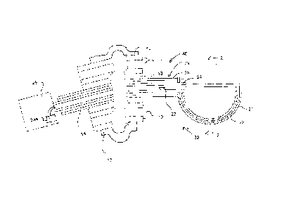
La présente invention concerne le transfert d'un embryon d'un animal femelle dans un autre animal en déterminant la présence d'un embryon dans l'utérus d'un animal donneur par imagerie ultrasonore et insertion d'un endoscope par voie vaginale dans l'utérus à un emplacement adjacent à l'embryon. Un outil de l'endoscope se projette dans une position pour extraire l'embryon lavé dans un récipient de l'outil qui est alors fermé par déplacement d'une partie de fermeture pour enceindre l'embryon et extraire l'endoscope afin de retirer l'embryon et de le transférer à un animal receveur. La pression du fluide dans le récipient peut être surveillée pour maintenir une pression requise correspondant généralement à celle rencontrée à l'intérieur de l'utérus.


Abrégé anglais


An embryo from a female animal is transferred to another animal by
determining presence of an embryo in the uterus of a donor animal by ultra-
sonic
imaging and inserting an endoscope vaginally into the uterus to a location
adjacent
the embryo. A tool of the endoscope projects to a position to extract the
embryo
washed into a container of the tool which is then closed by moving a closure
part to
enclose the embryo and extracting the endoscope to remove the embryo for
transfer
to a recipient animal. The fluid into the container can be controlled in
pressure to
maintain a required pressure generally matching that inside the uterus.

Revendications

Note : Les revendications sont présentées dans la langue officielle dans laquelle elles ont été soumises.


19
CLAIMS:
1. A method for raising a mammalian animal comprising:
transferring an embryo from a uterus of a female mammalian donor
animal in pregnancy to a uterus of a female mammalian recipient animal;
and raising the embryo to full term in the recipient animal;
wherein the embryo is transferred by steps comprising:
providing an ultrasonic imaging system and an endoscope
having a camera;
waiting in the pregnancy of the donor animal until the embryo is
observable in an ultrasonic image taken by the ultrasonic imaging system of
the
uterus of the donor animal and is visible in an image generated by the camera
of the
endoscope of the uterus of the donor animal;
determining a presence and location of the embryo on a wall of
the uterus of the donor animal from said ultra-sonic image;
inserting a remote end of the endoscope vaginally into the
uterus of the donor animal to a location adjacent the embryo;
the remote end of the endoscope carrying a scoop member;
operating the scoop member to cause the embryo to enter the
scoop member from the wall of the uterus;
the remote end of the endoscope carrying an enclosing member
for covering the scoop member to enclose the embryo;
operating the enclosing member to enclose the embryo within

20
the scoop member after the embryo has been scooped up into the scoop member;
extracting the endoscope to remove the embryo in the scoop
member from the uterus of the donor animal;
and transferring the embryo to the uterus of the recipient animal.
2. The method according to claim 1 wherein operating movement
of the scoop member includes rotation to scoop up the embryo from the wall of
the
uterus.
3. The method according to claim 1 or 2 wherein the scoop
member and the enclosing member are moved to a fully closed position.
4. The method according to any one of claims 1 to 3 wherein the
scoop member and the enclosing member comprise two hemi-spherical parts which
are rotated from a first open position with one inside the other to a closed
spherical
position.
5. The method according to any one of claims 1 to 4 including
supplying fluid to the scoop member through a fluid supply duct.
6. The method according to any one of claims 1 to 5 wherein the
scoop member has a transverse dimension of at least 1.0 cm.
7. The method according to any one of claims 1 to 6 wherein the
presence of the embryo is detected at a time period in the range 9-13 days
after
insemination.
8. A method for raising a mammalian animal comprising:
transferring an embryo from a uterus of a female mammalian donor

21
animal in pregnancy to a uterus of a female mammalian recipient animal;
and raising the embryo to full term in the recipient animal;
wherein the embryo is transferred by steps comprising:
providing an endoscope having a camera;
determining a presence and location of the embryo on a wall of
the uterus of the donor animal;
inserting a remote end of the endoscope vaginally into the
uterus of the donor animal to a location adjacent the embryo on the wall of
the
uterus of the donor animal;
operating the endoscope to move the remote end to a position
within the uterus adjacent the embryo on the wall;
the remote end of the endoscope including an elongate carrying
member projecting outwardly from the remote end so as to extend toward the
wall;
the elongate carrying member carrying a scoop member forming
a bowl;
the elongate carrying member being movable in rotation around
an axis longitudinally of the elongate carrying member;
operating the scoop member including rotation to scoop up the
embryo from the wall of the uterus;
providing an enclosing member for covering the scoop member
to enclose the embryo;
operating the enclosing member to enclose the embryo within

22
the scoop member after the embryo is scooped up;
extracting the endoscope to remove the embryo in the scoop
member from the uterus of the donor animal;
and transferring the embryo to the uterus of the recipient animal.
9. An apparatus for transferring an embryo from a uterus of a
female donor mammalian animal in pregnancy to a uterus of a female recipient
mammalian animal and raising the embryo to full term in the recipient animal,
the
apparatus comprising:
an endoscope having a camera;
the endoscope having a remote end of the endoscope arranged to be
inserted vaginally into the uterus of the donor animal to a location adjacent
the
embryo on the wall of the uterus of the donor animal;
the remote end of the endoscope being operable to move the remote
end to different positions within the uterus;
an elongate carrying member extending from the remote end of the
endoscope;
a scoop member forming a bowl carried on the elongate carrying
member;
the elongate carrying member being movable in rotation around an
axis longitudinally of the elongate carrying member for operating the scoop
member
including rotation to scoop up the embryo from the wall of the uterus;
and an enclosing member operable for covering the scoop member to

23
enclose the embryo after the embryo is scooped up.
10. The apparatus according to claim 9 wherein there is provided a
fluid supply duct for supplying fluid to the scoop member.
11. The apparatus according to claim 10 wherein the fluid supply
duct opens into the scoop member.
12. The apparatus according to claim 10 or 11 wherein there is
provided a pressure sensor for controlling pressure of the fluid inside the
container.
13. The apparatus according to any one of claims 9 to 12 wherein
the scoop member has a transverse dimension of at least 1.0 cm.
14. An apparatus for transferring an embryo from a uterus of a
female donor mammalian animal in pregnancy to a uterus of a female recipient
mammalian animal and raising the embryo to full term in the recipient animal,
the
apparatus comprising:
an endoscope having a camera;
the endoscope having a remote end of the endoscope arranged to be
inserted vaginally into the uterus of the donor animal to a location adjacent
the
embryo on the wall of the uterus of the donor animal;
the remote end of the endoscope being operable to move the remote
end to different positions within the uterus;
an elongate carrying member extending outwardly from the remote end
of the endoscope;
a scoop member carried on the elongate carrying member to scoop up

24
the embryo from the wall of the uterus;
and an enclosing member operable by relative movement of the
enclosing member and the scoop member in a direction longitudinally of the
elongate carrying member to enclose the embryo after the embryo is scooped up.
15. The apparatus according to claim 14 wherein there is provided a
fluid supply duct for supplying fluid to the scoop member.
16. The apparatus according to claim 15 wherein the fluid supply
duct opens into the scoop member.
17. The apparatus according to claim 15 or 16 wherein there is
provided a pressure sensor for controlling pressure of the fluid inside the
container.
18. The apparatus according to any one of claims 14 to 17 wherein
the scoop member has a transverse dimension of at least 1.0 cm.

Description

Note : Les descriptions sont présentées dans la langue officielle dans laquelle elles ont été soumises.


CA 02833782 2016-04-11
EMBRYO TRANSFER
This invention relates to an apparatus for embryo transfer from one
female mammal to another. The description hereinafter primarily relates to
mares
where the commercial operation of transfer methods is desirable, but
ineffective; but
can relate to any female mammal.
BACKGROUND OF THE INVENTION
Embryo transfer (ET) is the process of harvesting an embryo or
embryos from a donor and transferring it to a recipient. The process can be
clone
surgically or non-surgically with the latter being the preferred method in
bovine and
equine species and the former being the preferred technique in smaller species
such
as porcine, ovine, caprine and canine.
Application for ET in the mare is commercially done for three main
reasons; a donor mare in competition can produce and transfer an embryo and
still
compete while a recipient mare carries her genetics to a term pregnancy, to
produce
multiple pregnancies in one year, to transfer to a recipient mare when the
donor
mare is considered a high risk for pregnancy complications.
In cattle superovulation is a successful procedure where with hormone
therapy multiple (average 6 but numbers as high as 40 reported) embryos can be
harvested (flushed) in any one procedure. Unfortunately mares do not respond
successfully to superovulation so a single procedure yields at best only one
embryo.
The exception is when a mare naturally double ovulates and thus two potential

CA 02833782 2016-04-11
2
embryos might be retrieved. The average success of achieving a pregnancy
through embryo transfer in the mare is 25%. The average cost per attempt is
from
$7000 to $12000 and the industry reports embryo transfer in mares to be
approximately $250 000 000 annually worldwide. This means that $187 500 000 is
spent with no results. This poor success has hindered the process of embryo
transfer in mares from becoming a more main stream procedure.
The traditional method for transferring an embryo in mares is to
aseptically pass a catheter through the vulva, vagina and cervix and into the
uterine
body. A cuff is inflated to seal the cervical uterine junction. The uterus is
flushed
with approximately 4 liters of specialized solution. The solution is filtered
through a
20micron filter. The filter is emptied into petri dishes and then the dishes
are
searched with microscopy to find an embryo. If found the embryo is isolated
and
washed in another specialized solution and then loaded into a transfer
pipette. The
recipient mare is aseptically prepared for transfer and the transfer pipette
is passed
through the vulva, vagina and cervix and into the uterine body where the
embryo is
deposited.
Special concern for timing is required for a successful pregnancy from
an ET procedure. The procedure is considered to start at day 0 which is when
the
mare is observed to have ovulated. The sperm must be present in the fallopian
tubes prior to ovulation. Fertilization takes place in the fallopian tubes
shortly after
ovulation and the embryo remains there for 5 days after which time it moves
into the
uterus. Flushing or retrieving the embryo is normally done at day 7 which
allows for

CA 02833782 2016-04-11
3
adequate time for the embryo to reach the uterus for it cannot be retrieved
from the
fallopian tube. After day 8 the embryo hatches from its protective shell
called the
zona pellucita which then makes the embryo more fragile to handle. So 7 days
post
ovulation achieves the highest success rates thus far. A uterus is dynamic and
changes through the female cycle. For this reason a recipient mare must be
synchronized with the donor mare and her uterus must be close to 7 days post
ovulation which adds another level of difficulty to the procedure. At the time
a flush
is performed it is unknown whether there is a viable embryo present or not.
Attempts have been made to transfer embryos at later stages of
development such as 11 to 14 days post ovulation. At 11 days post ovulation
the
embryo is visible to a highly trained practitioner using ultra-sonography.
This would
seem to be ideal as retrieval would only be attempted if there was a pregnancy
visualized. Unfortunately no success has been achieved at this stage. It is
hypothesised that the embryos were too fragile and didn't survive the
procedure.
SUMMARY OF THE INVENTION
It is one object of the invention to provide an improved apparatus for
use in embryo transfer.
According to one aspect of the invention there is provided 1. A
method for raising a mammalian animal comprising:
transferring an embryo from a uterus of a female mammalian donor
animal in pregnancy to a uterus of a female mammalian recipient animal;
and raising the embryo to full term in the recipient animal;

CA 02833782 2016-04-11
4
wherein the embryo is transferred by steps comprising:
providing an ultrasonic imaging system and an endoscope
having a camera;
waiting in the pregnancy of the donor animal until the embryo is
observable in an ultrasonic image taken by the ultrasonic imaging system of
the
uterus of the donor animal and is visible in an image generated by the camera
of the
endoscope of the uterus of the donor animal;
determining a presence and location of the embryo on a wall of
the uterus of the donor animal from said ultra-sonic image;
inserting a remote end of the endoscope vaginally into the
uterus of the donor animal to a location adjacent the embryo;
the remote end of the endoscope carrying a scoop member;
operating the scoop member to cause the embryo to enter the
scoop member from the wall of the uterus;
the remote end of the endoscope carrying an enclosing member
for covering the scoop member to enclose the embryo;
operating the enclosing member to enclose the embryo within
the scoop member after the embryo has been scooped up into the scoop member;
extracting the endoscope to remove the embryo in the scoop
member from the uterus of the donor animal;
and transferring the embryo to the uterus of the recipient animal.
This is preferably done by providing two hemi-spherical parts which

CA 02833782 2016-04-11
rotate, or one of which rotates relative to the other from a first open
position with one
cupped inside the other to a closed spherical position sealing around the
edges of
the two parts. Other closure systems can be used for example a sliding sleeve
arrangement around an inner tube which has a hole to collect the embryo.
5 Optionally there is provided a fluid supply duct for supplying
fluid to the
tool where the fluid supply duct opens into the closed container. In this
case, there
can be provided a pressure sensor for controlling pressure of the fluid inside
the
container to match that in the uterus. Pressure control may or may not be
necessary
within the closed scoop.
Preferably the container has a transverse dimension of at least 1cm
and preferably of the order of 1.5 cm.
Preferably the presence of the embryo is detected at a time period of
the order of 11 days after insemination.
According to a second aspect, the invention provides a method for
raising a mammalian animal comprising:
transferring an embryo from a uterus of a female donor mammalian
animal in pregnancy to a uterus of a female recipient mammalian animal;
and raising the embryo to full term in the recipient animal;
wherein the embryo is transferred by steps comprising:
providing an endoscope having a camera;
determining a presence and location of the embryo on a wall of

CA 02833782 2016-04-11
6
the uterus of the donor animal;
inserting a remote end of the endoscope vaginally into the
uterus of the donor animal to a location adjacent the embryo on the wall of
the
uterus of the donor animal;
operating the endoscope to move the remote end to a position
within the uterus adjacent the embryo on the wall;
the remote end of the endoscope including an elongate carrying
member projecting outwardly from the remote end so as to extend toward the
wall;
the elongate carrying member carrying a scoop member forming
a bowl;
the elongate carrying member being movable in rotation around
an axis longitudinally of the elongate carrying member;
operating the scoop member including rotation to scoop up the
embryo from the wall of the uterus;
providing an enclosing member for covering the scoop member
to enclose the embryo;
operating the enclosing member to enclose the embryo within
the scoop member after the embryo is scooped up;
extracting the endoscope to remove the embryo in the scoop
member from the uterus of the donor animal;
and transferring the embryo to the uterus of the recipient animal.
According to a third aspect, the invention provides an apparatus for

CA 02833782 2016-04-11
7
transferring an embryo from a uterus of a female donor mammalian animal in
pregnancy to a uterus of a female recipient mammalian animal and raising the
embryo to full term in the recipient animal, the apparatus comprising:
an endoscope having a camera;
the endoscope having a remote end of the endoscope arranged to be
inserted vaginally into the uterus of the donor animal to a location adjacent
the
embryo on the wall of the uterus of the donor animal;
the remote end of the endoscope being operable to move the remote
end to different positions within the uterus;
an elongate carrying member extending from the remote end of the
endoscope;
a scoop member forming a bowl carried on the elongate carrying
member;
the elongate carrying member being movable in rotation around an
axis longitudinally of the elongate carrying member for operating the scoop
member
including rotation to scoop up the embryo from the wall of the uterus;
and an enclosing member operable for covering the scoop member to
enclose the embryo after the embryo is scooped up.
According to a fourth aspect, the invention provides an apparatus for
transferring an embryo from a uterus of a female donor mammalian animal in
pregnancy to a uterus of a female recipient mammalian animal and raising the
embryo to full term in the recipient animal, the apparatus comprising:

CA 02833782 2016-04-11
8
an endoscope having a camera;
the endoscope having a remote end of the endoscope arranged to be
inserted vaginally into the uterus of the donor animal to a location adjacent
the
embryo on the wall of the uterus of the donor animal;
the remote end of the endoscope being operable to move the remote
end to different positions within the uterus;
an elongate carrying member extending outwardly from the remote end
of the endoscope;
a scoop member carried on the elongate carrying member to scoop up
the embryo from the wall of the uterus;
and an enclosing member operable by relative movement of the
enclosing member and the scoop member in a direction longitudinally of the
elongate carrying member to enclose the embryo after the embryo is scooped up.
The apparatus can have the further features defined above and may
be associated with an ultra sound imaging system for detecting and locating
the
embryo.
Based on the augmentation and refinement to the method of embryo
described herein, retrieval at 9 to 13 days and more preferably 11 or 12 days
can
achieve success rates in excess of 80%.

CA 02833782 2016-04-11
9
In the equine uterus a cascade of events begins in which, if no embryo
is present, the uterus undergoes changes and the reproductive system starts
the
process toward ovulation. If a viable embryo is present in the uterus it
blocks the
hormone and chemical pathways that initiate the cascade to ovulation thus
pregnancy is maintained. At the time of transfer on day 11 or 12 the
recipient's
reproductive track is already undergoing changes that may make it unable to
maintain a pregnancy.
In the method of using the present invention, the recipient and the
donor both are bred at the same time and an embryo is removed from the
recipient
mare and exchanged for an embryo from the donor mare. The delicate nature of
the
embryo is a very important consideration for success of this procedure so
highly
specialized equipment has been designed to overcome this.
Using this modified embryo transfer apparatus the procedure is cost
effective and is a more attractive arrangement of breeding in the equine
industry.
BRIEF DESCRIPTION OF THE DRAWINGS
One embodiment of the invention will now be described in conjunction
with the accompanying drawings in which:
Figure 1 is a vertical cross sectional view through a tool for use in an
endoscope for extraction of an embryo, showing the tool in an initial open
position.
Figure 2 is a cross-sectional view along the lines 2-2 of Figure 1.
Figure 2A is a cross-sectional view along the lines 2-2 of Figure 1
showing the tool in the closed position after collection of an embryo.

CA 02833782 2016-04-11
Figures 3 and 4 show the operation of the tool.
Figure 5 is a vertical cross sectional view through a second
embodiment of tool for use in an endoscope for extraction of an embryo,
showing
the tool in an initial open position for insertion through the tube of the
endoscope.
5 Figure 6 is a top plan view through of the tool of Figure 5 in the
closed
position after insertion through the tube of the endoscope and collection of
the
embryo.
In the drawings like characters of reference indicate corresponding
parts in the different figures.
10 DETAILED DESCRIPTION
Endoscopes are a well known device widely used in surgery and other
procedures and comprise a tube with a camera and illumination which can be
passed through an opening into the interior of the body and which can be
manipulated to different positions. A central bore allows a tool to be passed
through
the tube for acting on the interior, many different tools are available.
Typically a
collar is provided which can be inflated to locate the tube at a required
portion and to
seal the opening relative to the tube. Arrangements of this type are widely
used and
well known to persons skilled in the art so that further details are not
required.
Figure 1 shows a recovery tool for use as part of a modified endoscope
40 including a tube 10 with a camera lens 11 and an illumination source 12
carried
on the tube and including fiber optic communication from a distal end 10A of
the
tube 10 to control systems of the endoscope and the near end for operation by
the

CA 02833782 2016-04-11
11
user. The tube 10 is arranged so that it can be passed through an opening, in
this
case the vagina into the interior of the body. The tube includes components
(not
shown) which allow the end 10A to be manipulated to different positions. One
or
more central bores 13 or Ports allow a tool 14 to be passed through the tube.
Typically a collar 15 is provided which can be inflated to locate the tube at
a required
position and to seal the opening, in this case the uterus, relative to the
tube.
Through one of the working ports 13 of the endoscope is inserted a
grasping tool 30. The grasping tool is small globe 20, approximately 1.5 cm in
diameter. The globe has two hemispherical halves 21 and 22 one of which
rotates
by sliding slides inside the other. The components are made out of surgical
grade
stainless steel.
When closed as shown in Figure 2 the two parts 21 and 22 form a
sealed unit or globe 20 with a sealing edge 23. The inner part 22 is rotated
around
the axis of the sphere by an operating element 24 at the end of the tube 10
which is
operated by a control at the near end of the tube from the open position where
the
inner part is wholly within the outer part to form a hemi-spherical scoop to a
closed
position in which the globe is closed and sealed. This globe also has a fluid
port 25
within it so fluid can be added or withdrawn from the globe 20. The fluid
supply
system 26A of a control unit 26 for supply to the port 25 passes through the
tube 14
and includes an inline pressure sensor 27 sensitive to the internal pressure
in the
line and therefore within the closed globe 20. The supply 26A of the control
unit 26
can be operated so that holding fluid can be supplied or removed to adjust the

CA 02833782 2016-04-11
12
internal pressure in the globe 20 to match the same pressure as that of a
normal
uterine environment for that stage of embryo. The pressure within the uterus
can be
measured in situ or can be predetermined from historical measurements.
The special tool described above can in some embodiments be used
with a stock endoscope. The tool can alternatively be a permanent "biopsy
tool"
which is manufactured by assembly into place in an endoscope from a typical
supplier, but where the tool is not be able to be removed after it is
manufactured into
the scope. This is due to the fact that the typical globe is too large for the
portal
through the tube of a typical endoscope.
The tool operates in a similar manner to an ice cream scoop. In the
open position one half of the globe 20 is rotated inside the other half as
shown in
Figure 2A. The neck 29 of the tool that passes through the endoscope portal 13
is
formed from two flexible tubes 25 and 24 one inside the other. The outer tube
25 is
fixed to the bottom half 21 of the globe and the inner tube 24 is fixed to the
top half
of the globe. At the operator end the user operates the device 26 by
activating a
turning movement to the inner tube 24 so that it rotates the top half of the
globe to
close it. The inner tube 24 also provides the fluid port which is optional.
When the tool is first inserted as shown in Figure 4 in the open position
and the collar 15 inflated to hold the tool in place, once the tool is passed
into visual
proximity of the embryo it can be used to pick up the embryo.
Endoscopes have the ability to pass fluid through the port 13 or
through a separate special port (not shown) to dilate the inside of the tube
14 or

CA 02833782 2016-04-11
13
open the lumen of the uterus.
Thus most endoscopes have a small port adjacent to the lens. This
port typically has a very small metal deflector that directs water across the
lens to
clean it should it become obscured with mucus or other debris. The air
required can
also be passed through this port. There is a pump on the power unit that works
the
scope. At the operator end there is a two stage valve that is normally worked
by the
index finger. With light depression air is pumped through the port adjacent to
the
lens that is normally used for insufflation to allow for dilation which
enhances
passage of an endoscope. If the valve is fully depressed fluid is pumped
through to
clean the lens.
This fluid supply through the endoscope is used to open the uterus and
to infuse a small amount of fluid into the uterus to float the embryo away
from
the tissue of the uterus wall so that it can be simply picked up with the
scoop. Air or
air and fluid may be used to insufflate the uterus to allow for better
visualization and
pull the majority of the endometrium away from the embryo. Fluid may then be
used
to completely free the embryo or the tool can be used to pick up the embryo at
that
point, if its positioning is good and endometrial contact is minimal.
Once the embryo is in view the cuff 15 will be inflated so that if further
fluid is infused the embryo will not float away. When insufflation is normally
done
there is constant loss of air along the outside of the scope but once the
embryo is in
view, the cuff 15 is inflated so constant insufflation is no longer needed and
dilation
of the uterus can be static.

CA 02833782 2016-04-11
14
It is necessary to control the supply and volume of fluid to prevent the
embryo from floating too far away. In normal instances, because the pressure
in the
inflation collar 15 is kept low, the natural closure/collapse of the tissue of
the uterus
around the collar and the tool keeps a partial seal around the instrument and
provides a slope running away from the collar 15 to prevent the embryo from
falling
into the area of the collar 15 where it become impossible to retrieve. The
injection of
fluid through the endoscope typically is required because of the fragile and
movable
nature of the embryo. In Figure 4, the inflation collar 15 is close to the end
of the
endoscope at the location of the tool since this better locates the tube 14
and allows
better control over movement of the tool. The third fluid supply tube 25 is
optional
but when provided acts to bathe the embryo.
When the embryo has been picked up, the tool is retracted from the
donor animal and moved to the recipient. Once the embryo is placed in the
recipient and the globe re-opened to release the embryo, fluid can be infused
into
the bottom of the globe and the embryo floated out.
The complete procedure using the apparatus herein is as follows:
1. The donor mare is synchronized in her estrous cycle with
recipient mare sufficiently that they are in synchronism; or the recipient can
be as
much as 24 hours ahead or 72 hours but preferably not more than 48 hours
behind
the donor mare in her ovulation.
2. Both mares are bred on their synchronized ovulation as per
normal breeding methods.

CA 02833782 2016-04-11
3. At earliest possible time post ovulation an embryo is searched
for via ultrasonography in both the donor and recipient mares. Currently this
is
carried out at day 11 post ovulation when the embryo is sufficiently large to
be
determined.
5 4. Once pregnancy is confirmed by the ultra-sound image in
both
the donor and recipient the embryo transfer and exchanged is commenced.
5. The donor and recipient mares are prepared pre-embryo
recovery for a normal aseptic embryo recovery technique. Ideally the recipient
mare
is pregnant but that is not absolutely necessary. This transfer can still be
attempted
10 if the recipient is not pregnant but still in synchrony with the donor.
6. The procedure starts with the recipient where the recipient is
sedated for ease of recovery and transfer.
7. In the recipient, a first technician operates the ultrasound
imaging system to locate and document where the embryo is residing.
15 8. A second technician passes the recovery scope vaginally
using
normal aseptic palmed delivery to the cervix and the cervix is digitally
enlarged and
the scope is then advanced through the cervix and the operators hand is
removed.
The scope is then advanced until it appears on the ultrasound adjacent to the
embryo. The ultrasonographer may or may not stop at this time. One the embryo
is
found via ultrasound the ultrasound is removed and the perineum washed
thoroughly and the scope is passed into position.
9. Once the scope is in view with the embryo, the uterine horn is

CA 02833782 2016-04-11
16
insufflated by air or air and fluid supply enough to free the majority of
endometrial
contact with the embryo. The uterus is infused with the fluid through the
supply tube
25 with a fluid, such as a commercially available embryo recovery medium, to
float
the embryo. The inflation cuff 15 on the end of the recovery scope is arranged
to
prevent washing the embryo away. The embryo is captured with the grasping tool
20 on the recovery scope. The grasping tool 20 on the working end 10A of the
recovery scope 10 is now a closed and is infused with the commercially
available
embryo holding fluid. The recovery scope is withdrawn from the uterus. From
the
recipient animal, the embryo is discarded or kept for other purposes.
10. The step 9 is repeated with the donor mare.
11. The recovery scope is washed with warmed alcohol and then 1
liter of warmed saline
12. The recipient mare is sedated again if necessary and her
perineum washed again.
13. The recovery scope, now containing the donor's embryo is
passed using normal aseptic palmed delivery to the cervix and the cervix is
digitally
enlarged
14. The recovery scope is
then advanced to the location from where
recipients own embryo was removed. The embryo is deposited in the uterus at
the
bifurcation of the uterine horns. The grasping tool 20 is opened and the
embryo is
either dumped by turning the whole tool by the base tube 29 to invert the cup
or
expelled with fluid. The recovery scope is withdrawn and the procedure is
complete.

CA 02833782 2016-04-11
17
The mare is checked via ultra sound immediately after the procedure
for embryo placement. The mare is checked by ultrasound imaging at 6 and 24
and
48 hours post-transplant for embryo viability and procedure success.
Turning now to Figures 5 and 6, a second embodiment of the tool 50 is
shown which is formed of an outer sleeve 51 slidable on the outer surface of a
tube
52 inside the sleeve 51. The tube 52 has an end portion 52A projecting beyond
an
end of the sleeve 51 which connects to a cylindrical stainless steel tip
member 54.
The tip member is typically 2 to 3 cms long and has an elongate slot 53 in one
side
leading to a hollow interior 53A. The tip member includes a portion 53B which
is
necked down to a reduced diameter onto which the end portion 52A of the tube
52 is
engaged. The hollow interior 53A of the tip member 54 communicates
with the
interior of the tube 52 allowing access to the interior of the tube 52. The
tube 52 and
the sleeve 51 are both formed of a medical grade plastics material allowing
some
flexibility. The stainless steel tip member 54 has an end closure portion 54A
which
= 15 closes the front end of the tip member. The metal tip member thus has
a hole in the
hollow interior that communicates with the slot 53 so that fluid can be passed
from
the inner tube 52 into the slot 53. The outer face 54B of the closure portion
54A is
domed or hemi-spherical to provide a smooth rounded surface of a transverse
diameter or the order of 1.0 cms. The diameter of the tip member is lightly
larger
than that of the outer surface of the cylindrical portion of the tip member 54
to
provide a shoulder 55 surrounding the end of the tube to provide an abutment
for the
end of the sleeve. In operation the endoscope is manoeuvered to the required

CA 02833782 2016-04-11
18
position and the tool 50 inserted through the bore 13 to the location of the
embryo.
As described previously, fluid can be supplied through the tube 52 through the
opening 53 to the uterus to wash out the embryo from its position. When
exposed,
the embryo is scooped by rotating the tool so that the opening 53 is moved to
the
embryo to allow it to enter into the interior of the the metal tip within the
slot and not
enter the tube. When the embryo is captured, the sleeve is moved
longitudinally to
close the hole by covering the tip portion 52A up to the tip member 54
allowing the
tool and captured embryo to be extracted. The fluid control systems previously
described are used to ensure the protection of the captured embryo.

Dessin représentatif
Une figure unique qui représente un dessin illustrant l'invention.
États administratifs

2024-08-01 : Dans le cadre de la transition vers les Brevets de nouvelle génération (BNG), la base de données sur les brevets canadiens (BDBC) contient désormais un Historique d'événement plus détaillé, qui reproduit le Journal des événements de notre nouvelle solution interne.

Veuillez noter que les événements débutant par « Inactive : » se réfèrent à des événements qui ne sont plus utilisés dans notre nouvelle solution interne.

Pour une meilleure compréhension de l'état de la demande ou brevet qui figure sur cette page, la rubrique Mise en garde , et les descriptions de Brevet , Historique d'événement , Taxes périodiques et Historique des paiements devraient être consultées.

Historique d'événement

Description Date
Le délai pour l'annulation est expiré 2023-12-20
Lettre envoyée 2023-06-19
Lettre envoyée 2022-12-20
Lettre envoyée 2022-06-20
Inactive : TME en retard traitée 2021-06-23
Paiement d'une taxe pour le maintien en état jugé conforme 2021-06-23
Représentant commun nommé 2019-10-30
Inactive : TME en retard traitée 2019-07-18
Lettre envoyée 2019-06-19
Lettre envoyée 2019-02-25
Inactive : Transfert individuel 2019-02-12
Inactive : Lettre officielle 2018-07-06
Exigences relatives à la révocation de la nomination d'un agent - jugée conforme 2018-07-06
Requête visant le maintien en état reçue 2018-06-28
Inactive : TME en retard traitée 2018-06-28
Demande visant la révocation de la nomination d'un agent 2018-06-28
Inactive : Correspondance - PCT 2018-06-28
Requête en rétablissement reçue 2018-06-28
Lettre envoyée 2018-06-19
Accordé par délivrance 2017-04-04
Inactive : Page couverture publiée 2017-04-03
Préoctroi 2017-02-22
Inactive : Taxe finale reçue 2017-02-22
Un avis d'acceptation est envoyé 2017-01-18
Lettre envoyée 2017-01-18
Un avis d'acceptation est envoyé 2017-01-18
Inactive : Approuvée aux fins d'acceptation (AFA) 2017-01-09
Inactive : Q2 réussi 2017-01-09
Inactive : Supprimer l'abandon 2016-12-21
Inactive : Abandon. - Aucune rép dem par.30(2) Règles 2016-10-27
Modification reçue - modification volontaire 2016-08-17
Lettre envoyée 2016-07-08
Exigences de rétablissement - réputé conforme pour tous les motifs d'abandon 2016-07-08
Réputée abandonnée - omission de répondre à un avis sur les taxes pour le maintien en état 2016-06-20
Inactive : Dem. de l'examinateur par.30(2) Règles 2016-04-27
Inactive : Rapport - CQ réussi 2016-04-26
Lettre envoyée 2016-04-18
Modification reçue - modification volontaire 2016-04-11
Exigences de rétablissement - réputé conforme pour tous les motifs d'abandon 2016-04-11
Requête en rétablissement reçue 2016-04-11
Inactive : Abandon. - Aucune rép dem par.30(2) Règles 2015-08-10
Inactive : Acc. réc. de correct. à entrée ph nat. 2015-07-13
Lettre envoyée 2015-03-06
Inactive : RE du <Date de RE> retirée 2015-03-06
Inactive : RE du <Date de RE> retirée 2015-03-05
Lettre envoyée 2015-03-05
Inactive : RE du <Date de RE> retirée 2015-03-05
Inactive : Dem. de l'examinateur par.30(2) Règles 2015-02-10
Demande de remboursement reçue 2015-02-04
Inactive : Acc. réc. de correct. à entrée ph nat. 2015-02-04
Inactive : Rapport - Aucun CQ 2015-01-28
Inactive : Page couverture publiée 2013-12-06
Inactive : Acc. récept. de l'entrée phase nat. - RE 2013-11-28
Lettre envoyée 2013-11-28
Modification reçue - modification volontaire 2013-11-28
Inactive : CIB en 1re position 2013-11-27
Inactive : CIB attribuée 2013-11-27
Demande reçue - PCT 2013-11-27
Exigences pour l'entrée dans la phase nationale - jugée conforme 2013-10-21
Exigences pour une requête d'examen - jugée conforme 2013-10-21
Toutes les exigences pour l'examen - jugée conforme 2013-10-21
Déclaration du statut de petite entité jugée conforme 2013-10-21
Demande publiée (accessible au public) 2012-12-27

Historique d'abandonnement

Date d'abandonnement Raison Date de rétablissement
2018-06-28
2016-06-20
2016-04-11

Taxes périodiques

Le dernier paiement a été reçu le 2016-07-08

Avis : Si le paiement en totalité n'a pas été reçu au plus tard à la date indiquée, une taxe supplémentaire peut être imposée, soit une des taxes suivantes :

  • taxe de rétablissement ;
  • taxe pour paiement en souffrance ; ou
  • taxe additionnelle pour le renversement d'une péremption réputée.

Veuillez vous référer à la page web des taxes sur les brevets de l'OPIC pour voir tous les montants actuels des taxes.

Historique des taxes

Type de taxes Anniversaire Échéance Date payée
Taxe nationale de base - petite 2013-10-21
Requête d'examen (RRI d'OPIC) - petite 2013-10-21
TM (demande, 2e anniv.) - petite 02 2014-06-19 2014-04-09
TM (demande, 3e anniv.) - petite 03 2015-06-19 2015-05-22
Rétablissement 2016-04-11
Rétablissement 2016-07-08
TM (demande, 4e anniv.) - petite 04 2016-06-20 2016-07-08
Taxe finale - petite 2017-02-22
TM (brevet, 5e anniv.) - petite 2017-06-19 2017-05-23
Annulation de la péremption réputée 2019-06-19 2018-06-28
TM (brevet, 6e anniv.) - petite 2018-06-19 2018-06-28
Enregistrement d'un document 2019-02-12
Annulation de la péremption réputée 2019-06-19 2019-07-18
TM (brevet, 7e anniv.) - petite 2019-06-19 2019-07-18
TM (brevet, 8e anniv.) - petite 2020-06-19 2020-04-30
TM (brevet, 9e anniv.) - petite 2021-06-21 2021-06-23
Surtaxe (para. 46(2) de la Loi) 2021-06-23 2021-06-23
Titulaires au dossier

Les titulaires actuels et antérieures au dossier sont affichés en ordre alphabétique.

Titulaires actuels au dossier
EXCIPIO TECHNOLOGIES INC.
Titulaires antérieures au dossier
QUINN A. GAVAGA
Les propriétaires antérieurs qui ne figurent pas dans la liste des « Propriétaires au dossier » apparaîtront dans d'autres documents au dossier.
Documents

Pour visionner les fichiers sélectionnés, entrer le code reCAPTCHA :


Pour visualiser une image, cliquer sur un lien dans la colonne description du document. Pour télécharger l'image (les images), cliquer l'une ou plusieurs cases à cocher dans la première colonne et ensuite cliquer sur le bouton "Télécharger sélection en format PDF (archive Zip)" ou le bouton "Télécharger sélection (en un fichier PDF fusionné)".

Liste des documents de brevet publiés et non publiés sur la BDBC .

Si vous avez des difficultés à accéder au contenu, veuillez communiquer avec le Centre de services à la clientèle au 1-866-997-1936, ou envoyer un courriel au Centre de service à la clientèle de l'OPIC.


Description du
Document 
Date
(aaaa-mm-jj) 
Nombre de pages   Taille de l'image (Ko) 
Revendications 2013-10-21 3 79
Dessins 2013-10-21 3 54
Description 2013-10-21 15 531
Abrégé 2013-10-21 1 58
Dessin représentatif 2013-10-21 1 15
Description 2013-10-22 15 513
Revendications 2013-10-22 3 77
Abrégé 2013-10-22 1 16
Page couverture 2013-12-06 1 42
Description 2016-04-11 18 600
Abrégé 2016-04-11 1 16
Revendications 2016-04-11 6 165
Dessin représentatif 2017-03-06 1 8
Page couverture 2017-03-06 1 39
Accusé de réception de la requête d'examen 2013-11-28 1 176
Avis d'entree dans la phase nationale 2013-11-28 1 202
Rappel de taxe de maintien due 2014-02-20 1 113
Courtoisie - Lettre d'abandon (taxe de maintien en état) 2016-07-08 1 171
Avis de retablissement 2016-07-08 1 167
Accusé de réception de la requête d'examen 2015-03-06 1 176
Accusé de réception de la requête d'examen 2015-03-05 1 176
Courtoisie - Lettre d'abandon (R30(2)) 2015-10-05 1 163
Avis de retablissement 2016-04-18 1 169
Avis du commissaire - Demande jugée acceptable 2017-01-18 1 164
Courtoisie - Certificat d'enregistrement (document(s) connexe(s)) 2019-02-25 1 106
Avis concernant la taxe de maintien 2018-07-06 1 180
Quittance d'un paiement en retard 2018-07-06 1 162
Avis de rappel: Taxes de maintien 2019-03-20 1 119
Avis concernant la taxe de maintien 2019-07-18 1 183
Quittance d'un paiement en retard 2019-07-18 1 166
Courtoisie - Réception du paiement de la taxe pour le maintien en état et de la surtaxe (brevet) 2021-06-23 1 432
Avis du commissaire - Non-paiement de la taxe pour le maintien en état des droits conférés par un brevet 2022-08-02 1 541
Courtoisie - Brevet réputé périmé 2023-01-31 1 537
Avis du commissaire - Non-paiement de la taxe pour le maintien en état des droits conférés par un brevet 2023-07-31 1 540
PCT 2013-10-21 3 112
Correspondance 2015-02-04 2 58
Accusé de correction d'entrée en phase nationale 2015-07-13 2 52
Demande de l'examinateur 2016-04-27 3 206
Taxes 2016-07-08 1 26
Taxe finale 2017-02-22 2 51
Paiement de taxe périodique / Rétablissement 2018-06-28 2 25
Correspondance reliée au PCT / Changement de nomination d'agent 2018-06-28 1 17
Courtoisie - Lettre du bureau 2018-07-06 1 22
Courtoisie - Lettre du bureau 2018-07-06 1 32
Paiement de taxe périodique 2019-07-18 1 26
Courrier retourné 2019-08-16 2 1 625
Courrier retourné 2019-08-16 2 1 640
Paiement de taxe périodique 2021-06-23 1 27