Note : Les descriptions sont présentées dans la langue officielle dans laquelle elles ont été soumises.
CA 02836645 2015-07-08
DEVICE AND METHOD FOR FIXATION
FOR BONE OR SOFT TISSUE DEFORMITY OF DIGITS
[0001]
FIELD
[0002] This disclosure relates to medical devices.
BACKGROUND
[0003] Fingers and toes may become deformed. A hammertoe is a
deformity of the
tissues surrounding the bony structures of the lesser toes. The patient's toes
elevate and cause
discomfort while wearing shoes. For example, poorly fitting shoes can cause a
hammertoe
deformity of the proximal interphalangeal (PIP) joint of any of the lesser
toes causing it to be
permanently bent. Osteoarthritis, rheumatoid arthritis, stroke, Charcot-Marie-
Tooth disease or
diabetes may cause muscle, nerve, or joint damage which may also deform one or
more joints.
[0004] Depending on the degree of deformity and the patient's
history, a doctor may
determine that fixation of the affected digit is appropriate to correct the
deformity. For example,
Wright Medical Technologies of Arlington, TN provides the "PRO-TOE' line of
bone implants
for correcting a hammertoe deformity. A threaded member has a longitudinal
axis and threads
extending from the proximal end to the distal end. A toothed blade is
integrally attached to the
distal end of the threaded member. The blade extends in a radial direction
away from the
longitudinal axis. The blade has an outer edge with a plurality of teeth on
it.
[0005] Improved fixation devices and methods are desired for correcting
joint
deformities, such as hammertoe and the like.
SUMMARY
[0006] In some embodiments, a fixation device comprises a first
clamping member
having an adjustable clamping collar and a first curved support portion
attached to the adjustable
clamping collar. The first clamping member is adapted to receive a digit
therethrough. The first
curved support portion is adapted to support an inferior surface of a proximal
phalanx of the
1
CA 02836645 2013-12-16
digit. A distal member is adjustably attachable to the first clamping member.
The distal member
has a second curved support surface adapted to support an inferior surface of
a distal phalanx of
the digit and a curved distal end adapted to apply a compressive force in a
proximal direction to a
distal end of the distal phalanx.
[0007] In some embodiments, a fixation device comprises a proximal clamping
member
and a distal clamping member. Each of the proximal and distal clamping members
has an
adjustable clamping collar adapted to receive a digit therethrough. First and
second coaxial
helical members are provided, opposing each other. Each of the helical members
has a respective
proximal end fixedly attached to the proximal clamping member and a respective
distal end
fixedly attached to the distal clamping member.
[0008] In some embodiments, a fixation device comprises a proximal
clamping member
and a distal clamping member. Each of the proximal and distal clamping members
has an
adjustable clamping collar adapted to receive a digit therethrough. A
plurality of longitudinal
spacers each have a proximal end attached to the proximal clamping member and
a distal end
attached to the distal clamping member. A plurality of spring members have a
proximal end
attached to the proximal clamping member and a distal end attached to the
distal clamping
member. Each spring member is located adjacent to and radially inward from a
respective
longitudinal spacer. Each longitudinal spacer constrains its respective spring
member to bow
radially inwards.
[0009] In some embodiments, a fixation device comprises a tube comprising a
contractible tubular woven mesh configured to contract radially under
longitudinal tension. At
least one helical yarn or fiber is fastened at or near a first end of the tube
and woven helically
through the mesh and extends from a second end of the tube opposite the first
end of the tube,
such that the yarn is capable of applying radial compression to the tube when
placed under
tension.
[0010] In some embodiments, a fixation device comprises a tube
comprising a
contractible tubular woven mesh configured to contract radially under
longitudinal tension. A
plurality of sleeves are arranged around an outer surface of the tube, the
sleeves smaller in
diameter than the tube. At least one rib is removably insertable in at least a
respective one of the
plurality of sleeves. The at least one rib is formed of a material that is
more rigid than a material
of the tubular woven mesh.
2
CA 02836645 2013-12-16
[0011] In some embodiments, a bone implant comprises a helical
threaded member
having first and second ends and a longitudinal central opening extending from
the first end to
the second end. The longitudinal central opening has a longitudinal axis. At
least one blade is
integrally attached to the first end of the helical threaded member. The blade
extends in a radial
direction away from the longitudinal axis. The blade has an outer edge with a
plurality of teeth
thereon.
[0012] In some embodiments, a bone implant comprises a central shaft
having first and
second ends and a longitudinal axis. A first set of blades are integrally
attached to the first end of
the central shaft. The first set of blades extend in a radial direction away
from the central shaft.
Each of the first set of blades having an outer edge with a plurality of teeth
thereon. A second set
of blades are integrally attached to the second end of the central shaft. The
second set of blades
extend in the radial direction away from the central shaft. Each of the second
set of blades has an
outer edge with a plurality of teeth thereon. The second set of blades are
rotationally offset from
the first set of blades.
BRIEF DESCRIPTION OF THE DRAWINGS
[0013] FIGS. 1-4 show an embodiment of an external fixation /
correction device for
deformity of a toe or finger.
[0014] FIG. 5 shows a second embodiment of an external fixation /
correction device for
deformity of a toe or finger.
[0015] FIGS. 6-10 show a third embodiment of an external fixation!
correction device
for deformity of a toe or finger.
[0016] FIG. 11 shows a drill guide suitable for use with one of the
fixation / correction
devices of FIGS. 1-10. FIG. 11A shows a detail of an adjustment mechanism for
the drill guide
of FIG. 11. FIG. 11B shows a detail of another adjustment mechanism for the
drill guide of FIG.
11.
[0017] FIGS. 12 to 15 show a fourth embodiment of an external
fixation! correction
device for deformity of a toe or finger.
[0018] FIGS. 16-19 show an embodiment of a bone implant for
correcting deformity of a
toe or finger.
[0019] FIG. 20 shows a variation of the bone implant of FIG. 16.
3
CA 02836645 2013-12-16
[0020] FIG. 21 shows a second variation of the bone implant of FIG.
16.
[0021] FIG. 22 shows a third variation of the bone implant of FIG.
16.
[0022] FIGS. 23-24 show a fourth variation of the bone implant of
FIG. 16.
[0023] FIGS. 25-29 show a method of installing the bone implant of
FIG. 23.
[0024] FIGS. 30-32 show a second embodiment of a bone implant for
correcting
deformity of a toe or finger.
[0025] FIGS. 33-34 show a method of installing the bone implant of
FIG. 30.
DETAILED DESCRIPTION
[0026] This description of the exemplary embodiments is intended to
be read in
connection with the accompanying drawings, which are to be considered part of
the entire
written description. In the description, relative terms such as "lower,"
"upper," "horizontal,"
"vertical,", "above," "below," "up," "down," "top" and "bottom" as well as
derivative thereof
(e.g., "horizontally," "downwardly," "upwardly," etc.) should be construed to
refer to the
orientation as then described or as shown in the drawing under discussion.
These relative terms
are for convenience of description and do not require that the apparatus be
constructed or
operated in a particular orientation. Terms concerning attachments, coupling
and the like, such
as "connected" and "interconnected," refer to a relationship wherein
structures are secured or
attached to one another either directly or indirectly through intervening
structures, as well as
both movable or rigid attachments or relationships, unless expressly described
otherwise.
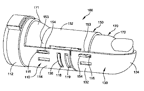
[0027] FIGS. 1 to 3 show an embodiment of an external fixation device 100.
FIG. 2 is
an exploded view. FIG. 3 shows the assembled device. Once assembled and
applied to a digit
(e.g., toe ), the device 100 achieves axial compression, radial compression
and superior / inferior
stabilization. The device 100 can be used by itself for non-invasive
treatment, or as a fixation
and support device during recovery from a surgical procedure. FIG. 1 shows the
assembled
device in use for correcting a hammertoe deformity.
[0028] The device 100 has a proximal first clamping member 110 having
an adjustable
clamping collar 112 and a first curved support portion 114 attached to the
adjustable clamping
collar 112. The first clamping member 110 is adapted to receive a digit 170
(e.g., a toe or finger)
therethrough. The first curved support portion 114 is adapted to support an
inferior surface of a
proximal phalanx 171 of the digit 170. The circular clamping collar 112 allows
for radial
4
CA 02836645 2013-12-16
compression on the proximal side of the joint-line. The collar 112 as shown
has a ratchet
mechanism with ramped teeth which permit tightening, but retain the clamp in
position once
tightened.
[0029] Although FIG. 1 shows ratchet type clamping collar 112 , other
clamping collars
can be used. For example, some embodiments include a clamping mechanism of a
type used in
cable ties. Some embodiments include a hook and locking latch type clamping
collar. Some
embodiments include a worm drive clamping mechanism (similar to the drive of a
hose clamp).
Other embodiments include circular springs of various configurations, which
the doctor can
pinch to expand, and which are biased to contract and provide compression upon
release. Other
embodiments include hook and loop fasteners on ends of a band encircling the
clamping collar
112. These are only examples of clamping mechanisms, and other embodiments
include other
types of clamping mechanisms.
[0030] A distal member 130 is adjustably attachable to the first
clamping member 110.
The distal member 130 has a second curved support surface 132 adapted to
support the inferior
surface of a distal phalanx of the digit 170 and a curved distal end 134
adapted to apply a
compressive force in a proximal direction to a distal end of the distal
phalanx 172.
[0031] In some embodiments, the distal member 130 has a hemispherical
distal surface
which is sized and shaped to receive the most distal end of a digit (toe or
finger). In some
embodiments, the superior member has cantilever arms 153, each having a
locking tab 154
adapted to mate with the respective slot 116 of the clamping member 110.
[0032] An superior member 150 is configured to be attached to the
first clamping
member 110 and the distal member 130. The superior member 150 has a curved
surface 152
adapted to apply a force against a superior surface of the digit 170. When the
joint is flexed, the
superior member 150 assists in correcting that flex. In some embodiments, the
superior member
150 has spring-like material properties, a circular profile, and tabs 154,
which interlock with
slots 116 in both the clamping member 110 and distal member 130. Some
embodiments include
a plurality of locking tabs 154 or a plurality of slots 116 inferiorly , to
allow incremental increase
of compression. Each respective slot 116 is configured to receive the at least
one tab 154 with the
superior member 150 at a respectively different location relative to the
proximal member 110 &
distal member 130, for applying a respectively different compressive force in
the inferior
5
CA 02836645 2013-12-16
direction. For example, in the embodiment of FIG. 1, clamping member 110 has
two slots 116,
and superior member 150 has tab 154.
[0033] The superior member has means for locking the superior member
to the first
clamping member and the distal member. In some embodiments, the superior
member 150 has
one of the group consisting of a slot 116 and a locking tab 154, either of
which provides a
locking means. At least one of the first clamping member 110 and the distal
member 130 has the
other of the group consisting of a slot 116 and a locking tab 154 for engaging
the slot or locking
tab of the superior member. For example, in the embodiment of FIG. 1, clamping
member 110
and distal member 130 each have a slot 116, and superior member 150 has
locking tab 154. In
an alternative embodiment, the superior member 150 has slots, and the clamping
member 110
and distal member 130 each have a locking tab for mating with the slots of the
superior member
150. In another embodiment, the superior member has a slot at one end and tab
at the other end;
one of the clamping member 110 and the distal member 130 has a tab and the
other has a slot, for
mating with the respective slot and tab of the superior member 150.
[0034] In some embodiments, the clamping member 110, distal member 130 and
superior
member 150 are made of padded stainless or titanium alloy.
[0035] In some embodiments, one of the first clamping member 110 and
the distal
member 130 includes at least one locking tab 136, and the other of the first
clamping member
110 and the distal member 130 includes a plurality of slots 118, 119. Each
respective slot 118,
119 is configured to receive the at least one locking tab 136 with the distal
member 130 at a
respectively different location relative to the proximal member 110, for
applying a respectively
different compressive force in the proximal direction. For example, in the
embodiment of FIG.
1, clamping member 110 has two slots 118 and 119, and distal member 130 has
locking tab 136.
In some embodiments, the locking tab 136 has ramped surface for easy
insertion, and for
retaining the distal member 130 in position relative to the clamping member
110, absent an
affirmative user action to release the tab 136 from the slot 118 or 119.
[0036] In some embodiments, the slots 116 are substantially longer
than the width of tabs
154, so that the superior member 150 can move along the longitudinal (proximal-
distal) axis.
This allows the selection of slot 118 or 119 to determine the engagement depth
of the distal
member 130 relative to the clamping member 110, for control of the compression
force against
the distal end 172 of the digit 170.
6
CA 02836645 2013-12-16
[0037] An exemplary method of using the device 100 is as follows:
[0038] 1. The user secures the device 100 to the proximal side 171 of
the joint, using the
adjustable clamping collar 112 on the clamping member 110.
[0039] 2. The user assembles the distal member 130 to the clamping
member 110 by
engaging the tabs 154 of the cantilever arms 153 into the slots 116 of the
clamping member 110.
[0040] a. The engagement depth of the tabs 136 determines the amount
of axial
compression. Various engagement depth options are provided with multiple
slots. The
embodiment of FIG. 1 has two slots 118, 119, providing two engagement depth
options. Other
embodiments include other numbers of slots for this purpose (e.g., one, three,
four or the like).
[0041] 3. The user assembles the superior member 150 to the clamping member
110 and
distal member 130 by compressing the tabs 154 of the superior member 150
inward and aligning
the tabs 154 with the mating slots 116 of the clamping member 110 and distal
member 130.
[0042] a. The amount of superior compression is determined by the
mating features
selected (overlapping in the superior I inferior axis by selection of one of
the slots 116), the
degree of semi-circularity (i.e., the angle of the sector of a cylinder that
the superior member 150
subtends), as well as the material properties of the superior member 150..
[0043] FIG. 4 shows another embodiment of a fixation device 200. A
first clamping
member 210 has an adjustable clamping collar (not shown) at its proximal end
and a first curved
support portion 214 attached to the adjustable clamping collar. The device 200
of FIG. 4 can
include any type of clamp 112 described above with respect to the device 100
of FIGS. 1-3, and
solely for brevity, descriptions thereof are not repeated. The first clamping
member 210 is
adapted to receive a digit (not shown). The first curved support portion 214
is adapted to support
the inferior surface of a proximal phalanx of the digit. A distal member 230
is adjustably
attachable to the first clamping member 210. The distal member 230 has a
second curved support
surface 232 adapted to support an inferior surface of a distal phalanx of the
digit and a curved
distal end 234 adapted to apply a compressive force in a proximal direction to
a distal end of the
distal phalanx. A superior member 250 is configured to be attached to the
first clamping
member 210 and the distal member 230. The superior member 250 has a curved
surface 252
adapted to apply a force against a superior surface of the digit.
[0044] The device 200 of FIG. 4 differs from the embodiment of FIGS. 1-3 in
that it has
a different means for locking the superior member to the first clamping member
and the distal
7
CA 02836645 2013-12-16
member. Device 200 has a fully adjustable locking means including at least one
suture, strap or
band 220 on each side (medial and lateral) of the device 200. The at least one
suture, strap or
band 220 is formed of a strong, flexible material, such as nitinol or ultra-
high molecular weight
polyethylene (UHMWPE) suture material, or nylon. The suture, strap or band 220
is woven
through the slots 216, 217 in the clamping member 210 and distal member, and
attached to the
tabs 254 of the superior member 250. The tabs 254 are inserted through one of
the slots 216,
selected to control the force applied by superior member 250.
[0045] This embodiment of the device 250 can be placed on the patient
while fully
assembled and then the suture, strap or band 220 tightened until the desired
compression is
reached. The mechanism provides a locking feature such that compression is not
lost through
repetitious motion.
[0046] In some embodiments, the means for locking also includes a
release, or
unloading, method in the event that the desired compression decreases with
time.
[0047] Although FIG. 4 shows a suture, strap or band 220, in other
embodiments other
mechanisms are substituted, such as, but not limited to:
[0048] a. Cable tie configurations
[0049] b. Enhanced hook and loop fastener for increased holding
strength
[0050] c. Strap / buckle mating device
[0051] d. Super-elastic ribbon / Shape set to distances that provides
compression,
stretched to fit over surgical site and allowed to return to its natural state
providing the desired
compression.
[0052] e. UHMWPE Suture with locking technology integrated in mating
features
[0053] FIG. 5 shows an embodiment of a fixation device 300,
comprising a proximal
clamping member 310 and a distal clamping member 311. Each of the proximal and
distal
clamping members 310, 311 has an adjustable clamping collar 112 adapted to
receive a digit 170
therethrough. The device 300 of FIG. 5 can include any type of clamp 112
described above with
respect to the device 100 of FIGS. 1-3, and solely for brevity, descriptions
thereof are not
repeated.
[0054] The device 300 has first and second coaxial helical members
322 and 324
opposing each other. For example, in FIG. 5, from left to right, helical
member 322 is wound in
a clockwise direction around a longitudinal axis of the device 300, and
helical member 324 is
8
CA 02836645 2013-12-16
wound in a counter-clockwise direction around the longitudinal axis. Each of
the helical
members 322, 324 has a respective proximal end fixedly attached to the
proximal clamping
member 310 and a respective distal end fixedly attached to the distal clamping
member 311. In
some embodiments, the helical members 322, 324 are joined to the clamping
members 310, 311
by be laser-welded or other suitable technique. Each of the first and second
coaxial helical
members 322, 324 is in the form of helical band having an inner support
surface arranged to be
wrapped around the digit 170.
[0055] The helical members 322, 324 have spring-like material
properties. Flexibility at
the joint can be controlled by the stiffness in the material selection for the
helical members. In
some embodiments, the helical members 322, 324 comprise spring steel or
nitinol.
[0056] In use, the clamping members 310, 311 are positioned on
opposing ends of the
joint-line at the surgical/treatment site.
[0057] 1. The user positions one clamp proximally with respect to
the joint line. The
user secures the proximal clamp 312 of member 310 to fix the location of
device 300.
[0058] 2. The user extends the free clamp 312 axially in the distal
direction, stretching
the helical members 322, 324 beyond their relaxed lengths.
[0059] 3. The user secures the distal clamp 312 of member 311 in the
extended position.
[0060] Once installed, the helical members 322, 324 react similar to
an extension spring
as they are strained in extension between two fixed members 310, 311. This
results in active
compression across the joint acting concurrently with the radial compression
created by the
clamping action. Device 300 allows some flexion, but, selection of the
diameter of the helical
members 322, 324 allows the designer to limit the amount of flexion the device
300 affords.
[0061] FIGS. 6-11 show an embodiment of a fixation device 400,
comprising a proximal
clamping member 410 and a distal clamping member 411. Each of the proximal and
distal
clamping members 410, 411 has an adjustable clamping collar 112 (not shown in
FIGS. 6-11)
adapted to receive a digit therethrough. The device 400 of FIGS. 6-11 can
include any type of
clamp 112 described above with respect to the device 100 of FIGS. 1-3, and
solely for brevity,
descriptions thereof are not repeated.
[0062] Device 400 has a plurality of longitudinal spacers 420. Each
longitudinal spacer
420 has a proximal end attached to the proximal clamping member 410 and a
distal end attached
to the distal clamping member 411. Each longitudinal spacer 420 has at least
one threaded end,
9
CA 02836645 2013-12-16
and one of the proximal and distal clamping members 410, 411 has a respective
thread
configured to receive the threaded end for adjusting a separation between the
proximal and distal
clamping members. In some embodiments, the clamping members have are
countersunk to
receive nuts 422 for receiving the threads of the spacers 420 (See FIGS. 9 and
10). In some
embodiments, spacers 420 are threaded throughout their lengths. In other
embodiments, the
spacers 420 are threaded at each end, and have a smooth surface in between,
configured to
receive a tightening instrument (e.g., a wrench) to adjust a distance between
the proximal and
distal clamping members 410, 411.
[0063] Device 400 has a plurality of spring members 430. Each spring
member 430 has
a proximal end attached to the proximal clamping member 410 and a distal end
attached to the
distal clamping member 411. For example, as shown in FIG. 9, each end of each
spring member
430 fits in a respective slot 412 of clamping members 410 and 411. Each spring
member 430 is
located adjacent to and radially inward from a respective longitudinal spacer
420. Each
longitudinal spacer 420 constrains its respective spring member 430 to bow
radially inwards.
[0064] In the embodiment of FIGS. 6-11, the plurality of longitudinal
spacers 420
includes four longitudinal spacers, and the plurality of spring members 430
include four spring
members arranged approximately evenly around a circumference of the proximal
clamping
member and distal clamping member. Other embodiments include different numbers
of spacers
420 and spring members 430 (e.g., 2 or 6).
[0065] The device 400 achieves radial compression. The adjustable clamps
create a site
of fixation on either side of the joint-line. Rotating the spacers 420, with
their threaded ends,
creates linear extension or retraction of the distance between the clamping
members 410, 411.
[0066] When the spacers 420 are rotated to reduce the distance
between the clamping
member 410, 411, the spring members 430 bow and flex inwards as the clamping
members 410,
411 translate towards each other, creating radial stability. Device 400
achieves simultaneous
radial stability and axial compression at the joint-line.
[0067] FIG. 11 shows the device 400 with a detachable drill guide 500
detachably
connected to the distal clamping member 411. The detachable drill guide 500
has a guide
portion 510 adapted to guide a drill along a proximal-distal axis should an
intramedullary device
be appropriate. The guide portion 510 has a tapered lead in guide with an
opening 512, through
which the drill is inserted.
CA 02836645 2013-12-16
[0068] The drill guide 500 comprises a support structure including at
least a pair of radial
arms 515, 517 and a longitudinal arm 516 for positioning the guide portion 510
a variable
distance away from the distal clamping member 411. The radial arms 515, 517
and longitudinal
arm 516 are extendible for varying a radial offset and a longitudinal
displacement of the guide
portion 510 relative to the distal clamping member 411.
[0069] Some embodiments provide fine-tuning and adjustable height and
length to ensure
that the guide is in the desired location in both the longitudinal and
superior/inferior directions.
Once the desired location is determined, the adjustable arms lock into place.
This achieves
accurate and consistent placement of temporary fixation devices or pre-drills.
Various
mechanisms can be used to provide adjustability. For example, in some
embodiments, as shown
in FIG. 11A, the drill guide 510 and cylindrical body 520 are an assembly,
wherein the
cylindrical body 520 replaces the post 515 of FIG. 11. The post 521 has
detents 522 at
predetermined offsets from each other. The drill guide 510 has at least one
spring plunger 523
threaded into the cylindrical body 520 perpendicular to the tapered guide 510.
The spring
plungers are biased to engage the post detents, retaining the drill guide 510
in position & will
disengage upon sufficient force supplied in the direction of the post axis to
allow positional
adjustments of the drill guide. In another embodiment, as shown in FIG. 11B,
the threaded post
531 is attached to a rod 530 & drill guide 510 as an assembly. The
longitudinal post 516 is
attached to a threaded nut 532 in which the nut is constrained in translation
coaxially and
perpendicular to the threaded axis. As the threaded nut 532 is turned, the
assembly comprising
the threaded post 531, rod 530 & drill guide 510 will move along the threaded
body's axis per
the threaded pitch. In other embodiments (not shown), the threaded post 531
mates with a worm
gear attached to the post 516. As the worm gear is turned the assembly
comprising the threaded
post 531, rod 530 & drill guide 510 will move along the threaded body's axis
per the threaded
pitch.
[0070] The drill guide 500 has an attachment mechanism 520 which
allows it to be
secured to one of the clamping members 411. In some embodiments, the
attachment mechanism
is a partial collar 520 which matches a portion of the distal clamping member
411. In other
embodiments (not shown), the attachment mechanism is a complete ring, matching
the shape of
clamping member 411.
11
CA 02836645 2015-07-08
[0071] Although the drill guide is only shown in FIG. 11, the drill
guide can be used with
any of the devices 100, 200, or 300 described above.
[0072] The material of drill guide 500 can be comprised of either
radiopaque or
radiolucent materials. A radiolucent material (e.g., hard plastic or glass
filed polymer) may be
desired if the assembly is to be imaged by fluoroscopy in situ, and the
physician does not want
the drill guide to appear in the image. A physician may desire a radiopaque
material if he/she
wishes to establish the position of the drill guide 500 with respect to the
bone under fluoroscopy,
for example.
[0073] FIGS. 12-15 show an embodiment of a fixation device 600
comprising a tube 610
comprising a contractible tubular woven mesh configured to contract radially
under longitudinal
tension. Such contractible tubular woven meshes are commonly referred to as
"Chinese finger
traps," and are described, for example, in U.S. Patents 2,783,758, 3,872,861
and 5,649,541.
[0074] The contractible tubular woven mesh 610 has at least one
helical yarn or fiber 630
fastened at or near a first end 611 of the tube 610 and woven helically
through the mesh and
extending from a second end 612 of the tube 610 opposite the first end 611 of
the tube, such that
the yarn 630 is capable of applying radial compression to the tube when placed
under tension.
The helical yarn or fiber 630 is a separate yarn or fiber from those used to
form the contractible
tubular woven mesh 610. In some embodiments, the helix of the yarn or fiber
630 winds around
the circumference of the tube 610 with a different period that the fibers
which constitute the
mesh of tube 610. In some embodiments, the helical yarn or fiber 630 comprises
a different
material from the material of the tube 610. The helical yarn or fiber provides
a drawstring
[0075] In some embodiments, the at least one helical yarn or fiber
630 includes two
opposing yarns or fibers 630 extending in opposite directions around a
circumference of the tube
610. That is, one is configured as a right hand helix and the other is a left
hand helix, so that
viewed in a direction of the longitudinal axis of the tube 610, one helix
winds clockwise around
the tube, and the other helix winds counter-clockwise around the tube. In some
embodiments, the
yarn or fiber 630 includes a circular winding (in a plane perpendicular to the
longitudinal axis of
the tube 610) at each end of the tube 610, so that pulling the string causes
both radial
compression and cinching of the ends of the tube 610. The helix crosses in the
middle of the
12
CA 02836645 2013-12-16
fixation device 600 such that when cinched, the contraction of the helical
yarn or fiber 630
causes a bent joint to straighten through the application of a compressive
force along the helix.
[0076] The device 600 is slipped over the digit to be treated, and
the at least one helical
yarn or fiber 630 is (are) pulled and tied or fastened. The at least one
helical yarn or fiber 630
act as a drawstring, cinching the tube 610 and placing the tube in radial
compression. In some
embodiments, the device 600 is used to straighten the digit (e.g., toe) for
percutaneous drilling
into the end of the toe. The mesh will compress the toe while holding it rigid
in line for drilling.
This device 600 can also be used for minor adjustment until the soft tissue
releases.
[0077] In some embodiments, the user attaches a weight to the yarns
or fibers 630 to
maintain compression during a surgical procedure. In other embodiments, the
device 600 is used
to provide stability post-surgery for a length of time by pulling, cinching
and tying off the helical
yarns or fibers 630. The device 600 can provide compression and help correct
deformity.
[0078] In some embodiments, the device 600 is used alone to provide
compression and
support. In some embodiments, additional support and rigidity is provided by
inclusion of
optional sleeves 620. In some embodiments, a plurality of sleeves 620 are
arranged around an
outer surface of the tube 610. The sleeves 620 are substantially smaller in
diameter than the tube
610. Any number of sleeves 620 may be included. In some embodiments, four, six
or eight
sleeves 620 are uniformly distributed about the circumference of the tube 610.
In some
embodiments, the sleeves 620 comprise the same material as the mesh of tube
610. In other
embodiments, the sleeves comprise a different material from tube 610.
[0079] Each of the plurality of sleeves 620 has a respective first
end fixed at or proximate
to a first end 611 of the tube 610 and a respective second end fixed at or
proximate to a second
end 612 of the tube 610 opposite the first end 611. Each sleeve 620 has a
portion that is freely
movable relative to the tube 610, the portion being between the first end and
second end of each
sleeve. In some embodiments, the sleeves are only fixed (e.g., by sewing) at
their ends to the
respective ends 611, 612 of the tube 610, and the sleeves are free to move
relative to the tube at
all intermediate locations along the lengths of the sleeves. In other
embodiments, the sleeves
620 are fixed at both ends and at one or more intermediate points along their
length to the outer
surface of the tube 610. In other embodiments, the sleeves 620 are sewn at or
near one end of
the tube 620, and the other end of each sleeve 620 is free to move relative to
the tube 610.
13
CA 02836645 2013-12-16
[0080] In some embodiments, the user can optionally insert at least
one removably
insertable rib 622 in at least a respective one of the plurality of sleeves
620. In some
embodiments, the ribs are inserted after pulling the helical yarns or fibers
630 to cinch the tube
610. The at least one rib 622 is formed of a material that is more rigid than
a material of the
tubular woven mesh. The rib can comprise any of a variety of materials, such
as wood, plastic or
a more rigid material.
[0081] FIG. 15 shows the device 600 after the user inserts six ribs
622 in the respective
sleeves 620. The physician can determine on an individual basis how many ribs
to insert, if any,
and where to put the ribs to achieve desired rigidity in a directional manner.
Thus, the physician
can select placements of the ribs to increase rigidity in the lateral-medial
direction, or in the
superior-ventral direction.
[0082] FIGS. 12-15 show an embodiment of device 600 having both the
helical yarns or
fibers 630 and the sleeves 620. In other embodiments, the device includes a
tube 610 with the
sleeves 620, but without the helical yarns or fibers 630. In other
embodiments, as shown in
FIGS. 15A-15C, the device 650 includes a tube 610 with the helical yarns or
fibers 630, but
without the sleeves 620. When cinched, the helical yarns or fibers 630 causes
a bent joint of
digit 170 to straighten through the application of a compressive force along
the helix.
[0083] Although FIGS. 12-15C show the device 600 used alone, the
device 600 can be
used in combination with any of the devices shown in FIGS. 1-11. In
particular, in some
embodiments, the device 300 (FIG. 5) or 400 (FIGS. 6-11) can be applied over
the device 600.
The physician applies device 600 and cinches the helical yarns or fibers 630,
and optionally
inserts one or more ribs 622 in sleeves 620. Then the physician places the
device 300 or 400
over the digit and tightens the clamps 312 or 112 at each end of the device
300 or 400. The
physician attaches the drill guide 500 and performs the drilling (e.g., for K-
wire insertion). This
is just one example, and the device 600 can be used with other external
fixation devices to
provide compression and support during surgical procedures.
[0084] FIGS. 16-19 show an embodiment of a bone implant 700 suitable
for correcting a
deformity such as a hammertoe. This device 700 addresses the common secondary
procedure
stabilize the metatarsophalangeal (MTP) joint by releasing the joint capsule
and employing a
temporary fixation wire. The bone implant 700 comprises a helical threaded
member 710 having
14
CA 02836645 2013-12-16
first and second ends 704, 706 and a longitudinal central opening 708
extending from the first
end 704 to the second end 706. The longitudinal central opening has a
longitudinal axis 702.
[0085] At least one blade 720 integrally attached to the first end
704 of the helical
threaded member 710. The blade 720 extends in a radial direction away from the
longitudinal
axis 702. The blade 720 has an outer edge with a plurality of teeth thereon
722. The blade 720
has an outer edge with a plurality of teeth thereon 722. In some embodiments,
as shown in FIGS.
16-19, the implant 700 has two blades evenly spaced and symmetrically arranged
to extend in
opposite radial directions away from the longitudinal axis 702. A central tube
730 with a central
cannula 732 runs along the central axis for a portion of the length of the
helical threaded
member.
[0086] In some embodiments, as shown in FIGS. 16-19, the helical
threaded member 710
has a cork-screw shape. The central longitudinal opening (referred to herein
as a cannula) of the
helical threaded member 710 is open to the exterior of the device. The central
opening 708 is
continuous with the cannula 732 of the central tube 730 and the cannula 724
which extends to
the end of blades 720. This configuration is analogous to a cannula diameter
greater than the
minor diameter of a screw. The cork screw configuration allows implantation
over a k-wire,
which is used to address metatarsophalangeal (MTP) joint soft-tissue
contracture. The cork-
screw configuration of helical threaded member 710 allows both axial
compression (FIG. 17)
and extension (FIG. 18) and perpendicular bending flexion (FIG. 19), similar
to the range of
motion of a coiled spring. This provides additional flexibility in the joint
as well as enhanced
bone integration within the threads of the helical threaded member 710. The
degree of flexibility
is a function of material properties and geometry and can be controlled and
optimized.
[0087] In some embodiments, implant 700 comprises a material having
super-elastic
material properties, such as nitinol. In other embodiments, the material is
selected to include
shape memory properties. Shape memory alloys, such as Nickel Titanium
(nitinol), undergo a
phase transformation in their crystal structure when cooled from the stronger,
high temperature
form (Austenite) to the weaker, low temperature form (Martensite). When heated
after
deformation, the shape memory material recovers its original shape. For
example, an implant 700
formed of a material with shape memory is set in the expanded state (FIG. 18),
and implanted.
Then the device 700 compresses when introduced into the body due to
temperature increase
(FIG. 17). This ensures compression at the joint while maintaining some
flexibility. Also, nitinol
CA 02836645 2013-12-16
exhibits superelasticity if deformed in an environment above their
transformation temperatures
and will change phase from austenite to stress-induced martensite, allowing it
to be strained from
¨2-6% with nearly constant stress & return from martensite to austenite during
unloading.
[0088] In the configuration as shown, the cantilevered blades 720
deflect inwards when
radial force is applied, allowing compatibility with an undersized preparation
hole. When
inserted in the bone, the cantilever blades 720 flex outward increasing
fixation in the bone. The
outward spring force of the blades 720 is a function of the material
properties and geometry, and
can be controlled and optimized.
[0089] FIG. 20 shows an embodiment of a device 800 which is similar
to the device 700
of FIGS. 16-19, except that a single blade 820 with teeth 822 is provided. The
bone implant 800
comprises a helical threaded member 810 having first and second ends 804, 806
and a
longitudinal central opening 808 extending from the first end 804 to the
second end 806. Device
800 is partially cannulated, providing greater flexibility along the helical
threaded member 810,
along the axis 802 from the first end 804 to the second end 806. Compared to
the device 700, the
single blade configuration of blade 820 provides greater rigidity when
inserted in the bone. In
some embodiments, the device 800 comprises a superelastic, shape memory alloy,
such as
nitinol, providing the expansion/contraction properties of the device 700, but
with greater
rigidity.
[0090] FIG. 21 shows an embodiment of a partially cannulated device
900 which is
similar to the device 800 of FIG. 20, except that four perpendicular blades
920 with teeth 922 are
provided in a continuous, cross-blade configuration. The bone implant 900
comprises a helical
threaded member 910 having first and second ends 904, 906 and a longitudinal
central opening
908 extending from the first end 904 to the second end 906. Compared to the
devices 700 and
800, the cross-blade configuration of blades 920 provides greater rigidity
when inserted in the
bone. The cross-blade configuration can provide a greater degree of fixation.
The cross-blade
configuration allows implant pre-drill (circular) preparation instead of
broaching. Using an
undersized pre-drill step, the crossed blades 920 achieve fixation by
circumferential interference
with the surrounding bone. In some embodiments, the device 900 comprises a
superelastic shape
memory alloy, such as nitinol, providing the expansion/contraction properties
of the device 700
described above but with greater rigidity & fixation
16
CA 02836645 2013-12-16
[0091] FIG. 22 shows an embodiment of a fully cannulated device 1000
which is similar
to the device 700 of FIGS. 16-20, except that four perpendicular blades 1020
are provided in a
cross-blade configuration, evenly spaced around the longitudinal axis. The
central opening 1008
is continuous with the cannula (not shown in FIG. 22) of the central tube 1030
and the cannula
1024 which extends to the end of blades 1020. A K-wire or the like can be
placed through the
central opening 1008 of helical threaded member 1010, along the axis 1002 from
the first end
1004 to the second end 1006. Compared to the device 700, the four blade
configuration of
blades 1020 can provide a greater degree of fixation. The cross-blade
configuration allows
implant pre-drill (circular) preparation instead of broaching. Using an
undersized pre-drill step,
the crossed blades 1020 achieve fixation by circumferential interference with
the surrounding
bone. In the configuration of FIG. 22, the four cantilevered blades 1020
deflect inwards when
radial force is applied, allowing compatibility with an undersized preparation
hole. When
inserted in the bone, the cantilever blades 1020 flex outward increasing
fixation in the bone. The
outward spring force of the blades 1020 is a function of the material
properties and geometry,
and can be controlled and optimized. In some embodiments, the device 1000
comprises a
superelastic, shape memory alloy, such as nitinol.
[0092] FIGS. 23 and 24 show an embodiment of a fully cannulated
implant 1100 which
is similar to the device 1000 of FIG. 22, except that the helical threaded
member 1110 of implant
1100 has a minor diameter larger than a diameter of the longitudinal central
opening 1124, so
that the helical threaded member 1110 has a central tube 1140 with a
continuous inner surface
1142 around the longitudinal central opening (cannula) 1124. In implant 1100,
four
perpendicular blades 1120 with teeth 1122 are provided in a cross-blade
configuration, evenly
spaced around the longitudinal axis 1102. In other embodiments, only two
cantilever blades
(similar to blades 720 in FIG. 16) are provided, but the rest of the implant
1100 is otherwise the
same. In some embodiments, the device 1000 comprises a superelastic, shape
memory alloy,
such as nitinol.
[0093] A K-wire or the like can be placed through the central opening
1124 of helical
threaded member 1110, along the axis 1102 from the first end 1104 to the
second end 1106.
Compared to the device 700, the four blade configuration of blades 1120 can
provide a greater
degree of fixation. The cross-blade configuration allows implant pre-drill
(circular) preparation
17
CA 02836645 2013-12-16
instead of broaching. Using an undersized pre-drill step, the crossed blades
1120 achieve
fixation by circumferential interference with the surrounding bone.
[0094] FIGS. 25-29 show a method for installing the implant 1100. The
same sequence
of steps is performed for any of the fully cannulated implants, such as
implant 700 (FIG. 16), and
implant 1000 (FIG. 22). Note that in the views of FIGS. 25-27, the proximal
direction is left and
the distal direction is right, but in the views of FIGS. 28 and 29, the
proximal direction is right
and the distal direction is left
[0095] In FIG. 25, the proximal bone (phalanx) 2503 is pre-drilled to
receive the K-wire
1126, and the physician broaches the middle phalanx 2502.
[0096] Then, the physician drills distally through middle phalanx 2502 and
through the
tip of the toe 2501 with the K-wire 1126, as shown in FIG. 26.
[0097] Once the K-wire is exposed, a drill 2510 (shown in FIG. 27) is
attached to the
distal end of the K-wire 1126. The K-wire 1126 is backed out towards the
distal end of the bone
2501, until the proximal tip of the K-wire 1126 is sub-flush with the joint
line (i.e., withdrawn
past the proximal end of middle phalanx 2502. In some embodiments, the K-wire
is drawn past
the end of the middle phalanx 2502 by a distance greater than a length of the
blades 1120.)
[0098] The helical threaded portion 1110 of the implant 1100 is then
advanced into the
proximal phalanx 2503 until the implant 1100 is fully seated. Once the implant
1100 is fully
seated, the physician closes the joint, forcing the blades 1120 into the
previously broached canal
as shown in FIG. 28.
[0099] With a correctly aligned joint (optionally using one of the
external fixation
devices shown in FIGS. 1-15), the physician advances the K-wire 1126 in the
proximal direction,
through the cannulated implant 1100 into the MP joint. They physician caps the
K-wire 1126
with a Jurgan ball 1128, completing the installation.
[00100] FIGS. 30-34 show a bone implant 1200 comprising a central shaft
1210 having
first and second ends and a longitudinal axis. A first set of blades 1220 are
integrally attached to
the first end of the central shaft 1210. The first set of blades 1220 extends
in a radial direction
away from the central shaft 1210. Each of the first set of blades 1220 has an
outer edge with a
plurality of teeth 1222 thereon.
[0100] A second set of blades 1250 with teeth 1252 are integrally attached
to the second
end of the central shaft 1210. The second set of blades 1250 extend in the
radial direction away
18
CA 02836645 2013-12-16
from the central shaft 1210. Each of the second set of blades 1250 has an
outer edge with a
plurality of teeth 1222 thereon. The second set of blades are rotationally
offset from the first set
of blades.
[0101] In some embodiments, each one of the second set of blades 1250
is rotationally
spaced midway between an adjacent pair of the first set of blades 1210. For
example, in the
implant of FIGS. 30-34, there are four first blades 1220 and four second
blades 1250. The
angular spacing between each second blade 1250 and the adjacent first blades
1210 on either side
is 45 degrees. FIGS. 31 and 32 show the angular offsets between the two sets
of blades 1220,
1250.
[0102] In some embodiments, the bone implant 1200 has a cannula 1224
extending along
the longitudinal axis 1202 from a first end of the bone implant to a second
end of the bone
implant. In other embodiments, the implant is solid, with no cannula. In some
embodiments, the
device 1000 comprises a superelastic, shape memory alloy, such as nitinol.
[0103] In some embodiments, an instrument is provided that inserts a
broach in the bone
on one side of the joint in a first orientation, and then is rotated +/- 45
degrees to broach the bone
on the other side of the joint in a second orientation rotationally offset
from the first orientation.
The instrument has a shape to match the cross-blade configuration 1220 of the
implant 1200. In
other embodiments, a K-wire channel is pre-drilled into the bone prior to
inserting the implant,
and no broach is required. In either case, the physician uses a safety tool to
handle the implant
1200. The safety tool has a gripping handle and a head shaped to receive
either the blades 1220
or the blades 1250, so the physician is not harmed by the blades 1220, 1250.
[0104] The inventors have determined that one of the sources of
problems in hammertoe
implants is implant loosening after insertion. The rotational offset between
blades 1220 and
blades 1250 provides a different orientation on the distal end and the
proximal end to help
prevent against the blades from loosening.
[0105] FIGS. 33-34 show the method of insertion. First, the bones
2501-2503 are pre-
drilled as shown and described above with reference to FIGS. 25-27, and the
physician inserts
the K-wire 1126 across the joint. The alignment may be checked by fluoroscopy
to confirm
where to insert the implant. The K-wire 1126 is backed out beyond the proximal
end of the
middle phalanx 2502.
19
CA 02836645 2015-07-08
[0106] As shown in FIG. 33, the physician inserts the blades 1220 of
implant 1200 in the
proximal phalanx 2503, where the K-wire pre-drilled hole is visible. The
physician can use the
above-mentioned safety tool for this purpose.
[0107] Then as shown in FIG. 34, the physician takes the PIP joint
and places the
broached or pre-drilled side of the middle phalanx 2502 over the blades 1250
and presses the
middle phalanx into place, with the implant now embedded in both the middle
phalanx 2502 and
the proximal phalanx 2503.
[0108] If the implant 1200 is cannulated, then the K-wire 1126 is
advanced through the
implant in the same manner described above with reference to FIG. 29, and a
Jurgan ball 1128 is
attached.
[0109] The scope of the claims should not be limited by the preferred
embodiments set
forth in the examples, but should be given the broadest interpretation
consistent with the
description as a whole.
20