Note : Les descriptions sont présentées dans la langue officielle dans laquelle elles ont été soumises.
CA 02837463 2013-11-26
DESCRIPTION
HIGH-SPEED SCREENING APPARATUS FOR A, RAMAN ANALYSIS-BASED
HIGH-SPEED MULTIPLE DRUG
Technical Field
The present invention relates to a Raman analysis-based
apparatus for screening multiple drugs at a high speed.
W Background Art
Drug development is an advanced country-type strategic
process requiring a massive commitment of time and money of
more than ten years and eight hundred million dollars,
respectively. A well-developed social infrastructure is also
necessary for drug development.
Broadly, the process of drug development can be divided
into the discovery of a drug target by basic research, the
selection of effective and lead materials by compound
screening, the determination of a candidate drug, clinical
research through pre-clinical work/clinical phase 1, and
commercialization through clinical phases 2 and 3.
Of a total of 35,000 genes discovered thus far as drug
targets, approximately 500 are currently under research for
drug development, with a steady expansion of the development
subject since the Human Genome Project. Once a drug target is
1
CA 02837463 2013-11-26
selected, development of a screening method that is the most
suitable and effective should be undertaken. The
screening
method can be divided into an in vitro assay and a cell-based
assay. Major
pharmaceutical companies possess libraries of
compounds, typically amounting in number to ten of thousands
to hundreds of millions, as screening targets, and such a
number of compounds are employed from an early screening
stage.
A great expense for this screening process has given rise
M to making every effort to design effective screening methods
and develop high-speed and minimized apparatuses and reagents
which allow the screening of as many compounds as possible
within a short period of time.
A screening process for many compounds must be
technically simple with high reproducibility. When a drug
target is an enzyme, a relative easy approach is possible
thanks to an abundant number of screening methods and reagents
established therefor. However, because most of the biological
processes taking place within cells are associated with
interaction with proteins, a screening method based on
interaction with proteins is the most effective among analysis
methods for developing lead compounds. Great weight is given
to such screening methods for the following reasons: a protein
functions as it associates with another protein in vivo; a
change in gene and protein expression, in intracellular
2
CA 02837463 2013-11-26
location, and/or in structure through post-translational
modification induces an altered interaction between proteins,
resulting in a change in the activity and regulation of
intracellular metabolisms and signaling pathways; and an
abnormal protein interaction attributed to a genetic mutation
directly leads to the onset of a disease. There
are
technologies for detecting protein interactions, including
FRET (Fluorescence Resonance Energy Transfer), BRET
(Bioluminescence Resonance Energy Transfer) and FP
W (Fluorescence Polarization), and a technical advance has also
been achieved in apparatuses to which the technologies are
applicable. In
recent years, HCS (high-content screening)
with automated high resolution microscopy has been introduced,
whereby after cells are incubated with substances in multiwell
plates, such as 96-well, 384-well plates, etc., phenomena
associated with the quantitative change and transport of
proteins within cells can be rapidly observed in a
quantitative manner. HCS is
now arising as the most
interesting biological research method for world-leading
pharmaceutical companies or research institutes because it
allows the quantitative analysis of biological parameters,
such as protein interaction, Ca++ influx, etc., which are
difficult to screen on a large scale with conventional
methods, over the simple information obtained using
conventional enzyme detection methods or reporter systems, for
3
CA 02837463 2013-11-26
example, on enzyme activity, promoter strength, protein
levels, etc.
Typically, a procedure for drug screening comprises
preparation of compound aliquots, dilution, mixing of
screening components, culturing and detection, analysis of
screening data, and reporting on results. A high-throughput
screening (hereinafter referred to as "HTS") system is used to
rapidly process such a serial procedure. Advanced
pharmaceutical companies are known to possess a compound
W library consisting of hundreds of millions of different
compounds, and whenever a novel drug target is discovered, the
companies take advantage of the HTS system in screening the
compound library against the drug target. Thus,
major
pharmaceutical companies have accumulated tremendous data on
biological activities of hundreds of millions of compounds,
thus far. In order to more rapidly and effectively screen the
compound library against thousands of drug targets, a curve-
fitting tool capable of performing various functions including
a QC function, error checking for overlapped data, calculation
of relative activity (% activity), and extraction of
biochemical parameters, such as ICK, K, and K, is needed.
In this regard, HTS which allows much data to be produced by
one screening process is required. This new
technology,
aiming to overcoming problems associated with the conventional
technology, is basically designed to evaluate synthetic
4
CA 02837463 2013-11-26
,
compounds randomly on a mass scale through automation, and can
reduce the time taken to determine candidate drugs as much as
possible in association with automated synthesis of new
materials (CCL), molecular design and systematic information
management.
Prerequisites for HTS with a capacity of screening more
than 10,000 different compounds a day are summarized as
follows:
M (1) Rapidity: Given a higher screening speed, an HTS can
screen a higher number of compounds, and thus can complete its
performance within a shorter time and at a lower expense.
(2) Expense: Reagents used in the screening process
account for a large portion of the total screening expense. A
measure must be taken toward financial retrenchment.
(3) Miniaturization: Miniaturization is not only one of
the best measures to cut expenses for reagents, but can also
reduce the time taken to perform a screening process.
Besides, it can reduce laboratory space necessary for the
instruments.
(4) Automation: Automation increases reproducibility of
results as well as the speed of screening. Particularly, it
makes a great contribution to the reduction of experimental
error.
(5) Screening sensitivity: The sensitivity of a detection
5
CA 02837463 2013-11-26
method is directly relevant to the quantity of samples to be
used. High detection sensitivity is required because it takes
a longer time to screen samples of lower sensitivity.
(6) Non-radioactive method: As high as 50 % of the HTS
methodologies developed thus far use radioactive substances.
However, radioactive substances produce waste which must be
specifically cared for, and thus are disadvantageous in terms
of space, time and finances.
(7) Simplicity: Because a method operating with
W filtration, separation, washing, distinction, and solid-state
extraction requires additional expense and processes, the
screening process should be simplified in a liquid state as
much as possible.
Pharmaceutical companies have made enormous investments
in the development of chemical approaches to compounds, and
HTS technology. As a result, the number of drug candidates
has sharply increased. Then, the candidates excavated through
the primary screening process (discovery and evaluation of
target, and excavation of candidates) are subjected to a
secondary screening process (optimization of candidates) which
is much lower in yield than is the primary screening process.
The difference of yield between the primary and secondary
screening processes incurs a significant bottleneck phenomenon
in the development of new drugs. Hence, it is an important
challenge throughout new drug development to increase the
6
CA 02837463 2013-11-26
efficiency of secondary screening to a level in harmony with
the primary screening process without deteriorating the
quality of data generated in the secondary screening.
High-content screening (HCS) can be defined as a
"technology for functionally and complexly screening various
targets inside living cells on the basis of highly temporally
and spatially resolved fluorescence images." Among
fundamental technologies of HCS are a cell-based assay, real-
time fluorescent imaging of living cells with high temporal
W and spatial resolution, and a high-speed and high-content
automated assay. Representative of HCS analysis instruments
is the Opera system of Perkin-Elmer shown in FIG. 1. Formal
cell analysis data obtained by the Opera system is as shown in
FIG. 2. In this regard, first, images of tens of aggregated
cells are obtained within a field, and cell nuclei and walls
are discriminated among the images, during which images of
some cells are removed on the program while leaving
significant cell images. Finally,
two-color images are
obtained as seen in FIG. 2.
The high-content screening technology has been based on
fluorescence assay, so far. However, fluorescent labels used
in fluorescence assay weaken in fluorescence intensity
(photobleaching), and exhibit interference between different
fluorescent labels because excitation light with a very narrow
wavelength range is used while the fluorescent light has a
7
CA 02837463 2013-11-26
very broad range of wavelengths. In
addition, there are an
extremely limited number of available fluorescent substances.
Therefore, there is a need for a new method for effective
high-speed drug screening that exhibits sharp spectrum peaks
without causing interference between fluorescent substances,
thus allowing the detection of multiple drugs.
In recent years, Raman spectroscopy has attracted
extensive attention.
Inter alia, Surface Enhanced Raman Scattering (SERS) is a
W spectroscopic method which utilizes the phenomenon whereby,
when molecules are adsorbed on a roughened surface of a metal
nanostructure such as a gold or silver nanoparticle, the
intensity of Raman scattering is dramatically increased to the
level of 106 - 108 times compared with normal Raman signals.
As light passes through a transparent medium, molecules or
atoms of the medium scatter the light. In this
regard, a
small fraction of the photons undergoes inelastic scattering,
known as Raman scattering. For
example, a fraction of the
incident photons interact with the molecules in such a way
that energy is gained or electrons are excited into higher
energy levels, so that the scattered photons have a different
frequency from that of the incident photons. Because
the
frequencies of the Raman scattering spectrum account for the
chemical compositions and structural properties of the light
absorbing molecules in a sample, Raman spectroscopy, together
8
CA 02837463 2013-11-26
with the nanotechnology which is currently being quickly
developed, can be further developed for highly sensitive
detection of a single molecule. In
addition, there is a
strong expectation that an SERS sensor can be importantly used
as a medical sensor. The SERS
effect is in relation with
plasmon resonance. In this
context, metal nanoparticles
exhibit apparent optical resonance in response to incident
electromagnetic radiation due to the collective coupling of
conduction electrons within the metal. Thus, nanoparticles of
W gold, silver, copper and other specific metals can
fundamentally serve as nanoscale antenna for amplifying the
localization of electromagnetic radiation.
Molecules
localized in the vicinity of these particles show far greater
sensitivity to Raman spectroscopy.
Accordingly, many studies are being actively carried out
about using SERS sensors to detect biomarkers including genes
and proteins for early diagnosis of various diseases. Raman
spectroscopy has various advantages over other methods (e.g.,
infrared spectroscopy). While
infrared spectroscopy can
detect strong signals from molecules which have a dipole
moment, Raman spectroscopy allows strong signals to be
detected even from non-polar molecules in which induced
polarizability is modulated. Hence,
almost all organic
molecules have their own Raman shifts (cm-1). In
addition,
being free from the interference of water molecules, Raman
9
CA 02837463 2013-11-26
spectroscopy is suitable for use in the detection of
biomolecules including proteins, genes, etc. Due to
low
signal intensity, however, the stage of development of Raman
spectroscopy has not yet reached the level where it can be
used in practice in spite of research spanning a long period
of time.
Since its discovery, Surface-Enhanced Raman Scattering
(SERS) has continually been developed to such a level so as to
W detect signals at a molecular level from randomized aggregates
of fluorescent dye-absorbed nanoparticles (Science 1997,
275(5303), 1102; Phys rev lett 1997, 78(9), 1667). Since
then, many studies of SERS enhancement with various
nanostructures (nanoparticles, nanoshells, nanowires) have
been reported. In order to utilize SERS as a highly sensitive
detection method for a biosensor, Mirkin et al. reported
highly sensitive DNA analysis by using DNA-modified gold
nanoparticles, with a detection limit of 20 fM (2002, Science,
297, 1536). However,
there have been almost no advances in
preparing single molecule SERS active substrates based on the
salt-induced aggregation of silver (Ag) nanoparticles having
Raman active molecules (e.g., Rhodamine 6G) since the first
study. A report has it that only a fraction (less than 1%) of
heterogeneously aggregated colloids has single molecule SERS
activity (J Phys Chem B 2002, 106(2), 311). Like this,
CA 02837463 2013-11-26
randomly roughened surfaces provide a multitude of interesting
essential data associated with SERS, but this strategy is
fundamentally impossible to reproduce because even a small
change in surface morphology leads to a significant change of
enhancement. Recently,
Fang et al. reported a quantitative
measurement of the distribution of site enhancements in SERS.
The hottest SERS-active sites (EF > 109) accounted for only 63
sites out of a total of 1,000,000 sites, but contributed 24%
to the overall SERS intensity (Science, 2008, 321, 388). In
W these regards, assembling SERS-active nanoparticles into well-
defined and reproducible hot SERS nanostructures would lead to
a highly reliable, sensitive assay for biomolecules and be
greatly useful for use in xenodiagnosis and in vivo imaging
techniques.
Leading to the present invention, intensive and thorough
research into the high-speed screening of multiple drugs in
association with Raman spectroscopy, conducted by the present
inventors, resulted in the finding that when exposed to a
sample containing one or more analytes, a nanoparticle labeled
with an analyte-recognizing biomolecule functionalized
thereon, comprising a core and a shell with a nanogap formed
therebetween, is used to produce Raman signals if it is
irradiated with an excitation laser beam, and that specific
Raman wavelengths can be obtained from the Raman signals by
11
CA 02837463 2015-07-29
filtration through multiple Raman filters, detected with a
high SERS enhancement factor by a detector, and color coded to
generate color-coded Raman images, whereby multiple drugs can
be screened at high speeds with high reproducibility and
reliable quantifiability.
Disclosure
Technical Problem
It is an object of the present invention to provide a
W high-speed screening apparatus of multiple drugs using Raman
spectroscopy by which multicolors are coded for Raman signals.
It is another object of the present invention to provide
a high-speed screening method of multiple drugs, using the
apparatus.
Technical Solution
The one object of the present invention may be
accomplished by providing a high-speed screening apparatus for
multiple drugs using surface-enhanced Raman scattering,
comprising:
an excitation module, composed of a lens, a mirror, and a
pinhole, for introducing light from a light source into a
microscope;
12
CA 02837463 2015-02-05
a microscope module for acquiring an image of a sample,
comprising a motion controller for controlling a position of
the well plate well to well, a filtration unit composed of one
or more Raman filters for filtering Raman wavelengths against
light scattered from the sample when the sample is irradiated
with excitation light from the light source, and a CCD camera
operating in non-scanning manner for sequentially receiving
light beams passing through the filtration unit;
an image processing module for coding colors for a set of
W images obtained from a point containing a sample to produce
cell or tissue images, and for displaying the cell or tissue
images, said point being positioned by the motion controller;
and
a storage chamber for storing one or more core-gap-shell
nanoparticles selectively associated with the one or more
analytes present in a sample,
wherein the CCD camera takes in non-scanning manner one
or more Raman images of the sample in the individual well of
the well plate as said individual wells are sequentially
brought into a photographing site by the motion controller,
wherein each of the one or more core-gap-shell
nanoparticles comprises a core and a shell surrounding the
core, with a nanogap formed therebetween, said nanogap
containing an optically active molecule therein,
wherein the core consists of a metal exhibiting surface
CA 02837463 2015-02-05
plasmon resonance, and the shell consists of a metal
exhibiting surface plasmon resonance,
wherein the optically active molecule is a molecule
consisting of an atom selected from the group consisting of C,
H, 0, N, S, and a combination thereof.
The other object of the present invention may be
accomplished by providing a high-speed screening method for
multiple drugs using the apparatus above, comprising:
a step 1 of adding core-gap-shell nanoparticles to a
sample to be analyzed;
a step 2 of obtaining one or more Raman images from the
sample by irradiating a laser beam on the sample to generate
Raman scattered light, filtering the Raman scattered light
through a filtration unit composed of one or more Raman
filters to extract a Raman wavelength of interest, and
detecting the Raman spectrum using a CCD camera operating in
non-scanning manner; and
a step 3 of coding colors for the Raman images of the
sample to generate cell or tissue images and displaying the
cell or tissue images,
wherein the laser beam has a diameter which can take
Raman images in a non-scanning manner from individual well of
a well plate,
wherein the CCD camera takes in non-scanning manner one
or more Raman images of the sample in the individual wells of
13a
CA 02837463 2015-02-05
the well plate as the individual wells are sequentially
brought into a photographing site by the motion controller,
wherein each of the one or more core-gap-shell
nanoparticles comprises a core and a shell surrounding the
core, with a nanogap formed therebetween, said nanogap
containing an optically active molecule therein,
wherein the core consists of a metal exhibiting surface
plasmon resonance, and the shell consists of a metal
exhibiting surface plasmon resonance,
W wherein the optically active molecule is a molecule
consisting of an atom selected from the group consisting of C,
H, 0, N, S, and a combination thereof.
Advantageous Effects
Ob
CA 02837463 2013-11-26
As described hitherto, the screening apparatus and method
of the present invention is not designed to detect
autofluorescence, but to measure Raman signals generated from
core-gap-shell nanoparticles, so that it exhibits no
interference between fluorescent labels. The core-
gap-shell
nanoparticles show very strong surface-enhanced Raman
scattering (SERS) signals, with an SERS enhancement factor of
up to about 1012, and are proven to be highly reproducible. In
addition, the use of a CCD camera as a detector allows the
W apparatus and method of the present invention to screen
multiple drugs at a high speed because the CCD camera, which
operates in a non-scanning manner, can photograph individual
wells of well plates momentarily and can take pictures of
other wells in association with the operation of the motion
controller. Further, the apparatus and method of the present
invention can code multiple colors for Raman images, and are
effectively applicable to the screening of various drugs.
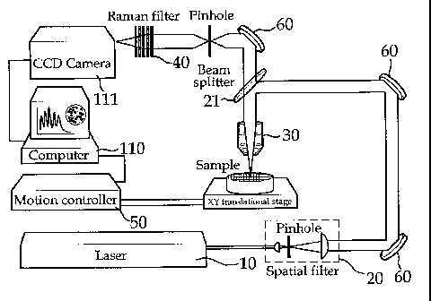
Description of Drawings
FIG. 1 is a photograph of a conventional fluorescence-
based high-content screening analysis instrument.
FIG. 2 is a 2-color image of cells obtained by a
conventional fluorescence-based high-content screening
analysis instrument.
FIG. 3 is a conceptual view of a Raman-based high-speed
14
CA 02837463 2013-11-26
screening apparatus of multiple drugs according to the present
invention.
FIG. 4 shows a core-gap-shell nanoparticle useful for the
Raman spectroscopy-based high-speed screening method of
multiple drugs.
FIG. 5 shows surface-enhanced Raman scattering spectra
measured by the apparatus of the present invention using
nanoparticles synthesized in Synthesis Examples 1 to 3.
FIG. 6 shows wavelength ranges of narrow band pass
W filters for selectively filtering Raman light scattered from
the nanoparticles of Synthesis Examples 1 to 3.
FIG. 7 shows Raman images detected after the selective
filtration of Raman signals scattered from the nanoparticles
synthesized in Synthesis Examples 1 to 3 through respective
narrow band pass filters.
FIG. 8 shows Raman images selectively filtered through
respective narrow band pass filters optimized for the
nanoparticles of Synthesis Examples 1 to 3, and a merged image
thereof.
FIG. 9 is a schematic diagram of a PEG-coated
nanoparticle synthesized in Synthesis Example 4, 5 or 6.
FIG. 10 shows images of cells incubated without (a)
(control) and with (b) (test group) the PEG-coated
nanoparticles synthesized in Synthesis Example 5, as measured
by the apparatus of the present invention using two narrow
CA 02837463 2013-11-26
band pass filters ("Filter 1" and "Filter 2").
FIG. 11 shows cell images of three sections of the test
group incubated with the PEG-coated nanoparticles of Synthesis
Example 4, as measured by the apparatus of the present
invention using two narrow band pass filters ("Filter 1" and
"Filter 2").
FIG. 12 shows cell images of three sections of the test
group incubated with the PEG-coated nanoparticles of Synthesis
Example 5, as measured by the apparatus of the present
W invention using two narrow band pass filters ("Filter 1" and
"Filter 2").
Mode for Invention
Below, a detailed description will be given of the
present invention.
In accordance with one aspect thereof, the present
invention addresses a high-speed screening apparatus of
multiple drugs using surface-enhanced Raman scattering,
comprising:
an excitation module, composed of a lens, a mirror, and a
pinhole, for introducing light from a light source into a
microscope;
a microscope module for acquiring an image of a sample,
comprising a motion controller for controlling a position of
the sample, one or more Raman filters for filtering Raman
16
CA 02837463 2013-11-26
wavelengths against light scattered from the sample when the
sample is irradiated with excitation light from the light
source, and a detector for sequentially receiving light beams
passing through the Raman filters; and
an image processing module for coding colors for one or
more images obtained at a point containing a sample to produce
cell or tissue images, and for displaying the cell or tissue
images.
Below, a description will be given of preferred
embodiments of the present invention in conjunction with FIG.
3.
Throughout the accompanying drawings, the same reference
numerals are used to designate the same or similar components.
Further, in the description of the present invention, when it
is determined that the detailed description of the related art
would obscure the gist of the present invention, the
description thereof will be omitted.
FIG. 3 is a conceptual view of a Raman-based high-speed
screening apparatus of multiple drugs according to the present
invention.
The Raman-based high speed screening apparatus of
multiple drugs according to the present invention may be
divided into an excitation module, a microscope module, and an
17
CA 02837463 2013-11-26
,
image processing module. It
should be apparent to those
skilled in the art that the functional modules are intended
simply for concrete descriptions thereof, but not to divide
them into exclusive and independent parts, and that the
functional modules may be overlapped in certain regions or two
or more functional modules may participate in one region.
Excitation Module
In the apparatus of the present invention, the excitation
M module functions to introduce a laser beam generated from a
light source (LS) 10 into a microscope.
The LS 10 may generate a near infrared (NIR) laser or a
visible laser. The visible laser is light with a wavelength
of from 400 to 700 nm. In one embodiment, the visible laser
has a wavelength of 514.5 nm. In
the biotechnology field,
Raman images have been obtained mainly using an NIR laser
since the use of visible light as a light source induces
autofluorescence, which brings about a reduction in the
intensity of Raman signals.
However, because Raman signal
strength is in inverse proportion to a fourth power of
wavelength, a visible laser can increase the intensity of
Raman signals further than can an NIR laser. In
addition,
optical devices utilizing visible light are more advanced than
those using NIR light.
Hence, if it can reduce
autofluorescence, the use of visible lasers has an advantage
18
CA 02837463 2013-11-26
over that of NIR lasers in optimizing an optical system.
After being generated by the LS 10, a laser beam passes
through a spatial filter 20 so that the beam diameter expands.
Through a plurality of lenses, a mirror, and a pinhole, the
beam is collimated to have a diameter of about 10 mm, and then
introduced into a microscope module.
Microscope Module
In the apparatus of the present invention, the microscope
module comprises a motion controller 50 for controlling the
position of a sample, a Raman filtration unit 40 consisting of
one or more Raman filters for filtering Raman wavelength light
against scattered light from the sample when the sample
irradiated with excitation light from a laser beam, and a
detector 111 for sequentially receiving light beams passing
through the Raman filtration unit 40.
After entry into a microscope, the laser beam is
reflected by a light separation unit 21 and is directed toward
a microscope objective (MO) lens 30. As the light separation
unit 21, a beam splitter, a dichroic mirror, or a detachable
mirror may be used.
The number of the Raman filters for filtering Raman
wavelength light is in the order of 1 to 20, and preferably in
the order 5 to 20.
The Raman filtration unit may be a band pass filter, and
19
CA 02837463 2013-11-26
preferably includes, but is not limited to, a narrow band pass
filter.
So long as it operates as a scanning type or non-scanning
type, any detector may be employed in the present invention.
For example, PMT (photomultiplier tube) detectors or APD
(avalanche photodiode) detectors, all operating in a scanning
manner, may be employed, while a CCD (charge-coupled device)
camera is representative of available detectors operating in a
non-scanning manner.
The sample may be a cell containing an analyte. Examples
of the analyte of interest include amino acids, peptides,
polypeptides, proteins, glycoproteins,
lipoproteins,
nucleosides, nucleotides, oligonucleotides, nucleic acids,
saccharides, carbohydrates, oligosaccharides, polysaccharides,
fatty acids, lipids, hormones, metabolites, cytokines,
chemokines, receptors, neurotransmitters, antigens, allergens,
antibodies, substrates, metabolites, co-factors, inhibitors,
drugs, phatmaceuticals, nutrients, prions, toxins, poisons,
explosives, pesticides, chemical warfare agents, biohazardous
agents, radioisotopes, vitamins, heterocyclic aromatic
compounds, carcinogens, mutagens, narcotics, amphetamines,
barbiturates, hallucinogens, waste products and contaminants.
In addition, when the analyte is a nucleic acid, it may be
exemplified by genes, viral RNA and DNA, bacterial DNA, fungal
DNA, mammalian DNA, cDNA, mRNA and DNA fragments,
CA 02837463 2013-11-26
oligonucleotides, synthetic oligonucleotides, modified
oligonucleotides, single- and double-stranded nucleic acids,
and natural and synthetic nucleic acids.
Separately, the
sample may be associated with a core-gap-shell nanoparticle
shown in FIG. 4 so as to amplify Raman signals. The
association may be achieved by exposing core-gap-shell
nanoparticles stored in a chamber (not shown) of the apparatus
to the sample.
The core-gap-shell is designed to have a biomolecule
W functionalized on the surface of the shell which can recognize
the analyte of interest. When the
core-gap-shell
nanoparticles are exposed to a sample, they selectively bind
to the analyte of interest and can be ready for imaging.
Among the biomolecules functionalized on the
nanoparticles may be antibodies, antibody fragments,
genetically modified antibodies, single-chain antibodies,
receptor proteins, ligand proteins, enzymes, inhibitor
proteins, lectins, cell adhesion proteins, oligonucleotides,
polynucleotides, nucleic acids, and
aptamers.
Functionalization may be accomplished by, but is not limited
to, attaching a biomolecule onto a nanoparticle via an
electrostatic force, or by binding a biomolecule to a
nanoparticle directly or via a linker.
In the present invention, the core-gap-shell nanoparticle
comprises a core, a shell surrounding the core, and a nanogap
21
CA 02837463 2013-11-26
formed between the core and the shell. In the nanoparticle,
the core is connected with the shell via a nanobridge or is
not connected with the shell, with the nanogap containing an
optically active molecule therein.
So long as it consists of an atom selected from among C,
H, 0, N, S, and a combination thereof, any optically active
molecule may be used in the present invention. In addition, a
metal ion, a chelator of metal ions, or a metal nanoparticle
may be employed. In
detail, a signal substance used in the
W present invention is a broad concept encompassing fluorescent
organic molecules, non-fluorescent organic molecules,
inorganic nanoparticles, and Raman active molecules, and
refers to a chromogenic labeling substance without limitations
imparted thereto. Preferred is a Raman active molecule. As
used herein, the term "Raman active molecule" refers to a
molecule that facilitates the detection and measurement of an
analyte by a Raman detection apparatus after the nanoparticle
of the present invention is bound to at least one analyte.
Raman active molecules available for Raman spectroscopy may be
organic atoms or molecules, or inorganic atoms or molecules.
Examples of the Raman active molecules useful in the present
invention include, but are not limited to, FAM, Dabcyl, TAMRA,
TRITC (tetramethyl rhodamine -5-isothiocyanate), MGITC
(malachite green isothiocyanate), XRITC (X-rhodamine-5-
isothiocyanate), DTDC (3,3-diethylthiadicarbocyanine iodide),
22
CA 02837463 2013-11-26
TRIT (tetramethyl rhodamineisothiol), NBD (7-nitrobenz-2-1,3-
diazole), phthalic acid, terephthalic acid, isophthalic acid,
para-aminobenzoic acid, erythrosine, biotin, digoxigenin, 5-
carboxy-4',5'-dichloro-2',7'-dimethoxy, fluorescein, 5-
carboxy-2',4',5',7'-tetrachlorofluorescein, 5-
carboxyfluorescein, 5-carboxyrhodamine, 6-carboxyrhodamine, 6-
carboxyteteramethyl aminophthalocyanine, azomethine, cyanines
(0y3, Cy3.5, 0y5), xanthine,
succinylfluorescein,
aminoacridine, quantum dots, carbon isotopes, cyanides,
W thiols, chlorine, bromine, methyls, phosphorous, and sulfur.
For use in the nanostructure of the present invention, the
Raman active molecule is required to show a clear Raman
spectrum and must be associated or related with different
kinds of analytes. Preferred are molecules that detect higher
Raman signals by being resonant with excitation laser
wavelengths used for Raman analysis.
The optical active molecule may be confined within the
nanogap. In this
regard, the optically active molecule is
modified via a covalent bond or electrostatic attraction with
the biomolecule functionalized on the nanoparticle so that it
is positioned in an interior gap.
Alternatively, the
optically active molecule may be attached onto the surface of
the core particle via a covalent bond or electrostatic
attraction irrespective of the biomolecule. Modification with
the biomolecule has the advantage of controlling the position
23
= CA 02837463 2013-11-26
of the optically active molecule. In
detail, if it is
modified at a position near the end of the biomolecuie
attached onto the core, the optically active molecule may be
located near the core. In this manner, the optically active
molecule can be positioned within the nanogap. Raman signals
may vary depending on the position of the optically active
molecule. For example, when the optically active molecule is
positioned in the interior gap, the strongest Raman signals
can be detected, with high uniformity and reproducibility.
Herein, kinds of the optically active molecule confined
within the nanogap of the core-gap-shell determine certain
Raman peaks generated. The Raman peaks are detected through
corresponding Raman filters by a detector, such as CCD, to
acquire images of the sample (cell). These images are color
coded by a computer program and then displayed.
The teLm "core," as used herein, refers to a spherical or
sphere-like particle with a diameter of 1 - 900 nm, consisting
of a metal exhibiting surface plasmon resonance, such as gold,
silver or copper.
As used herein, the term "shell" refers to a coating
layer surrounding the core, composed of a metal exhibiting
surface plasmon resonance. The shell ranges in thickness from
0.1 to 900 nm and preferably from 1 nm to 100 nm. Between the
core and the shell, a space, called a nanogap, is formed.
Gold, silver or copper may be used as the metal exhibiting
24
CA 02837463 2013-11-26
surface plasmon resonance.
As used herein, the tetm "nanogap" means a space formed
between the core and the shell. The thickness of the nanogap
is preferably in the order of 0.01 to 100 nm. The core may be
discriminated from the shell by the nanogap. The core and the
shell may not contact each other where the nanogap is formed
while contacting each other through a nanobridge. That is,
the "nanogap" does not mean a space by which the core and the
shell are completely separated from each other.
The tetm "nanobridge," as used herein, refers to a bridge
with a diameter of 0.5 to 20 nm through which the core is
connected with the shell. The
nanoparticle may comprise a
"nanobridged nanogap" or a "nanobridgeless nanogap."
The term "optically active molecule," as used herein,
refers to a molecule that produces Raman scattering beams in
response to excitation light. Located between the core and
the shell, both exhibiting surface plasmon resonance, the
optically active molecule exerts a maximum surface-enhanced
Raman scattering effect.
In accordance with a preferred embodiment of the present
invention, the core-gap-shell nanoparticle may be selected
from the group consisting of i) a nanoparticle consisting of a
gold core and a silver shell with a nanogap formed between the
gold core and the silver shell, ii) a nanoparticle consisting
of a silver core and a gold shell with a nanogap formed
CA 02837463 2013-11-26
=
,
between the silver core and the gold shell, iii) a
nanoparticle consisting of a gold core and a gold shell with a
nanogap formed between the gold core and the gold shell, and
iv) a nanoparticle consisting of a silver core and a silver
shell with a nanogap formed between the silver core and the
silver shell. Most preferable is a nanoparticle consisting of
a gold core and a gold shell with a nanogap formed
therebetween. No particular limitations are imparted to the
morphology of the core.
M In the nanoparticle, the core may be connected with the
shell via a nanobridge. That is, a shell may be established
over the core in such a way that the shell touches the core
surface in some parts to form nanobridges, and the nanobridged
nanogap is formed along the core surface.
The number of
nanobridges is not particularly constrained so long as it
guarantees the formation of the nanogap.
Preferably, the
nanobridge has a diameter of from 0.5 nm to 20 nm.
The
nanobridge functions to stably maintain the core-shell
structure and increase the signal of SERS.
The optically active molecule, positioned in the nanogap
between the core and the shell, exerts a maximum surface-
enhanced Raman scattering (SERS) effect with the help of the
plasmonic coupling at the nanogap between the core and the
shell, thereby amplifying Raman signals.
Particularly, the
nanogap structure can be synthesized with high
26
CA 02837463 2013-11-26
reproducibility. In
addition, the nanogap structure brings
about exceptional improvements in the quantifiability of
signals, the reproducibility of data, the ease and convenience
of synthesis, the expense, and the stability of probes.
The light emitted from the sample transverses the light
separation unit 21 and then travels toward the Raman
filtration unit 40 before detection by the detector 111.
The Raman filtration unit may comprise one or more Raman
filters through which only specific Raman wavelengths can
W pass, preferably 1 to 20 Raman filters, and more preferably 5
to 20 Raman filters. The
light with different Raman
wavelengths, emitted from the sample, passes through a series
of Raman filters for respective Raman wavelengths, so that
specific Raman wavelengths are detected by the detector to
obtain 1 to 20 multiple images.
As stated above, the Raman filtration unit may employ a
band pass filter, and preferably a narrow band pass filter.
The detector 111, for example, a CCD camera operating in
a non-scanning manner, may be provided with a zoom lens to
adjust magnification. Given a zoom lens, the detector can be
improved in optical microscopic function, and allows for the
observation of more concrete optical images.
Turning to the motion controller 50, it functions to
locate the sample at a precise position fit to the focal point
of the incident light by moving a stage on which a well plate
27
= CA 02837463 2013-11-26
containing the sample is loaded in the X or Y axis direction.
After multiple images are obtained from one point (well)
containing the sample according to the number of the Raman
filters, another point is moved into the focal point by the
motion controller 50 and is used for Raman imaging. In
association with the motion controller, a detector operating
in a non-scanning manner, for example, a CCD (charge-coupled
device), can take Raman images from individual wells at a high
speed, thus allowing for high-speed screening.
In addition, the microscope module may be provided with
an atmosphere maintainer (not shown) for maintaining the
atmosphere of the external chamber in which the sample is
positioned. The atmosphere maintainer may control conditions
of the chamber, such as temperature, humidity, pH and the
like.
Image Processing Module
The image processing module functions to code colors for
the single or plural Raman images obtained from the points, to
convert the color-coded Raman images into cell or tissue
images, and to display the cells or tissue images.
Preferably, the image processing module is a computer.
The data obtained in the CCD camera is processed, and may be
stored in a main memory unit. Data on emission profiles for
standard analytes may also be stored in a main memory or ROM.
28
CA 02837463 2013-11-26
The processor may compare emission spectra from analytes on a
Raman-active substrate to discriminate kinds of the analytes.
In addition, the processor analyzes the data from the detector
to determine identities and/or concentrations of various
analytes. In the image processing module, different computers
may be used for respective specific tasks. Thus,
different
system structures may be employed in different embodiments of
the present invention. After collection thereof, the data is
subjected to analysis. To facilitate data analysis, a high-
performance digital computer may be recruited. The computer
may be suitably programmed for analyzing and reporting
collected data in addition to accommodating and storing the
data.
Respective different colors are coded for one or more
Raman peaks detected through one or more Raman filters using
software. The
color-coded Raman images thus obtained are
converted into and displayed as images of cells or biotissues
on a monitor.
As described above, the apparatus of the present
invention can generate highly-resolved, surface-enhanced Raman
scattering (SERS) spectra from one or more analytes present in
a sample (e.g. cells) after one or more core-gap-shell
nanoparticles are selectively associated with the analytes.
When employing a detector operating in a non-scanning manner,
for example, a CCD (charge-coupled device) camera, the
29
CA 02837463 2013-11-26
apparatus of the present invention can screen multiple drugs
at a high speed because the CCD camera can photograph many
wells within a short period of time in concert with the
operation of the motion controller.
It should be apparent to those skilled in the art that
although many specified elements such as concrete components
are elucidated with reference to the drawings illustrating the
apparatus of the present invention, those skilled in the art
M will appreciate that various modifications, additions and
substitutions are possible, without departing from the scope
and spirit of the invention.
In accordance with another aspect thereof, the present
invention addresses a method for screening multiple drugs at a
high speed using surface enhanced Raman scattering,
comprising:
adding the core-gap-shell nanoparticles to a sample to be
analyzed (step 1);
obtaining one or more Raman images from the sample by
irradiating a laser beam on the sample to generate Raman
scattered light, filtering the Raman scattered light through
one or more Raman filters to extract a Raman wavelength of
interest, and detecting the Raman spectrum using a detector
(step 2); and
CA 02837463 2013-11-26
coding colors for the Raman images of the sample to
generate cell or tissue images and displaying the cell or
tissue images (step 3).
In step 1, a reagent containing core-gap-shell
nanoparticles is added to a sample comprising cells.
For use in step 1, the core-gap-shell nanoparticles are
designed to have a biomolecule, capable of recognizing an
analyte of interest, which is functionalized on the surface of
the shell. When the core-gap-shell nanoparticles are exposed
W to a sample, the biomolecule binds to the analyte of interest,
and thus can be ready for Raman imaging.
As described above, the core-gap-shell nanoparticle may
be selected from the group consisting of i) a nanoparticle
consisting of a gold core and a silver shell with a nanogap
formed between the gold core and the silver shell, ii) a
nanoparticle consisting of a silver core and a gold shell with
a nanogap formed between the silver core and the gold shell,
iii) a nanoparticle consisting of a gold core and a gold shell
with a nanogap foLmed between the gold core and the gold
shell, and iv) a nanoparticle consisting of a silver core and
a silver shell with a nanogap formed between the silver core
and the silver shell. Most
preferable is a nanoparticle
consisting of a gold core and a gold shell with a nanogap
formed therebetween.
In step 1, exposure of the core-gap-shell nanoparticles
31
CA 02837463 2013-11-26
to an analyte may be performed inside or outside the screening
apparatus of the present invention.
Step 2 is designed to produce and capture one or more
Raman images of the analyte of interest. In this regard, a
laser beam is irradiated on the sample to generate Raman
scattered light which is then directed toward one or more
Raman filters. After
passage through the Raman filters,
specific Raman wavelengths are detected by a detector, for
example, a CCD camera.
In the screening apparatus of the present invention, the
Raman filtration unit may comprise one or more Raman filters
through which only specific Raman wavelengths can pass,
preferably 1 to 20 Raman filters, and more preferably 5 to 20
Raman filters. The
light with different Raman wavelengths,
0 emitted from the sample, passes through a series of Raman
filters for respective Raman wavelengths, so that specific
Raman wavelengths are detected by the detector to obtain 1 to
multiple images
As stated above, the Raman filtration unit may employ a
20 band pass filter, and preferably a narrow band pass filter.
The detector, for example, a CCD camera, may be provided
with a zoom lens to adjust the magnification. Given a zoom
lens, the detector allows for the observation of optical
images in more detail.
Next, in step 3, colors are coded for the Raman images
32
CA 02837463 2013-11-26
obtained in step 2, and the color-coded Raman images are
converted into cell or tissue images which are then presented
on a display.
According to Raman peaks, 1 to 20 colors are coded for
the Raman images obtained in step 2 to produce color-coded
Raman images ranging in multiplexity from 1 to 20 colors.
Designed not to detect autofluorescence but to measure
Raman signals generated from core-gap-shell nanoparticles, the
W screening apparatus and method of the present invention
exhibit no interference between fluorescent labels. The core-
gap-shell nanoparticles show very strong surface-enhanced
Raman scattering (SERS) signals, with an SERS enhancement
factor of up to about 1012, and are proven to be highly
reproducible. In
addition, the use of a CCD camera as a
detector allows the apparatus and method of the present
invention to screen multiple drugs at a high speed because the
CCD camera, which operates in a non-scanning manner, can
photograph individual wells of well plates momentarily, and
can take pictures of other wells in association with the
operation of the motion controller. Further,
the apparatus
and method of the present invention can code multiple colors
for Raman images, and is effectively applicable to the
screening of various drugs.
33
CA 02837463 2013-11-26
A better understanding of the present invention may be
obtained through the following examples which are set forth to
illustrate, but are not to be construed as limiting the
present invention.
<SYNTHESIS EXAMPLES 1 TO 3> Synthesis of Core-Gap-Shell
Nanoparticles
A DNA strand was used as a Raman-dye modification
W platform with highly accurate position-controlling capability
to synthesize an NNP (nanobridged nanogap particle) with a
nanobridge-supported interior gap, as follows.
DNA-modified gold nanoparticles (20 nm in diameter; DNA
sequences: [31-HS-
(CH2)3-(Dabcy1)-An-PEG18-AAACTCTTTGCGCAC-5')
for Synthesis Example 1, (3'-HS-(CH2)3-(Cy3)-A10-PEG18-
AAACTCTTTGCGCAC-5'] for Synthesis Example 2, and [3'-HS-(CH2)3-
(TAMRA)-A10-PEG18-AAACTCTTTGCGCAC-5'] for Synthesis Example 3)
were prepared according to literature procedures (S. J. Hurst,
A. K. R. Lytton-Jean, C. A. Mirkin, Anal. Chem. 78, 8313
(2006)). To form gold shells around these DNA-modified gold
nanoparticle cores, DNA-modified gold nanoparticles in a
phosphate-buffered solution (0.3 M NaC1, 10 mM PB, pH 7.4)
were reacted with a gold precursor (HAuC14), a reductant
(NH2OH-HC1) and 1 % poly-N-vinyl-2-pyrrolidone (PVP; MW
40,000), followed by gently vortexing at room temperature for
34
CA 02837463 2013-11-26
30 min. Amounts of the gold precursor and the reductant were
controlled based on the amount of the seeds (DNA-modified gold
nanoparticles, 1 nM) to monitor a nanoparticle morphology
change during the course of gold shell formation.
In this regard, the DNA-modified gold nanoparticle
solution (100 pL; 1 nM in 0.3 M PBS) was mixed with 50 pL of a
1% PVP solution. The resulting solution was then mixed with
1.5, 5.2, 10.3 or 30.4 pL of hydroxylamine hydrochloride
solution (10 mM) and 1.5, 5.2, 10.3 or 30.4 pL of chloroauric
W acid solution (5 mM), respectively. Depending on the amount
of reagents used, various nanostructures were formed.
<SYNTHESIS EXAMPLES 4 TO 6> Synthesis of PEG-Coated Core-
Gap-Shell Nanoparticles
PEG was applied to the shell surface of each of the
nanoparticles synthesized in Synthesis Examples 1 to 3 so as
to render the particles well-dispersible in a cell culture
media and thus more suitable for use in cellular experiments
("Dabcyl" (Synthesis Example 4), "Cy3" (Synthesis Example 5),
"TAMRA"( Synthesis Example 6); refer to FIG. 9).
mPEG-SH (MW -5 kDa) was applied to the shell surface of
the nanoparticles to prepare PEG-coated gold-silver core-shell
nanoparticles (Synthesis Examples 4 to 6) with reference to
'W. Peter Wuelfing, Stephen M. Gross, Deon T. Miles, and Royce
= CA 02837463 2013-11-26
W. Murray, J. Am. Chem. Soc. 120, 12696 (1998).
<EXPERIMENTAL EXAMPLE 1> Evaluation of Surface-Enhanced
Raman Scattering Spectrum
SERS spectra were recorded by the apparatus of the
present invention, that is, the in-house nano-Raman
spectroscope equipped with an inverted optical microscope
(Axiovert 200, Zeiss) using the nanoparticles synthesized in
W Synthesis Examples 1 to 3.
First, 20 pL of each of the solutions containing the
nanoparticles of Synthesis Examples 1 to 3 was applied to a
cover glass slip by spin coating to construct a sample for
spectral measurement. An
excitation laser beam with a
wavelength of 660 nm was directed at an energy of from 50 nW
to 1 mW into an oil-immersion microscope objective (x100, 1.3
numerical aperture; x50, 0.5 numerical aperture; Zeiss), which
focuses the beam into the sample to generate Raman signals.
The background Raman signals were collected on a liquid-
nitrogen-cooled (-125 C) CCD (charge-coupled device). All of
the data was baseline-corrected to afford SERS spectra. The
results are shown in FIG. 5.
FIG. 5 shows surface-enhanced Raman scattering spectra
recorded by the apparatus of the present invention using
nanoparticles synthesized in Synthesis Examples 1 to 3.
36
CA 02837463 2013-11-26
As can be seen in the SERS spectra of FIG. 5, the
nanoparticles synthesized in Synthesis Examples 1 to 3
generate their respective inherent Raman peaks.
In addition, in order to search for narrow band pass
filters which selectively pass the Raman light scattered from
the solutions containing the nanoparticles of Synthesis
Examples 1 to 3 therethrough, the spectra obtained using an
excitation laser of 660 nm were divided in nm units on the X-
axis to deteLmine the detail specifications of narrow band
M pass filters for filtering peaks and signals selected from the
Raman spectra of the nanoparticles of Synthetic Examples 1 to
3, and the results are given as follows.
"Filter 1," optimized to nanoparticles of Synthetic
Example 1: center=707 nm, FWHM=1.5 nm
"Filter 2," optimized to nanoparticles of Synthetic
Example 2: center=715 nm, FWHM=1.5 nm
"Filter 3," optimized to nanoparticles of Synthetic
Example 3: center=740 nm, FWHM=1.5 nm
FIG. 6 shows wavelength ranges of narrow band pass
filters for selectively filtering Raman light scattered from
the nanoparticles of Synthesis Examples 1 to 3.
As is understood from the data of FIG. 6, the
nanoparticles synthesized in Synthesis Examples 1 to 3 have
respective inherent Raman wavelength ranges, which enable the
establishment of narrow band pass filters optimized to the
37
= CA 02837463 2013-11-26
nanoparticles.
Further, to examine whether the nanoparticles synthesized
in Synthesis Examples 1 to 3 are selectively imaged only by
specific narrow band pass filters, an excitation laser of 660
nm was irradiated on solutions of the nanoparticles
synthesized in Synthesis Examples 1 to 3, and the Raman light
was sequentially directed towards the narrow band pass filters
("Filter 1", "Filter 2" and "Filter 3").
The results are
given in FIG. 7. In addition, respective images obtained from
M the nanoparticles of Synthesis Examples 1 to 3 through narrow
band pass filters optimized thereto were merged, and the
results are given in FIG. 8.
FIG. 7 shows Raman images detected after the selective
filtration of Raman signals scattered from the nanoparticles
synthesized in Synthesis Examples 1 to 3 through respective
narrow band pass filters.
FIG. 8 shows Raman images selectively filtered through
respective narrow band pass filters optimized for the
nanoparticles of Synthesis Examples 1 to 3, and a merged image
thereof.
As is apparent from data of FIGS. 7 and 8, the
nanoparticles synthesized in Synthesis Examples 1 to 3 were
selectively imaged only when the narrow band pass filters
optimized thereto were employed. Moreover, a Raman image was
obtained by merging the Raman scattered beams obtained from
38
CA 02837463 2013-11-26
the nanoparticles of Synthesis Examples 1 to 3.
<EXPERIMENTAL EXAMPLE 2> Evaluation of Multicolor-Coded
Cell Image
Multicolor-coded cell images were obtained by the
apparatus of the present invention using the nanoparticles
synthesized in Synthesis Examples 4 and 5.
In this regard, HeLa cells (cervix adenocarcinoma cell
line) was seeded at a density of 20,000 cells/well into 96-
well plates and maintained for 20 - 24 hrs in an incubator.
Then, the cells were washed with PBS and incubated for 6 hrs
with a cell medium containing the nanoparticles synthesized in
Synthesis Example 4 or 5 in an incubator. The
cells were
again washed with PBS, and fixed for 15 min with a chilled
fixation buffer (BD Cytofixlm). After removal of the fixation
buffer, the cells were washed twice with PBS, and stored in
PBS in a refrigerator until use. A 660 nm excitation laser
was irradiated onto the samples to generate Raman scattered
beams which were allowed to pass through the narrow band pass
filters ("Filter 1" and "Filter 2"). The resulting images are
given in FIGS. 10 to 12.
FIG. 10 shows images of cells incubated without (a)
(control) and with (b) (test group) the PEG-coated
nanoparticles synthesized in Synthesis Example 5, as measured
39
CA 02837463 2013-11-26
by the apparatus of the present invention using two narrow
band pass filters ("Filter 1" and "Filter 2").
FIG. 11 shows cell images of three sections of the test
group incubated with the PEG-coated nanoparticles of Synthesis
Example 4, as measured by the apparatus of the present
invention using two narrow band pass filters ("Filter 1" and
"Filter 2").
FIG. 12 shows cell images of three sections of the test
group incubated with the PEG-coated nanoparticles of Synthesis
W Example 5, as measured by the apparatus of the present
invention using two narrow band pass filters ("Filter 1" and
"Filter 2").
As can be seen in FIGS. 10 to 12, Raman images of cells
were obtained only through "Filter 2" because it selectively
transmitted the signals of the PEG-coated nanoparticles
synthesized in Synthesis Example 5. It is
understood that
these images were not attributed to the autofluorescence of
cells, but to Raman signals scattered from the PEG-coated
nanoparticles associated with the cells.
<Description of the Reference Numerals in the Drawings>
10: Light Source 20: Spatial Filter
21: Beam Splitter 30: Objective Lens
40: Raman Filter 50: Motion Controller
CA 02837463 2013-11-26
60: Mirror 110: Computer
111: Detector (CCD camera)
41