Note : Les descriptions sont présentées dans la langue officielle dans laquelle elles ont été soumises.
CA 02837568 2013-10-18
WO 2012/151432
PCT/US2012/036374
CRANIOTOMY PLUGS
CLAIM OF PRIORITY
100011 This application claims priority to U.S. Provisional Application No.
61/482,039 filed
May 3, 2011, the disclosure of which is hereby incorporated by reference in
its entirety.
FIELD OF THE INVENTION
100021 The invention relates to cranial plugs used to fill small openings in
the cranium or
the burr holes made to facilitate cutting out a skull flap. In a further
aspect, the invention
relates to plugs and methods which enhance bone growth and the consequent
healing of the
skull flap and the skull.
BACKGROUND OF THE INVENTION
100031 Surgical access to the brain for neurosurgical procedures is created by
removing a
portion of the patient's skull, a procedure termed a craniotomy. The
craniotomy is determined
by the location of the pathology within the brain, the safest/easiest access
route and the
degree of exposure required for the procedure. Once the location is
determined, the first step
is to create an initial perforation of the full thickness of the skull.
Special skull perforators are
available to create perfectly round holes but most surgeons simply use a
rounded, end-cutting
burr to create the perforation. Typically the perforation is in the range of
about 11-15
millimeters (mm) in diameter. A surgeon may choose to create more than one
perforation
around the perimeter of the planned craniotomy. Some surgeons prefer a single
perforation
and others use more than one, but there is no standard number. Once this hole
is created, it
allows the insertion of a rotary powered surgical instrument (e.g., a
craniotome) which is
used to create a continuous cut (kerf) around the perimeter of the craniotomy.
This kerf
begins and ends at the perforation when there is one perforation or it runs
from one
perforation to another when more than one perforation is made in the skull.
The kerf is made
with a side cutting burr which is shielded from the dura (outer covering of
the brain) by a foot
plate on the craniotome. The foot plate extends below and forward of the
cutting burr and the
surgeon keeps the tip of the foot plate in contact with the inner surface of
the skull as he
performs the craniotomy. The typical kerf is made freehand with an
approximately 2 mm
CA 02837568 2013-10-18
WO 2012/151432
PCT/US2012/036374
diameter burr. The shape of the craniotomy is therefore highly variable and
the kerf is not
always oriented perpendicular to the skull. The kerf may be larger than 2 mm
in some areas
as well. Over the course of the kerf, the skull thickness will vary, typically
over the range of
3-8 mm in adults.
100041 Once the cut is complete, the skull flap is removed from the skull and
placed on the
sterile back table for reinsertion at the end of the procedure. After
completion of the soft
tissue surgery (typically 1-6 hours), the skull flap is inserted back into the
craniotomy and
fixated to prevent movement and restore the original contour of the skull. The
surgeon may
bias the skull flap toward one side or another to create bone-to-bone contact
in a particular
area or he may leave a gap around the entire flap. The scalp is then closed
and the patient is
sent to the neurosurgical intensive care unit for recovery.
100051 If complications develop while the patient is in the hospital, there
may be the need
for emergency access to the brain through the craniotomy site. In addition,
some patients may
return for subsequent craniotomies in the same region, particularly in cases
of recurrent
tumors. Postoperative imaging studies (MRI or CT) are generally conducted on
all patients.
There is no clear evidence that the skull flap ever completely heals (solid
bony union) in
adults. It is more likely that a combination of new bone formation and fibrous
connective
tissue fills the gap between the skull and the skull flap.
100061 From a surgeon's perspective, the method of reattaching the bone flap
must be safe,
simple to use, be rapidly applied, permit emergent re-entry, not interfere
with postoperative
imaging studies, provide stable fixation and have an acceptably low profile.
The ideal method
would result in complete fusion of the bone flap to the native skull with no
long term
evidence of prior surgery.
100071 Current methods of reattaching the skull flap include drilling a series
of small holes
in the edge of the skull and the edge of the flap. Sutures are then passed
through the
corresponding holes and the flap is secured back into the skull opening from
which it was
taken. Because the fit is not exact due to the material removed by the
craniotome, the flap can
sag and sit slightly below the surface of the skull resulting in a depressed
area that is obvious
through the skin.
2
CA 02837568 2013-10-18
WO 2012/151432
PCT/US2012/036374
100081 Another common reattachment method substitutes stainless steel wire for
the suture
material and fewer holes are used. There is still the risk of a cosmetically
objectionable
depressed area resulting. Metallic cranial fixation is (generally) only ever
removed if it
becomes symptomatic or if it interferes with subsequent surgeries.
100091 More recently, surgeons have begun to use the titanium micro plates and
screws that
were developed for internal fixation of facial and finger bones. While this
method results in a
more stable and cosmetic result, it is relatively expensive, does not insure
fusion and leaves
foreign bodies at the surgical site.
100101 All of these methods take ten minutes to one hour of additional surgery
after the soft
tissue (brain) surgery.
[0011] There is another method in which a titanium rivet (or clamp) is placed
inside the
skull with the stem of the rivet (clamp) passing between the skull and the
flap. A large "pop
rivet" type tool is used to force an upper titanium button down over the stem
of the rivet,
locking the flap and the skull in place between the upper and lower buttons.
Three or four of
these rivets and buttons are used to secure the flap in place. This method can
be faster than
other methods and less expensive than the titanium plates, but more expensive
than sutures or
wires. Just as with titanium plates and screws, fusion is not assured and
foreign bodies remain
in the patient.
100121 According to the present invention we have developed cranial plugs for
burr holes or
cranial perforations. The cranial plugs of the invention are secure and
cosmetically
acceptable. The plugs also can enhance bone growth in a manner which causes
healing by
means of bone-to-bone reattachment of the skull flap to the skull.
SUMMARY OF THE INVENTION
[0013] The cranial plugs of the invention are used to plug small circular
openings in the
skull such as those made by a surgeon to gain access for surgery or to insert
a cutting
instrument such as a craniotome to cut out a skull flap. Sometimes more than
one small hole
is made in the skull to facilitate cutting out a skull flap and, following
surgery, the cranial
plugs can be used to fill each of those holes, sometimes referred to as burr
holes. The cranial
3
CA 02837568 2013-10-18
WO 2012/151432
PCT/US2012/036374
plugs can be used by themselves, or in combination with fasteners, such as
those described in
U.S. Patent No. 8,080,042, the disclosure of which is hereby incorporated by
reference in its
entirety. The cranial plug design described herein also can be filled or
partially filled with
medication, bone paste, bone growth enhancers and the like.
100141 The cranial plugs of the present invention do not contain a top flange
¨ they arc
simply inserted into the burr hole, the sidewalls of the plug conform to the
sidevvalls of the
burr hole and act as spring fingers to hold the plug in place and, in certain
embodiments,
provide openings through which the bone paste can move sideways to contact the
bone. This
also provides pathways for fusion.
100151 The present invention is therefore directed to a cranial plug for a
burr hole or a
cranial perforation made in a skull during brain surgery, the cranial plug
having a bottom
portion and side walls extending upwards from the bottom portion.
100161 In certain embodiments, the present invention is directed to a cranial
plug for a burr
hole or a cranial perforation made in a skull during brain surgery, the
cranial plug having a
bottom portion and side walls extending upwards from the bottom portion, the
bottom portion
and the side walls forming a concave shape.
100171 In other embodiments, the present invention is directed to a cranial
plug for a burr
hole or a cranial perforation made in a skull during brain surgery, the
cranial plug having a
bottom portion and side walls, wherein the cranial plug is substantially flat
prior to insertion
into a burr hole or a cranial perforation and wherein the cranial plug is
concave, with the side
walls extending upwards from the bottom portion when the cranial plug is
inserted into a burr
hole or a cranial perforation.
BRIEF DESCRIPTION OF THE DRAWINGS
100181 The appended drawings are not intended to illustrate every embodiment
of the
invention but they are representative of embodiments within the principles of
the invention.
The drawings are for illustrative purposes and are not drawn to scale.
4
CA 02837568 2013-10-18
WO 2012/151432
PCT/US2012/036374
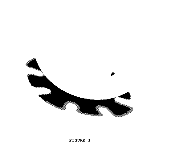
[0019] FIG. 1 is a perspective view of an embodiment of a cranial plug of the
present
invention,
100201 FIG. 2 is a perspective view of an embodiment of a cranial plug of the
present
invention.
10021] FIG. 3 is a side view of an embodiment of a cranial plug of the present
invention.
100221 FIG. 4 is bottom view of an embodiment of a cranial plug of the present
invention.
[00231 FIG. 5 is an alternate side view of an embodiment of a cranial plug of
the present
invention.
(00241 FIG. 6 is a perspective top view of an embodiment of a cranial plug of
the present
invention.
100251 FIG. 7 is a top view of an embodiment of a cranial plug of the present
invention.
[0026] FIG. 8 is a perspective view of an embodiment of a cranial plug of the
present
invention.
100271 FIG. 9 is a view of an embodiment of a cranial plug of the present
invention when
inserted into a burr hole.
[0028] FIG. 10 is an embodiment of a cranial plug of the present invention
when the plug is
substantially flat prior to insertion into a burr hole.
10029] FIG. 11 is a perspective view of an alternate embodiment of a cranial
plug of the
present invention.
100301 FIG. 12 is a perspective view of an alternate embodiment of a cranial
plug of the
present invention.
CA 02837568 2013-10-18
WO 2012/151432
PCT/US2012/036374
100311 FIG. 13 is a perspective view of an alternate embodiment of a cranial
plug of the
present invention.
100321 FIG. 14 is an embodiment of a cranial plug of the present invention
when the plug is
substantially flat prior to insertion into a burr hole.
DETAILED DESCRIPTION
100331 The cranial plugs of the present invention may be made, in part or in
its entirety,
with bioabsorbable material. The term "bioabsorbable material" as used herein
includes
materials which are partially or completely bioabsorbable in the body.
100341 Suitable bioabsorbable materials include collagen, polyglycolide,
poly(lactic acid),
copolymers of lactic acid and glycolic acid, poly-L-lactide, poly-L-lactate;
crystalline plastics
such as those disclosed in U.S. Pat. No. 6,632,503 which is incorporated
herein by reference;
bioabsorbable polymers, copolymers or polymer alloys that are self-reinforced
and contain
ceramic particles or reinforcement fibers such as those described in U.S. Pat.
No. 6,406.498
which is incorporated herein by reference; bioresorbable polymers and blends
thereof such as
described in U.S. Pat. No. 6,583,232 which is incorporated herein by
reference; copolymers
of polyethylene glycol and polybutylene terephthalate, and the like. The
foregoing list is not
intended to be exhaustive. Other bioabsorbable materials can be used based
upon the
principles of the invention as set forth herein. Some of the most common
include Poly-L-
lactic acid (PLLA) Poly-DL-lactic acid (PDLLA) Polyglycolic acid (PGA)
Polydioxanone
(PDS) Polyorthoester (POE) Poly-C-capralactone (PCL).
10035] Bioactive materials can be admixed with the bioabsorbable materials,
impregnated
in the bioabsorbable materials and/or coated on the outer surface thereof.
Bioactive materials,
including natural and/or synthetic materials, also can be used to fill
cavities in the cranial
plugs. These materials can include, for example, bioactive ceramic particles,
bone chips or
paste, platelet rich plasma (PRP), polymer chips, synthetic bone cement,
autologous
materials, allograft, cadaveric materials, xenograft, nanoparticles,
nanoemulsions and other
materials employing nanotechnology, capsules or reinforcement fibers. And they
can contain,
for example, antimicrobial fatty acids and related coating materials such as
those described in
Published U.S. Patent Publication No. 2004-0153125; antibiotics and
antibacterial
6
CA 02837568 2013-10-18
WO 2012/151432
PCT/US2012/036374
compositions; immunostimulating agents; tissue or bone growth enhancers and
other active
ingredients and pharmaceutical materials known in the art.
100361 The cranial plugs of the invention which are made with bioabsorbable
material can
be made by molding, extrusion, heat shrinking or coating the bioabsorbable
material on a
base which has been provided with attachment means such as those described in
Published
U.S. Patent Publication No. 2006-0142772 which is incorporated herein by
reference. When
the bioabsorbable material will have functional mechanical properties which
are not made
from the base material, the bioabsorbable material can be molded onto the base
in the desired
shape. Alternatively, the bioabsorbable material also can be coated, shrink
wrapped or
molded onto the base. If necessary, the bioabsorbable material can be machined
to the desired
shape and/or dimensions.
100371 As will be apparent to those skilled in the art, the sizes of the plugs
of the invention
can be varied to meet their intended applications. The shapes can take various
forms in
addition to those illustrated without deviating from the principles of the
invention. And the
sizes, lengths and widths can be varied for particular applications within the
principles of the
invention set forth herein.
100381 When the plug is inserted into the burr hole, at least a portion of the
plug would
cover the outer layer of the brain, the dura. The intent is to create a
"floor" in the opening to
aid in containment of autologous and other bioactive substances with the goal
of creating
fusion across the burr hole. Without this "floor", these substances could
easily be pushed
beneath the skull, into the space between the dura and inside of the skull.
This would result
in bioactive substances where they are not intended and it would also force
the surgeon to use
a larger volume of these materials than is necessary. The latter is a concern
because bioactive
substances are costly, or when harvested autologously, are available in
limited quantity.
100391 Any of the plugs disclosed in this application could be produced in
either a
preformed shape (e.g., Figures 1-8) or a flat condition (e.g., Figure 10).
Either style would
produce the desired result provided the material was sufficiently flexible. In
a preferred
embodiment, the installed plug provides gaps, holes or slots in the sidewall
to allow bone
graft or bioactive materials to directly contact the sidewalls of the burr
hole thereby
improving fusion. Alternately the sidewalls can be solid and the plug material
could be a
7
CA 02837568 2013-10-18
WO 2012/151432
PCT/US2012/036374
rapidly dissolving material or a bioabsorbable material impregnated with a
bioactive
substance. Figures 12 and 13 show an embodiment with and without holes in a
side wall.
100401 In the flat condition, the plugs may be pre-scored or embossed to cause
them to
fold/collapse in a predetermined manner. Such an embodiment is shown in Figure
14.
100411 In an alternate embodiment (not shown), the cranial plug may be in
essentially flat
condition and tucked under the skull to cover the dura in the area(s) directly
below the burr
hole(s) before the skull flap is reattached. The plug/dura cover is made from
a sheet of
generally even thickness and can be in one piece or more than one piece. The
cranial plug of
this design would not be inserted into the burr hole and would not have any
significant
contact with the sidewalls of the burr hole.
8
