Note : Les descriptions sont présentées dans la langue officielle dans laquelle elles ont été soumises.
ARTICULATING OPHTHALMIC SURGICAL PROBE
la Field of the Invention
This invention relates to ophthalmic surgical equipment and more particularly
to posterior segment ophthalmic surgical probes.
Background of the Invention
15 Microsurgical
instruments typically are used by surgeons for removal of tissue
from delicate and restricted spaces in the human body, particularly in surgery
on the
eye, and more particularly in procedures for removal of the vitreous body,
blood, scar
tissue, or the crystalline lens. Such instruments include a control console
and a
surgical handpiece with which the surgeon dissects and removes the tissue.
With
zo respect to posterior
segment surgery, the handpiece may be a vitreous cutter probe, a
laser probe, or an ultrasonic fragmenter for cutting or fragmenting the tissue
and is
connected to the control console by a long air- pressure (pneumatic) line
and/or power
cable, optical cable, or flexible tubes for supplying an infusion fluid to the
surgical
site and for withdrawing or aspirating fluid and cut/fragmented tissue from
the site.
25 The cutting, infusion,
and aspiration functions of the handpiece are controlled by the
remote control console that not only provides power for the surgical
handpiece(s)
(e.g., a reciprocating or rotating cutting blade or an ultrasonically vibrated
needle), but
also controls the flow of infusion fluid and provides a source of vacuum
(relative to
atmosphere) for the aspiration of fluid and cut/fragmented tissue. The
functions of the
30 console are controlled
manually by the surgeon, usually by means of a foot-operated
switch or proportional control.
During posterior segment surgery, the surgeon typically uses several
handpicces or instruments during the procedure. This procedure requires that
these
instruments be inserted into, and removed out of the incision. This repeated
removal
35 and insertion can
cause trauma to the eye at the incision site. To address this concern,
Page 1
CA 2842440 2018-10-12
CA 02842440 2014-01-17
WO 2013/019859
PCT/1JS2012/049160
hubbed cannulae were developed at least by the mid-1980s. These devices
consist of
a narrow tube with an attached hub. The tube is inserted into an incision in
the eye up
to the hub, which acts as a stop, preventing the tube from entering the eye
completely.
Surgical instruments can be inserted into the eye through the tube, and the
tube
protects the incision sidewall from repeated contact by the instruments. In
addition,
the surgeon can use the instrument, by manipulating the instrument when the
instrument is inserted into the eye through the tube, to help position the eye
during
surgery.
Many surgical procedures require access to the sides or forward portion of the
io retina. In order to reach these areas, the surgical probes must be pre-
bent or must be
bendable intra-operatively. Various articulating optical surgical probes for
providing
laser and/or illumination light are known. See for example, U.S. Patent No.
5,281,214
(Wilkins, et al.) and U.S. Patent No. 6,984,130 (Scheller, et a/.). The
articulation
mechanism, however, adds extra complexity and expense. One flexible laser
probe
is needing no articulation mechanism is commercially available, but this
device uses a
relatively large diameter optical fiber sheathed in a flexible tube comprising
the distal
tip, resulting in a large bend radius and large distal tip diameter with
significant bend
stiffness. These characteristics require that the distal tip contain a non-
bent straight
portion for ease of insertion of the bent portion, which must flexibly
straighten as it
20 passes through the hubbed cannula. The straight portion of the distal
tip allows the
bent portion to flexibly pass through the hubbed cannula before the distal
cannula of
the handpiece enters the hubbed cannula, to allow maximum bending clearance of
the
flexible portion, thereby minimizing the bending strain and corresponding
frictional
insertion forces. Such a large bend radius, large diameter flexible tube, and
straight
25 distal tip causes the useable portion of the fiber to extend a
relatively long distance
from the distal tip of the probe and limits access of the probe.
A further disadvantage in the known art is the flexibility of the distal
cannula,
which is a function of the material properties and cross sectional moment of
inertia, as
determined by the gauge size of the outside diameter of the cannula to fit
within the
30 hubbed cannula, and the inside diameter of the cannula to accept the
flexible tube.
For any given material, the outer and inner diameters of the cannula determine
the
flexibility of the cannula. This flexibility limits the surgeon's ability to
use the
instrument to manipulate the position of the eye during surgery.
Page 2
A flexible-tip probe is disclosed in U.S. Patent Application Publication
2009/0093800
(Auld, et al.) that does not require a straight portion of flexible tube,
which thus provides a
more compact useable tip length, thereby allowing greater access to internal
posterior
structures of the eye without compromising insertion forces. The flexible-tip
probe provides
increased rigidity of the distal cannula to facilitate manipulation of the
position in the eye
during surgery. While this probe provides a relatively smaller cross section
as compared to the
previous probes, such as those disclosed by Scheller et al., it does not
provide controllable
articulation over a range of angles in the manner those probes do.
Brief Summary of the Invention
Certain exemplary embodiments can provide an articulating optical surgical
probe,
comprising: a handle; a cannula extending from the handle, the cannula having
a diameter
of 20 Ga or less; a slotted tip at a distal end of the cannula, the slotted
tip comprising a
plurality of slots spaced evenly along a length of the slotted tip; at least
one optical fiber
extending through the handle, the cannula, and the slotted tip to a distal end
of the slotted
tip; and a pull-wire immovably secured to a first fixed object in the handle
and suspended
between the first fixed object and a second fixed object in the handle in a
substantially
straight orientation, the pull-wire further extending through the cannula and
coupled to the
distal end of the slotted tip, wherein a deflection of the pull-wire from the
substantially
straight orientation causes an increased tension on the pull-wire which causes
the slotted
tip to deviate from a straight position to a bent position; a sliding pin
located between the
first object and the second object within the handle, wherein the pull-wire is
suspended
relative to the sliding pin when the sliding pin is in a first position such
that the sliding pin
does not deflect the pull-wire from the substantially straight orientation
when in the first
position; wherein an advancing movement of the sliding pin to an additional
position
causes a deflection of the suspended pull-wire and an associated increase in
the tension of
the pull-wire, the increased tension causing the slotted tip to deviate from a
straight
position to a bent position; and wherein the slotted tip is formed from a
resilient material
that will restore to the substantially straight orientation when the increased
tension exerted
by the pull-wire is released.
Page 3
CA 2842440 2019-06-18
Other embodiments describe an articulating optical surgical probe that
includes a
handle sized to fit in a single hand and a single rigid cannula extending from
the handle having
a diameter of 20 Ga or less. The probe further includes a slotted tip at a
distal end of the cannula
and at least one optical fiber extending through the handle, the single rigid
cannula and the
slotted tip, and a pull-wire secured to the slotted tip. When the pull-wire
exerts tension on the
slotted tip, the slotted tip will deviate from straight to a bend angle
controlled by the tension
in the pull-wire. The slotted tip is formed from a resilient material that
will restore to the
straight position when the tension exerted by the pull-wire is released.
Other objectives, features and advantages of the present invention will become
apparent with reference to the drawings, and the following description of the
drawings and
claims.
Brief Description of the Drawings
FIG. 1 is a schematic of an articulating optical surgical endoprobe, according
to a
particular embodiment of the present invention;
FIG. 2 illustrates an end view of an example of a slotted tip 20 according to
a
particular embodiment of the present invention;
FIGs. 3A-3H illustrate various slot designs for a slotted tip according to
particular
embodiments of the present invention; and
Page 3a
CA 2842440 2018-10-12
CA 02842440 2014-01-17
WO 2013/019859
PCMJS2012/049160
FIGs. 4A-4K illustrate various mechanisms for increasing the tension in the
pull-wire 22 according to particular embodiments of the present invention.
Detailed Description of the Invention
Various embodiments of the present invention may avoid difficulties
associated with previous articulating optical surgical probes. In particular,
certain
embodiments of the present invention may provide a single rigid cannula with a
small
diameter not only capable of insertion into very small incisions but also
capable of
io articulating in a controlled fashion through a range of angles. Thus,
such
embodiments of the present invention combine the advantages of a relatively
rigid
articulating optical surgical probe with the controllable articulation of dual
cannula
probes that require a larger diameter.
Particular embodiments of the present invention include a single rigid cannula
is with a slotted tip of resilient material secured to a pull wire.
Tension in the pull wire
causes the slotted tip to bend in a particular direction, while releasing the
tension
allows the resilient tip to restore to its straight position. Pull-wire
technology has
been used previously to deviate a distal end of a surgical catheter, but not
in a small-
diameter, rigid cannula used in handheld optical surgical probes nor with the
degree
zo of angular movement used in the relatively small spaces found within
the interior of
an eye. Consequently, the application of pull-wire tension in the context of
hand-held
surgical probes is uniquely advantageous. In particular embodiments of the
present
invention, one or more of the optical fibers used in the endoprobe 10 may also
be used
as the pull-wire.
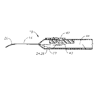
25 FIG. 1 is a schematic of an articulating optical surgical endoprobe
10,
according to a particular embodiment of the present invention, with a handle
12
suitable for being held in a single hand and a cannula 14. (For ease of
illustration, the
handle 12 and cannula 14 are not shown to scale and certain external features
of the
handle 12, such as the control mechanism for an internal pull-wire, are not
shown.)
30 The proximal end of the endoprobe 10 is connected to one or more
light sources (not
shown) that provide laser and/or illumination light by connection to at least
one
optical fiber running through the interior of the endoprobe 10.
Page 4
CA 02842440 2014-01-17
WO 2013/019859
PCT/1JS2012/049160
The cannula 14 is formed of a rigid biocompatible material, such as stainless
steel. Endoprobes, according to various embodiments of the present invention,
use a
"single" rigid cannula, referring to the fact that there is no other
relatively rigid, self-
supporting cannula formed separately inside or outside the single cannula
and/or
independently movable with respect to the single cannula. However, the term
"single" is not intended to exclude the use of multiple layers or coatings to
form the
single cannula, nor does it exclude the use of soft polymer sleeves or sheaths
that
conform to the shape of the cannula. The cannula 14 has a slotted tip 20 at a
distal
end (referring to the end farthest from the surgeon during use). The slotted
tip 20 may
io articulate in a selected direction in a controllable manner by applying
tension to a pull
wire secured within the slotted tip 20 (not shown in FIG. 1).
The slotted tip 20 is formed from a resilient material, referring to a
material
that can restore to the straight orientation after the tension from the pull-
wire is
removed. The resilient material for the slotted material may be, for example,
nitinol,
is which can be both sufficiently rigid to insert through an incision hub
and sufficiently
resilient to restore after articulation. Other metals, such as spring steel,
or other
materials with similar properties known in the art could be used. Depending on
the
particular slot configuration of the slotted tip, it may be possible to use
relatively rigid
materials that are not exceptionally elastic, such as stainless steel, Ni-base
super-
20 alloys, Co-Cr alloys, or the like without applying so much force as to
exceed the yield
point and permanently deform the material. The resilient materials may
themselves
be biocompatible, or they could be enclosed in another material, such as a
polymer
sheath, to prevent contact with tissue. The cannula 14 and the slotted tip 20
can be,
but need not be, formed from the same material. The cannula 14 and/or the
slotted tip
25 20 may also be coated with a stiffening material, such as synthetic
diamond or metal
plating (e.g., chromium), to provide improved stiffness for insertion into an
incision
hub and to reduce the likelihood of breakage.
FIG. 2 illustrates an end view of an example of a slotted tip 20 according to
a
particular embodiment of the present invention. In the embodiment depicted in
FIG.
30 .. 2, a pull-wire 22 is secured to what is shown as the top side of the
slotted tip 20. The
slotted tip 20 encloses two optical fibers 24 and 26, an illumination fiber 24
having a
diameter of 183 jm and a laser fiber 26 having a diameter of 108 ttm. Allowing
for
Page 5
CA 02842440 2014-01-17
WO 2013/019859
PCT/1JS2012/049160
the width of the slotted tip 20, this allows for the diameter of the cannula
14 to be
made smaller relative to dual cannula systems.
FIGs. 3A-3H illustrate various slot designs for the slotted tip 20 according
to
particular embodiments of the present invention (respectively labeled as
"20A,"
"20B," etc., and collectively referred to as "slotted tip 20"). In FIG. 3A,
slots deeper
than the radius of the slotted tip 20A are cut into the side of the slotted
tip 20A toward
which the slotted tip 20A is to bend. Shallow slots are cut into the opposite
side
permitting that side to bend as well. FIGs. 3B and 3C illustrate straight and
curved
"keyhole" slots having a wider base as the slot extends deeper into the
respective
io slotted tips 20B and 20C. The wider base reduces the amount of force
required to
deviate the slotted tip 20B or 20C to its curved position, potentially making
the
endoprobe 10 more comfortable to use.
FIGs. 3D-3G illustrate slot designs that can be used for more rigid tip
materials to allow the slotted tip 20 to resiliently restore to the straight
position after
being deviated into the curved position. In FIG. 3D, slots that are generally
oblong
along the length of the cannula 14 are set opposed to shallow back slots,
which tends
to reduce the force required to deviate the slotted tip 20D to the curved
position. In
FIG. 3E, a continuous spiral cut, allowing the slotted tip 20E to bend, is
interspersed
with back slots (in this case, keyhole slots), causing the slotted tip 20E to
bend in the
direction of the back slots. FIG. 3F shows a spiral cut pattern wherein the
spiral path
is perpendicular to the longitudinal axis of the slotted tip 20F on one side,
causing the
slotted tip 20F to preferentially deviate toward the side where the spiral
path is
perpendicular. FIG. 3G shows a spiral cut pattern with the cut widened
selective on
one side, causing slotted tip 20G to selectively deviate toward the side where
the
spiral cut is wider.
FIG. 3H shows a slotted tip 20H formed out of a wound wire of material, such
as by winding material drawn into wires around a mandrel. At a proximal and
distal
end of the slotted tip 20H the coils of the wound wire are welded together. In
the
intermediate region between the proximal and distal ends, one side of the tube
has
widened interstitial slots formed between the coils of the wound wire, causing
the
slotted tip 20H to selectively deviate toward the widened slots when tension
is applied
via the pull-wire. Forming the slotted tip 20 of a wound wire of material may
have
advantages by permitting the use of materials that can more easily be formed
into
Page 6
CA 02842440 2014-01-17
WO 2013/019859
PCT/1JS2012/049160
wires than tubes. Although a single wound wire is shown in FIG. 311, multiple
wire
strands could also be used.
FIGs. 4A-4K illustrate various mechanisms for increasing the tension in the
pull-wire 22 according to particular embodiments of the present invention. In
FIGs.
4A and 4B, the pull-wire 22 is wound on a pinion 40 secured between a control
button
42 and a base 44. The pinion 40 comprises two surfaces, a smaller diameter
surface r
which rolls between the control button 42 and a base 44, and a larger diameter
surface
R about which the pull wire 22 winds. The radial difference between the
smaller and
larger diameter surfaces r and R results in a differential displacement Al in
the pull
io wire as the pinion 40 rotates and translates. By selecting appropriate
diameters for the
smaller and larger diameter surfaces r andR, a relatively small amount of pull
wire
displacement d/ can be achieved during a relatively large amount of control
button
translation, providing the user with precise control over the deflection in
the slotted
tip 20. In one embodiment, the smaller diameter surface r comprises gear teeth
with
mating gear teeth on the control button 42 and the base 44. This may reduce
the
likelihood of slippage.
FIGs. 4C and 4D illustrate a lever arm 50 with a sliding actuation pin 52 held
in place by a fixed pin 54 at a pivot of the arm. A control button (not shown)
can be
used to advance the sliding pin 52, permitting the proximal portion of the
lever arm 50
.. to rise, thus rotating a lanyard 56 at a distal end of the lever arm 50 to
apply tension to
the pull-wire 22. FIGs. 4E and 4F show a pull-wire 22 threaded over a sliding
pin 60
and a first fixed pin 62 and anchored to a second fixed pin 64. Advancing a
control
button 66 attached to the sliding pin 60 increases the tension in the pull-
wire 22.
FIGs. 4G and 411 illustrate a pull-wire 22 threaded over a sliding pin 70 that
is
.. directed in a generally upward direction by a guide track 72 as a control
button 74 is
advanced. The path of the guide track 72 determines how the tension in the
pull-wire
22 varies as the control button is advanced, thus providing a smooth and
controlled
increase in tension. In the case of a linear guide, like the one illustrated
in FIG. 4G
and 4H, the pull-wire take up will occur in the latter portion of the
advancement of the
control button 74. In the alternative configuration shown in FIG. 41, the
guide track
72 is reshaped to provide greater take-up of the pull-wire at the beginning of
the
advancement by the control button 74 to produce a more balanced increase in
tension
throughout the stroke of the control button 74. In FIG. 4J, the guide track 72
inclines
Page 7
CA 02842440 2014-01-17
WO 2013/019859
PCT/1JS2012/049160
even more sharply so that most of the tension increase takes place early in
the stroke
of the control button 74. FIG. 4K illustrates an alternative embodiment of the
guide
track 72 with detents 80, allowing for distinct "stops" along the path
corresponding to
different angles of the slotted tip 20. A shelf or surface with detents can be
also be
used with any of the various embodiments of endoprobe 10 using a sliding pin
or
similar actuation mechanism, including any of the embodiments shown in FIGs.
4A-
4K.
While certain embodiments of the present invention have been described
above, these descriptions are given for purposes of illustration and
explanation.
Variations, changes, modifications and departures from the systems and methods
disclosed above that would be apparent to one skilled in the art may be
adopted
without departure from the scope of the present invention as recited in the
following
claims.
Pagc 8