Note : Les descriptions sont présentées dans la langue officielle dans laquelle elles ont été soumises.
CA 02844124 2014-02-04
WO 2013/025339 PCT/US2012/048913
SURGICAL ACCESS SYSTEM INCLUDING
SURGICAL PORTAL APPARATUS AND ADHESIVE PATCH
CROSS-REFERENCE TO RELATED APPLICATIONS
[0001] This application claims the benefit of and priority to U.S.
Provisional Patent
Application No. 61/522,790, filed August 12, 2011, the entire disclosure of
which is incorporated
by reference herein.
BACKGROUND
Technical Field
[0002] The present disclosure relates generally to a surgical access
system for use in a
surgical procedure, and more particularly, to a surgical access system
including a surgical portal
apparatus and an adhesive patch for forming a fluid tight seal with both
tissue and one or more
surgical objects positioned therethrough.
Background of Related Art
[0003] Today, many surgical procedures are performed through small
incisions in the
skin, as compared to the larger incisions typically required in traditional
procedures, in an effort
to reduce both trauma to the patient and recovery time. Generally, such
procedures are referred
to as "endoscopic", unless performed on the patient's abdomen, in which case
the procedure is
referred to as "laparoscopic". Throughout the present disclosure, the term
"minimally invasive"
should be understood to encompass both endoscopic and laparoscopic procedures.
[0004] During a typical minimally invasive procedure, surgical objects,
such as surgical
access devices, e.g., trocar and cannula assemblies, or endoscopes, are
inserted into the patient's
body through the incision in tissue. In general, prior to the introduction of
the surgical object
- 1 -
CA 02844124 2014-02-04
WO 2013/025339 PCT/US2012/048913
into the patient's body, insufflation gases are used to enlarge the area
surrounding the target
surgical site to create a larger, more accessible work area. Accordingly, the
maintenance of a
substantially fluid-tight seal is desirable so as to prevent the escape of the
insufflation gases and
the deflation or collapse of the enlarged surgical site.
[0005] To this end, various surgical ports with valves and seals are used
during the
course of minimally invasive procedures and are widely known in the art. The
small incisions,
however, are typically enlarged for specimen removal from the patient's body.
The enlarged
opening prohibits continued use of the surgical port therethrough as the
opening has become too
large to maintain a fluid-tight seal with the surgical port. Additional
incisions may be required
for continued access of the surgical site with a surgical port.
[0006] A continuing need exists for a surgical access system that can
facilitate the
accessibility of an underlying tissue site with relative ease and with minor
inconvenience for a
clinician. It would be advantageous to provide a surgical access system that
would allow for
continued use, or re-use, of a surgical port after an incision is enlarged
while maintaining a fluid
tight seal with the enlarged incision.
SUMMARY
[0007] The present disclosure relates to surgical access systems and
methods of using the
same during a surgical procedure. The surgical access system includes a portal
member and a
patch. The portal member includes at least one longitudinal port for passage
of a surgical object.
The portal member is formed from a compressible material and is adapted to
transition from a
first expanded condition to a second compressed condition such that an outer
surface is adapted
for a substantial sealing relation with an opening in tissue upon insertion of
the portal member
- 2 -
CA 02844124 2014-02-04
WO 2013/025339 PCT/US2012/048913
therethrough. The patch includes a non-porous substrate having a tissue facing
surface including
an adhesive for positioning over the opening in the tissue and sealing a
surface thereof The
patch is adapted for sealed reception of the portal member.
[0008] In accordance with one embodiment of the present disclosure, to
access a surgical
site, a patch including a non-porous substrate having a tissue facing surface
including an
adhesive is placed over an opening in tissue and seals a surface thereof. A
portal member is then
positioned through an aperture in the patch. The portal member includes at
least one longitudinal
port for passage of a surgical object. The portal member is formed from a
compressible material
and is adapted to transition from a first expanded condition to a second
compressed condition
such that an outer surface is adapted for a substantial sealing relation with
the aperture of the
patch and the opening in the tissue upon insertion of the portal member
therethrough.
[0009] In accordance with another embodiment of the present disclosure,
to access a
surgical site, a portal member including at least one longitudinal port for
passage of a surgical
object is placed in an opening in tissue. The portal is formed from a
compressible material and is
adapted to transition from a first expanded condition to a second compressed
condition such that
an outer surface is adapted for a substantial sealing relation with the
opening in the tissue upon
insertion of the portal member therethrough. Surgical objects are placed
through the at least one
longitudinal port. The portal member is removed from the opening in the tissue
and the opening
of the tissue is enlarged. A patch including a non-porous substrate having a
tissue facing surface
including an adhesive is positioned over the enlarged opening in the tissue
and seals a surface
thereof The portal member may then be placed through the patch in sealed
relation therewith.
- 3 -
CA 02844124 2014-02-04
WO 2013/025339 PCT/US2012/048913
BRIEF DESCRIPTION OF THE DRAWINGS
[0010] Various embodiments of the present disclosure are described
hereinbelow with
reference to the drawings, wherein:
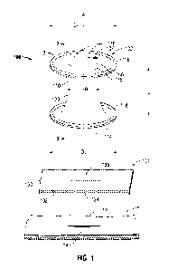
[0011] FIG. 1 is a front perspective view of a surgical access system
including a portal
member and a patch positioned relative to tissue in accordance with the
principles of the present
disclosure;
[0012] FIG. 2 is a cross-sectional view of the portal member of FIG. 1
taken along line 2-
2 of FIG. 1 illustrating a longitudinally extending port of the seal anchor
member;
[0013] FIG. 3A is a top view of a patch in accordance with another
embodiment of the
present disclosure;
[0014] FIG. 3B is a top view of a patch in accordance with yet another
embodiment of
the present disclosure;
[0015] FIG. 4A is a cross-sectional view of the patch of FIG. 3A in
accordance with one
embodiment of the present disclosure;
[0016] FIG. 4B is a top view of a patch in accordance with another
embodiment of the
present disclosure; and
[0017] FIG. 4C is a top view of a patch in accordance with yet another
embodiment of
the present disclosure.
DETAILED DESCRIPTION OF EMBODIMENTS
[0018] In accordance with the present disclosure, a surgical access
system is utilized to
access a surgical site. The surgical access system includes a surgical portal
apparatus, e.g., a
portal member, adapted for insertion into an opening, e.g., an incision, in
tissue to form a fluid-
- 4 -
CA 02844124 2014-02-04
WO 2013/025339 PCT/US2012/048913
tight seal with the tissue and an adhesive patch that creates an artificial
surface or interface
through which the portal member may be placed.
[0019] Particular embodiments of the present disclosure will be described
herein with
reference to the accompanying drawings. As shown in the drawings and as
described throughout
the following description, and as is traditional when referring to relative
positioning on an object,
the term "proximal" or "trailing" refers to the end of the apparatus that is
closer to a clinician and
the term "distal" or "leading" refers to the end of the apparatus that is
farther from a clinician. As
used herein, the term "patient" should be understood as referring to a human
subject or other
animal, and the term "clinician" should be understood as referring to a
doctor, nurse, or other
care provider and may include support personnel. In the following description,
well-known
functions or constructions are not described in detail to avoid obscuring the
present disclosure in
unnecessary detail.
[0020] One type of minimal invasive surgery described herein is multiple
instrument
access through a single surgical port. Multiple instrument access through a
single surgical port is
a minimally invasive surgical procedure, which permits a clinician to operate
through a single
entry point, typically the patient's navel. The disclosed multiple instrument
access through a
single surgical port procedure may involve insufflating the body cavity and
positioning a portal
member within, e.g., the navel of the patient. Examples of surgical
instruments or objects which
may be introduced through the portal member include clip appliers, graspers,
dissectors,
retractors, staplers, forceps, laser probes, photographic devices, trocars,
cannulas, endoscopes,
laparoscopes, arthroscopes, tubes, electrosurgical cutting, coagulating, and
ablation devices, and
other tools within the purview of those skilled in the art.
- 5 -
CA 02844124 2014-02-04
WO 2013/025339 PCT/US2012/048913
[0021] Referring now to the drawings, in which like reference numerals
identify identical
or substantially similar parts throughout the several views, FIG. 1
illustrates a surgical access
assembly 100 including a portal member 102 and a patch 104 that may be used in
any
endoscopic, laparoscopic, and/or open surgical procedure in accordance with
the principles of the
present disclosure. Portal member 102 includes an elongated body 110 defining
a longitudinal
axis "A" and including trailing (or proximal) and leading (or distal) ends 112
and 114,
respectively. Portal member 102 includes at least one longitudinal port 116,
in embodiments, a
plurality of longitudinal ports 116, extending along axis "A" between trailing
and leading ends
112 and 114, respectively, and through the elongated body 110. At least one or
more ports 116
are dimensioned to receive a surgical object, such as a cannula 106
therethrough. Upon
introduction through a respective port 116, the inner surface portions 117
(FIG. 2) defining the
port 116 establish and maintain a substantially sealed relation about the
surgical object. Cannula
106 may be inserted through the at least one longitudinal port 116 and
provides a fluid-tight seal
with the port 116. Cannula 106 provides an access port 107 including a valve
(not shown) for
passage of surgical instruments, e.g., endoscopic instruments, therethrough.
[0022] Trailing and leading ends 112 and 114 may define flange segments
118, which
may be integrally formed with portal member 102. Trailing end 112 of portal
member 102
defines a first diameter D1 and leading end 114 defines a second diameter D2.
In embodiments,
the respective first and second diameters D1, D2 of the trailing and leading
ends 112 and 114 are
substantially equivalent, as seen in FIG. 1. In other embodiments, diameters
D1, D2 may be
different. As depicted in FIG. 1, trailing and leading ends 112 and 114 define
substantially
planar surfaces. However, it is also contemplated that either or both of
trailing and leading ends
112 and 114, respectively, may define surfaces that are substantially arcuate
to assist in the
- 6 -
CA 02844124 2014-02-04
WO 2013/025339 PCT/US2012/048913
insertion of portal member 102 within a tissue opening 12 defined by tissue
surfaces 14 and
formed in tissue "T", e.g. an incision.
[0023] Elongated body 110 defines a radial dimension "R" and extends
longitudinally
between trailing and leading ends 112 and 114, respectively, to define an
axial dimension or
length "L". The radial dimension "R" of elongated body 110 varies along the
axial dimension,
or length, "L" thereof to aid in anchoring portal member 102 within tissue
"T". Other
embodiments in which the radial dimension "R" remains substantially uniform
along the axial
dimension "L" thereof is also within the scope of the present disclosure.
[0024] The radial dimension "R" of elongated body 110 may be appreciably
less than the
respective diameters D1, D2 of trailing and leading ends 112 and 114 such that
the portal member
102 defines an "hour-glass" shape or configuration to assist in anchoring
portal member 102
within tissue "T". However, in alternate embodiments, the radial dimension "R"
of elongated
body 110 may be substantially equivalent to the respective diameters D1 and/or
D2 of trailing and
leading ends 112 and 114. In cross section, elongated body 110 may exhibit any
suitable
configuration, e.g. substantially circular, oval, or oblong.
[0025] Portal member 102 may be made from a disposable, compressible,
and/or flexible
type material, such as, for example, a suitable foam or gel material having
sufficient compliance
to deform and establish a seal about one or more surgical objects, and also
establish a sealing
relation with the tissue. The compressible material may be sufficiently
compliant to
accommodate off axis motion of the surgical object. In one embodiment, the
material is a foam
including a polyisoprene material. In embodiments, the material may be
fabricated from an
elastomer such as a soft urethane gel, silicone gel, thermoplastic elastomer,
or the like.
- 7 -
CA 02844124 2014-02-04
WO 2013/025339 PCT/US2012/048913
[0026] Portal member 102 is adapted for insertion within a tissue tract
"T", e.g., through
the abdominal or peritoneal lining in connection with a laparoscopic surgical
procedure. Portal
member 102, however, is adapted for insertion within any opening in a
patient's skin, e.g., an
incision or any naturally occurring orifice. The presently disclosed portal
member 102 and
surgical access system 100 may be used with a surgically created incision, a
naturally occurring
opening, or in non-laparoscopic procedures.
[0027] When inserted within an opening 12 in tissue "T", portal member
102 is adapted
to establish a substantial seal within the opening 12, i.e., with the tissue
surfaces 14 defining the
opening 12. Portal member 102 is dimensioned to provide a fluid tight seal
with an opening 12
of about 2 mm to about 18 mm. In embodiments, portal member 102 is about 5 mm
to about 20
mm in diameter in its unbiased, expanded condition. During insertion, portal
member 102 may
be compressed from its first, expanded condition to a second, compressed
condition to permit at
least partial passage through the opening 12 in tissue "T". Once within the
opening 12, portal
member 102 will return toward the first, expanded condition with the outer
wall 120 of the portal
member 102 establishing a seal with the tissue "T" defining the opening 12.
[0028] Portal member 102 may include an insufflation conduit (not shown)
mounted
within one of ports 116 and connectable to a source of insufflation gases to
permit passage of
gases, e.g., CO2, to maintain the pneumoperitoneum. Other suitable portal
members which may
be utilized with the surgical access system 100 of the present disclosure
including, for example,
those disclosed in commonly assigned U.S. Patent Application Pub. No.
2009/0093752, entitled
"Seal Anchor for Use in Surgical Procedures", the entire contents of which is
hereby
incorporated by reference herein.
- 8 -
CA 02844124 2014-02-04
WO 2013/025339 PCT/US2012/048913
[0029] Patch 104 includes a non-porous substrate 130 including a tissue
facing surface
132 including an adhesive 134 that is adapted to adhere to tissue "T". Non-
porous substrate 130
of patch 104 may be a film, foam, mesh, fibrous sheet, or composite thereof
adapted to adhere
and seal tissue.
[0030] Non-porous substrate 130 is fabricated from suitable materials
such that the patch
104 has sufficient tensile strength to support the portal member 102 during
use in a surgical
procedure; is sufficiently inert to avoid foreign body reactions when retained
on tissue "T" for
long periods of time; and is easily sterilized to prevent the introduction of
infection when the
patch 104 is placed against opening 112 of tissue "T".
[0031] Examples of suitable materials include, for example, polyolefins
such as
polyethylene (including ultra high molecular weight polyethylene) and
polypropylene including
atactic, isotactic, syndiotactic, and blends thereof; polyethylene glycols;
polyethylene oxides;
polyisobutylene and ethylene-alpha olefin copolymers; fluorinated polyolefins
such as
fluoroethylenes, fluoropropylenes, fluoroPEGSs, and polytetrafluoroethylene;
polyamides such
as nylon, Nylon 6, Nylon 6,6, Nylon 6,10, Nylon 11, Nylon 12, and
polycaprolactam;
polyamines; polyimines; polyesters such as polyethylene terephthalate,
polyethylene naphthalate,
polytrimethylene terephthalate, and polybutylene terephthalate; polyethers;
polybutester;
polytetramethylene ether glycol; 1,4-butanediol; polyurethanes; acrylic
polymers; methacrylics;
vinyl halide polymers such as polyvinyl chloride; polyvinyl alcohols;
polyvinyl ethers such as
polyvinyl methyl ether; polyvinylidene halides such as polyvinylidene fluoride
and
polyvinylidene chloride; polychlorofluoroethylene; polyacrylonitrile;
polyaryletherketones;
polyvinyl ketones; polyvinyl aromatics such as polystyrene; polyvinyl esters
such as polyvinyl
acetate; etheylene-methyl methacrylate copolymers; acrylonitrile-styrene
copolymers; ABS
- 9 -
CA 02844124 2014-02-04
WO 2013/025339 PCT/US2012/048913
resins; ethylene-vinyl acetate copolymers; alkyd resins; polycarbonates;
polyoxymethylenes;
polyphosphazine; polyimides; epoxy resins; aramids; rayon; rayon-triacetate;
spandex; silicones;
and copolymers and combinations thereof
[0032] Patch 104 is dimensioned to surround opening 12 in tissue "T" such
that the patch
104 adheres to the surrounding tissue "T" to create a seal around opening 12.
Accordingly, patch
104 may be any suitable shape or size, such as rectangular as illustrated in
FIG. 1, circular as
illustrated in FIG. 3A, among other shapes within the purview of those skilled
in the art. In
embodiments, patch 104 may be cut to a desired size and shape. Patch 104 may
also be
substantially planar (FIG. 1) or concave (FIG. 4A) to seal opening 12 of
tissue "T".
[0033] Adhesive 134 may be applied to a portion of the patch 104, such as
coated on the
entire tissue facing surface 132 of patch 104 (FIG. 1), or around a periphery
thereof (FIG. 4A).
It is envisioned that the adhesive may be applied in a random or systematic
pattern around the
tissue facing surface 132 of patch 104. In embodiments, adhesive 134 is pre-
applied to patch
104, while in other embodiments, adhesive 134 may be applied to patch 104
prior to application
to tissue "T". Additionally, or alternatively, the patch 104 may include
mechanical means for
binding to tissue. In embodiments, the patch may include mechanical grips or
hooks to achieve,
or enhance, adhesivity to tissue.
[0034] The adhesive is a biocompatible material capable of effecting
temporary
attachment between the patch and tissue. Adhesives which may be utilized with
the surgical
access system of the present disclosure include, but are not limited to,
adhesive which cure upon
tissue contact, which cure upon exposure to ultraviolet (UV) light, which are
two-part systems
which are kept isolated from one another and cure upon coming into contact
with one another,
which are pressure sensitive, which are any combinations thereof, or any other
known suitable
- 10 -
CA 02844124 2014-02-04
WO 2013/025339 PCT/US2012/048913
adhesive. Examples of adhesives include, for example, silicones, acrylics,
polyurethanes,
polyesters, polyamides, and rubber-based adhesives. In embodiments, a hydrogel
is utilized as
an adhesive. Hydrogels are materials that absorb solvents (such as water),
undergo rapid
swelling without discernible dissolution, and maintain three-dimensional
networks capable of
reversible deformation. Hydrogels may also be utilized as a two-part adhesive
system in which
the hydrogel is a network of crosslinked molecules formed by reacting first
and second hydrogel
precursors. The first and second hydrogel precursors include functional
groups, e.g.,
nucleophilic or electrophilic functional groups, which combine to form a
crosslinked polymeric
product as a result of electrophilic-nucleophilic reactions. Hydrogels
include, for example, those
using synthetic precursors within the purview of those skilled in the art,
such as those used in
o o
commercially available products such as FocalSeal from Genzyme, Inc., Coseal
from
o
Angiotech Pharmaceuticals, and DuraSeal from Confluent Surgical, Inc. Other
examples of
adhesives which can be employed include protein derived, aldehyde-based
adhesive materials,
for example, the commercially available albuminiglutaraldehyde materials sold
under the trade
TM
designation BioGlue by Cryolife, Inc., and cyanoacrylate-based materials sold
under the trade
designations IndermilTTM
and Derma BondTM by Tyco Healthcare Group, LP and Ethicon
Endosurgery, Inc., respectively.
[0035] The adhesive patch facilitates insertion of the portal member
within a tissue
opening when the opening is an irregular shape or too large to solely
accommodate the portal
member in sealing relation, e.g., in an incision larger than about 20 mm, for
example, larger than
about 25 mm to about 30 mm. As illustrated in FIG. 1, an aperture 136 (shown
in phantom) may
be cut by a clinician into patch 104 to a desired size and shape. Aperture 136
is cut into patch
- 11 -
CA 02844124 2014-02-04
WO 2013/025339 PCT/US2012/048913
104 by creating a slit in the non-porous substrate 130 or cutting a desired
shape corresponding to
the shape of the portal member 102, for example.
[0036] Alternatively, the patch may include a pre-formed or pre-patterned
aperture. In
one embodiment, such as shown in FIG. 3A, patch 204 includes a pre-formed
aperture 236. The
diameter of aperture 136, 236 should be substantially equal or smaller than
the diameter of
elongated body 110 of portal member 102 such that a fluid-tight fit is formed
between the portal
member 102, patch 104, 204, and tissue "T". In another embodiment, as
illustrated in FIG. 3B,
patch 204 may include one or more pre-patterned apertures 236', 236", and 236"
' designated by
perforation lines 237', 237", and 237", respectively, extending around the
perimeter of
apertures 236', 236", and 236". The strength of the patch 204 is reduced at
each perforation
line 237', 237", and 237" ' so that the material within the perforation lines
237', 237", and/or
237" ' may be pressed, torn, or otherwise removed to form the aperture 236',
236", or 236". In
this manner, the appropriate size of the aperture may be selected at the time
of use,
corresponding to the size of the portal member. In embodiments, the inner,
first perforation line
237' may be configured to tear when a relatively low force is applied to the
patch 204 in the
vicinity of the perforation line 237', while the second perforation line 237"
may tear when a
higher lever of force is applied to the patch 204, and the third perforation
line 237" ' may tear
when an even higher level of force is applied to the patch 204 to minimize
inadvertent separation
at the perforation lines.
[0037] In embodiments, as illustrated in FIGS. 4A-4C, non-porous
substrate 230 of patch
204 may include a reinforcement member 238, 239, and 240, respectively, to
provide the desired
strength to the patch 104, to prevent the aperture 236 from expanding, and to
support the portal
member 102 (FIG. 1) during use. Non-limiting examples of the reinforcement
member includes
- 12 -
CA 02844124 2014-02-04
WO 2013/025339 PCT/US2012/048913
meshes, monofilaments, multifilament braids, staple fibers, and combinations
thereof In
embodiments, the reinforcement member 238 may be an additional woven or non-
woven
structure disposed within at least a portion, or entirely through, the non-
porous layer 230 as
illustrated in FIG. 4A, or the reinforcement member 239 may be positioned on a
surface of the
non-porous layer 230, as illustrated in FIG. 4B, to form a multi-layered
structure. In some
embodiments, patch 204 may be reinforced by stitching the periphery of
aperture 236 with a
monofilament or multifilament thread 240, e.g., a suture, as illustrated in
FIG. 4C.
[0038] In use, the operator of the surgical access system 100 will insert
the portal
member 102 into the opening 12 of tissue "T" such that portal 102 is disposed
within opening 12.
Flanges 118 of the portal member 102 may aid in anchoring the portal member
102 in tissue "T".
Surgical instruments, such as a cannula, may then be inserted into
longitudinal port 116 of portal
member 102 and procedures, e.g., minimally invasive procedures, may be
performed. To
remove or pass a specimen through the tissue "T" (e.g., the permanent removal
of diseased
internal anatomy and/or temporary removal of portions of the colon to be
manipulated outside of
the body before being returned to inside the body), the portal member 102 is
removed and the
opening 12 is enlarged to allow for passage of the specimen therethrough.
Thereafter, patch 104
may be applied to the enlarged opening 12 and portal member 102 may be
inserted therethrough
so that the procedure proceeds in a manner described above without requiring
multiple incisions
for accessing the surgical site.
[0039] It should be understood that the surgical access system 100 may
also be used
where a relatively large opening exists that is too large to accommodate a
portal member.
[0040] Persons skilled in the art will understand that the devices and
methods specifically
described herein and illustrated in the accompanying drawings are non-limiting
exemplary
- 13 -
CA 02844124 2014-02-04
WO 2013/025339 PCT/US2012/048913
embodiments. It is envisioned that the elements and features illustrated or
described in
connection with one exemplary embodiment may be combined with the elements and
features of
another without departing from the scope of the present disclosure. As well,
one skilled in the
art will appreciate further features and advantages of the system based on the
above-described
embodiments. Accordingly, the present disclosure is not to be limited by what
has been
particularly shown and described, except as indicated by the appended claims.
- 14 -