Note : Les descriptions sont présentées dans la langue officielle dans laquelle elles ont été soumises.
W02013/019943 CA 02844147 2014-02-03
PCT/US2012/049317
METHODS FOR MEASURING HDL SUBPOPULATIONS
This application claims priority of U.S. Provisional Application No.
61/515,101,
filed August 4, 2011, the contents of which are incorporated herein by
reference.
Throughout this application, various publications are cited. The disclosure of
these publications is hereby incorporated by reference into this application
to
describe more fully the state of the art to which this invention pertains.
Field of the Invention
The present invention relates to methods and kits for measuring HDL and
diagnosing cardiovascular disease and other HDL-related diseases in a
subject. This invention exploits the physical proximity between two protein
epitopes to identify and quantify discrete HDL subpopulations present in
heterogeneous mixtures, and measure changes in HDL subpopulations as a
result of disease or treatment.
Background of the Invention
Cardiovascular Disease and HDL
Cardiovascular disease is a leading cause of morbidity and mortality,
particularly in developed nations such as the United States, Western
European countries and East Asian countries. The incidence of mortality due
to cardiovascular disease in these regions has decreased in last 30 years
(Braunwald, E., N. Engl. J. Med. 337:1360, 1997; Hoyert, D. L., et al.,
WO 2013/019943 CA 02844147 2014-02-03
PCT/US2012/049317
"Deaths: Preliminary Data for 2003" in National Vital Statistics Reports.
Hyattsville: National Center for Health Statistics, 2005; Unal B., et. al.,
Circulation 109:1101, 2004). Factors contributing to improved patient
outcome include improved cardiovascular diagnostics, reduction of major
modifiable cardiovascular risk factors and advanced medical technologies to
treat acute coronary syndrome. Despite these advances, however,
cardiovascular disease remains a leading cause of morbidity and mortality in
developed countries (see Hoyert D. L., et al., National Vital Statistics
Reports,
2005; Ueshima, H., et. al., Circulation, 118:2702, 2008).
In the end, most cardiovascular deaths result from acute coronary syndromes,
including unstable angina pectoris and acute myocardial infarction (see Shah,
P. K., Am. J. Cardiol., 79:17, 1997). Coronary syndromes often arise from
acute coronary thrombosis, itself typically the result of disruption or
rupture of
the fibrous cap of a lipid-laden atherosclerotic plaque (see Munger, M.A. and
Hawkins, D. W., J. Am. Pharm. Assoc., 44(Suppl 1):S5, 2003). The
understanding of the mechanisms mediating atherosclerotic plaque formation,
progression and subsequent rupture remains limited. At the cellular level, the
pathophysiology of the disease remains in constant evolution, albeit at such a
slow pace that it takes years if not decades to reveal itself in the clinical
setting. From the moment the genotypic blueprint is set to the environmental
inducement brought about through lifestyle choices, the disease has a
beginning and an end. The factors that influence this trajectory are numerous
and at the molecular level remain mostly undefined. In the absence of
detailed molecular knowledge, it is safe to say that the physiological state
at
any juncture during the progression of this disease is different than at any
other point. This can be observed experimentally as wide-ranging biological
indicators such as biomarkers, cellular events and functional activities vary
over the course of the disease. Particular indicators that precede disease
symptoms are often referred to as risk factors that may be predictive of the
2
W02013/019943 CA 02844147 2014-02-03
PCT/US2012/049317
pending disease state. Some predictive indicators are closely associated with
the disease while others may be intrinsically involved with the disease and
its
development.
Involvement of plasma cholesterol in the development of atherosclerotic risk
and subsequent cardiovascular disease has been validated in both human
and animal models alike. Elevated LDL cholesterol and total cholesterol are
directly related to an increased risk of cardiovascular disease (Anderson, K.
M., et. al., JAMA 257:2176, 1987). The positive relationship between the
concentration of low-density lipoprotein cholesterol (LDL-C) and the future
risk
of cardiovascular events has been observed in many large-scale population
studies, and the benefits of reducing LDL-C levels has been proven in
numerous intervention studies. The apparent effects of aggressive LDL-
lowering are exemplified by various statin treatments leading to risk
reductions of over 25-35%, and further declines in LDL-C levels by co-
administration of drugs targeting LDL-C levels through independent
mechanisms of action (including ezetimibe and resins) could result in plaque
regression.
In contrast, it has been established that the risk of cardiovascular disease
is
inversely proportional to plasma levels of HDL-C and the major HDL
apolipoprotein, apoA1 (Gordon, D. J., et al., N. Engl. J. Med 321:1311, 1989).
Studies have shown that high HDL-C levels are associated with longevity
(Barzilai, N., et al.. JAMA 290:2030, 2003). Consistent with these findings,
an
abnormally low HDL-C level is a well-accepted risk factor for the development
of clinically significant atherosclerosis (particularly common in men with
premature atherosclerosis) (Gordon, D. J., et al., N. Engl. J. Med. 321:1311,
1989; Wilson, P. W., et al., Arteriosclerosis 8:737, 1988).
3
W02013/019943 CA 02844147 2014-02-03
PCT/US2012/049317
Early demonstration of the inverse relationship between HDL-C levels and
cardiovascular risk can be found in the Framingham Heart Study, which
showed that individuals with HDL-C levels of less than 35 mg/dL at the
beginning of the study had a future coronary risk of greater than four times
that of individuals with HDL-C levels over 65 mg/dL (Wilson, P. Wõ et al.,
Amer. J. Cardio(., 46:649, 1980). Other prospective population studies
including PROCAM, Helsinki Heart Study and Multiple Risk Factors
Intervention Trial support the view that risk associated with lower HDL-C is
independent of LDL-C levels, and raising levels of HDL-C should be
considered as important a therapeutic target as lowering LDL-C. The
increased risk associated with a low HDL-C can be seen at all concentrations
of LDL-C (Gordon, T., et. al., Am. J. Med., 62:707, 1977). Post hoc analyses
of stable CHD and ACS in prospective trials indicate that both HDL-C and
triglyceride levels are associated with high risk even at recommended LDL-C
goals (Olsson, A. G., et. al., Eur. Heart J., 26:890 2006; Mil(er, M., et.
al., J.
Am. Coll. Cardiol., 51:724, 2008; Barter, P., et. al., N. Eng. J. Med.,
357:1301,
2007). These studies suggest that for every HDL-C increase of 1 mg/dL, the
risk for a CHD event is reduced by 2-5% (Chapman M. J., et al., Curr. Med.
Res. Opin. 20:1253, 2004). Thus, a strategy of targeting both high LDL-C and
low HDL-C is supported by the results of the INTERHEART Study which
showed that the ratio of apoB to apoAl (reflecting LDL to HDL ratio)
demonstrated considerable power for predicting future myocardial infarction in
a broad population of differing ethnic origin (Yusuf S., et. al., Lancet
364:973,
2004).
Despite the growing epidemiological evidence indicating that HDL-C is a
cardiovascular risk marker and raising HDL-C levels can reduce that risk,
ambiguity and debate continue to challenge the concept of HDL as a risk
marker or therapeutic target (see Chapman, M. J., et. al., Eur. Heart J.,
2011,
Apr 29 online). Large failures of HDL-modifying drug trials undermine the
4
W02013/019943 CA 02844147 2014-02-03
PCT/US2012/049317
confidence of researchers and clinicians alike (Tall, A. R., Arterioscler.
Thromb. Vasc. Biol., 27:257, 2007; Horowitz, J. D., et. al., Cardiovasc. Drug
Ther., 25-69, 2011; AIM-HIGH Investigators, Am. Heart J., 161:471, 2011)
and have left researchers searching for explanations.
Cholesterol numbers are expressed as different units of measurement in
different countries. The United States uses milligrams as the standard for
measuring cholesterol, and levels in the blood are expressed as milligrams
per deciliter (mg/dL). In Canada, millimoles per liter (mmol/L) are used in
measuring cholesterol numbers, and the same goes for many parts of Europe.
In the United States, good cholesterol numbers for the average, healthy
person are less than 200 mg/dL. Once a person gets to 200 mg/dL, he is
considered to have borderline-high levels of cholesterol. At levels of over
240
mg/dL, the person is considered to have high cholesterol. In Canada and
many European countries, good cholesterol numbers are those under 5.2
mmol/L. Above 5.2 mmol/L. and up to 6.2 mmol/L is considered borderline
high. Once a person's levels move above 6.2 mmol/L of blood, his levels of
cholesterol are considered high. Sometimes, cholesterol numbers are
categorized by the type of cholesterol. In the United States, LDL levels of
less
than 70 mg/dL are considered best for those at higher risk for developing
heart disease, which corresponds to 1,8 mmol/dL in Canada and many parts
of Europe. An LDL level of 100 to 129 mg/dL in the United States and 2.6 to
3.3 mmol/L is considered close to optimal for those at lower or average risk
of
developing heart disease. HDL-C levels are considered good at 60 mg/dL
and above in the United States, and more than 1.5 mmol/L in Canada and
European countries. The range from 40 to 59 mg/dL (1.3 to 1.5 mmol/L) may
be considered acceptable for HDL numbers, depending on gender and other
risk factors for heart disease. Anything below 50 mg/dL (1.3 mmol/L) is
considered poor for women. Levels of HDL-C below 40 mg/dL (1 mmol/L) are
considered poor for men.
W02013/019943 CA 02844147 2014-02-03
PCT/US2012/049317
The current version of the Framingham Risk Score was published in 2002
(see "Third Report of the National Cholesterol Education Program (NCEP)
Expert Panel" Circulation, 106:3143 2002). The publishing body is the Adult
Treatment Panel III (ATP III), an expert panel of the National Heart, Lung,
and
Blood Institute, which is part of the National Institutes of Health (NIH),
USA.
The Framingham/ATP III criteria were used to estimate CHD risk in the USA.
Data from 11,611 patients from a very large study, the NHANES III, were
used. The Risk Score is estimated using the 10-year risk for coronary heart
disease (CHD). The updated version included age range, gender, total
cholesterol, LDL cholesterol, HDL cholesterol, blood pressure, hypertension
treatment and smoking, and it excluded diabetes, because diabetes
meanwhile was considered to be a CHD Risk Equivalent. Some patients
without known CHD have a risk of cardiovascular events comparable to that
of patients with established CHD. Cardiology professionals refer to such
patients as having a CHD Risk Equivalent. These patients should be
managed as patients with known CHD. Diabetes is accepted as a CHD Risk
Equivalent.
Guidelines receive regular review and constant revision compelled by ongoing
and growing scientific knowledge of the disease. Recent recommendations of
the European Atherosclerosis Society (EAS) Consensus Panel (see
Chapman, M. J., et. al., Eur. Heart J., 32:1345, 2011) include targeting
elevated low HDL-C < 1 nrimol/L (40 nrig/dL) and/or triglyceride-rich
lipoproteins (TRLs) > 1.7 mmol/L (150 mg/dL). These recommendations will
facilitate reduction in the substantial cardiovascular risk that persists in
patients with cardiometobolic abnormalities at LDL-C goal.
The mechanisms by which HDL prevents cardiovascular disease are the
subject of current scientific research. As a predictive risk factor and then
as a
6
W02013/019943 CA 02844147 2014-02-03
PCT/US2012/049317
functional contributor to atherosclerosis, the role of HDL itself likely
varies
during the progression of the disease and the associated physiological state
of the individual. The biological functions, attributed to the lipoprotein
particle
population, which are important to the prevention of plaque formation, could
in
fact be significantly different than those HDL activities critical to reducing
inflammation of the arterial wall and unrelated still to the role HDL plays
during
recruitment of platelets to the growing thrombus. On an individual basis,
levels of these various activities likely differ. Preceding the onset of the
disease, it is supposed that a state of dyslipidemia has been established
which is characterized by an imbalance in favor of circulating levels of
proatherogenic, cholesterol-rich apoB-containing particles rather than the
antiatherogenic apoA1-containing HDL. Mechanisms related to lipoprotein
disequilibrium, such as HDL-mediated protection of LDL from oxidation and
lipid exchange between HDL and LDL, may be overwhelmed by such
governing principals as mass action. Some believe that HDL protects against
LDL oxidative modification that may be a trigger to the initiation and
progression of atherosclerosis (Parthasarathy, S., et al., Biochim. Biophys.
Acta, 1044:275, 1990; Barter, P. J., et al., Circ. Res. 95: 764, 2004). Others
believe that the athero-protective activity of HDL comes from removing
cholesterol from artery wall macrophages (Tall, A. R., et al., J. Clin.
Invest.,
110:899, 2002; Oram, J. F., et. al., Arterioscler. Thromb. Vasc. Biol.,
23:720,
2003). Resulting endothelial dysfunction includes arterial stiffness,
extracellular matrix signaling, and induced NO-dependent vasorelaxation
(Havlik, R. J., et. al., Am. J. Cardiol., 87:104, 2001; Ortiz-Munoz, G., et.
al.,
FASEB J. 23:3129, 2009; Nofer, J. R., et. al., J. Clin. Invest. 113:569,
2004).
Other studies indicate that inflammation is the key process underlying the
pathology given that inflammation is a systemic response directed at
decreasing toxic effects of harmful agents and repairing vessel endothelial
damage (Ross, R., et. al., N. Engl. J. Med., 340:115,1999). A variety of
specific functions associated with HDL have been attributed to its anti-
7
W02013/019943 CA 02844147 2014-02-03
PCT/US2012/049317
inflammatory activities, including prevention of endothelial inflammation,
recruitment of circulating leukocytes resulting in plaque formation followed
by
recruitment of platelets forming a thrombus (see Toth, P. P., J. Clin.
Lipidol.,
4:376, 2010; Asztalos, B. F., et. al., Curr. Opin. Lipidol., 22:176, 2011).
The pleiotropic and polygenic nature of cardiovascular disease makes for
complex disease etiology, which can obfuscate both prediction and diagnosis.
Since the initial studies measuring HDL-C and LDL-C (Eder, H. A., Am. J.
Med. 23:269, 1957), methodologies have advanced along with technology,
and predictive correlations have improved with ever more complex medical
statistical analysis (Modern Medical Statistics: A Practical Guide Brian S.
Everitt Wiley 2003). Even so, there continues to be a necessity for improved
methods for early assessment of cardiovascular disease and risk.
The Measurement and Properties of HDL
The principal of the surrogate lipid marker cholesterol to classify and
quantify
lipoprotein particles has been the historical stalwart for over fifty years.
Variations include calculating non-HDL-C, which accounts for cholesterol in
lipoprotein classes in addition to LDL, including VLDL and intermediate
density lipoproteins (IDL). An extension of this methodology uses lipoprotein
cholesterol ratios such as LDL-C:HDL-C to improve clinical correlations
(Grover, S. A., et. al., Epidemiology 14:315 2002) or total cholesterol:HDL-C.
More recently, risk metrics have been employed such as measuring apoA1, a
protein surrogate for HDL, or apoB, the surrogate marker for LDL, which may
better reflect lipoprotein particle numbers rather than their cholesterol load
(Knopp, R. H., Am. J. Med. 83:75 1987; Contois, J. H., et. al., Clin. Chem.,
42:507, 1996; Contois, J. H., et. al., Clin. Chem., 42:515, 1996). These
approaches rely on immuno-turbidimetric or -nephelometric assays
(Marcovina, S. M., et. al., Clin. Chem. 39:773, 1993), provide an alternative
8
W02013/019943 CA 02844147 2014-02-03
PCT/US2012/049317
means of measuring those lipoprotein classes, and offer a different
perspective given the physiochemical nature of the lipoprotein constituent and
the methods used to measure it. Lipoproteins measured using surrogate
proteins rather than lipids are reported to be less susceptible to
postprandial
effects and fluctuations. Similarly, proponents of the apoB:apoAl ratio
believe it to be the single best predictor of coronary risk (Walldius, G., et.
al.,
Clin. Chem. Lab Med. 42:1355, 2004; Holzmann, M. J, et al., Ann Med. 2010
Nov 30 in press). A comprehensive prospective cohort study designed to
compare the clinical utility of all said measurements and numerous ratio
metric permutations was performed to investigate prediction of coronary heart
disease in men and women. The study concluded that the apoB:apoA1 ratio
for predicting CHO was comparable with that of traditional lipid ratios, but
did
not offer incremental utility over total cholesteroi:HDL-C (ingeisson, E., et.
al.,
J. Amer. Med. Assoc., 298:776, 2007).
Other approaches to clinical measures of lipoprotein particle concentration
involve sizing and counting using nuclear magnetic resonance (Otvos, J., Clin.
Cardiol. 22:1121, 1999). This method offers an additional level of resolution
by
expanding HDL into three particle subpopulations founded on particle
diameter. This method reported discordance between individuals when
comparing LDL-C and LDL particle levels which they attributed to
disproportionate cholesterol distribution between large and small LDL (Otvos,
J. D., et. al., J. Clin. Lipidol., 5:105, 2011). Lastly, both analytical
ultracentrifugation and electrophoretic methods used in research settings
have led to fractionation of HDL into several subpopulations based on distinct
physiochemical property differences (Anderson, D. W., et. al., Biochim
Biophys Acta 493:55, 1977, Chapman, M. J., et. al., J. Lipid Res., 22:339,
1981, Kontush, A., et. al., Arterioscler. Thromb. Vasc. Biol. 23:1881, 2003,
Asztalos, B. F., et. al., Biochim. Biophys. Acta 1169:291, 1993).
9
W02013/019943 CA 02844147 2014-02-03
PCT/US2012/049317
Liquid chromatography-mass spectrometry (LC-MS) is also used in the study
of proteomics, where again components of a complex mixture must be
detected and identified in some manner. The bottom-up proteomics LC-MS
approach is a common method to identify proteins and characterize amino
acid sequences and post-translational modifications (Aebersold, R. and Mann,
M. Nature 422:198, 2003; Chait, B. T., Science 314:65, 2006). Proteins can
be purified first or the crude protein extract digested directly, followed by
one
or more dimensions of separating the peptides by liquid chromatography
coupled to mass spectrometry (a technique known as shotgun proteomics)
(Washburn, M. P., et. al., Nat. Biotechnology 19:242, 2001; Wolters, D. A.,
et.
al., Anal. Chem. 73:5683, 2001). By comparing the masses of the proteolytic
peptides or their tandem mass spectra with those predicted from a sequence
database, peptides can be identified and multiple peptide identifications
assembled into a protein identification (Nesvizhskii, A. l., Methods Mol.
Biol.
367:87, 2007; Nesvizhskii, A. l., et. al., Nat. Methods 4:787, 2007). Samples
of complex biological fluids like human serum may be run in a modern LC-
MS/MS system and result in over 1000 proteins being identified, provided that
the sample was first separated using physiochemical properties such as
density gradient ultracentrifugation, SDS-PAGE or HPLC. Such approaches
have been used to identify and quantify proteins associated with lipoprotein
particle fractions HDL and LDL.
HDL has unique and measurable physiochemical properties that arise as a
direct result of the quantity and relative amounts of its two major
constituents,
protein and lipid (Rosenson, R. S., et. al., Clin. Chem. 57:392, 2011). Both
of
these two common constituents can be further divided into specific molecular
entities. For lipids, seven classes, including fatty acyls, glycerolipids,
glycerophospholipids, sphingolipids, sterol lipids, prenol lipids,
saccharolipids
and polyketides, are recognized by the LIPIDS MAPS consortium (Fahy, E.,
et. al., J. Lipid Res., 50:S9, 2009). At the molecular level, there are -
30,100
WO 2013/019943 CA 02844147 2014-02-03
PCT/US2012/049317
distinct lipid entities identified in nature of which ¨200 have been detected
in
fractions of HDL and are referred to as the HDL lipidome. The human plasma
proteome has been curated to date to contain 1,175 distinct genes resulting in
7,614 unique protein products (Anderson, N. L., et. al., Mol. Cell.
Proteomics,
3:311, 2004). The protein fraction of HDL could consist of ¨110 different
members, either bound or associating with the lipoprotein particle (Karlsson,
H. et. al., Proteomics 5:1431, 2005; Rezaee, F., et. al, Proteomics, 6:721,
2006: Hortin, G. L., et. al., Biochem. Biophys. Res. Commun., 340:909, 2006;
Heller, M., et. al., Proteomics 5:2619, 2005; Vasair, T., et. al., J. Clin.
Inv.,
117:746, 2007; Davidson, W. S., et. al., Arterioscler. Thromb. Vasc. Biol,
29:870, 2009; Davidson, P., et. al., Arterioscler. Thromb. Vasc. Biol.,
30:156,
2009). The specific list of proteins associated with HDL is dependent upon
the methodology used to separate this lipoprotein subclass away from a
serum/plasma sample prior to analysis, given that the separation methodology
can result in loss or gain of constituents (Heller, M., et. al., Proteomics
5:2619,
2005; Gordon, S. M., et. al., J. Prot. Res. 9:5239, 2010). The consequence of
this observation is that the proteins associated with HDL can vary as a result
of the isolation technique.
The totality of all constituents in a single HDL particle combine to generate
a
physiochemical state. in the physiochemical state reside measurable
properties including hydrodynamic radii, volume, charge, and affinity. Such
properties influence migration rates used in separation technologies
employed, and include, for example, density, size/charge ratio and
hydrophobicity. Separation of one particle from another is a direct
consequence of differences in their physiochemical states which are defined
by the content of their constituents. Typical methods of separating HDL
particles from other exogenous contaminants include density
ultracentrifugation, gel electrophoresis, gel filtration chromatography and
affinity chromatography (Mendez, A. J., et al., J. Biol. Chem. 266:10104,
11
W02013/019943 CA 02844147 2014-02-03
PCT/US2012/049317
1991; Guerin, M., et. al., Arterioscler. Thromb. Vasc. Biol. 21:282, 2001; Li,
Z.,
et. al., J. Lipid Res., 35:1698, 1994; Gordon, S. M., et. al., J. Prot. Res.
9:5239, 2010; Krimbou, L., et. al. J. Lipid Res., 44:884, 2003).
HDL particle diversity and heterogeneity is a direct result of the fact that
the
distribution of both the lipid and protein constituents are in disequilibrium
with
the HDL particle population as a whole and to each other (Li, Z., et. al., J.
Lipid Res., 35:1698, 1994; Kontush, A., et. al., Arterioscler. Thromb. Vasc.
Biol. 24:526, 2004; deSouza J. A. et. al., Atherosclerosis 197:84, 2008;
Davidson W. S, et. al. Arterioscler. Thromb. Vasc. Biol. 29:870, 2009; Garcia-
Sanchez, C., et. al., Clinica Chimica Acta, 412:292, 2011). By definition,
this
means that any given HDL particle contains only a subset of lipidome and
proteome constituents. The molar concentration of individual proteome
members in the serum is much lower than that of HDL, suggesting that
specific proteome members exist only in subpopulations of HDL (Anderson,
L., J. Physiol. 563:23, 2005). Furthermore, it indicates that any two
particles
can be distinguished from each other by their lipid and protein constituents
and by the relative amounts of those molecular entities. Two HDL particles
containing the exact same proteome and lipidome, but differing in quantities,
can be distinguished from one another by such properties as size or volume.
Similarly, two particles could have similar physiochemical properties (such as
=
size, density or migration rate) but contain very different proteome and
lipidome constituents.
HDL, when considered as a single entity, is a biologically active complex that
contains a plethora of functional activities. In this context, HDL is
historically
recognized for its antiatherogenic and vasculoprotective activities.
Particular
focus on its role in cholesterol efflux and reverse-cholesterol transport
(RCT),
as well as its anti-thrombotic, anti-inflammatory, anti-oxidative, endothelial
repair and vasodilation roles, are all believed to be critical activities
12
WO 2013/019943 CA 02844147 2014-02-03
PCT/US2012/049317
contributing to the beneficial and cardio-protective role this lipoprotein
class
plays (see Kontush, A. and Chapman M. J., Pharmacological Rev., 58:342,
2006; deGoma, E. M., et al., J. Am. Coll. Cardiol. 51;2199; 2008 Navab, M., et
al. Nat. Rev. Cardiol. 8:222, 2011). A relationship between HDL and other
metabolic-related diseases (including modulation of glucose metabolism,
antiapoptotic activity against pancreatic beta cells, platelet function, stem
cell
maturation and embryogenesis) have been demonstrated. HDL also is
involved in innate immunity. HDL demonstrates specific anti-infective
activities (Vanhollebeke B. and Pays E., Mol. Microbiol., 76:806, 2010) and a
variety of infections modulate HDL (Baker, J., et. al., J. Infect. Dis.,
201:285,
2010; Barlage, S., et. al., Intensive Care Med., 35:1877, 2009). This
association may be a direct consequence given the number of HDL proteome
members involved in innate immunity (Vasair, T., et. al., J. Clin. Inv.,
117:746,
2007) and the utilization of HDL metabolic pathways in infection mechanisms
(Scarselli, E., EMBO J. 21:5017, 2002; Shi, S. T., et al., Virology 292:198,
2002).
Evidence shows that HDL particles separated from each other based on their
physiochemical qualities result in an apportioning of functional activity
(Kontush, A., et. al., Atheroscler. Thromb. Vascl. Biol., 24:526, 2004;
Shiflett,
A. M., et. al., J. Biol. Chem. 280:32578, 2005). In other words, particles of
different physiochemical states preferentially contain identifiable and
specific
measurable functional activities. Such segregation of functional activity with
physiochemical properties indicates that bioactivity is particle type-
specific.
Given that particle physiochemical properties are the direct consequence of
the constituent lipidome and proteome associated with the particle, it may be
understood that an HDL particle's activity is the direct result of the
absolute
composition of all constituents. As such, it can be inferred that measuring
the
particle's constituents can identify a specific biological activity of the
particle
once it has been defined.
13
W02013/019943 CA 02844147 2014-02-03
PCT/US2012/049317
One of the most important aspects of HDL particle analysis is correct
collection and storage of the sample set (Dunn, W. B., et. al., Nature
Protocols 6:1060, 2011). Beyond this, sample handling may result in various
technical complications in a method-dependent manner. As a consequence
of HDL particle population heterogeneity and the compositional nature of the
particle, analytical methods used to assess HDL that depend on separation by
physiochemical properties are susceptible to limitations. The separation
process causes the HDL particle to degrade from its natural state in an
unpredictable manner. The separation process results in the loss or gain of
constituents (Whiteaker, J. R., et. al., J. Proteome Res., 6:828, 2007). The
separation process does not resolve the desired end-product from
contaminating materials. The separation process does not deiiver the
necessary precision to resolve HDL subpopulations into distinct groups of
particles of identical constituents. Methods designed to limit these issues
offer a refined view of HDL, the entity, and provide clearer insights into HDL
biology.
Antibodies, Antigens and Immunoassays
An antigen is any substance that the immune system can recognize as
foreign. At the molecular level, an antigen is characterized by its ability
bind
at the antigen-binding site of an antibody. Antigens are usually proteins or
polysaccharides. Polypeptides, lipids and nucleic acids can also function as
antigens. Small molecules, called haptens, can also act as antigens but
typically must be chemically coupled to large carrier proteins such as bovine
serum albumin or keyhole limpet hemocyanin (Wu, C. and Cinader, B., J. Exp.
Med. 134:693, 1971). Vaccines are examples of immunogenic antigens
intentionally administered to induce acquired immunity in the recipient
(Immunobiology: The Immune System in Health and Disease, 5" ed., 2001;
14
W02013/019943 CA 02844147 2014-02-03
PCT/US2012/049317
Janeway, C.A., Travers, P., Walport, M. and Shlomchik, M. J., Garland
Science, NY, 2001). Although antigens are usually thought to be derived from
non-self antigens, immunogens derived from host sequences can act as
antigens and can induce acquired immunity which produces antibodies
capable of binding host proteins.
An epitope is also known as an antigenic determinant. The part of an
antibody that recognizes the antigen epitope is called the antigen-binding
site
of an antibody, or paratope. It is a small region in the antibody's Fv region
and is approximately 15-22 amino acids, contributed from both the antibody's
heavy and light chains (Immunology, 5th ed., 2003 pp.57-75; Goldsby, R.,
Kindt, T. J., Osborne, B. A. and Kuby, J., W. H. Freeman and Co., NY). The
epitopes of protein antigens are divided into two categories, linear epitopes
and conformational epitopes, based on their structure and interaction with the
paratope. (Huang, J., and Honda, W., BMC Immunology 7:7, 2006). A linear
epitope interacts with the paratope based on primary structure, a continuous
sequence of amino acids from the antigen. In contrast, a conformational
epitope is typically composed of discontinuous sections of the antigen's amino
acid sequence that are brought together upon three-dimensional protein
folding. These epitopes interact with the paratope based on tertiary structure
and the 3-D surface shape and features of the antigen. In some instances, a
conformational epitope can be composed of a continuous sequence of amino
acids constrained to a specific tertiary structure. A large number of antibody-
antigen interactions have conformational epitopes (Flanagan, N., Genet.
Engineer. Biotech. News, 31:x2011; Banik, S. R. and Doranz, B. J., Genet.
Engineer. Biotech. News. 3:25, 2010).
Since antigens are usually proteins that are too large to bind as a whole to
any antibody, only a small portion of the protein ¨ a specific epitope ¨ is
bound by the paratope. When used to induce an adaptive immune response,
W02013/019943 CA 02844147 2014-02-03
PCT/US2012/049317
one immunogenic protein results in a polyclonal B cell response producing
many different antibodies to that single antigen (Immunology, 5th ed., 2003
pp.57-75; Goldsby, R., Kindt, T. J., Osbome, B. A. and Kuby, J., W. H.
Freeman and Co. NY). The protein is recognized by multiple antibodies that
interact with different epitopes. These epitopes can reside in distinct
regions
of the protein found spatially separated from one another while in other
instances, multiple, distinguishable and overlapping epitopes can be
identified
(Mateau, M. J., et. al., J. Gen. Virol., 71:629, 1990).
Epitope mapping is the process of identifying the binding epitope of an
antibody to its target antigen (Cunningham B. C. and Wells J. A., Science
244:1081, 1989; Zhou, Y., and Chait, B. T., Anal. Chem., 66:3723, 1994;
Komoda, H., et. al., J. Immunological Methods, 183:27, 1995). In some
instances, the binding of one antibody to its epitope can prevent the binding
of
another antibody. Beyond direct overlap of two epitopes, other issues,
including steric hindrance caused by neighboring antibody molecules and the
distance between an antibody and the support surface, may be at fault (Bin,
L., et. al., Analyst, 121:29R, 1996). Identification and characterization of
the
binding sites of antibodies can aid in the discovery and development of new
therapeutics, vaccines, and diagnostics (Gershoni, J. M., et. al., BioDrugs,
21:
145, 2007; Epitope Mapping: a practical approach (A practical approach
series), 2001; Westwood, O. M. R. and Hay, F. C., Oxford University Press,
Oxford).
An analyte that binds to an antibody is often called an antigen, and assays
that use an antibody to measure the analyte are referred to as
immunoassays. In addition to binding specificity, the other key feature of all
immunoassays is a means to produce a measurable signal in response to a
specific binding. One type of assay is a homogeneous immunoassay (or less
frequently called non-separation assay). These assays are designed in such
16
WO 2013/019943 CA 02844147 2014-02-03
PCT/US2012/049317
a way that a binding event effects a change in the signal produced by the
label. Immunoassays in which the signal is affected by binding can often be
run without a separation step. Such immunoassays can frequently be carried
out simply by mixing the reagents and sample and making a physical
measurement. Assays of this nature may be founded in the principles of time-
resolved fluorescence (TRF) and fluorescence resonance energy transfer
(FRET) (Mathis, G., Clin. Chem., 39:1953, 1993; Mathis, G., J. Biomol.
Screen., 4:309, 1999). The other category of immunoassay is referred to as
an enzyme immunoassay (EIA) (van Weeman, B. K. and Schuurs, A. H,
FEBS Lett., 15:23 1971), also known as an enzyme-linked immunosorbent
assay (ELISA) (Engvall, E. and Perlman, P., Immunochemistry, 8:871, 1971).
This type of assay requires that either the antigen or antibody be immobilized
on any suitable rigid or semirigid support. Supports may consist of filters,
chips, plates, slides, wafers, fibers, magnetic or nonmagnetic beads, gels,
tubing, plates, polymers, microparticles or cylinder (Cantarero, L. A., et.
al.,
Anal. Biochemistry, 105:375, 1980; Kellar, K. L., et. al., Cytometry, 45:27,
2001; U.S. Patent No. 7,510,687). The substrate can have a variety of
surface forms, such as wells, trenches, pins, channels, and pores to which the
polypeptides are bound. For example, a chip, such as a biochip, may be a
solid substrate having a generally planar surface to which a detection reagent
is attached. Also, for example, a variety of chips are available for the
capture
and detection of lipoprotein proteome members, from commercial sources
such as Ciphergen Biosystems (Fremont, Calif.), Packard BioScience
Company (Meriden Conn.), Zyomyx (Hayward, Calif.), and Phylos (Lexington,
Mass.). An example of a method for producing such a biochip is described in
U.S. Pat. No. 6,225,047. These assays are considered separation assays,
given that quantitation of binding events follows the separation of free and
bound antibody-antigen complexes. Either the sample can be bound non-
specifically by adsorption to the support or specifically by binding a primary
(capture) antibody to the support first. Immunoassays of this variety are
17
W02013!019943 CA 02844147 2014-02-03
PCT/US2012/049317
called indirect, sandwich and competitive ELISA. They depend on the use of
an analytical reagent that is associated with the antibody and acts as a
detectable label. A large variety of labels have been successfully used
including, for example, radioactive elements; enzymes; fluorescent,
phosphorescent, and chemiluminescent dyes; latex and magnetic particles;
dye crystalites, gold, silver, and selenium colloidal particles; metal
chelates;
coenzymes; electroactive groups; oligonucleotides; stable radicals; and
others.
Several ELISA immunoassay formats are known (Tijssen, P., Burson, R. H.
and van Knippenberg, P. H. 1985, Laboratory Techniques in Biochemistry and
Molecular Biology: practice and theory of enzyme immunoassays, Elsevier
Scientific Publishing Co., NY). In an indirect immunoassay, the enzyme acts
as an amplifier, as only a few bound enzyme-linked antibodies are needed
since the linked enzyme molecule produces many signal molecules. Within
common sense limitations, the enzyme can go on producing color indefinitely,
but the more antigens present, the more secondary (detection) antibody with
enzyme will bind, and signal will develop faster. A major disadvantage of the
indirect ELISA is that immobilization of the antigen is non-specific. So,
proteins in the sample may adhere to the solid support and an antigen must
compete with other anaiytes in the sample for binding. This can result in
diminished signal if the proportion of antigen in the sample is small. The
direct or sandwich-ELISA provides a solution to this problem, by starting with
a capture antibody which is specific for the test antigen and selectively
binds a
site on the antigen in a sample mixture. This approach preferably immobilizes
only the desired antigen and in principle concentrates the analyte. The
antigen in the unknown sample is first bound to the antibody site, and then
the
detection antibody binds to the capture-antibody-antigen complex. The
amount of detection antibody bound to capture-antibody-antigen complex
generates the measure signal. The resulting measure will be directly
18
W02013!019943 CA 02844147 2014-02-03
PCT/US2012/049317
proportional to the concentration of the antigen. As a prerequisite for this
assay format, the binding epitope for the capture antibody must be distinct
from that of the detection antibody. In a competitive-ELISA, an unlabeled
antibody is bound to the antigen. The antibody-antigen complex is added to
an antigen coated solid-support and the unbound antibody is washed away.
A labeled secondary antibody, which is capable of recognizing the primary
antibody is added and generates the signal. The remaining unbound antigen
in the unknown sample competes with labeled antigen to bind the antibodies.
The amount of labeled antigen bound to the antibody is then measured. In
this method, the response will be inversely related to the concentration of
antigen in the unknown because the higher the sample antigen concentration,
the weaker the signal. The primary advantage of a competitive ELISA over
other formats is the ability of the assay to use crude or impure samples and
still selectively bind any antigen that may be present. Some competitive
ELISA formats rely on enzyme-linked antigen rather than enzyme-linked
antibody. The labeled antigen competes for primary antibody binding sites
with the sample antigen. The more antigens in the sample, the less labeled
antigen is retained in the well and the weaker the signal. It is common that
the antigen is not first positioned in the well.
Immunoassays are used to measure an analyte which is frequently contained
in a complex mixture of substances. Analytes in biological liquids (for
example, serum or urine) are frequently assayed using immunoassay
methods (VoIler, A., et. al., Bull. World Health Org., 53:55, 1976). Such
assays are based on the unique ability of an antibody to bind with high
specificity to one or a very limited group of molecules. Immunoassays can be
carried out for either member of an antigen/antibody pair. For antigen
analytes, an antibody that specifically binds to that antigen can frequently
be
prepared for use as an analytical reagent. When the analyte is a specific
antibody, its cognate antigen can be used as the analytical reagent. In either
19
W02013/019943 CA 02844147 2014-02-03
PCT/US2012/049317
case, the specificity of the assay depends on the degree to which the
analytical reagent is able to bind to its specific binding partner to the
exclusion
of all other substances that might be present in the sample to be analyzed
(Boscato, L. M. and Stuart, M. C., Clin. Chem., 32:1491, 1986; Boscato, L. M.
and Stuart, M. C., Clin. Chem. 34:27 1988). In addition to the need for
specificity, a binding partner must be selected that has a sufficiently high
affinity for the analyte to permit an accurate measurement. The affinity
requirements depend on the particular assay format that is used (Tijssen, P.,
Burson, R. H. and van Knippenberg, P. H. 1985, Laboratory Techniques in
Biochemistry and Molecular Biology: Practice and Theory of Enzyme
Immunoassays, Elsevier Scientific Publishing Co., NY).
Regardless of the method used, interpretation of the signai produced in an
immunoassay requires reference to a calibrator that mimics the characteristics
of the sample medium. For qualitative assays, the calibrators may consist of
a negative sample with no analyte and a positive sample having the lowest
concentration of the analyte that is considered detectable. Quantitative
assays require additional calibrators with known analyte concentrations.
Comparison of the assay response of a real sample to the assay responses
produced by the calibrators makes it possible to interpret the signal strength
in
terms of the presence or concentration of analyte in the sample (Findlay, J.
W. A., et. al., J. Pharmaceutical and Biomedical Analysis, 21:1249, 2000).
W02013/019943 CA 02844147 2014-02-03
PCT/US2012/049317
Summary of the Invention
This invention provides a method for measuring the amount of a high density
lipoprotein (HDL) subpopulation present in a sample, wherein each particle of
the HDL subpopulation being measured is characterized by the presence of a
plurality of defined protein epitopes, the method comprising performing a
quantitative antibody-based assay on the sample, wherein (i) the assay
employs one or more capture/detection antibody pairs, (ii) the capture and
detection antibodies in each pair are directed to different protein epitopes
present on each particle of the HDL subpopulation, and (iii) each antibody
pair
is directed to a different set of epitopes than is each other antibody pair,
thereby measuring the amount of the HDL subpopulation in the sample.
This invention also provides a method for measuring the amount of each of a
plurality of high density lipoprotein (HDL) subpopulations present in an HDL-
containing sample, wherein each particle of each of the HDL subpopulations
being measured is characterized by the presence of a plurality of defined
protein epitopes, the method comprising performing a quantitative antibody-
based assay on the sample, wherein, for each HDL subpopulation being
measured, (i) the assay employs one or more capture/detection antibody
pairs, (ii) the capture and detection antibodies in each pair are directed to
different protein epitopes present on each particle of the HDL subpopulation,
and (iii) each antibody pair is directed to a different set of epitopes than
is
each other antibody pair, thereby measuring the amount of each of the HDL
subpopulations present in the sample.
This invention further provides a method for determining whether a subject is
afflicted with a disorder characterized by an abnormal amount of a defined
high density lipoprotein (HDL) subpopulation, wherein each particle of the
HDL subpopulation is characterized by the presence of a plurality of defined
21
CA 02844147 2014-02-03
WO 2013/019943
PCT/US2012/049317
protein epitopes, the method comprising (a) performing a quantitative
antibody-based assay on an HDL-containing sample from the subject,
wherein (i) the assay employs one or more capture/detection antibody pairs,
(ii) the capture and detection antibodies in each pair are directed to
different
protein epitopes present on each particle of the HDL subpopulation, and (iii)
each antibody pair is directed to a different set of epitopes than is each
other
antibody pair, thereby measuring the amount of the HDL subpopulation in the
subject's sample; and (b) comparing the measured amount of HDL
subpopulation in the subject's sample with a known standard correlative with
the presence and/or absence of the disorder, thereby determining whether the
subject is afflicted with the disorder.
This invention provides a method for determining the iikeiihood of a subject's
becoming afflicted with a disorder, wherein the disorder's likelihood of onset
is
characterized by an abnormal amount of a defined high density lipoprotein
(HDL) subpopulation, and wherein each particle of the HDL subpopulation is
characterized by the presence of a plurality of defined protein epitopes, the
method comprising
(a) performing a quantitative antibody-based assay on an HDL-containing
sample from the subject, wherein (i) the assay employs one or more
capture/detection antibody pairs, (ii) the capture and detection
antibodies in each pair are directed to different protein epitopes present
on each particle of the HDL subpopulation, and (iii) each antibody pair
is directed to a different set of epitopes than is each other antibody
pair, thereby measuring the amount of the HDL subpopulation in the
sample; and
(b) comparing the measured amount of HDL subpopulation in the subject's
sample with a standard correlative with a known likelihood of the
disorder's onset,
22
W02013/019943 CA 02844147 2014-02-03
PCT/US2012/049317
thereby determining the likelihood of the subject's becoming afflicted with
the
disorder.
This invention also provides a method for measuring the success of a high
density lipoprotein (HDL)-modifying treatment on a subject, wherein the
treatment's success is characterized by a change in the amount of a defined
HDL subpopulation, and wherein each particle of the HDL subpopulation is
characterized by the presence of a plurality of defined protein epitopes, the
method comprising
(a) performing a quantitative antibody-based assay on an HDL-containing
sample from the subject during or after treatment, wherein (i) the assay
employs one or more capture/detection antibody pairs, (ii) the capture
and detection antibodies in each pair are directed to different protein
epitopes present on each particle of the HDL subpopulation, and (iii)
each antibody pair is directed to a different set of epitopes than is each
other antibody pair, thereby measuring the amount of HDL
subpopulation in the sample; and
(b) comparing the measured amount of HDL subpopulation in the subject's
sample with a known standard correlative with a successful treatment
outcome,
thereby measuring the treatment's success.
This invention further provides a method for characterizing a high density
lipoprotein (HDL) particle with respect to the presence of one or more sets of
defined protein epitopes, the method comprising performing an antibody-
based assay on a population of the HDL particles to determine the presence
and/or amount of each set of the defined protein epitopes, wherein (i) the
assay employs one or more capture/detection antibody pairs, (ii) the capture
and detection antibodies in each pair are directed to different protein
epitopes
present on each particle of the HDL subpopulation, and (iii) each antibody
pair
23
W02013/019943 CA 02844147 2014-02-03
PCPUS2012/049317
is directed to a different set of epitopes than is each other antibody pair,
thereby characterizing the HDL particle.
This invention still further provides a method for identifying a subpopulation
of
high density lipoprotein (HDL) whose abnormal concentration in a subject
correlates with a particular disorder, comprising
(a) measuring the amounts of one or more HDL subpopulations present in
an HDL-containing sample from a subject afflicted with the disorder,
wherein each particle of each of the HDL subpopulations being
measured is characterized by the presence of a plurality of defined
protein epitopes, the method comprising performing a quantitative
antibody-based assay on the sample, wherein, for each HDL
subpopulation being measured, (i) the assay employs one or more
capture/detection antibody pairs, (ii) the capture and detection
antibodies in each pair are directed to different protein epitopes present
on each particle of the HDL subpopulation, and (iii) each antibody pair
is directed to a different set of epitopes than is each other antibody
pair, thereby measuring the amounts of the HDL subpopulations
present in the subject's sample,
(b) comparing the measured amounts of HDL subpopulations in the
subject's sample with a known standard correlative with the amounts of
the respective HDL subpopulations present in a healthy subject, and
(c) for each of the measured HDL subpopulations, determining whether
the amount of the HDL subpopulation differs from that in the known
standard,
whereby any such difference indicates that an abnormal concentration of the
HDL subpopulation correlates with the disorder.
Finally, this invention provides kits for performing the instant methods
described herein. Each kit comprises (i) a solid substrate suitable for use in
24
W02013/019943 CA 02844147 2014-02-03
PCT/US2012/049317
performing an antibody-based assay; (ii) a capture antibody operably affixed
to the substrate; and (iii) in a separate compartment, a detection antibody,
wherein the capture and detection antibodies are directed to different protein
epitopes present on each particle of a predetermined HDL subpopulation.
W02013/019943 CA 02844147 2014-02-03
PCT/US2012/049317
Brief Description of the Figures
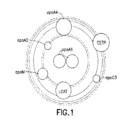
Figure 1: "Solar System" rendering of an HDL particle.
This image is a hypothetical model of an HDL particle. The HDL particle is
composed of two major constituents, lipids and proteins. Several major lipid
classes are represented as the large shaded concentric rings and each ring
reflects the percentage of a lipid which is in proportion to the relative ring
area. The overall diameter of the particle can be scaled and is designed to
replicate the measured diameter of an HDL particle. Proteome members are
denoted by smaller circles layered on top of the lipid rings and are labeled
by
gene name. Each protein molecule is represented by one circle and the area
of the circle is calculated to be proportional to the molecular weight of the
post-translation processed mature form of the protein and does not include
any mass increase resulting from glycosylation. The distance of the proteome
circles from the center of the particle is intended to account for apparent
affinity differences proteome members have for the lipoprotein particle.
Proteome members exhibiting the lowest affinity for the particle would be
arranged furthest from the center. Such proteins would be classified as
having higher particle dissociation rates and are likely to exist in both an
HDL
particle bound and unbound state. Basic positioning of proteome members
around the radius of the particle and the relative distances to each other is
essentially arbitrary in this modeling view. With exception, apoA1 has been
shown to exist as a dimer and is represented by two adjacent circles to
reflect
this observation.
Both protein and lipid constituents can vary in each particle and the density
of
the particle is defined by the ratio of lipid to protein. The total amounts of
all
constituents define the diameter, volume and charge of the particle.
Variations in particle physicochemical properties are due to differences in
the
26
W02013/019943 CA 02844147 2014-02-03
PCT/US2012/049317
mix of constituents and their absolute levels. Each specific combination of
constituents and their particle levels serve as a self-contained set of
instructions which in turn dictates and directs the particle's physiological
activities.
Figure 2: A hypothetical representation of HDL particle suboopulation
heterocieneity.
An extrapolation of an HDL particle population displayed using the solar
system model and reflecting its heterogeneous nature. This figure provides a
hypothetical view that is limited in scope and detail but demonstrates common
particle features as well as distinct differences. The HDL particle population
exhibits the disequilibrium of the proteome members and lipidome to one
another and to the particle population as a whole.
Variations in lipid and protein constituents are reflected in the diameter,
shading and protein patterns for each particle. Fractionation techniques can
separate particles using physicochemical properties and in doing so result in
the apportioning of biological activities. HDL has a large number of measured
biological activities many of which associate with cardiovascular health. To
reconcile all of these reported observations, a model that depends on particle
heterogeneity to account for the variety of physiological activities requires
that
particle subpopulations, which can be defined by physicochemical
characterization or permutations of constituent molecules, perform particular
and specific functions. It is the totality of all particle subpopulations that
contributes to cardiovascular health, and alterations in subpopulation levels
or
specific constituents affect particle instructional blueprints that can
reflect
disease phenotypes.
27
W02013/019943 CA 02844147 2014-02-03
PCT/US2012/049317
Figure 3: HDL LipoPrint analysis of plasma sample.
Acrylamide gel electrophoresis of a plasma sample prepared by pooling fifty
reportedly healthy individuals was used to separate lipoprotein particle
fractions. HDL particles are separated principally based on particle size,
with
faster migration rates and larger distances from the origin for smaller sizes.
Lipoprotein particles are visualized by staining with the dye, Sudan black,
which quantitatively binds neutral lipids, primarily cholesteryl esters (Cp.
Slow migrating VLDL and LDL appear as the last peak to the far left of the
chromatograph as these two classes of lipoproteins do not resolve using the
HDL LipoPrint gel system. Fast migrating albumin, stained with coomassie
blue, to the far right is representative of the free protein fraction in
plasma.
Sudan black staining of cholesterol provides a distribution profile for that
lipid
class across the broad HDL fraction. The HDL is subdivided into three
fractions identified as large, intermediate and small which can be observed as
shading differences delineated by thick black vertical lines according to the
analysis software provided by the instrument's manufacturer. Peak fitting
(area under the curve; AUC) is calculated by the manufacturer's software
provided with the LipoPrint system to estimate relative amounts of the three
subpopulations sizes.
LipoPrint gel segments are labeled 1-20 below the chromatograph. Each
segment composed of a gel two millimeters in length. The entire gel is 40
millimeters in length starting from the trailing edge of VLDULDL peak in
fraction 1 to the leading edge of free albumin peak contained primarily in
fractions 18-20.
Lipoprotein particles were further isolated from each individual gel segment
by
buffer extraction and the isolated particles were reduced and denatured and
subjected to separation by SDS-PAGE using a 4-12% gradient gel. Following
28
W02013/019943 CA 02844147 2014-02-03
PCT/US2012/049317
transfer and immobilization on nitrocellulose, immunoblot analysis is
perfomned to characterize the sub-fraction distribution and relative amounts
of
the target protein.
The middle panel depicts the immunoblot analysis using an antibody specific
for apolipoprotein A-1 (ab27630). Staining of apoAl can be clearly observed
in fractions 4-20 and also in fractions 2 and 3 at much lower levels following
extended exposures. Each of the apoAl containing fractions contains varying
levels of apoAl protein. The significant level of apoAl in fractions 18-20 is
indicative of apoAl protein in very small lipoprotein particles or lipid-free
protein, both of which contain undetectable levels of cholesterol. The bottom
panel provides a generalized reference for categorizing particle
subpopulations into assigned fractions by particle size.
This experiment demonstrates that both apoAl protein and HDL-cholesterol
exist in diseguilibrium to each other. Both particle constituents are in
disequilibrium to the HDL particle population as a whole. Very large particles
contain large ratios of cholesterol to apoAl and small particles contain
larger
ratios of apoAl to cholesterol. Signal levels and distribution patterns for
both
cholesterol and apoAl represent profile averaging effects due to pooling of
the plasma sample prior to analysis. Individual samples exhibit signal
heterogeneity and variations in apoA1 and cholesterol distribution across the
HDL fractions.
Figure 4: HDL Lipoprint of a plasma sample characterized by several HDL
proteome members.
HDL LipoPrint electrophoresis of a pooled plasma sample divided into 10
segments. LipoPrint gel segments are labeled 1-10 below the chronnatograph.
Each segment is composed of a gel four millimeters in length. The entire gel
29
W02013/019943 CA 02844147 2014-02-03
PCT/US2012/049317
is 40 millimeters in length starting from the trailing edge of the VLDL/LDL
peak
in fraction 1 to the leading edge of the free albumin peak contained primarily
in fraction 10.
Lipoprotein particles were further isolated from each individual gel segment
by
buffer extraction and the isolated particles were reduced and denatured and
subjected to separation by SDS-PAGE using a 4-12% gradient gel. Following
transfer and immobilization on nitrocellulose, immunoblot analysis is
performed to characterize the sub-fraction distribution and relative amounts
of
the target protein.
Various commercial antibodies (Tables 3 and 4) targeting several HDL
proteome members (Table 1) were used for immunoblot analysis. The
following proteome examples: apoA1 (HDL110), apoA2 (H00000336-M03),
CLU (mab2937), SerpinF1 (mab1177), SerpinA1 (mab1268), KNG1
(mab15692) and SerpinF2 (nnab1470) were tested and demonstrate various
distribution patterns for HDL particle sub-fractions separated by particle
size.
The proteome distribution disequilibriurn is observable with these proteome
member examples which reflect both broad and restricted distribution patterns
across HDL particle sub-fractions and represent profile averaging effects due
to the sample consisting of pooled plasma samples from fifty individuals.
This physicochemical separation process does resolve particles into
homogeneous sub-populations, and therefore fractions characterized as
positive for one or more proteome member do not establish that any two
proteome members reside on the same particle. Each sub-fraction still
contains multiple particle species that co-migrate under these specific
separation conditions, indicating that further resolution of particle sub-
populations is possible.
W02013!019943 CA 02844147 2014-02-03
PCT/US2012/049317
Figure 5: Representation of proteome distribution diseguilibrium in
lipoprotein
particles.
Five HDL particle subpopulations are represented as circles labeled as 2b,
2a, 3a, 3b and 3c (large to small) using standard HDL particle nomenclature.
Lipoprotein particles can be fractionated and identified by various
physicochemical properties including size and density, but for the purpose of
this example, those differences are simply illustrated by circle diameter.
Attached to the perimeter of the circle is a variety of unique shapes. Five
different proteins are depicted and collectively they represent the HDL
proteome. Each proteome member also has two specific epitopes (shaded
patches) that are considered unique to the individual protein and different
from all other epitopes. The "constellation" of proteome members surrounding
each of the five particles (2b, 2a, 3a, 3b, and 3c) is similar but also
contains
several differences. For example, one proteome member is shared by all
particles (circle) while another protein (triangle) is found only on the two
largest HDL particle subpopulations (2b and 2a). This drawing exhibits a set
of proteome members that are in disequilibrium to the particle population and
to each other.
Figure 6: Sandwich ELISA-based measurements of lipoprotein particle
proteome.
Historically, and due to the basic principles of sandwich ELISA-based
measurement, the technique requires two different antibodies targeting an
individual protein, which are indicated as bound to one protein (circle). The
antibodies must recognize unique and non-overlapping epitopes and the
binding of one antibody must not interfere with the binding of the second.
One antibody, bound to a solid support, serves to capture the target protein
31
WO 2013/019943 CA 02844147 2014-02-03
PCT/US2012/049317
while the second detection antibody provides the means of generating a
signal. The amount of target protein bound by both antibodies should be
proportional to the signal generated, thus providing a means of quantifying
the
protein. In this drawing, the example proteome member (circle) can exist in
HDL particle-bound form or in an unbound state. In some instances, the HDL
proteome member may be bound to other classes of lipoproteins such as LDL
and VLDL, and displaying the proteome not bound to an HDL particle can also
represent such a situation. A comparison of Tables 1 and 2 offers examples
of lipoproteins for which this may be true.
Sandwich ELISA measurements such as this are incapable of discerning the
bound or unbound state of the target protein unless (1) the lipoprotein
particles are first separated into their prospective subpopuiations prior to
measurement, or (2) either the capture or detection antibody is conformation-
dependent and has the capacity to bind the target protein only in instances
where the protein adopts the desired conformation in a specific
subpopulation-restricted manner. Using routine sandwich-ELISA methods,
the quantification of the target protein is aimed at determining the total
amount
of the protein in the sample.
Figure 7: Method for measuring HDL subpopulations.
This figure exemplifies several fundamental concepts demonstrating the
distinct nature of the method to measure HDL subpopulations in this
application. (1) This method relies on the fact that HDL proteome member
distribution is in disequilibrium to each other and to the particle population
as
a whole. (2) The distribution of proteome members across the particle
population includes individual members that are bound to all particle
subpopulations and other proteome members that demonstrate varying
degrees of HDL particle subpopulation restriction. (3) This sandwich ELISA
32
W02013/019943 CA 02844147 2014-02-03
PCT/US2012/049317
methodology requires, but is not limited to, the use of one antibody to each
of
the proteome members to be measured.
Using the example presented and the availability of one antibody capable of
recognizing each of the five HDL proteome members, a series of sandwich
ELISA assays can be devised to identify different HDL subpopulations in a
sample composed of a heterogeneous mixture of HDL particles. In the
bottom portion of the figure, all possible proteome pairs within each of the
five
particle subpopulations are represented. Each particle subpopulation can be
identified by the proteome pair in which both proteome members exist
together on the same particle. The total number of possible pairs is a
function
of the number of proteome members bound. The capture antibody, which is
capable of binding the target protein in the context of any particle, will
produce
a measurable signal only when the detection antibody is also bound to its
target protein held in close proximity on the same particles where both
proteome members reside.
Figure 8: Surrogate markers for HDL subpopulations.
Set theory can be used to identify surrogate markers for specific
subpopulations. Signals from paired proteome measurements in Figure 7 are
rendered using a Venn diagram to demonstrate the use of inclusion and
exclusion criteria to identify specific HDL subpopulations. Five groups are
labeled as 2b, 2a, 3a, 3b, and 3c. The largest lipid-rich HDL particles,
commonly referred to as HDL2, consist of the two subpopulations 2b and 2a
and the smaller lipid poor HDL particles, called HDL3, consist of three
subpopulations 3a, 3b and 3c. Two proteome pairs can be used to identify
larger HDL2 particles (intersection 2b and 2a), while the smaller more dense
HDL3 particles include one proteome pair (intersection of 3a, 3b and 3c). In
addition to HDL2 and HDL3 specific particles, various other proteome pairs
33
W02013/019943 CA 02844147 2014-02-03
PCT/US2012/049317
can serve as surrogate measurements for particle subpopulations of greater
homogeneity. Specific to this example are two proteome pairs restricted to
the largest HDL 2b particles, and the smallest particle subpopulation contains
a single proteome pair that does not exist in any other subpopulation.
This methodology permits the use of restricted proteome particle distribution
to identify subpopulations of increasingly defined homogeneity, as
combinations of restricted distributions can be overlapped to identify
increasingly refined subsets of particles. In a similar fashion, this method
offers the means to identify proteome pairs that do not typically exist in
normal
healthy individuals. Such is the case for one proteome pair which can be
observed in the upper left hand corner of the figure. This proteome pair
resides outside the boundary of all five particle subsets in the diagram. Such
instances, where both proteins and applicable antibodies exist, offer the
prospect of identifying surrogate markers for HDL subpopulations that are
considered atypical. HDL particles and associated proteome pairs of this
nature may occur as a result of underlying genetics or disease states, and
this
method offers a means for their identification and measurement.
This method provides a means to expand the number of particle
subpopulations that can be identified by adding increasing numbers of
proteome members from Table 1. Furthermore, this method can utilize the
overlapping restricted distribution of two proteome members to measure
expanded subsets of particles that cannot be distinguished by a single
proteome member.
34
W02013/019943 CA 02844147 2014-02-03
PCT/US2012/049317
Figure 9: Method provides for geometric expansion of surrogate markers for
HDL subpopulations.
The use of proteome-paired signals to identify HDL subpopulations provides
the prospect of geometrically expanding the repertoire of measurements for
each new antibody added for use in the proteome pair sandwich ELISA. This
example incorporates the drawing from figure 7 (upper panel) for comparison.
The lower panel displays a second antibody recognizing an alternative
epitope from the first on the protein designated by the circle. The
substitution
of a different antibody recognizing a second unique epitope on the protein
results in additional sandwich ELISAs available from the same proteome
pairs, resulting in an increase of the number of possible novel measurements
in proportion to the number of proteome members present. Such
measurements may result in no observable signal difference and in such
instances can only offer independent testing of the first measurement or
introducing the second antibody provides an alternative set of measurements
depending on the nature of the epitope recognized. This method can
increase the number of unique proteome-paired measurements by a factor
equivalent to the number of proteome members bound to the particle, thus
providing the means to geometrically expand the number of potential
surrogate markers for an HDL particle.
Figure 10: Method provides for expansion of surrogate markers for HDL
subpopulations.
The top panel displays the components of a sandwich ELISA which include a
capture antibody (lg-C) attached to a solid support (SS). A protein antigen
composed of two unique and non-overlapping epitopes and a detection
antibody (Ig-D) coupled to an agent capable of producing a measurable signal
(*). In some cases the role of the capture and detection antibodies can be
W02013/019943 CA 02844147 2014-02-03 PCT/US2012/049317
reversed and the resulting signals from both configurations are equivalent.
The success of such experimentation is often considered a validation of the
assay components and the subsequent measurement they produce. A
measurement of this nature is independent of other proteins in the mixture
and represents a typical sandwich ELISA.
The middle panel is an illustration of a sandwich ELISA in which the roles of
the antibody pair cannot be reversed and doing so will alter the absolute
values of the measurement for a given sample. Excluding technical
restrictions, such as the inability of the antibody to serve in the capture
role
due to non-productive coupling to the solid support or to act as a detection
antibody as a result of loss or altered binding following labeling signal-
generating agent, other molecular explanations are possible. An example is
the recognition of post-translational modifications that occurs in only a
percentage of the antigen being measured such as a phosphorylation event.
In this instance when the lg-C binds the common epitope to all antigen
molecules and the lg-D binds an epitope of limited distribution, a productive
signal is generated only from a subset of the total antigen bound to the Ig-C.
Increasing the concentration of the antigen will not alter that ratio, as the
amount of non-productive antigen binding increases to the same degree as
productive antigen binding until the sandwich ELISA reaches saturation.
=
When the lg-C and Ig-D are reversed, only the productive antigen is bound
and the signal is dependent solely on the concentration of protein containing
the epitope of limited distribution. The sandwich ELISA does not saturate at
the same concentration of total (productive and non-productive antigen), and
the difference in signal between each sandwich ELISA goes to unity as the
limited distribution epitope increased to all antigens.
The bottom panel illustrates the unique nature of this method of measuring
proteome pairs, and the Ig-C and Ig-D bind epitopes on two different
36
WO 2013/019943 CA 02844147 2014-02-03
PCT/US2012/049317
proteome members. In this situation, the lg-C and ig-D cannot be reversed
for the same reasons as described for the example above but also accounts
for the antigen epitope distribution within the HDL population as well as the
bound/unbound considerations described in Figure 6. This specific relational
dimension cannot be captured when both the lg-C and lg-D interact with
unique non-overlapping epitopes on the same antigen. What was a
measurement of two independent antigens has been transformed into a
relational intramolecular measurement which characterizes two antigens and
the four antibodies involved. The eight distinct measurements of HDL
subpopulations are a result of both limited epitope distribution associated
with
the antigen and the distribution disequilibrium of the two proteome members
have to each another, Only in instances in which both epitopes exist on all
proteome members in the sample and both proteome members maintain
identical particle distribution profiles, including HDL particle bound and
unbound fractions, does this model not hold true.
Figure 11: A hypothetical array of antibodies in a 96-well format to measure
HDL subpopulations.
This rendering displays a collection of antibody pairs organized into ninety-
six
distinct measurements of HDL subpopulations. This assay construct consists
of a labeled network of shaded boxes overlaid on a 96-well (circles) plate
template. Plate rows are labeled with letters (A-H) to the left of the plate
and
columns are labeled above the plate with numbers (1-12). Each well contains
two boxes located in diagonal corners. The upper left box identifies a capture
antibody by proteome and epitope using a letter and number code. The box
in the lower right corner identifies the detection antibody by proteome and
epitope using the letter and number code.
37
W02013/019943 CA 02844147 2014-02-03
PCT/US2012/049317
Labeling of proteome epitopes is essentially arbitrary, but in this example,
the
boxes labeled with the letter "Z" represent a non HDL proteome
cardiovascular control. Proteorne members are designated by a letter (A-J)
and unique epitopes by a number. In this illustration, eight antibodies
targeting proteome member A contribute to fifty-one sandwich ELISA
measurements. Eighteen of these pairs are designed to measure proteome
member A to itself using unique and non-overlapping epitopes. Thirty-three
measurements utilizing antibodies to proteome member A also involve
antibodies to other proteome members. In real terms proteome member A
would likely be apoA1 and antibodies selected for their ability to recognize
both conformational-dependent and -independent epitopes. Antibodies could
serve strictly as Ig-C (A1, A3) or as Ig-D (A2), while others could serve in
both
roles (A4-A8). Similar design rules would hold true for other HDL proteome
members. Some assays (A1/A4, A1/A5, A1/A6, and A1/A7) utilize a single Ig-
C and four different Ig-D. Other assays (A4/A2, A5/A2, A6/A2 and A7/A2)
utilize different lg-C and a common lg-D while other assays (A6/A7, A7/A6)
use the same antibody pair with roles reversed. A similar design would be
used to measure to proteome members to each other (A9/B2, A9/B3, A4/B2,
A4/B3, A5/B1, A5/B3, A6/B4, A7/64, B1/A9, B3/A9, B1/A2, B3/A2,
B2/A5,133/A5, B4/A6 and B4/A7) where proteome member B would represent
apoA2. The remaining wells on the plate depict series of antibody
combinations that target various proteome members (Table 1), all of which
would have far more limited particle distribution profiles than apoA1 or
apoA2.
This method is designed to construct a measurement-matrix for determining
the amount of HDL subpopulations in an HDL-containing sample by
performing a quantitative assay on a plurality of HDL proteome epitopes. This
systematic analysis utilizes the distribution disequilibrium found between two
proteome members within the HDL subpopulations and exploits the
relationship those epitopes have through the use of common antibodies in
38
W02013/019943 CA 02844147 2014-02-03
PCT/US2012/049317
multiple sandwich ELISA assays. This method replaces the typical
independent intermolecular measurements of sandwich ELISA, where both
antibodies interact with unique epitopes on a single protein, with a series of
relational intramolecular measurements based on many antibodies used in
various combinations in multiple assays.
Figure 12: Determining whether a subiect is afflicted with a disorder
characterized by an abnormal amount of an HDL suboopulation.
Subject samples containing HDL are measured using the antibody array
matrix to quantify the relative levels of HDL subpopulations defined by the
combination of antibody pairs. Ninety-six measurements generate signal
intensity levels which are reflected by grayscale shade from least (white) to
greatest (black). The subpopulation profile for the subject is the composite
view of multiple HDL proteome ELISA signals taken concurrently. The levels
and patterns are hypothetical and offer a visual representation of measured
differences in samples from individuals afflicted with diseases that affect
HDL
proteome member levels or their association with HDL particle
subpopulations.
Samples from individuals afflicted with disorders characterized by abnormal
amounts of an HDL subpopulation or abnormal levels of a proteome member
can generate signal levels that are higher or lower than that of a healthy
individual. Two diseases, atherosclerosis and type-2 diabetes, are examples
of afflictions affecting HDL proteome levels (Kontush, A., et. al.,
Arterioscler.
Thromb. Vasc. Biol. 24:526, 2004; Lyons, T. J., et. al. Invest. Opthomology
and Visual Sci. 45:910, 2004; Vaisar, T., et. al., J. Clin. Inv., 117:746,
2008;
Green, P. S., et al., Circulation 118:1259, 2008). It is unclear from existing
data whether the observed changes in measured protein reflect modulations
of protein on the HDL particles or changes in levels of HDL particles
39
W02013/019943 CA 02844147 2014-02-03
PCT/US2012/049317
containing those proteins (Corsetti, J. P., et. al., PLOS One 7:e39110, 2012).
This subtle difference represents a key feature in assessing HDL and an
important dimension that this method brings to correlating HDL to disease
states.
Figure 13: Detection of HDL using a sandwich ELISA assay.
Twenty-six antibodies directed at twelve HDL proteome members from Table
1 and one non-immune antibody control (IgG-C) are labeled along the
ordinate and abscissa. Cells (shaded) representing a tested sandwich ELISA
can be identified by pairing a capture (ordinate) and a detection (abscissa)
antibody using HDL-containing samples. Antibodies designated by their
catalogue number according to Tables 3 and 4 as well as their clone
identification ([clone]) where applicable, are grouped according to the
proteome member they target. Sandwich ELISA assays generating a strong
signal (dark), weak (intermediate) and no signal ("N" light). Cells (white)
indicate antibody pairs not tested. All antibodies were evaluated by
immunoblot analysis with HDL samples to confirm their capacity to recognize
their cognate proteome member prior to sandwich ELISA testing.
Excluding the IgG-C non immune control, these antibodies comprise a
possible 650 unique antibody pairs if a single antibody cannot serve in both
the capture and detection role. Of these, 92 sandwich ELISA's were
performed, of which 56 generated measurable signals and 36 did not. Some
antibodies performed either capture or detection roles. Other antibodies did
not work in either position despite pairing with antibodies validated to work
in
this assay format. Several sandwich ELISA assays utilizing antibody pairs
interacting with epitopes on the same protein (apoA1, apoB, apoE) generated
signals. Several detection antibodies demonstrated the capacity to work with
multiple capture antibodies targeting the same proteome member, and,
W02013/019943 CA 02844147 2014-02-03
PCT/US2012/049317
multiple capture antibodies worked with a common detection antibody.
Antibody pairs targeting different proteome members exhibited signals
indicating proximity of both proteins on the same particle. Limited testing of
antibodies derived from Tables 3 and 4 identified sandwich ELISA assays that
place the following proteome members on the same particle: apoA1/apoA2,
apoA1/apoB, apoA1/apoE, apoA1/CLU, apoA1/KNG1, apoA1/SerpinA1,
apoA1/SerpinC11 apoA1/SerpinFl, and constitute novel ELISA-based
measurements of HDL not previously observed.
Antibodies are commercially available and not previously evaluated for use in
this sandwich ELISA format. It is expected that not all antibodies or antibody
combinations should work. In some instances, technical limitations such as
non-productive coupling to the solid support or labeling with a signal
generating molecule may be an issue. In other cases, sandwich ELISA-
validated antibodies were unable to pair and may reflect epitope availability
problems or that both epitopes do not exist on the same particle. Instances of
steric hindrance due to overlapping epitopes are also likely.
41
W02013/019943 CA 02844147 2014-02-03
PCT/US2012/049317
Detailed Description of the Invention
This invention provides an accurate tool for measuring HDL in a sample. The
invention is useful for determining whether a subject is at risk of
developing, is
suffering from or is shifting between cardiovascular disorders. The methods
are based on the physical relationship between two distinct proteins or
epitopes held in proximity to one another as part of a single lipoprotein
particle. That is, this invention exploits the physical proximity between two
protein epitopes to identify and quantify discrete HDL subpopulations present
in heterogeneous mixtures, and measure changes in HDL subpopulations as
a result of disease or treatment.
Definitions
In this application, certain terms are used which shall have the meanings set
forth as follows.
As used herein, the term "antibody" includes, without limitation, (a) an
immunoglobulin molecule comprising two heavy chains and two light chains
and which recognizes an antigen; (b) polyclonal and monoclonal
immunoglobulin molecules; and (c) monovalent and divalent fragments
thereof. Immunoglobulin molecules may derive from any of the commonly
known classes, including but not limited to IgA, secretory IgA, IgG and IgM.
IgG subclasses are also well known to those in the art and include, but are
not
limited to, human IgG1 , IgG2, IgG3 and IgG4. Antibodies can be both
naturally occurring and non-naturally occurring. Furthermore, antibodies
include chimeric antibodies, wholly synthetic antibodies, single chain
antibodies, and fragments thereof. Antibodies may be human, humanized or
nonhuman.
42
W02013/019943 CA 02844147 2014-02-03
PCT/US2012/049317
As used herein, the term "capture antibody" includes, for example, the primary
antibody used in a homogeneous immunoassay or an ELISA immunoassay.
The capture antibody is immobilized on a solid support, such as a polystyrene
microtiter plate, bead or cylinder.
As used herein, the term "cardiovascular disease", also referred to as
"cardiovascular disorder" and "CVD", includes, without limitation, heart and
blood vessel diseases, such as atherosclerosis, coronary heart disease,
cerebrovascular disease, and peripheral vascular disease. Cardiovascular
disorders also include, for example, myocardial infarction, stroke, angina
pectoris, transient ischemic attacks, and congestive heart failure.
Cardiovascular disease, such as atherosclerosis, usually results from the
accumulation of fatty material, inflammatory cells, extracellular matrices and
plaque. Clinical symptoms and signs indicating the presence of CVD may
include one or more of the following: chest pain and other forms of angina,
shortness of breath, sweatiness, Q waves or inverted T waves on an EKG, a
high calcium score by CT scan, at least one stenotic lesion on coronary
angiography, and heart attack.
As used herein, the term "defined protein epitope" includes, without
limitation,
an epitope defined structurally (e.g., by primary amino acid sequence and/or
atomic coordinates) and/or functionally (e.g., able to bind to a defined
monoclonal antibody, ideally with a Kd of 10-8M or lower).
As used herein, the term "detection antibody" includes, for example, the
secondary antibody used in a homogeneous immunoassay or an ELISA
immunoassay. The detection antibody is typically immobile, and contains a
label that produces a measurable signal.
43
W02013/019943 CA 02844147 2014-02-03
PCT/US2012/049317
As used herein, the term "high density lipoprotein", also referred to as
"HDL",
includes, without limitation, a particle as exemplified in Figure 1 that is
made
from protein and lipid, and that (i) has a density of from 1.06 to 1.21 g/mL,
(ii)
has a diameter from 7.1 nm to 12.6 nm, and (iii) contains at least one of
apoA1, apoA2 and apoE (alternatively referred to as ApoA1, ApoA2 and
ApoE, respectively). Examples of HDL include 1-IDL3 (having a density of
from 1.06 to 1.10 g/mL), and HDL2 (having a density from 1.10 to 1.21 g/mL).
As used herein, the term "HDL subpopulation" means a subset of all HDL.
Preferably, the HDL subset differs from all other HDL subsets by the presence
or absence of a particular protein or protein epitope.
As used herein, the term "sample", when used with respect to HDL, includes
any biological substance present within, or obtainable from, a subject. These
substances include, without limitation, blood, bone marrow, urine, saliva,
synovial fluid, cerebrospinal fluid or tissue, lesions, ulcers and tumors.
Samples may optionally be treated, purified and/or fractionated. For example,
when a sample is obtained via fractionation, the fractionation of components
may take place in column chromatography by a difference in affinity between
a stationary phase and a mobile phase, or by the principals of a gradient.
Other fractionation methods include separation by differences in mass,
solubility or density that may be induced by methods such as freezing, pH
change, organic extraction, precipitation or electrophoretic mobility.
As used herein, the term "subject" includes, without limitation, a mammal such
as a human, a non-human primate, a dog, a cat, a horse, a sheep, a goat, a
cow, a rabbit, a pig and a rodent.
44
W02013/019943 CA 02844147 2014-02-03
PCT/US2012/049317
Embodiments of the Invention
This invention provides a method for measuring the amount of a high density
lipoprotein (HDL) subpopulation present in a sample, wherein each particle of
the HDL subpopulation being measured is characterized by the presence of a
plurality of defined protein epitopes, the method comprising performing a
quantitative antibody-based assay on the sample, wherein (i) the assay
employs one or more capture/detection antibody pairs, (ii) the capture and
detection antibodies in each pair are directed to different protein epitopes
present on each particle of the HDL subpopulation, and (iii) each antibody
pair
is directed to a different set of epitopes than is each other antibody pair,
thereby measuring the amount of the HDL subpopulation in the sample.
The HDL subpopulation measured by this method can be characterized by
any naturally occurring permutation of proteins within the HDL proteome.
Members of the HDL proteome are set forth, for example, in Table 1. In a
preferred embodiment, the HDL subpopulation being measured is
characterized by the presence of ApoA1 protein, ApoA2 protein and/or ApoE
protein. Typically, amounts of HDL in a sample such as blood are measured
in mg/dL. Moreover, the "amount" of HDL subpopulation measured by this
method can be either the absolute amount (e.g., 10 mg of HDL per dL of
blood) or a relative amount (e.g., 1.5 times the concentration of HDL present
in normal blood).
Examples of samples containing the HDL subpopulation being measured are
set forth above. In a preferred embodiment, the sample is blood, plasma,
serum or urine, all preferably from a human.
In this method, the plurality of defined protein epitopes can be present on
the
same protein. In this scenario, the plurality of defined protein epitopes are
W02013/019943 CA 02844147 2014-02-03
PCT/US2012/049317
preferably present on one of ApoA1 protein, ApoA2 protein and ApoE protein.
Alternatively, the plurality of defined protein epitopes can be present on two
or
more proteins. In this scenario, the plurality of defined protein epitopes are
preferably present on two or more proteins in the HDL proteome set forth in
Table 1.
The subject invention employs antibody-based assays to measure HDL
subpopulations. Such methods and the antibodies they employ are well
known in the art, and are exemplified above. Moreover, the antibodies that
can be used in this invention are also well known, and are exemplified in
Tables 3 (anti-apoA1 antibodies) and 4 (antibodies directed to various
members of the HDL proteome). In one embodiment, the quantitative
antibody-based assay is a radioimmunoassay (RIA) or an enzyme
immunoassay (EIA). Preferably, the EIA is an enzyme-linked immunosorbent
assay (ELISA), a homogeneous time resolved fluorescence assay (HTRF) or
an electrochemiluminescence assay (ECL).
This invention also provides a method for measuring the amount of each of a
plurality of high density lipoprotein (HDL) subpopulations present in an HDL-
containing sample, wherein each particle of each of the HDL subpopulations
being measured is characterized by the presence of a plurality of defined
protein epitopes, the method comprising performing a quantitative antibody-
based assay on the sample, wherein, for each HDL subpopulation being
measured, (i) the assay employs one or more capture/detection antibody
pairs, (ii) the capture and detection antibodies in each pair are directed to
different protein epitopes present on each particle of the HDL subpopulation,
and (iii) each antibody pair is directed to a different set of epitopes than
is
each other antibody pair, thereby measuring the amount of each of the HDL
subpopulations present in the sample.
46
W02013/019943 CA 02844147 2014-02-03
PCT/US2012/049317
Envisioned is a method wherein a large number of HDL subpopulations is
measured concurrently (as are biomolecules using known chip array
technology) or in close temporal succession. The number of HDL
subpopulations measured by this method can be any number, such as 100,
500, 1,000, 10,000, or more. In one embodiment, the number of HDL
subpopulations measured is at least 16. Preferably, the number of HDL
subpopulations measured is at least 96.
In this method, the amounts of HDL subpopulations can be measured either
sequentially or concurrently. However, the method preferably involves
concurrently measuring the amount of each of the plurality of HDL
subpopulations present in the HDL-containing sample.
Also, in this method, the HDL subpopulations being measured can constitute
any collection of subpopulations (e.g., grouped by disease state or
characterizing proteins). In a preferred embodiment, at least one of the HDL
subpopulations being measured is characterized by the presence of ApoAl
protein, ApoA2 protein and/or ApoE protein.
This invention further provides a method for determining whether a subject is
afflicted with a disorder characterized by an abnormal amount of a defined
high density lipoprotein (HDL) subpopulation, wherein each particle of the
HDL subpopulation is characterized by the presence of a plurality of defined
protein epitopes, the method comprising (a) performing a quantitative
antibody-based assay on an HDL-containing sample from the subject,
wherein (i) the assay employs one or more capture/detection antibody pairs,
(ii) the capture and detection antibodies in each pair are directed to
different
protein epitopes present on each particle of the HDL subpopulation, and (iii)
each antibody pair is directed to a different set of epitopes than is each
other
antibody pair, thereby measuring the amount of the HDL subpopulation in the
47
WO 2013/019943 CA 02844147 2014-02-03
PCT/US2012/049317
subject's sample; and (b) comparing the measured amount of HDL
subpopulation in the subject's sample with a known standard correlative with
the presence and/or absence of the disorder (e.g., HDL measurements
previously taken from healthy and afflicted subjects), thereby determining
whether the subject is afflicted with the disorder.
In this method, the amount of defined HDL in an afflicted subject can be
either
higher or lower than in a healthy subject In one embodiment, the amount of
the defined HDL subpopulation in an afflicted subject is higher than (e.g., by
5%, 10%, 20%, 50%, 100%, or more) the amount of the defined HDL
subpopulation in a healthy subject. In this scenario, the disorder can be, for
example, dyslipidemia, hypertension, diabetes mellitus, coronary artery
disease (CAD) or coronary heart disease (CHD).
In another embodiment, the amount of the defined HDL subpopulation in an
afflicted subject is lower than (e.g., by 5%, 10%, 20%, 50%, or more) the
amount of the defined HDL subpopulation in a healthy subject. In this
scenario, the disorder can be, for example, dyslipidemia, atherosclerosis,
diabetes mellitus, obesity-induced dyslipidemia, coronary artery disease
(CAD), coronary heart disease (CHD) or chronic kidney disease (CKD).
This invention still further provides a method for determining the likelihood
of a
subject's becoming afflicted with a disorder, wherein the disorder's
likelihood
of onset is characterized by an abnormal amount of a defined high density
lipoprotein (HDL) subpopulation, and wherein each particle of the HDL
subpopulation is characterized by the presence of a plurality of defined
protein
epitopes, the method comprising
(a) performing a quantitative antibody-based assay on an HDL-containing
sample from the subject, wherein (i) the assay employs one or more
capture/detection antibody pairs, (ii) the capture and detection
48
W02013/019943 CA 02844147 2014-02-03
PCT/US2012/049317
antibodies in each pair are directed to different protein epitopes present
on each particle of the HDL subpopulation, and (iii) each antibody pair
is directed to a different set of epitopes than is each other antibody
pair, thereby measuring the amount of the HDL subpopulation in the
sample; and
(b) comparing the measured amount of HDL subpopulation in the subject's
sample with a standard correlative with a known likelihood of the
disorder's onset (e.g., HDL measurements previously taken from
healthy, at-risk and/or afflicted subjects),
thereby determining the likelihood of the subject's becoming afflicted with
the
disorder.
In one embodiment, the amount of the defined HDL subpopulation in a subject
likely to become afflicted is higher than (e.g., by 5%, 10%, 20%, 50%, 100%,
or more) the amount of the defined HDL subpopulation in a subject less likely
to become afflicted. In this scenario, the disorder can be, for example,
dyslipidemia, hypertension, diabetes mellitus, coronary artery disease (CAD)
or coronary heart disease (CHD).
In another embodiment, the amount of the defined HDL subpopulation in a
subject likely to become afflicted is lower than (e.g., by 5%, 10%, 20%, 50%,
or more) the amount of the defined HDL subpopulation in a subject less likely
to become afflicted. In this scenario, the disorder can be, for example,
dyslipidemia, atherosclerosis, diabetes mellitus, obesity-induced
dyslipidemia,
coronary artery disease (CAD), coronary heart disease (CHD) or chronic
kidney disease (CKD).
This invention also provides a method for measuring the success of a high
density lipoprotein (HDL)-modifying treatment on a subject, wherein the
treatment's success is characterized by a change in the amount of a defined
49
W02013/019943 CA 02844147 2014-02-03
PCT/US2012/049317
HDL subpopulation, and wherein each particle of the HDL subpopulation is
characterized by the presence of a plurality of defined protein epitopes, the
method comprising
(a) performing a quantitative antibody-based assay on an HDL-containing
sample from the subject during or after treatment, wherein (i) the assay
employs one or more capture/detection antibody pairs, (ii) the capture
and detection antibodies in each pair are directed to different protein
epitopes present on each particle of the HDL subpopulation, and (iii)
each antibody pair is directed to a different set of epitopes than is each
other antibody pair, thereby measuring the amount of HDL
subpopulation in the sample; and
(b) comparing the measured amount of HDL subpopulation in the subject's
sample with a known standard correlative with a successfui treatment
outcome,
thereby measuring the treatment's success.
The treatment whose success is measured by this method can be any form of
treatment, whether pharmaceutical or otherwise (e.g., lifestyle changes and
surgery). Pharmaceutical treatments include, for example, cholesterol-
lowering medications, antiplatelet agents (e.g., aspirin, ticlopidine,
clopidogrel), glycoprotein 11b-Illa inhibitors (such as abciximab,
eptifibatide or
tirofiban), antithrombin drugs (blood-thinners such as heparin), beta-
blockers,
nitrates (e.g., nitroglycerin), calcium-channel blockers, and medications for
reducing blood pressure (e.g., ACE inhibitors and diuretics).
In a preferred embodiment, the HDL-modifying treatment is the administration
of a statin. Statins are well known in the art, and include, for example,
atorvastatin (Lipitor and Torvaste), fluvastatin (Lescole), lovastatin
(Mevacore, Altocor , Altopreve), pitavastatin (Livalo , Pitavae), pravastatin
W02013/019943 CA 02844147 2014-02-03
PCT/US2012/049317
(Pravache, Selektine, Lipostae), rosuvastatin (Crestor ) and simvastatin
(Zocor , Lipexe), ezetimibe/sirnvastatin (Vytorin , EzetroP).
By way of example, in a post hoc cohort study, statins were shown to raise
HDL (measured as HDL-cholesterol (HDL-C) and apoA1), and these
elevations were maintained in the long-term (McTaggart, F. and Jones, P.,
Cardiovasc. Drugs Ther. 22:321, 2008). In patients afflicted with
hypercholesterolemia, statins raise HDL-C by approximately 4% to 10%, with
the percentage change greatest in patients having low HDL-C baseline levels
(including patients having the common combination of high triglycerides (TG)
and low HDL-C). Another study compared the effects of five different statins
(namely, atorvastatin, simvastatin, pravastatin, lovastatin and fluvastatin)
on
the lipid, lipoprotein, and apoA1-containing high-density lipoprotein (HDL)
subpopulation profiles of 86 coronary heart disease (CHD) patients (Asztalos,
B. F., et al., Atherosclerosis 164:361, 2002). This study identified the most
effective agents for altering the HDL subpopulation profiles in CHD patients
to
more closely resemble those found in healthy individuals. Finally, in patients
afflicted with coronary artery disease, 12 months of combined atorvastatin and
extended-release niacin therapy partially reversed the adverse changes in
HDL3 protein composition (Green, P. S., et. al., Circulation 118:1259, 2008).
This invention further provides a method for characterizing a high density
lipoprotein (HDL) particle with respect to the presence of one or more sets of
defined protein epitopes, the method comprising performing an antibody-
based assay on a population of the HDL particles to determine the presence
and/or amount of each set of the defined protein epitopes, wherein (i) the
assay employs one or more capture/detection antibody pairs, (ii) the capture
and detection antibodies in each pair are directed to different protein
epitopes
present on each particle of the HDL subpopulation, and (iii) each antibody
pair
51
W02013/019943 CA 02844147 2014-02-03
PCT/US2012/049317
is directed to a different set of epitopes than is each other antibody pair,
thereby characterizing the HDL particle.
This method can be used to characterize any type of HDL particle. In a
preferred embodiment, the antibody-based assay is performed on a
population of the HDL particles selected from HDL2a, HDL2b, HDL3a,
HDL3b, HDL3c, pre-f31 and pre-f32.
This invention still further provides a method for identifying a subpopulation
of
high density lipoprotein (HDL) whose abnormal concentration in a subject
correlates with a particular disorder, comprising
(a) measuring the amounts of one or more HDL subpopulations present in
an HDL-containing sample from a subject afflicted with the disorder,
wherein each particle of each of the HDL subpopulations being
measured is characterized by the presence of a plurality of defined
protein epitopes, the method comprising performing a quantitative
antibody-based assay on the sample, wherein, for each HDL
subpopulation being measured, (i) the assay employs one or more
capture/detection antibody pairs, (ii) the capture and detection
antibodies in each pair are directed to different protein epitopes present
on each particle of the HDL subpopulation, and (iii) each antibody pair
is directed to a different set of epitopes than is each other antibody
pair, thereby measuring the amounts of the HDL subpopulations
present in the subject's sample,
(b) comparing the measured amounts of HDL subpopulations in the
subject's sample with a known standard correlative with the amounts of
the respective HDL subpopulations present in a healthy subject, and
(c) for each of the measured HDL subpopulations, determining whether
the amount of the HDL subpopulation differs from that in the known
standard,
52
W02013/019943 CA 02844147 2014-02-03
PCT/US2012/049317
whereby any such difference indicates that an abnormal concentration of the
HDL subpopulation correlates with the disorder.
This method is, in essence, a way to find novel correlations between
particular
disorders and HDL subpopulations. Each correlation can then form the basis
for a diagnostic test for such disorder, whereby an abnormal concentration of
the relevant HDL subpopulation indicates an affliction with the disorder. In a
preferred embodiment, the disorder is dyslipidemia, obesity-induced
dyslipidemia, hypertension, diabetes mellitus, coronary artery disease (CAD),
coronary heart disease (CHD), vascular inflammation, atherosclerosis or
chronic kidney disease (CKD).
Finally, this invention provides kits for performing the instant methods
described herein. Each kit comprises (i) a solid substrate suitable for use in
performing an antibody-based assay; (ii) a capture antibody operably affixed
to the substrate; and (iii) in a separate compartment, a detection antibody,
wherein the capture and detection antibodies are directed to different protein
epitopes present on each particle of a predetermined HDL subpopulation.
Antibody-based diagnostic kits of all types and their methods of manufacture
and use are well known. In a preferred embodiment, the instant kit is suitable
for performing a radioimmunoassay (RIA) or an enzyme immunoassay (EIA).
Preferably, the EIA is an enzyme-linked immunosorbent assay (ELISA), a
homogeneous time resolved fluorescence assay (HTRF) or an
electrochemiluminescence assay (ECL). The inclusion of suitable solvents
and instructions for using these kits is envisioned.
In a preferred embodiment of the subject kits, the capture antibody is
directed
to an epitope present on a protein set forth in Table 1, and the detection
antibody is directed to an epitope present on one of ApoA1 protein, ApoA2
53
WO 2013/019943 CA 02844147 2014-02-03
PCT/US2012/049317
protein and ApoE protein, wherein the capture and detection antibodies are
directed to different epitopes. In another preferred embodiment, the capture
antibody is directed to an epitope present on one of ApoAl protein, ApoA2
protein and ApoE protein, and the detection antibody is directed to an epitope
present on a protein set forth in Table 1, wherein the capture and detection
antibodies are directed to different epitopes.
Numerous embodiments (preferred and otherwise) are set forth above in
connection with the instant methods and kits. Each embodiment explicitly set
forth for any of the instant methods or kits applies, mutatis mutandis, to
each
of the other instant methods and kits, unless stated otherwise.
This invention will be better understood by reference to the examples which
follow, but those skilled in the art will readily appreciate that the specific
examples detailed are only illustrative of the invention as described more
fully
in the claims which follow thereafter.
54
W02013/019943 CA 02844147 2014-02-03
PCT/US2012/049317
Examples
Example 1
This invention provides a method of determining a mammalian test subject's
risk of developing CVD by measuring apoA1 with a collection of antibodies,
where each paratope is distinct, whose epitopes are distinguishable and
interact in both conformation-dependent and -independent manner. The
measurements from the subject's sample are then compared to one or more
predetermined values measured in a control population of healthy subjects
and to a collection of samples representing specific disorders.
Rationale: The lipoprotein apoAl is the major constituent protein of HDL,
accounting for 60-70% of the protein mass. Each particle is wrapped in 2-5
apoA1 proteins depending upon the size of the particle and the lipid
composition (McLachlan, A. D., Nature, 267:465, 1977; Wu, Z., et. al., Nat.
Struct. Mol. Biol. 14:861, 2007; Huang, R., et. al., Nat. Struct. Mol. Biol.,
Online 13 March 2011). As the volume of the sphere changes with the gain or
loss of lipid molecules, so will the particle diameter and circumference. As a
consequence, the apoA1 molecules surrounding the lipid particle are also
changing conformation to accommodate varying sphere geometries.
Antibodies that recognize conformation-independent epitopes should have a
greater probability of binding all apoA1 regardless of particle size.
Analyzing
serum with a panel of these antibodies should provide a measure of total
apoA1 in the sample using a plurality of independent measurements and
limits the risk of omission that a single antibody pair will produce. Total
apoA1, whose level is predictive of CVD, can be used as a surrogate marker
for the entire HDL population. Antibodies that interact with apoA1 in a
conformation-dependent manner will recognize only those subsets of particles
wherein apoA1 adopts the conformation recognized by that distinct paratope.
W02013/019943 CA 02844147 2014-02-03
PCT/US2012/049317
Measurements based upon a panel of these antibodies will identify HDL
subpopulations based upon selected antibody pair values, whose levels vary
between individual samples in a disease-specific manner. In one aspect, the
present invention provides a diagnostic test in instances where paratope ¨
epitope interactions are not yet defined.
Methods: A serum sample from an individual and those of a predetermined
disease phenotype are subjected to a panel of capture-detection antibody
pairs as defined in Table 3. Each of the 37 anti-apoA1 mAbs is evaluated for
both its ability to work as a capture antibody and to act as a detection
antibody. The total possible number of measurements is 1332 if the same
antibody is not used for both capture and detection. Measurements are
deemed positive if the positive signal is concentration dependent, saturable,
reproducible and exhibits a linear response over a physiologically plausible
range of concentration of apoA1.
Results: Antibody pairs demonstrating specific and saturable signals in a
dose-dependent manner provide a measure of an existing HDL particle
population present in the sample at concentrations that exceed the lowest
level of detection that antibody pair affords. For all capture-detection
antibody
pairs resulting in a signal, analysis can be performed. Each antibody pair
signal value can be statistically compared to itself and each other across a
library of control samples and samples of known disease conditions. A select
set of measurements showing strong correlations to each other across a
sample set may represent a plurality of apoA1 epitopes associated with the
same or highly similar particle subpopulation. Antibody pair signals that do
not correlate with one another may be representative of independent particle
subpopulations. Signals demonstrating the least variability across similar
samples and the greatest variability between disease states are preferable for
establishing predictive biomarkers of CVD. Each antibody pair signal value
56
W02013/019943 CA 02844147 2014-02-03
PCT/US2012/049317
can be correlated to the surrogate marker total HDL-C surrogate level of a
serum sample. Antibody pair signals having significant correlation to HDL-C
are representative of subpopulations associated with large cholesterol-rich
particles including the HDL2 particle fraction. Antibody pair signals having
the
least correlation with HDL-C levels are representative of small dense lipid
poor HDL3 particle fraction which remains unaccounted for in the total HDL-C
number. The greater the discordance between HDL-C levels and antibody
pair signals, the more probable that the antibody pair is measuring an HDL
subpopulation whose contributions to the HDL profile are not captured in the
surrogate marker HDL-C.
Example 2
This invention provides a method of determining a mammalian test subject's
risk of developing CVD by measuring apoA2 with a collection of antibodies,
where each paratope is distinct, whose epitopes are distinguishable and
interact in both conformation-dependent and -independent manner. The
measurements from the subject sample are then compared to one or more
predetermined values measured in a control population of healthy subjects
and to a collection samples representing specific disorders.
Rationale: The lipoprotein apoA2 is the second most abundant protein on
HDL and is found in plasma as a monomer, homodimer, or heterodimer with
apolipoprotein D (Schmitz, G., et. al., J. Lipid Res., 24:1021, 1983; Yang, C.
Y., et al., Biochem. 33:12451, 1994; Gillard, B. K., et al. Biochem. 44:471,
2005). The differential equilibrium distribution between apoAl and apoA2
across HDL2 and HDL3 sub-fractions has long been recognized (Cheung, M.
C. and Albers, J. J., J. Lip. Res. 23:747, 1982). Most apoA2 is present on
apoA1-containing particles and structural studies indicate that apoA2 can
cause significant structural changes in apoA1 conformation, affecting both
57
WO 2013/019943 CA 02844147 2014-02-03
PCT/US2012/049317
particle remodeling and activity (Rye, K. A. et. al., J. Biol. Chem.,
278:22530,
2003; Boucher, J. et al., J. Lipid. Res. 45:849, 2004). In the plasma, apoA2
is
associated predominantly with smaller and less lipid-enriched HDL particles.
The denser HDL3 fraction has been shown to contain higher relative amounts
of apoA2 than the larger HDL2 particles with apoA1/apoA2 ratios of 3.7 and
4.8, respectively (Brewer, H. B., Jr., et. al., Methods Enzymol. 128:223,
1986).
The small dense fraction HDL3 demonstrates superior atheroprotective
activities when compared to HDL2 isolated from the same individuals (Zerrad-
Saadi, A., et. al., Arterioscler. Thromb. Vasc. Biol., 29:2169, 2009; Kontush,
A., et. al., Arterioscler. Thromb. Vasc. Biol. 27:1843, 2007; de Souza, J. A.,
et.
al., J. Cell. Mol. Med. 14:608, 2010), thus opening the possibility that
individuals with low levels of HDL3 are thought to be at risk for CVD (see
Kontush, A. and Chapman M. J. Nat. Clin Pract. 3:144, 2006).
Methods: A serum sample from an individual and those of a predetermined
disease phenotype are subjected a panel of capture-detection antibody pairs
directed at apoA2 from the list in Table 4. Each of the anti-apoA2 mAbs is
evaluated both for its ability to work as a capture antibody and to act as a
detection antibody. Measurements are deemed positive if the positive signal
is concentration-dependent, saturable, reproducible and exhibits a linear
response over a physiologically plausible range of apoA2 concentrations.
Combining the panel of working apoA2 capture-detection antibody pairs and
the antibody pairs identified as successfully generating a signal in Example 1
yields a mixed antigen measurement where one apoA1 and one apoA2 are
used as capture-detection antibody pairs. As in Example 1, each
apoA1iapoA2 mixed antigen antibody pair signal value can be statistically
compared to itself and others across a library of control samples and samples
of known disease condition.
58
W02013/019943 CA 02844147 2014-02-03
PCT/US2012/049317
Results: Antibody pairs demonstrating specific and saturable signals in a
dose-dependent manner provide a means of measuring an existing particle
population present in the sample at concentrations that exceed the lowest
level of detection that antibody pair affords. For all mixed antigen capture-
detection pairs resulting in a signal, analysis can be performed.
Measurements from apoA1lapoA2 paired antibodies should identify HDL
subpopulations containing both proteins. Antibody paired signal
demonstrating the least variability across similar samples and the greatest
variability between disease states are preferable for establishing predictive
biomarkers of CVD. The measurements from the subject sample are then
compared to one or more predetermined values measured in a control
population of healthy subjects and to a collection of control and disease
samples.
Example 3
This invention provides a method of determining a mammalian test subject's
risk of developing CVD through measuring a plurality of epitopes from the
HDL proteome members defined by Table 1 using a selection of HDL
proteome member antibodies from Table 4. Each antibody that binds a
distinct proteome member can be used in mixed antigen capture-detection
pairs with apoA1 or apoA2 if epitopes are distinguishable and non-
overlapping. The measurements from the subject sample are then compared
to one or more predetermined values measured in a control population of
healthy subjects and to a collection samples representing specific disorders.
Rationale: ,Both apoA1 and apoA2 are present on the majority of HDL
particles. Other proteome members demonstrate degrees of particle
restriction and selectivity (Davidson, W. S, et. al. Arterioscler. Thromb.
Vasc.
Biol. 29:870, 2009; Gordon, S. M., et. al., J. Proteome Res., 9:5239, 2010).
59
W02()13/019943 CA 02844147 2014-02-03
PCT/US2012/049317
Using a conformational-independent apoAl capture antibody from Example 1
paired with a detection antibody for another proteome member can provide a
measure of the relative concentration of the subpopulation containing both
proteins in the sample. Both of the HDL particle populations, those containing
the proteome member and those that do not, compete for binding to the same
apoA1 capture antibody resulting in diminished signal. In other instances,
proteome member capture antibodies will selectively bind only particle
subpopulations containing the proteome member, and can preferentially
concentrate particles containing the proteome member. Such a combination
can increase the lower limits of detection in instances where the
subpopulation defined by the proteome member is small. Antibody pair
signals generated by measuring apoA11proteome or proteomelapoA1 are
different and can be statistically compared to themselves and others across a
library of control samples and samples of known disease condition.
Methods: A serum sample from an individual and those of a predetermined
disease phenotype are subjected to a panel of capture-detection antibody
pairs derived from pairs successfully generating a signal in Example 1 or 2
with proteome-specific antibodies from Table 4. Each proteome mAb is
evaluated both for its ability to work as a capture antibody and to act as a
detection antibody. Measurements are deemed positive if the positive signal
is concentration-dependent, saturable, reproducible and exhibits a linear
response over a range of HDL concentrations. Mixed antigen antibody pair
signal values can be statistically compared to themselves and other signals
across a library of control samples and samples of known disease condition.
Results: Antibody pairs demonstrating specific and saturable signals in a
dose-dependent manner provide a means of measuring an existing particle
population present in the sample at concentrations that exceed the lowest
level of detection that an antibody pair affords. For all mixed antigen
capture-
W02013/019943 CA 02844147 2014-02-03
PCT/US2012/049317
detection pairs resulting in a signal, analysis can be performed.
Measurements from apoA1lproteome and proteomelapoAl paired antibodies
should identify HDL subpopulations containing both proteins. Antibody paired
signals demonstrating the least variability across similar samples and the
greatest variability between disease states are preferable for establishing
predictive biomarkers of CVD.
Example 4
This invention provides a method of determining a mammalian test subject's
risk of developing CVD by measuring specific apoA1 conformations
associated with levels of functional HDL subpopulations previously identified
by physiochemical properties. The measurements from the subject sample
are then compared to one or more predetermined values measured in a
control population of healthy subjects and to a collection of samples
representing specific disorders.
Rationale: Small lipid-poor prer31-HDL, a minor HDL sub-fraction consisting
of a discoidal-shaped particle containing apoA1, PL and unesterified
cholesterol, can be identified by 2-D gel electrophoresis (Asztalos, B. F., et
al., Arterioscler. Thromb. Vasc. Biol. 20:2670, 2000). Prep1-HDL is a
preferred acceptor of cellular cholesterol, an important first step in the
process
of reverse-cholesterol transport (Castro, G. R. and Fielding P. E.,
Biochemistry 27:25, 1988; Kawano, M., et. al., Biochem. 32:5025, 1993;
Huang, Y., et. al., Arterioscler. Thromb. Vasc. Biol. 13:445, 1993). Levels of
pre61-HDL are elevated in type 2 diabetes and indicative of patients with
hyperlipidemia and CAD (Hirayama, S., et. al., Diabetes Care 30:1289, 2007;
Miida, T., et. al., Clin. Chem 42:1992, 1996; Asztalos, B. F., et al.,
Arterioscler. Thromb. Vasc. Biol. 20:2670, 2000). Rather than characterizing
pre131-HDL using physiochemical separation, the particle population can be
61
W02013/019943 CA 02844147 2014-02-03
PCT/US2012/049317
measured using a capture antibody highly specific for an apoA1 conformation
found only in pref31-HDL paired with a conformational-independent apoA1
detection antibody identified from Example 1. Such conformation-dependent
antibodies include mAb 55201 (Miyazaki, 0, et. al., J. Lipid Res., 41:2083)
that recognizes an apoA1 epitope located between residues 140-210
(Sviridov, D., et. al., Arterioscler. Thromb. Vasc. Biol. 22:1482, 2002) or
the
mAb that recognizes apoA1 residues 1 37-1 44 of the mature protein uniquely
associated with prer31-HDL (Fielding, P. E., et. al., Biochemistry 33:6981,
1994). Elevated levels of pre01-HDL are a predictor of carotid atherosclerosis
(Suzuki, M. et. al., Clin. Chem. 51:132, 2005; Hirayama, S., et al., Diabetes
Care 30:1289, 2007; Tashiro, J., et. al., Atherosclerosis 204:595, 2009).
Methods: A serum sample from an individual and those of a predetermined
disease phenotype are measured using a specific conformation-dependent
antibody capable of recognizing apoA1 only when present in the prepl-HDL
subpopulation, paired with a conformation-independent apoA1 detection
antibody identified from Example 1. Measurements are deemed positive if the
positive signal is concentration-dependent, saturable, reproducible and
exhibits a linear response over a range of HDL concentrations. Mixed antigen
antibody pair signal values can be statistically compared to themselves and
other signals across a library of control samples and samples of known
disease condition.
Results: Individuals with normal ranges of total HDL-C and total apoA1 levels
can exhibit increased levels of prei31-HDL subpopulation. Individuals with
increased levels of pre01-HDL are at risk for CAD and may also have
compounding factors including dyslipidemia or diabetes.
62
W02013/019943 CA 02844147 2014-02-03
PCT/US2012/049317
Example 5
In one embodiment, the present invention provides a method of determining a
mammalian test subject's levels of functionally defective HDL particles
resulting from specific post-translational modifications of apoA1. The
measurements from the subject sample are compared to one or more
predetermined values measured in a control population of healthy subjects
and to a collection samples representing specific disorders.
Rationale: Post-translation modifications to apoA1 can result in functionally
defective HDL (see Kontush, A. and Chapman, M. J., Pharmacol. Rev.,
58:342, 2006). Oxidized lipids and proteins associated with lipoprotein
particles play a key role in atherogenesis (Barter, P. J., et. al., Circ.
Res.,
95:764, 2004; Nicholls, S. J., et. al., Trends Card. Med., 15:212, 2005;
Deigner, H. P. and Hermetter, A., Curr. Opin. Lipidol., 19:289, 2008).
Myeloperoxidase modifies apoA1 at specific susceptible residues (Met86,
Met112, Met148, and Tyr192) resulting in functionally defective HDL (Zheng,
L.. B., et. al., J. Clin. Invest. 114:529, 2004; Shao, B. G., et. al., Proc.
Natl.
Acad. Sci. USA 105:12224, 2008; Shao, B., et. al., Chem. Res. Toxicol.
23(3):447, 2010). Antibodies developed to detect modification to those
residues, MOA-I and mAb17 (Wang, X. S., et. al., J. Lipid Res. 50:586, 2009)
can be employed to measure the extent of oxidated apoA1 in HDL when
paired with an apoA1 capture antibody with a distinguishable and non-
overlapping epitope as identified in Example 1. Another example of post-
translational modification affecting HDL particle function is glycation (non-
enzymatic glycosylation) which is the result of the bonding of a sugar
molecule (fructose, glucose or galactose) with a protein or lipid molecule.
Glycation is considered an arbitrary process which differs from glycosylation
which involves enzyme-controlled addition of sugars to protein or lipid
molecules at defined sites. Glycation can impair the functioning of
63
WO 2013/019943 CA 02844147 2014-02-03
PCT/US2012/049317
biomolecules and this specific modification of apoAl results in impaired anti-
inflammatory activities of HDL (CaIvo, C., et. al., Clin. Chim. Acta, 217:193,
1993; Nobecourt, E., et. al., Arterioscler. Thromll Vasc. Biol., 30:766, 2010;
Park, K-H. and Cho, K-H., J. Gerontol., 66A:511, 2011). Methodology
devised to generate specific antibodies capable of detecting specific
glycation
modified proteins can be employed to develop similar measure for glycanated
apoAl (Steward, L. A., et. al., J. Immuno. Method., 140:145, 1991; Cohen, M.
P., et. al., Eur. J. Clin. Chem. Clin. Biochem., 31:707, 1993; Qin, X., et.
al.,
Diabetes, 53:2653, 2004). In another example, secreted apoAl exists as two
species in plasma, a pro-protein and mature protein form which differ by six
amino acid residues on the N-terminal end of the protein (Zannis, V. I., et.
al.,
Proc. Natl. Acad. Sci. USA, 80:2574, 1983; Stoffel, W., J. Lipid Res.,
25:1586,
1984).
Example 6
This invention provides a method of determining the effects of apoC3 levels
on a subject's risk for the disorders hypertriglyceridemia and CVD. The
measurements from the subject sample are compared to one or more
predetermined values measured in a control population of healthy subjects
and to a collection samples representing specific disorders.
Rationale: Human apoC3 is a protein constituent of both apoB-containing
lipoproteins and HDL (Shin, M. J. and Krauss R. M., Atherosclerosis 211:337,
2010). In addition to rapid transfer between particles, apoC3 redistributes
from triglyceride-rich lipoproteins (TRLs) to HDL and transfers back to newly
synthesized TRLs (see Jong, M. C., et al., Arterioscler. Thromb. Vasc. Biol.,
19:472, 1999; Ooi, E. M. M., Clinical Science, 114:611, 2008). Through a
genome-wide association study, APOC3 null mutation carriers were identified
who had reduced apoC3 levels and had lower fasting triglycerides and
64
W02013/019943 CA 02844147 2014-02-03
PCT/US2012/049317
postprandial serum triglycerides and increased HDL-C. Consistent with the
favorable protective lipid profile, APOC3 null mutation carriers were less
likely
to have detectable coronary artery calcification (PoIlin, T. l., et al.,
Science
322:1702, 2008). To test a subject for risks associated with dyslipidemia due
to apoC3 disequilibrium, the combination of capture-detection antibody pairs
can be used to measure the levels and disposition of apoC3 in a biological
sample. The signal associated with these measurements in the test subject is
compared to a predetermined value to determine if the subject is at greater
risk of developing or suffering from CAD than subjects with an amount of
apoC3 that is at, or higher than, the predetermined value. Moreover, the
extent of the difference between the test subject's apoA1lapoC3 and
apoA2lapoC3 levels in the biological sample and the predetermined value is
also useful for characterizing the extent of the risk, and thereby determining
which subjects would most greatly benefit from certain TG-lowering therapies.
Example 7
This invention provides a method of determining the level of one or more
lipoprotein proteome members selected from apoJ, PON1, PON3 and PAF-
AH, as a method of assessing HDL subpopulations containing anti-oxidative
activity. The measurements from the subject sample are compared to one or
more predetermined values measured in a control population of healthy
subjects and to a collection of samples representing specific disorders.
Example 8
This invention provides a method of determining the level of one or more
lipoprotein proteome members associated with HDL selected from AHSG,
A1BG, apoF, GC, PLTP, RBP4, serpinA3, serpinA8, serpinF2 and TTR. The
detected amount of the lipoprotein proteome member is compared to one or
W02013/019943 CA 02844147 2014-02-03
PCT/US2012/049317
more predetermined values of the lipoprotein proteome member(s) measured
in a control population of healthy subjects to evaluate the level of small
dense
HDL3. The measurements from the subject sample are then compared to
one or more predetermined values measured in a control population of
healthy subjects and to a collection samples representing specific disorders.
Example 9
This invention provides methods of screening a human subject who appears
healthy, or may be diagnosed as having a low HDL:LDL ratio and/or as being
at risk for CVD based on certain known risk factors such as high blood
pressure, high cholesterol, obesity, or genetic predisposition for CVD. The
methods described herein are especially useful to identify subjects at high
risk
of developing CVD, in order to determine what type of therapy is most suitable
and to avoid potential side effects due to the use of medications in low risk
subjects. For example, prophylactic therapy is useful for subjects at some
risk
for CVD, including a low fat diet and exercise. For those at higher risk, a
number of drugs may be prescribed by physicians, such as lipid-lowering
medications as well as medications to lower blood pressure in hypertensive
patients. For subjects at high risk, more aggressive therapy may be indicated,
such as administration of multiple medications.
Envisioned here is a method of detecting arterial disease, atherosclerosis,
and fatty lesions formed on the inside of the arterial wall. These lesions
promote the loss of arterial flexibility and lead to the formation of blood
clots.
The lesions may also lead to thrombosis, resulting in most acute coronary
syndromes. Thrombosis results from weakening of the fibrous cap, and
thrombogenicity of the lipid core. It is well recognized that atherosclerosis
is a
chronic inflammatory disorder (see Ross, R., N. Engl. J. Med. 340:115, 1999).
Chronic inflammation alters the protein composition of HDL, making it
66
W02013/019943 CA 02844147 2014-02-03
PCT/US2012/049317
atherogenic (see Barter, P. J., et al., Circ. Res. 95:764, 2004; Chait, A., et
al.,
J. Lipid Res. 46:389, 2005; Navab, M., et al., J. Lipid Res. 45:993, 2004; and
AnseII, B. J., et al., Circulation 108:2751, 2003). Accordingly, HDL-
associated
proteins that serve as lipoprotein proteome member indicators for CVD, and
atherosclerotic lesions in particular, may be derived from macrophages,
smooth muscle cells, and endothelial cells present in atherosclerotic lesions.
Accordingly, HDL-associated lipoprotein proteome members isolated from a
blood sample represent a biochemical "biopsy" of the artery wall or
endothelium lining the vasculature. It is likely that lesions that are most
prone
to rupture would increase their output of HDL due to the fact that enhanced
proteolytic activity destroys the extracellular matrix and promotes plaque
rupture. Indeed, short-term infusion of HDL into humans may promote lesion
regression (Nissen, S. E., et al., JAMA 290:2292, 2003), suggesting that HDL
can remove components of atherosclerotic tissue. Therefore, the proteins
included in the protein cargo associated with HDL, enriched in CVD subjects,
and also known to be present in lesion HDL from a population of CVD
patients, serve as lipoprotein proteome members that may be used to detect
the risk and/or presence of atherosclerotic plaques in an individual subject.
In another aspect, this invention provides assays comprising one or more
detection reagents capable of detecting at least the proximity of two
lipoprotein proteome members that is indicative of the presence or risk of
CVD in a subject. The lipoprotein proteome member is detected by mixing a
detection reagent that detects at least one lipoprotein proteome member
associated with CVD with a sample containing HDL-associated proteins, and
monitoring the mixture for detection of the lipoprotein proteome member with
a suitable detection method such as spectrometry, immunoassay, or other
method. In one example, the assays are provided as a kit. The kit can have,
for example, detection reagents for detecting at least two, three, four, five,
ten
67
W02013/019943 CA 02844147 2014-02-03
PCT/US2012/049317
or more HDL-associated lipoprotein proteome members in biological samples
from a test subject.
The kit also includes written indicia, such as instructions or other printed
material for characterizing the risk of CVD based upon the outcome of the
assay. The written indicia may include reference information, or a link to
information regarding the predetermined signal values for paired proteome
measurements of one, two, three, four, five, ten or more HDL-associated
lipoprotein proteome members from a reference population of healthy subject
samples, and an indication of a correlation between paired proteome
measurements of one or more HDL-associated lipoprotein proteome
members with samples from subjects having, or at risk of having, CVD.
In one example, the detection reagent comprises one or more antibodies
which specifically bind one or more of the lipoprotein proteome members
provided in Table 1 or 2 that may be used for the diagnosis and/or prognosis
of CVD characterized by the relative abundance of the lipoprotein proteome
member in the serum, or an HDL subfraction thereof. Standard values for
protein levels of the lipoprotein proteome members are established by
combining biological samples taken from healthy subjects. Deviation in the
amount of signal produced from an antibody pair between control subjects
and CVD subjects establishes the parameters for diagnosing and/or
assessing risk levels, or monitoring disease progression.
In another example, this invention provides a method of determining the
efficacy of a treatment regimen for treating and/or preventing CVD by
monitoring the presence of one or more lipoprotein proteome members in a
subject during treatment for CVD. The treatment for CVD varies depending
on the symptoms and disease progression. The general treatments include
lifestyle changes and medications, and may include surgery. Lifestyle
68
W02013/019943 CA 02844147 2014-02-03
PCT/US2012/049317
changes include, for example, weight loss, a low saturated fat, low
cholesterol
diet, reduction of sodium, regular exercise, and a prohibition on smoking.
Medications useful to treat CVD include, for example, cholesterol-lowering
medications, antiplatelet agents (e.g., aspirin, ticlopidine and clopidogrel),
glycoprotein Ilb-111a inhibitors (such as abciximab, eptifibatide or
tirofiban), or
antithrombin drugs (blood-thinners such as heparin) to reduce the risk of
blood clots. Beta-blockers may be used to decrease the heart rate and lower
oxygen use by the heart. Nitrates, such as nitroglycerin are used to dilate
the
coronary arteries and improve blood supply to the heart. Calcium-channel
blockers are used to relax the coronary arteries and systemic arteries, and,
thus, reduce the workload for the heart. Medications suitable for reducing
blood pressure are also useful to treat CVD, including ACE inhibitors,
diuretics
and other medical treatments.
69
Table 1
c
c
0 go
L. e 0 c
.41; ,T t- 1-12 4., "s= 11:,' z=-= =St Z.'
EGNM GenelD
UNIPROT-KB Protein Name ii cgs _1(?
4). 6 43 (3(- I Si' 6-
A1BG I P04217 Alpha-IB-glycoprotein
MINIM MUM 0
A2M 2 P01023 Alpha-2 macroglobulin
IIIIIIIIIIIIII r..)
=
AFM 173 P43652 Afamln
MI
i...J
AGT 183 P01019 Angiotensinogen (Serpin Peptidase Inhibitor
Clade A Member 8) In MINIM -o--
,¨,
AHSG 197 P02765 Alpha-2-HS-glycoprotein
I. IIIIII Ell o
o
ALB 213 P02768 Serum albumin
MIN IN MIMI. .1..
(..4
AMBP 259 P02760 Alpha-l-microglobulin (bikunin)
1111
MEM"
APCS 325 P02743 Amyloid P Component Serum (SAP)
IIII El IIII
AP0A1 335 P02647 apolipoprotein A-I
IIIMIIIIIIIIIMIIIIIIIMMI
APDA2 336 P02652 apolipoprotein A-II
MIMI IIMMIIIIIIIIII. NI
AP0A4 337 P06727 apolipoprotein A-IV
1.11111111M NINON II.
AP0A5 116519 Q6Q788 apollpoprotein A-V
NI I.
APOB 338 P04114 apolipoprotein B-100
III
MIME' IIII n
APOCI 341 P02654 apolipoprotein C-I
III M IIIIIIIII MI
2
APOC2 344 P02655 apolipoprotein C-I1
INN NI III MI IIII co
.i.
APOC3 345 P02656 apolipoprotein C-III
.1111111111 MN. IN .i.
H
APOC4 346 P55056 apolipoprotein C-IV
IIII
IIII II. .i.
-A
-.1
o APOD 347
P05090 apolipoprotein D IIIIIII .11. El El IO)
APOE 348 P02649 apolipoprotein E
MEI MIIMIIIIIIIIIMMI H
FP
APOF 319 Q13790 apolipoprotein F
III
IIII oi
APOH 350 P02749 apolipoprotein H (beta-2-glycoprotein I)
11111
IIII MEIN il iv
oi
APOLI 8542 014791 apolipoprotein L-I
1111..11111 MEM III u.)
APOM 55937 P095445 apolipoprotein M
MI. Ell MIME IIII
APO() 79135 Q98UR5 apolipoprotein 0
Ell
ATRN 8455 075882 Attractin
IN
BMPI 649 P13497 Bone morphogenetic protein 1
IIIII
ClQB 713 P02746 Complement Clq subcomponent subunit B
III
CIQC 714 P02747 Complement Clq subcomponent subunit C
IIII
"0
C1R 715 P00736 Complement CI r subcomponent
MI
n
CIS 716 P09871 Complement Cl s subcomponent
IMO" H
C2 717 P06681 Complement C2
IIIIII cn
i,..)
C3 718 P01024 Complement C3
MIN .11.11111111 o
.k
C4A 720 POCOL4 Complement C4-A
IIII III t.)
C4B 721 POCOL5 Complement C4-B
.11
11111. 4.
C4BPA 722 P04003 Complement C4 binding protein alpha
chain
III
NI 1111 i,=.)
7-1
C5 727 P01031 Complement C5 . . -
ENE
C6 729 PI3671 Complement C6
III
C7 730 P10643 Complement C7
El.
Table 1
C8B 732 P07358 Complement ai beta chain
M
C9 735 P02748 Complement C9
III IIIMMI
CETP 1071 P11597 Cholesteryl ester transfer protein
NEI II
CFB 629 P00751 Complement factor B
EIMINII.
CFH 3075 P08603 Complement factor H
1111 OMNI
0
CFI 3426 P05156 Complement factor I
III IIII tv)
CLEC3B 7123 P05452 Tetranectin
..,
t44
CLU 1191 P10909 Clusterin (apal)
INN 1.111.11111111111 -Z:7
...
CP 1356 P00450 Ceruloplasmin
MI MINIM v:p
v:
CPN2 1370 P22792 Caboxypeptidase N polypeptide 2
NM 4..
t44
CRP 1401 P02741 C-Reactive protein
III
F13B 2165 P05160 Coagulation factor XIII beta subunit
IIII
F2 2147 P00734 Prothrombin
1111111=111
F8A 8263 P23610 Coagulation factor VIII intron 22 protein
NI
FCN2 2220 015485 Ficolin-2
III .
FCN3 8547 075636 Picolin-3
II.
FGA 2243 P02671 Fibrinogen alpha chain
III NI1.11.1.11M n
FGB 2244 P02675 Fibrinogen beta chain
NI il 1111111.1 o
FGG 2266 P02679 Fibrinogen gamma chain
MEI= iv
co
.i.
FN1 2335 P02751 Fibronectin 1
.11.11.1 .i.
H
-4 GC 2638 P02774 Vitamin D-binding protein
IIII Ell IIMMI .i.
-A
1-,
GSN 2934 P06396 Gelsolin
III IIII iv
HP 3240 P00738 Haptoglobin
Mil MEM 0
H
FP
oI
I-1PR 3250 P00739 Haptoglobin-related protein
M IIIII.IIIIII III
HPX 3263 P02790 Hemopexin
III MEM= iv
o1
HRG 3273 P04196 Histidine-rich glycoprotein
1.1 MIMI u.)
IGFALS 3483 P35858 Insulin-like growth factor binding
protein acid labile subunit MN
ITIH1 3697 P19827 Inter-alpha-trypsin inhibitor heavy chain
H1 NMI
ITIH2 3698 P19823 inter-alpha-trypsin inhibitor heavy chain H2
In MIIINIM
ITIH3 3699 Q06033 Inter-alpha-trypsin inhibitor heavy chain
H3 MEI
KNG1 3827 P01042 Kinlnogen-1
In====.0
en
LCAT 3931 P04180 Lecithin-cholesterol acyltransferase
III III
LPA 4018 P08519 Apolipoprotein(a)
III NI
(,)
LRG1 11.6844 P02750
Leucine-rich alpha-2-glycoprotein III M
t4
0
WM 4060 P51884 Lumican
MINIM IN
MASP1 5648 P48740 Mannan-binding lectin serine protease 1
precursor NI -ii-i
4..
ORM1 5004 P02763 Alpha-1-acid glycoprotein 1 (Orosomucoid
1)M =t..)
ORM2 5005 P19652 Alpha-1-acid glycoprotein 2 (Orosomu .
coid 2)
= III IIII '---.1
PAFAH1B1 5048 P43034 Platelet-activating factor
acetylhydrolase IB subunit alpha M
PCY0X1 51449 Q9UHG3 Prenylcysteine oxidase 1
M
Table 1
PGLYRP2 114770 Q96PD5 Peptidoglycan recognition protein 2
1111 NI
PLA2G7 7941 Q13093 Platelet-activating factor
acetylhydrolase (PAFA) 1111
PLG 5340 P00747 Plasminogen
IIII MEI
PLTP 5360 P55058 Phospolipid transfer protein
II NMI IIII
PON1 5444 P27169 Serum paraoxonasefarylesterase 1
Nil 1111.111111111111111 0
PON3 5446 015166 Serum paraoxonase/lactonase 3
111 NI iµJ
c=
PPBP 5473 P02775 Platelet basic protein
111 111
c...)
PROS1 5627 P07225 Vitamin-K-dependent protein 5
1111 3i3
,--,
RBP4 5950 P02753 Retinol-binding protein RBP4
11111 1111II
sz
SAA1 6288 P02735 Serum amyloid A protein (SAA1 and SAA2)
1111 III =1. =MIN .1...
c.N
SAA4 6291 P35542 Serum amyloid A-4 protein
NMI al NI
SERPINA1 5265 P01009 Alpha-l-antitrypsin (Serpin Peptidase
Inhibitor Clade A Member 1) IN MINI MINIM NI
SERPINA3 12 P01011 Alpha-1-antichymotrypsin (Serpin
Peptidase Inhibitor Clade A Member 3) NM IIII
SERPINA4 5267 P29622 Kallistastin (Serpin Peptidase Inhibitor
Clade A Member 4) 1111 111
SERPINA6 866 P08185 Corticosteroid binding globulin (Serpin
Peptidase Inhibitor Clade A Member 6) 1111
5ERPINC1 462 P01008 Antithrombin 111 (Serpin Peptidase
Inhibitor Clade C Member 1) 11111111111
SERPIND1 3053 P05546 Heparin cofactor 2 ( Serpin Peptidase
Inhibitor Glade D Member 1) 11111111 1111
SERPINFI 5176 P36955 Pigment epithelium-derived factor (Serpin
peptidase inhibitor Clade F Member i) III 111 II n
0
iv
SERPINF2 5345 P08697 Alpha-2-antiplasmin (Serpin peptidase
inhibitor Clade F Member 2) In11111111111.1 CO
11.
-.1 SERPING1 710 P05155 Plasma protease C1 inhibitor (Serpin
peptidase inhibitor Clade G Member 1) IN 11111111111111 11.
H
W
11.
SEPP1 6414 P49908 Selenoprotein P
ill111
-A
TF 7018 P02787 Serotransferin
ELM 1111 iv
o
TFPI 7035 P10646 Tissue Factor Pathway Inhibitor
IMO H
11.
TTR 7276 P02766 Transthyretin
IIIIIIIIIIII 1111111111111 IIII o,
VTN 7448 P04004 Vitronectin
111 1111 SI MUM T
0
(.,
Vaisar, T., et. al., J. Clin. Invest., 177:746, 2007
Renee, F., et. al., Proteomics 6:721, 2006
Hortin, G. L, et. al., Biochem. Biophys. Res. Commun. 340:909, 2006
Karlsson, H., et. al., Proteomics 5:1431, 2005
Cheung, M. C, et. al., Biochem. 49:7314, 2010
Davidson, W. S., et. al. ATVB 29:870, 2009
"0
en
Gordon, S. M., et. al., J. Proteome Res., 9:5239, 2010
r-3
Collins, L A. and Olivier, M., Proteome Sciences 4:42,2010
ci)
Lament, M., et. al., J. Biol. Chem., 281:36289, 2006
N
0
O'Brien, P. J. et.al., Clin Chem., 51:351, 2005
it..)
Majek, P. et.al., J. Translational Medicine 9:84, 2011
Mange, A., A., eta, PloS One 7:e34107, 2012
r..4
...,
¨a
Ta ble 2
a,
8
,,'?
a. n7
s .e"-- fil' Z v
EGNM Genel0 UNIPROT-KB
Protein Name t2 c E
q,
,e
44
Z 43. a a (S.
AlBG 1 P04217 . Alpha-18-glycoprotein
an
42M 2 P01023 Alpha-2 macroglobulin
11111111. C../
AHSG 197 P02765 Alpha-2-HS-glycoprotein
al MI he
ALB 713 P02768 Serum albumin
INN. MIMI 0
,¨.
AMBP 259 P02760 Alpha-l-microgiobulin (bikunln)
imilal La
.--,
APCS 325 P02743 Serum amyloid P-component
=
=¨,
AP041 335 P02647 apolipoprotein A-I
1111111111111111111111111INI ,o
,o
AP0A2 336 P02652 apolipoprotein A-II
G4
AP0A4 337 P06727 apolipoprotein A-IV
1111111111111111111 MIN=
APOB 338 P04114 apolipoprotein B-100
IIIIIIIIIIIIIIIII IIIIIIIIIIII
APOC1 341 P02654 apolipoprotein C-I
all
APOC2 344 P02655 apolipoprotein C-II
111111111111111 Ell
APOC3 345 P02656 apolipoprotein C-111
111111111111111111111.11 III
APOC4 346 P55056 apolipoprotein C-0/
Ila
APOD 347 P05090 apolipoprotein 0
APOE 348 P02649 apollpoproteln E
1111111111111111111all all
APOF 319 0,13790 a polipoprotein F
all 0
IV
APOH 350 P02749 Beta-2-glycoprotein 1 (apollpoproteln H)
aims OD
11.
APOL1 8542 014791 apolipoprotein L-I
all In 11.
H
APO M 55937 P095445
apolipoprotein M IIIIIIIIIIIIII all 11.
APO() 79135 Q9BUR5 apollpoprotein 0
L.) 510048 6279 P05109
Protein 5100-A8 all o
COSI. 922 043866 CD5 antigen-like
NUM H
.P
I
ClQA /12 P02745 Complement component 1 q subunit A
all o
ClQB 713 P02746 Complement component 1 q subunit 8
alai. iv
i
C1QC 714 P02747 Complement component 1 q subunit C
Ila o
Lo
C1R 715 P00736 Complement Cl r subcomponent
MINION
C1S 716 P09871 Complement Cl s subcomponent
alai
C3 718 P01024 Complement component C3
Mil
C44 720 POCOL4 Complement C3
MOM
C4B 721 POCOL5 Complement C4-4
allau
C4BPB 722 P04003 Complement C4-8
C7 730 P10643 Complement C4 binding protein alpha chain
III
CFH 3075 P08603 Complement factor H
IIIIIIIIII
CFHR1 3078 0.03591 Complement factor hi-related protein 1
CFHR5 81494 Q98XR6 Complement factor 44-related protein 5
n
CLU 1191 P10909 Clusterin (apol)
MEIN =I III ,-q
CP 1356 P00450 Ceruloplasmin
allam
F134 2165 P00468 Coagulation factor F XIII alpha subunit
CA
N
F138 2165 P05160 Coagulation factor F XIII beta subunit
all 0
,....
F2 2147 P00734 Prothrom bin
allall ts.)
---..
0
FCN3 8547 075636 Ficolin-3
all 4.
FGA 2243 P02671 Fibrinogen alpha chain
IIII 11.1 all
t=.)
FGB 2244 P02675 Fibrinogen beta chain
Nal NMI -
-.1
I-GG 2266 P02679 Fibrinogen gamma chain
IIIII
FN1 2335 P02751 Fibronectin J.
all
GC 2638 P02774 Vitamin 7 binding protein
11111
=
Table 2
GP1BA 2811 P07359 Platelet glycoprotein lb alpha chain
(Glycocalicin) MN
FIBA1 3039 P69905 Hemoglobin subunit alpha
MI
HBB 3043 P68871 Hemoglobin subunit beta
111.
HP 3240 P00738 Haptoglobin
OM MIN
HPR 3250 P00739 Haptoglobin-related protein
MOE
HPX 3263 P02790 Hemopexin
=I IIIII 0
ITIH2 3698 P19823 inter-alpha-trypsin inhibitor heavy chain H2
111.11111 t,..)
ITIH3 3699 Q06033 Inter-al pha-trypsin inhibitor heavy chain H3
11111 0
i¨,
ITIH4 3700 Q14624 tnter-alpha-trypsin inhibitor heavy chain H4
-......
KNG1 3827 P01042 Kininogen-1
1111nil 0
,¨,
LGALS3BP 3959 Q08380 Galectin-3-binding protei n
VZ
LPA 4018 P08519 Apolipoproteln (a)
t.=4
LYSX 38122 P37161 Lysozyrne X
Mill
Oa 4069 P61626 Lysozyme C Ell
MASP2 10747 000187 Man nari-binding lectin serine protease 2
precursor IIIII
PCY0X1 51449 0.91.11-1G3
Prenylcysteine oxidase MIMI
PLA2G7 7941 Q13093 Platelet-activating factor acetylhydrolase
(PAFA) 1.11
PLG 5340 P08519 Plasminogen
MI
PON1 5444 P27169 Serum paraoxonaselarylesterase 1
Ell OM
CFP 5199 P27918 Properdin (complement facctor P)
MIMI
PROS' 5627 P07225 Vitamin-K-dependent protein S
MINIM n
SAA1 6288 P02735 Serum amyloid A protein (SAA1 and SAA2)
.1111
o
SAA4 6291 P35542 SAA4
MIMI MI
F'.)
SERPINA1 5265 P01009 Alpha-1-antitrypsin (Serpin Peptidase
Inhibitor Cade A Member 1) .111M111.111.110111
a)
.i.
SERPINF2 5345 P08697 Alpha-2-antiplasmin (Serpi n peptidase
inhibitor Clade F Member 2) 111111 .i.
H
---11 TF 7018 P02787 Serotransferin
a...
-.1
TGFB 7040 P01137 Transforming growth factor beta-1
1.111 iv
TTR 7276 P02766 . Tra nsthyretln
MI 0
H
VTN 7448 P04004 Vitronectin
MIMI .i.
i
o
iv
i
Karlsson, H., et, al., Proteomics 5:551, 2005
o
L...)
Sun, H-Y, et. al., Clinica Chimica Acta 411;336, 2010
Mancone, C., et. al., Proteomics 7;143, 2007
Stahlman, 14,, et. al., J. Lipid Res. 49:481, 2008
Dihazi,H., et.al., Nephrol. Dial. Transplant 23:2925, 2008
Collins, t.. A. and Olivier, M., Proteome Sciences 4:42, 2010
Lamant, M., et. al., J. Biol. Chem., 281:36289, 2006
Tew, D. G., et. al., Arterioscler. Thromb. Vasc. Biol. 16:591, 1996
=0
n
cr
k.....,
.¨
ts.)
"a-
4-
(...,
¨
-.1
W02013/019943 CA 02844147 2014-02-03 PCT/US2012/049317
Table 3
Clone ID Host Specificty Isotype Vendor Cat. #
1402 mouse human IgG1 Abcam ab20411
1405 mouse human IgG1 Abcam ab20735
1409 mouse human lgG1 Abcam ab20918
105 mouse human IgG1 Biodesign Intl. H61531M
513 mouse human IgG1 CalBiochem 178472
6001 mouse human IgG2a CalBiochem 178470
412 mouse human IgG1 EMD Millipore MAB010-A/11
A/13 mouse human IgG1 EMD Millipore MAB011-A/13
EP1368Y rabbit human n.d. Epitomics 1920-1
LS-B3047 mouse human IgG2a,kappa LifeScience Bio 057-10029
LS-C84251 mouse human IgG2a LifeScience Bio M55311
LS-C84252 mouse human IgG2a LifeScience Bio M808121
LS-C35007 mouse human IgG1 lifeScience Bio 1402
LS-C35008 mouse human IgG1 LifeScience Bio 1404
HDL 110 mouse human IgG2b Mabtech 3710-2-1000
,
HDL 44 mouse human IgG1 Mabtech 3710-3-1000
412 mouse human IgG1 Millipore MAB010-A/11
057-10029 mouse human IgG2a,kappa MYBIOSOURCE MBS311600
057-16001 mouse human IgG2a,kappa MYBIOSOURCE MBS311599
G2 mouse human IgG1 Novus Biologicals NB100-65491
12C8 mouse human IgG1,kappa Novus Biologicals NBP1-
05174
2G4 mouse human IgG1,kappa Novus Biologicals NBP1-
41969 .
6A9G6 mouse human IgG1 ProMab Mab-2008031-
1
5F4F5 mouse human IgG1 ProMab Mab-2008031-
2
A5.4 mouse human IgG1 Sant Cruz Biotechnology sc-
13549
3A11-1A9 mouse human IgG2 Sigma WH0000335M1
2Q2200 mouse human IgG1 United States Biological A2299-
08C
6F31 mouse human IgGlikappa United States Biological
A2299-26A
6F30 mouse human IgGlikappa United States Biological
A2299-26
2Q2199 mouse human IgG1 United States Biological A2299-
08B
5E12 mouse human IgG2a,kappa United States Biological
A2299-25A
8.F.15 mouse human IgG2a United States Biological A2299-
12
2Q2201 mouse human IgG1 United States Biological A2299-
08D
7K4 mouse human IgG1 United States Biological A2299-
09B
4A89 mouse human IgG2a,kappa United States Biological
A2299-08F
9L39 mouse human IgG2a,kappa United States Biological
A2299-09A1
10H10 mouse human IgG1 Yorkshire Bioscience R1003
7C1 mouse human IgG1 Yorkshire Bioscience R1005
Table 4
EGNM GenelD VAIIPROT-KB Protein Name Clone ID Host
Specfficty Preparation QUantIty Vendee
A1BG 1 P04217 Alpha-18-glycoprotein
PBS mouse human ascites 0.1m1 Novus Biologicals
A1BG 1 P04217 Alpha-113-glycourotein
1H1 mouse human IgG purified 1004 Novus
Biologicals
AlBG 1 P04217 Alpha-113-glycoprotein
4F6 mouSe human IgG purified 1004 Novus
Biologicals
A2M 2 P01023 Alpha-2 rnacroglobulin
2.8.742 mouse human purified by DEAE chromatography, 1 mg
United States Biological
A2M 2 P01023 Alpha-2 macroglobulin
91177 mouse human purified by DEAE chrornatography. 1mg
United States Biological
A2M 2 P01023 Alpha-2 macroglobutin
9H154 mouse human purified by Protein G affiniry
500pg United States Biological 0
hi
AFM 173 P43652 Afa min poly
mouse human ri.d. 50PB AnitbodieS-online.corn C
AGT 183 P01019 Angiotensinogen
(Serpin Peptidase Inhibitor Clade A Member 8) BGN/KA/221e mouse human
affinity purified 20Oug Novus Biologicals
tea
AGT 183 P01019 Angiotensinogen
(Serpin Peptidase Inhibitor Glade A Member 8) 7H144 mouse human
purified by Proten G affinity 100pg United States Biological ....õ
C
AGT 183 P01019 Angiotensinogen
(Serpin Peptidase Inhibitor Clade A Member 81 7C4 mouse human
purified 504 United States Biological i¨
AGT 183 P01019 Angiotensin I
BGN/KAPIH rnause human Protein A Purified 2004 Nevus
Biologicals
,40
AGT 183 P01019 Angiotensin It Ang
II E7 (BGN/KA/41.1 mouse human affinity purified 20014
Novus Biologicals .&µ
tea
AGT 183 P01019 Angiotensin II
BGN/0856/21 mouse human Protein A purified 200 pg
Novus Biologicals
AGT 183 P01019 AngiOtensin 11 1A10
mouse human IgG purified 100pg Novus Biologicals
AGT 1133 P01019 Angiotensin II 3C10
mouse human IgG purified 100pg Novus Biologicals
AHSG 197 P02765 Alpha-2-HS-glyc0Protem
162919 mouse human purified 5004 R&D SysteMs
AHSG 197 P02765 Alpha-2-HS-
glycoprotein 162922 mouse human purified 5004 R&D
Systems
AHSG 197 P02765 Alpha-2-HS-
glycoprotein 508 mouse human rgG purified 1004
Novus BiOlegicals
MSG 197 P02765 Alpha-2-
HS.glycoprotein 109 mouse human IV purified 100 g
Novus Biologicals
, AHSG 197 P02765 Alpha-2-H5-
glycoprotein 9H121 mouse human purified by Protein G
affinity 500i1B United States Biological
ALB 21.3 P02768 Serum albumin 2E11
mouse human purified by affinity chromatography 0.1m1
Orlgene Technelogles C)
AL8 213 P02768 Serum albumin 3F1
mouse human purified by aff luny chromatography 0.1mL
Origene Technologies
A18 213 P02768 Serum albumin 4D8
mouse human purified by affinity chromatography 0.1m1.
OrigeneTechnologles 0
IV
ALI 213 P02768 Serum albumin 469
mouse human purified by affinity chromatography 0.1rnL
Origene Technologies 00
ALB 213 P02768 Serum albumin AL-01
mouse human purified 50pg Mygiosource 11.
11.
ALB 213 P02768 Serum albumin
M603208 mouse human column chromatography purified n.d.
OfeSpan Biosciences
ALB 213 PO2768 Serum albumin A1-01
mouse human Purified 10058 United States
Biological 11.
--.1
ALB 213 P02768 Serum albumin
8GN/1328/52 mouse human purified IgG - liquid 200 g
MyBiosource
CT
' ALB 213 P02768 Serum albumin 15C7
mouse human purified, unlabeled 1mg MyBioscurce 0
ALB 213 P02768 Serum albumin 1A9
mouse human pi:rifled, unlabeled lmg Mygiosource H
11.
ALB 213 P02768 Serum albumin 1C8
mouse human purified, unlabeled lmg MyBlosource
ALB 213 P02768 Serum albumin 6611
mouse human purified, unlabeled 1mg Niy8iosource O
ALB 213 P02768 Serum albumin 5911
mouse human n,ò,. 20058Thermo Scientific
Pierce IV
ol
ALB 213 P02768 Serum albumin 15D2
mouse human n,d. 200pg Thermo Scientific Pierce
LO
ALB 213 P32768 Serum albumin 667
mouse human n,d. 2001.12 Thermo Scientific Pierce
ALB 223 P02768 Serum albumin 1552
mouse human n.d. 100pg Thermo Scientific Pierce
ALB 213 P02768 Serum albumin AL-01
rnouSe human n.d. 100Lig Thermo Scientific Pierce
ALB 213 P02768 Serum albumin 283
mouse human n.d. SOOK Thermo Scientific Pierce
ALB 213 P02768 Serum albumin 262
mouse human n.d. 500PB Thermo Scientific Pierce
ALB 213 P027611 Serum albumin 286
mouse human n.d. SOOpg Thermo Scientific Pierce
ALB 213 P02768 Serum albumin KT11
mouse human n.d. 1.004 Thermo Scientific Pierce
ALB 213 P02768 Serum albumin IC732
mouse human n.d. 100pg Thermo Scientific Pierce
ALB 213 P02768 Serum albumin 5D5
mouse human n,d. 200pg Thermo Scientific Pierce
ALB 213 P02768 Serum albumin AL-01
mouse human biotin conjugate Mpg Novus Biologicals
AMBP 259 P02760 Alpha-1-microglobulin
(bikunin) 10000 mouse human IgG purified 100pg Novus
Biologicals
*Ci
AMBP 259 P02760 Alpha-1-micro3lobulin
(bikunin) 20 mouse human IgG purified 100ug
Moms Biologicals r.)
AMBP 259 P02760 Alpha-1-microglobulin
(bikunln) 4A7 mouse human IgG silicified 100pg Novus
Biologicals
.......õ
AMBP 259 P02760 Alpha-1-rnicroglobulin
(bikUnin1 4C10 mouse human 180 purified 1013pg Novus
Biologicals
AMBP 259 P02760 Alpha-l-microglobulin
(bikunin) 4F2 mouse human IgG purified 100 g
Novus Biologicals C/)
b.)
AMBP 259 P02760 Alpha-1-microglobulln
(bikunin} 4F4 mouse human IgG purified 100 g
Novus Biologicals 0
AMBP 259 P02760 Alpha-1-microglobulin
(bikunin) 08 mouse human IgG purified 1004 Novus
Biologicals
taa
AMBP 259 P02760 Alpha-1-microglobulin
(biku min) 4312 mouse human IgG purified 10014
Novus Biologicals .....õ
0
AMBP 259 P02760 Alpha-l-microglobulin
(bikunin) 5532 mouse human IgG purified 100pg
Novus Biologicals 4e.
AMBP 259 P02760 Alpha-1-microglobulin
ibikunin) 1F7 mouse human IgG purified 10014
Novus Biologicals tee
AMBP 2S9 P02760 Alpha-1-microgksbulin
(bikunin) 9121 mouse human purified 100p,g
MySiosource *..,
,I
AMBP 259 P02760 Alpha-1-mkroglobulin
Ibikunin) 9122 mouse human purified 100pg
Myfilosource
AM8P 259 P02760 Alpha-1-microglabullh
(bikunin) 9i2.4 mouse human purified 100pg
MyBlosouroe
-
AMBP 259 P02760 Alpha-1-microglobulin
(bikunin) 9125 mouse human purified 100p,g
MyBiosource
AM8P 259 P02760 Alpha-1-microglobulin
(bikunin) 9i26 mouse human purified 100pg
MyBIosource
Table 4
AMBP 259 P02760 Alpha-1-microglobulin
(bikunin) 9128 mouse human purified 100eg
MyBiosource
AMBP 259 P02760 Alpha-1-rnicroglobulin
(bikunin) 9129 mouse hunian purified 100pg
MyBiosource
AMBP 259 P02760 Alpha-1.-microgiobulin
(bikunin) 3F1 mouse human IgG purified 100118 Novus
Biologicals
APCS 325 P02743 Amyloid P Component
Serum (SAP) 31148 mouse human purified by Protein A
affinity 50pg United States Biological
APCS 325 P02743 Amyloid P Component
Serum (SAP) 4F3 mouse human IgG purified 100155
Novus Biologicals
APCS 325 P02743 Amyloid P Component
Serum (SAP) 106161 mouse human purified by Protein A
affinity 200pg United States Biological
APCS 325 P(12743 Amyloid P Component
Serum (SAP) 3H48 mouse human purified by Protein A
affinity SUps United States Biological
APCS 325 P02743 Amyloid P Component
Serum (SAP) 5.4A mouse human purified 100 s
EMD Millipore 0
APCS 325 P02743 Amyloid P Component
Serum (SAP) 5.4 0.3B mouse human purified
100pg EMO Millipore b..)
=
APCS 325 P32743 Amyloid P Component
Serum (SAP). 506 mouse human culture supernatant
500u1 United States Biological se..
APCS 325 P02743 Amyloid P Component
Serum (SAP) 202175 mouse human purified by Protein
A affinity 100pg United States Biological .......,
APCS 325 P02743 Amyloid P Component
Serum (SAP) 20.2174 mouse human purified
by Protein A affinity 100pg United States Biological .k
. AP0A2 336 P02652 apolipoproteln A-II
057-10221 mouse human purified, unlabeled lmg
MyBiosource VZ,
VZ,
AP0A2 336 P02652 apolipoprotein A-II
453 mouse human IgG purified 100pg Novus
Biologicals 4a,
fer.0
AP0A2 336 P02552 apolipoprotein A-II
1H6 mouse human IgG purified 100pg Novus
Biologicals
AP0A2 336 P02652 apolipoprotein A-II
4A90 mouse human purified by Protein A affinity 1mg
United States Biological
AP0A2 336 P02652 apolipoprotein A-II
105185 rnouse human purified by Protein A affinity 100pg
United States Biological
AP0A2 336 P02652 apolipoprotein A-II
057-10221 mouse human purified by Protein A affinity lms
Antibodies-online.com
AP0A2 336 P02652 apollpoprotein A-II
7H205 rat human purified by Protein G affinity 100ps
United States Biological
AP0A2 336 P02652 apolipoprotein A-II
EP2912 rabbit human purified, unlabeled 0.1m4 Origene
AP0A2 336 P02652 apolipoprotein A-II
EP2913 rabbit human purified, unlabeled 0.1m1.
Origene .
AP0A2 336 P02652 apolipoprotein A-II
395906 rat human purified by Protein G affinity 100ttg
R&D Systems
AP0A4 337 P06727 apolipoprotein A-IV
106136,104C11 mouse human ascitic fluid containing 0.03%
sodium az 0.1mL Prolvlab Biotechnotogies
AP0A4 337 P06727 apolipoprotein A-IV
201C9 mouse human ascitic fluid
containing 0.03% sodium az 0.1ML ProMab Blotechnologles (-)
AP0A4 337 P06727 apolipoprotein A-IV
Poll, mouse human purified 50pg United States
Biological
APOAS 116519 060788 apolipoprotein
AV 10C457 mouse human purified 100pg
MyBiosource 0
APOAS 116519 06Q788 apolipoprotein A-V
2011-111, 1F168 rnouse human purified
antibody in PBS containing 0.03 1004 x 2 ProMab Biotechnologies N.)
OD
APDA5 116519 Q50788 apolipoprotein A-V
10509 mouse human n.d. 0.1mL Origene
Technologies ila=
APOAS 116519 060788 aponpoprotein A-
V 1F1E8 mouse human purified, unlabeled 100pg
MyBiosource ila=
H
APOAS 116519 a60,758 apolipoprotein A-V
2G1H11 mouse human purified, unlabeled 100pg
MyBiosource ila=
.--I
-1
---1 AP0A5 116519 060788 apollpoprotein A-V
1F1E8 mouse human n.d. 100pg lmrnuno-Biological
Laboratories
APOAS 116519 060788 apolipoprotein A-V
41-18H862 mouse human n.d. 0.1ml. Thermo
Scientific Pierce IV
0
AP0A5 116519 0607813 apolipoprotein
A-V 16509 mouse human n.d. 0.1m4 Thermo
Scientific Pierce H
AP0A5 116519 Q6Q788 apolipoprotein A-V
81289 mouse human purified by Protein G affinity
0.1m4 United States Biological ila=
ol
APOAS 116519 = Q6Q768 apolipoprotein A-Nr
131280 mouse human purified by Protein G affinity
13.1rnIL United States Biological
AP0A5 116519 Q6Q788 apolipoprotein A-V
10C456 mouse human immunoaffinity purified
1001.4 United States Biological IV
APOAS 116$19 060788 apolipoprotein A-V
10C457 mouse human immunoaffinIty purified
100ps United States Biological O
AP0A5 116519 C160788 apolipoproteln A-V
8A105 mouse human ascites 40pg,
5x4Cee United States Biological U..)
APOAS 116519 060788 apolipoprotein A-V
4H8118E2 mouse human Protein G purified 0.1ml. Novus
Biologicals
APOAS 116519 060788 apolipoprotein A-V
1G5G9 mouse human Protein G purified 0.1m4 Novus
Biologicals
APOB 338 P04114 apolipoprotein B-100
369717 mouse human purified 10D g R&D Systems
APOB 338 P04114 apolipoprotein B-100
10E1190 mouse human purified by DEAE chromatography. lmg
United States Biological
APOB 338 PI34114 apolipoproteln 9-100
7H145 mouse human purified by Protein G affinity 100ssg
United States Biological
APOB 338 P04114 apolipoprotein 8-100
369717 mouse human purified 100pg R&D Systems
APOB 338 P04114 apolipoprotein B-100
F2C9 (APOB-001) mouse human purified IgG - liquid 1ms
MyBiosource
APOB 338 P04114 apolipoprotein B-100
1011190 mouse human purified by DEAE chromatography.
lrrig United States Biological
APOB 338 P04114 apolipoprotein 8-100
C1.4 . mouse human purified
200ps/mi Santa Cruz Biotechnology 'V
APOB 338 P04114 apolipoprotein 8-100
APOB-001 mouse human affinity purified 1.0mg
Novus Biologicals n
APOB 338 P04114 apolipoprotein 5-100
LDL 11 mouse human biotinylated, supplied at
lmg/m1 250p.g Mabtech AB lq
APOB 338 P04114 apollpoprotein 6-100
(D4 20/17 mouse human purified, supplied at 1mg/m1 250pg
Mabtech AB
APOB 335 P04114 apolipoprotein 8-100
512 mouse human purified 5014 United
States Molt:mica! CA
ba
APOB 338 P04114 apolipoprotein B-100
558 mouse human n.d. 1M4 Thermo
Scientific Pierce GO
APOB 338 P04114 apolipoprotein B-100
12G10 mouse human n.d. lml. Thermo
Scientific Pierce me,
Nil
APOB 338 P04114 apolipoprotein B-100
4C11 mouse human n.d. lmi. Thermo Scientific Pierce
CZ
APOB 338 P04114 apolipoprotein B-100
2000 mouse human n.d. iml. Thermo
Scientific Pierce 4..
`4D
APOB 338 P04114 apollpoprotein 8-100
81 mouse human = purified 5001.4 EMD
Millipore Ci4
APOB 338 P04114 apolipoproteln B-100
788 mouse human n.d. 200pg Thermo
Scientific Pierce ea,
---1
APOB 338 P04114 apolipoproteln 8-100
7C2 mouse human n.d. 200pg Thermo Scientific
Pierce
APOB 338 F04114 apolipoprotein B-100
903 mouse hurnan n.d. 20014 Thermo Scientific
Pierce
APOC1 341 P02654 apolipoproteln C-1
2E2-1A3 mouse human IgG purified 100pg NOVUS
Biologicals
APOC1 341 P02654 apolipoprotein C-I
1013191 mouse human Purified by Protein A affinity 100pg
United States Biological
Table 4
APOC2 344 P02655 apolipoprotein CAI
APOC3 3.45 P02656 apolipoprotein C-III
202198 mouse human Ascites 100pg United States
Biological
APOC4 346 P55056 apolipoprotein C-IV
3010 mouse human IgG purified 100og Novus
Biologicals
APOD 347 P05090 apolipcprotein D
2B15 mouse human purified by ammonium sulfate precipita
01ml. United States Biological
APO 347 P05090 apolipoprotein D
36C6 nnOuse human supematant 0.2mL BIOLDGO
APOE 348 P02649 apolipoprotein E
WUE-4 mouse human protein G purified 0.1mL OrIgene
Technologies
APOE 348 P02649 apolipoprotein E
41.4 mouse human purified 200pg/mi Santa Cruz Biotechnology
0
APOE 348 P02649 apoSipoprotein E
40000 mouse human Protein G purified 0.1mL Novus
Biologicals
IN.)
APOE 348 P02649 apolipoprotein E
565 mouse hu man purified 200pg immuno-Biological
laboratories 0
APOE 348 P02649 apolipoprotein E
E887 mouse human biotinylated, supplied at 0,5 mg/rni 250pg
Mabtech AB ie..
toa
APOE 348 P02649 apolipoprotein E
3012 mouse human purified, unconjugated 100pg YO Proteins
AB -0-.
APOE 348 P02649 apolipoprotein E .
F3 mouse human affinity purified, unconjugated 0.2mL
Amrican Research Products
s.0
APOE 348 P02649 apolipoprotein E
165-E1 mouse human affinity purified, unconjugated 0.2mL
Amrican Research Products
APOE 348 P02649 apolipoprotein E
F46.1 mouse human. affinity purified,
unconjugated 0.2mL Amrican Research Products .6.=
Ger
APOE 348 P02649 apolipoprotein E
84365 mouse human purified by Protein G affinity 0.1mL
United States Biological
APOE 3.48 P02649 apollpoprotein E
7C10 mouse human purified by Protein A affinity 50pg
United States Biological
APOE 348 P02649 apolipoprotein E
5F194 mouse human purified by Protein G affinity 50P8
United States Biological
APOE 348 P02649 apolipoprotein E
5E211 mouse human purified by Protein G affinity 50pg
United States Biological
APOE 348 P02649 apolipoprotein E
E887 mouse human biotinylated, Supplied at 0,5 'nem! 25014
Mabtech AB
APOE 348 P02649 apolipoprotein E
E276 mouse human purified, supplied at 0,5 mg/m1 2505g
Mabtech AB
APOE 348 P02649 apolipoprotein E
E607 mouse human purified 0.1mL MyBiosource
APOE 348 P02649 a pollpoprotein E
2E115 mouse human purified by Protein A affinity 0.1mL
United States Biological
APOF 319 013790 apolipoprotein F
105 mouse human IgG purified 100pg Novus Biologicals
APOH 350 P02749 apolipoprotein H (beta-
2-glycoprotein 1) S-F7 mouse human purified . 100pg
EMD Millipore (-)
APOH 350 P02749 apolipoprotein H (beta-
2-giycoprotein 1) 3F10 mouse human IgG purified 0.2m1. Novus
Biologicals
APOH 350 P02749 apolipoprotein H (bete-
2-glycoprotein 1) 517038 mouse human purified
100pg R&D Systems 0
APOH 350 P02749 apolipoprotein H
(beta.2-glycoprotein 1) 102 mouse human tissue culture
supernatant - liquid 2m1. MyBiosource "
00
APOH 350 P02749 apolipoprotein H (bete-
2-glycoprotein 1) 9820 mouse human supernatant 2m1
United States Biological iP
iP
AP011 350 P02749 apolipoprotein H (beta-
2-glycoprotein 1) 11C82 motiSe human purified by
Immunoaffinity 100pg United States Biological H
APOL1 8542 014791 apolipoprotein L-I
104 mouse human ascites 0.IML Novus
Biologkals its
--.1
--1
CA APOM 55937 P095445 apolipoprotein
M 3H3 mouse human purified 0.1rnL OrIgeNe
Technologies
APOM 55937 P095445 apolipoprotein M 10C3G5 mouse
human Protein A purified 100pg Novus Biologicals 1\2
0
APOM 55937 P095445 apolipoprotein M 8F12C688 mouse
human Protein A purified 100pg Novus Biologicals I-
i
APOM 55937 P095445 apolipoprotein M 10C305 mouse
human purified antibody in PBS 100pg ProMab
Blotechnologies iP
APOM 55937 P095445 apolipoprotein M 8P12C688 mouse
human purified antibody In PBS 100og ProMab
Biotechnologies O
APOM 55937 P095445 apolipoprotein M 8063 mouse
human purified by Protein A affinity 1004g United States
Biological K2
APOM 55937 P095445 apolipoprotein M 8B64 mouse
human purified by Protein A affinity 10Oug United States
Biological O
APOM 55937 P095445 apolipoprotein M 8AI07 mouse
human purified by Protein A affinity 40pg United States
Biological U..)
APOM 55937 P095445 apolipoprotein M 1F10 mouse
human IgG purified 100pg Novus BlologicaLs
APOM 55937 PO95445 apolipoprotein M 255 mouse
human IgG purified 100pg Novus Biologicals
APOM 55937 P095445 apolipoprotein M 14.2 mouse
human IgG purified 100pg Novus Biologicals .
APOM 55937 P095445 apolipoproteln M 109 mouse
human IgG purified 100pg Novus Biologicals
APOM 55937 P095445 apolipoprotein M 2A8 mouse
human IgG purified 100pg Novus Biologicals
APOM 55937 P095445 apolipoprotein M 5C7 mouse
human IgG purified 100pg Novus Biologicals
APOM 55937 P095445 apolipoprotein M 4C7 rnOuse
human IgG purified 100pg Novus Biologicals
APOM 55937 P095445 apollpoprotein M .
6H3 mouse human IgG purified 100pg Novus Biologicals
APOO 79135 Q98UR5 apolipoprotein 0
2F1 mouse human, mouse ascitic fluid 0.1mL ProMab
Ellotechnologies
ATRN 8455 075882 Attractin 9H8
mouse human n.d. 0.1m1. Thermo Scientific Pierce
en
8MP1 649 P13497 Bone morphogenetic protein 1
C1Q 712 P02745 Complement CI q
subcomponent subunit? 389/2 mouse human Protein A purified
0.1mL Novus Biologicals
ClQA 712 P02745 Complement CI q
subcomporient subunit A poly mouse human purified
50pg United States Biological CA
t...1
C1Q 713 P02746 Complement CI. q
subcomponent subunit? 90135 mouse human purified by
Protein A affinity SOog United States Biological 0
C1QB 713 P02746 Complement C1 q
subcomponent subunit 8 PoiY mouse human purified
50og United States Biological 1¨L
b..)
C10 714 P02747 Complement CI q
subcomponent subunit? 108372 mouse human purified op
Protein A affinity 0.1mL United Stetes Biological 0
C1Q 714 P02747 Complement Cl q
subcomponent subunit? 106371 mouse human purified
100pg United States Biological .6.
V:
C1Q 714 P02747 Complement C1 q
subcomponent subunit? 9j110 mouse human purified
loom United States Biological t...)
C1QC 714 P02747 Complement Cl. q
subcomponent subunit C 5F91 mouse human purified 100pg
United States Biological
--.1
C1R . 715 P00736 Complement C1 r
subcomponent 269104 mouse human purified 100pg
R&D Systems
CIS 716 P09871 Complement CI s
subcomponent 306904 mouse human purified 100pg
R&D Systems
C15 716 P09671 Complement C1 s
subcomponent 9G173 mouse human Protein A purified
100ptg United States Biological
C1S 716 P09871 Complement Cl s
subcomponent 9G174 mouse human Protein A purified
100pg United States Biological
Table 4
C15 716 P09871 Complement C1 s
subcomponent 6F44 mouse human PrOtein A purified 100
g United States Biological
CIS 716 P09871 Complement C1 s
subcomponent 6F45 mouse human Protein A purified
100pg United States Biological
C2 717 P05681 Complement C2
269716 mouse human purified 10014 R&D Systems
C2 717 P06681 Complement C2 6F61
mouse human purified by Protein A affinity 100pg United
States Biological
C2 717 P06681 Complement C2 6F60
mouse human purified by Protein A affinity 3.00pg
United States Biological
C2 717 P06681 Complement C2 6F62
rnouse human purified by Protein A affinity 100'4 United
States Biological
C3 718 P01024 Complement C3 6F64
mouse human purified by Protein A affinity 100pg United
States Biological
C)
C3 718 P01024 Complement C3 81442
mouse human purified by Protein G affinity 504g United
States Biological
It4
C3 718 P01024 Complement C3 81444
mouse human purified 50pg United States
Biological 0
C3 718 P01024 Complement C3 81443
mouse human purified SOK United States Biological
i.e.
tki
C3 718 P01024 Complement C3 7C10
mouse human protein A/G purified, BSA.free lag Bloporto
Diagnostics -0-3
C3 718 P01024 Complement C3 238
mouse human protein A/G purified, BSA-free ling
Bloporto Diagnostics i¨i
Cs 718 P01024 Complement C3 6F6
mouse human protein A/G purified, 85A-free 1mg Bioporto
Diagnostics
0
C3 7113 P01024 Complement C3 1041
Mouse human affinity purified 1DOctg Novus Biologicals
.6.
Lei
C3 718 PD1074 Complement C3 2841
mouse human affinity purified 19048 Novus Biologicals
C3 718 P01024 Complement C3 474
mouse human IgG purified 50pg Novus Biologicals
C3 718 P01024 Complement C3 755
mouse human ig6 purified 50pg Novus Biologicals
C3 718 P01024 Complement C3 5F9
mouse human IgG purified 0.201 NOVus Biologicals
C3 718 P01024 Complement C3 6F65
mouse human purified by Protein A affinity 'DOR United
States Biological
C3 718 P01024 Complement C3 6E66
mouse human purified by Protein A affinity 160pg United
States Biological
C3 718 P01024 Complement C3 6F63
mouse human purified by Protein A affinity 100pg United
States Biological
C4A 720 POCOIA Complement C4-A
1013390 mouse human purified by Protein G affinity 200pg
United States Biological
C4A 720 POCOL4 Complement component
C4 25.3F4 mouse human protein A/G purified, BSA-free 1mg
Bioporto Diagnostics
C48 721 POCOLS Complement C4-6
25300 mouse human protein A/G purified, BSA-free 1mg
Bioporto Diagnostics (-)
C48 721 POCOLS Complement Component
4d 1013396 mouse human purified by Protein A affinity
10014 United States Biological
C48 721 POCOLS Complement Component
4d 168397 mouse human purified by Protein A
affinity 100pg United States Biological 0
C4B 721 POCCIL5 Complement Component
4c lComplement C4c, C4c) 1013394 mouse human purified by
Protein A affinity 100pg United States BiOlogical h.)
OD
CAB 721 POCOLS Complement Component
4c (Complement C4c. Car) 1C43395 mouse human purified by
Protein A affinity 100pg United States Biological iP
iP
C4B 721 POCOL5 complement C4 KT29
mouse human affinity purified loom Novus Biologicals
H
C45 721 POCOLS Complement C4 6F70
rnOuse human purified by Protein A affinity 100pg United
States Biological iP
--.1
----I C4B 721 POCOLS Complement C4 6F71
mouse human purified by Protein A affinity 100pg United
States Biological
0
C4BPA 722 P04003 Complement C4 binding
protein alpha chain 40823 mouse human Protein A
purified 50pg Novus Biologicals h.)
0
C4BPA 722 P04003 Complement C4 Binding
Protein 40824 mouse human purified IgG - liquid
100pg MyBiosource H
C4BPA 722 P04003 Complement C4 Binding
Protein 10B393 mouse human purified by
Protein A affinity 100pg United States Biological iP
C4BPA 722 P04003 Complement C4 Binding
Protein 1G9 mouse human IgG purified 100pg
Novus Biologicals O
C5 727 P01031 Complement C5
295009 mouse human blotin conjugate 25014 R&D Systems
"
CS 727 P01031 Complement C5
295603 mouse human purified 5048 R&D Systems O
C5 727 P01031 Complement c9 39725
mouse human Protein A purified 100pg NOVUS Billk/giCUIS
CO
CS 727 P01031 Complement C5 12F3
mouse human protein A/G purified, BSA-free lmg Bioporto
Diagnostics
CS 727 P011231 Complement c5 11F6
mouse human protein A/G purified, BSA-free 1rng
Bioporto Diagnostics
C5 727 p01031 Complement C5 40821
mouse human purified igG - liquid 100pg MyBiosource
CS 727 P01031 Complement CS HCC
5.1 mouse human 50158 (iyophillted) with 0.5% BSA +
0.09550pg MyBiosource
C5 727 P01031 Complement CS 557
mouse human Protein G purified 504g . Novus Biologicals
CS 727 P01031 Complement CS 81447
mouse human purified by Protein G affinity 50118 United
States Biological
C6 729 P13671 Complement C6 0568-
214.2.4.2 mouse human Protein A purified 100og Novus
Biologicals
C6 729 P13671 Complement C6
108400 mouse human purified by Protein A affinity 100 g
United States Biological
C6 729 P13671 CoMplement C6
10B2760 mouse human purified by Protein G affinity lml
United States Biological 'V
C7 730 P10643 Complement C7
1082761 mouse human purified by Protein G affinity 1m1
United States Biological n
C7 730 P10643 Complement C7
106401 MOUSB human purified by Proteln A affinity 100 g
United States Biological ,q
C7 730 P10643 Complement C7 030-
113.7.5.4 mouse human Protein A purified 100pg Novus
Biologicals
C8B 732 P07358 Complement C8
monoclonal mouse human Protein A column purified
n.d. lifeSpan Biosciences CA
b..a
C8B 732 P07358 Complement C8
106402 inOuse human purified by Protein A affinity 100pg
United States Biological 0
i¨i
o813 732 P07358 C.ompiement C8 056B-
373 mouse human purified IgG - liquid 1C0pg MyBlosource
t.)
C9 735 P02748 Complement C9 X197
mouse human n.d. 100pg Thermo Scientific
Pierce -0--
C9 735 P02748 Complement C9 22
mouse human protein A/G purified, BSA-free lmg Bioporto
Diagnostics .1-
0
C9 735 P02748 Complement C9 53
mouse human piotein A/G purified, BSA-free 1mg Bioporto
Diagnostics tei
C.9 735 P02748 Complement (.3
monoclonal mouse human Protein A purified 1004g
Novus Biologicals
--J
CETP 1071 P11597 Choksteryl Ester
Transfer Protein ATM192 = mouse human Protein A purified
0.1mL Novus Biologicals
CETP 1071 P11597 Cholesteryi Ester
Transfer Protein 502 mouse human purified 0.1mL United
States Biological
(Ill P 1071 P21597 Cholesteryi Ester
Transfer Protein aa143-160 mouse human ascites 0.1mL CM
Millipore
CFB 629 P00751 Complement Factor 13
40826 mouse human purified 1gG - liquid 100pg
MyBiosource
=
Table 4
CFB 629 P00751 Complement Factor Et
014111-33.7.4.3 mouse human purified IgG = liquid 1001w
MyBiosource
CFB 629 P00751 Complement Factor B
39730 mouse human Protein A purified 100pg Novus
Biologicals
CFB 629 P00751 Complement Factor B
10K167 mouse human purified. 100 g United States
Biological
CFB 629 P00751 Complement factor B
KT21 mouse human affinity purified 100pg Noyes
Biologicals
CFB 629 P00751 Complement Factor B
10K169 mouse human ascites 100pg United States
Biological
Cfil 629 P00751 Complement Factor 9
10K168 mouse human purified by Protein A affinity 100pg
United States Biological
CFB 629 P00751 Complement Factor B
6480 mouse human purified by Protein A affinity 100pg
United States Biological
0
CFB 629 P00751 Complement Factor B
6F79 mouse human purified by Protein A affinity 200pg
United States Biological
L,..)
CFB 629 P0O751 Complement Factor B
6F81 mouse human purified by Protein A affinity
100pg United States Biological 0
CFB 629 P00751 Complement Factor B
032B-22.1.X mouse human purified IgG - liquid 100pg
MyBiosource lee
to4
CFH 3075 P08603 Complement Factor H
6F84 mouse human purified by Protein A affinity 100pg
United States Biological
CFH 3075 P08603 Complement Factor H
6F85 mouse human purified by Protein A affinity
100pg United States Biological i¨i
VD
CFH 3075 P08603 Complement Factor H
6F86 mouse human purified by Protein A affinity
100pg United States Biological VD
CFI 3426 P05156 Complement Factor I
271203 mouse human purified 100pg R&D Systems
=fe.
ter
CFI 3426 P05156 Complement Factor I
6F87 mouse human purified by Protein A affinity 100pg
United States Biological
CFI 3426 P05156 CoOMplensent Factor I
6F86 mouse human purified by Protein A affinity 100pg
United States Biological
CFI 3426 P05156 Complement Factor I
183 mouse human Ig6 purified 100pg Novus
Biologicals
CFI 3426 P05156 Complement Factor I
OX-21 mouse human purified 50pg MyBiosource
CLEC3B 7123 P05452 Tetranectin 587
mouse human n.d. 200pg Thermo Scientific Pierce
CLEC3B 7123 P05452 Tetranectin 10E3
mouse human n.d. 200pg Thermo Scientific Pierce
CLEC3B 7123 P05452 Tetranectin 11F1
mouse human lyophilized supernatant lmL Accurate
Chemical & Scientific
CLEC3B 7123 P05452 Tetranectin 5B7
mouse human protein A/6 purified, BSA-free lmg Bioporto
Diagnostics
cacae 7123 P05452 Tetranectin 10E3
mouse human protein AiG purified, BSA-free Isrig
Bioporto Diagnostics
C1EC38 7123 P05452 Tetranectin 6F9
mouse human protein A/G purified, BSA-free lmg Bloporto
Diagnostics o
CLEC38 7123 P05452 Tetranectin 130-11
mouse human purified 200pg Accurate Chemical &
Scientific
CLEC3B 7123 P05452 Tetranectin 130-13
mouse human purified 200pg Accurate chemical &
Scientific 0
h.)
CLEC3B 7123 P05452 Tetranectin 130-14
mouse human purified 200pg Accurate Chemical &
Scientific 00
CLEC3B 7123 P05452 Tetranectin 6F193
mouse human purified by Protein A affinity 100pg United
States Biological iP
iP
C1EC3B 7123 P05452 Tetranectin 6F216
mouse human purified by Protein A affinity 100pg United
States Biological H
CLEC3B 7123 P05452 Tetranectin 6F192
mouse human purified by Protein A affinity 100pg United
States Biological iP
--.1
CO
0 CLU 1191 P10909 Clusterin 1A11
mouse human Supplied in PBS (pH 7.4( 0.1ML MyBiosource
CLU 1191 P10909 Clusterin 3R3/2
mouse human n.d. 100pg Thermo Scientifk Pierce
"
0
CLU 1191 P10909 Clusterin 350227
mouse human purified 100pg R&D Systems F-i
CLU 1191 P10909 CluSterin 350270
mouse human purified 500pg R&D Systems iP
CLU 1191 P10909 Clusterin 350207
Meuse human biotin conjugate 250pg R&D Systems O
h.)
CLU 1191 P10909 Clusterin 1082265
mouse human purified by Protein A affinity 100pg United
States Biological
CLU 1191 P10909 Clusterin 1411
mouse human affinity purified, unconjugated 0.1mL
AMriCan Research Products O
CLU 1191 P10909 Clusterin $i231
mouse human purified by affinity chromatography 100pg
United States Biological bi
CW 1191 P10909 Clusterin 7624
mouse human ' purified by Protein G affinity 0,1mL
United States Biological
CLU 1191 P10909 Ciusterin 6D316
mouse human purified 100pg United States Biological
ClU 1191 P10909 Clusterin 701
mouse human lyophilized supernatant 0.1mL Accurate Chemical
P.. Scientific
CLU 1191 P10909 Clusterin 2F12
mouse human IgG purified 100pg Novus Biologicals
CLU 1191 P10909 Clusterin 10K81
mouse human biotin conjugate 25014 United States
Biological
CLU 1191 P10909 Clusterin. 10103
mouse human purified by Protein G affinity 5001-18
United States Biological
CLU 1191 P10909 Clusterin Ha-3
mouse human n.d. 100pg Thermo Scientific Pierce
CLU 1191 P10909 Clusterin CLI-9
mouse human n.o. 50 pg Thermo Scientific Pierce
CLU 1191 P10909 Clusterin 41D
mouse human purified 1004g EMD Millipore IV
CLU 1191 P10909 Clusterin 6-5
mouse human purified 200pg/rni Santa Cruz
BloteChnology n
CLU 1191 P10909 Clusterin 0.7.113
mouse human purified by Protein G affinity 1004 United
States Biological
CP 1356 P00450 Cerulopiasmin 3811
mouse human n.d. 0.1mL Thermo Scientific Pierce
V/
CPN2 1370 P22792 Caboxypeptidase N polypeptide 2
t.a
CRP 1401 P02741 &Reactive protein
SA9 mouse human Protein G purified 0.1mL
Novus Biologicals 0
i¨,
CRP 1401 P02741 C-Reactive Protein
2611018 mouse human purified, protein A n.d.
AALTO BIO REAGENTS LTD. 1,1
CRP 1401 P02741 C-Reactive Protein
2611028 mouse human purified, protein A n.d.
AALTO BIO REAGENTS LTD. 0
CRP 1401 P02741 C=Reactive Protein
CRP103 mouse human purified, unlabeled lmg
MyBiosource .1...
VD
CRP 1401 P02741 C-ReariNe Protein
C7 mouse human Pi oteln A purified 200 g
Novus Biologicals te)
...
CRP 1401 P02741 C-Reactive Protein
C3 mouse human Protein A purified 2004
Novus Biologicals --A
CRP 1401 P02741 OReactive Protein . C1
mouse human Protem A purified 200 g Novus Biologicals
.
CRP 1401 P02742. C-ReactIve Protein
C2 mouse human Protein A purified 200pg Novus
Biologicals
CRP 1401 P02741 C-Reactive Protein
C4 mouse hurran Protein A purified 200pg Novus
Biologicals
CRP 1401 P02741 C-Reactive Protein
C5 mouse human Protein A purified 200pg Novus
Biologicals
Table 4
CRP 1401 P02741 C-Reactive Protein
C6 mouse human Protein A purified 50Dpg Novus
Biologicals
CRP 1401 P02741 C-Reactive
ProteinCRP169 rnouse human purified, unlabeled lrng
MyBicsource
CRP 1401 P02741 C-Reactive Protein
CRP30 mouse human purified, unlabeled lmg MyBiosource
CRP 1401 P02741 C-Reactive Protein
CRP36 mouse human purified, unlabeled lmg MyBiosource
CRP 1401 P02741 C-Reactive Protein
CRP11 mouse human purified, unlabeled 1mg MyBiosource
CRP 1.401 P02741 C-Reactive Protein
CRP135 mouse human purified, unlabeled 1nig
MyBiosource
CRP 1401 P02741 C-Reactive Protein
6402 mouse human purified almg Medix Biochemica
CiD
CRP 1401 P02741 C-Reactive Protein
6403 mouse human purified almg Media Blochemica
Ca)
CRP 1401 202741 C-Reactive Protein
6404 mouse human purified almg Media
Baochemica =
CRP 1401 P02741 C-Reactive Protein
6405 mouse human purifiee alrng Media Blochemica
fou
CRP 1401 P02741 C-Reactive Protein
6407 mouse human purified alma Media
Biochemica 'a
CRP 1401 P02741 C-Reactive Protein
232007 mouse human purified SDOpg R&D Systems
ea,
a0
CRP 1401 P02741 C-Reacthre Protein
B893M rriOuse human purified, unlabeled
1Mg MyBiosource a0
CRP 1401 P02741 [-Reactive Protein
232026 mouse human, mouse, porcine
purified 500pg R&D Systems .f.,.
Lai
CRP 1401 P02741 C-Reactive Protein
5A9 mouse human Protein G column purified n.d.
LifeSpan Biosciences
CRP 1401 P02741 C-Reactive Protein
101(173 mouse human purified by Protein A affinity 1001.15
United States Biological
CRP 1401 P02741 C-Reactive Protein
101(174 mouse human purified by Protein G affinity 100pg
United States Biological
CRP 1401 P02741 C-Reactive Protein
101(175 mouse human purified by Protein G affinity 100pg
United States Biological
CRP 1401 P02741 C-Reactive Protein
S878c mouse human IgG purified 1.0mg Novus
Biologicals
F138 2165 P05160 Coagulation factor XIII beta subunit
F138 2165 P05160 Factor Vila 3F177
mouse human purified Oilint. MyBlosource
F2 2147 P00734 Prothrombin AH7-5020
mouse human n.d. 100pg Cell Sciences
F2 21,17 P00734 Prothrombin AHP-5013
mouse human n.d. 100pg Cell Sciences
F2 2147 P00734 Prothrombin 5[40
mouse human Protein A purified 250pg Novus Biologicals
C)
F2 2147 200734 Prothrombin 569
mouse human Protein A purified 125pg Novus Biologicals
F2 2147 P00734 Prothrombin BDI095 mouse human
purified 500K3 MyBiosource 0
IV
FBA 8263 P23610
Coagulation factor VIII intron 22
protein 00
FEA 8263 P23610 Coagulation factor VIP!
intron 22 protein 24-2-C7 mouse human purified
0.5ml Accurate Chemical & Scientific ila.
FBA 8263 P23610 Coagulation factor VIII
intron 22 protein SPM180 mouse human ltd, n.d.
lifeapan Biosclences pu
FBA 8263 P23610 Coagulation factor VIII
intron 22 protein RFFVIIIC/10 mouse human purified lgG -
liquid 500ag MyBiosource ila.
FBA 8263 P23610 Coagulation factor Vi
II intron 22 protein RFFVII1[/5 mouse human purified IgG - liquid
500p.g MyBiosource
)--,
FBA 8263 P23610 Coagulation factor Vill
intron 22 protein 10C185 mouse human purified
50Oug United States Biological h)
0
FBA 8263 P23610 Coagulation factor Vill
intron 22 protein 10C186 mouse human purified
500pg United States Biological I¨'
F8A 8263 P23610 Coagulation factor VIII
intron 22 protein 10C187 mouse human supernatant 5mL United
States Biological
FBA 8263 P23610 Coagulation factor VIII
intron 22 protein AC-1A1 mouse human IgG purified
0.5mL Novus Biologicals O
f84 8263 P23610 Coagulation factor VIII
intron 22 protein RFF-VII/1 mouse human affinity
purified 500pg Novus Biologicals n)
oI
F8A 8263 P23610 Coagulation factor VIII
intron 22 protein RFF-VII/2 mouse human affinity purified 500pg
Novus Biologicals
Ld
F8A 8263 P23610 Coagulation factor VIII
intron 22 protein AC-1A1 mouse human n.d. 500 I Thermo
Scientific Pierce
F8A 8263 P23610 Coagulation factor VIII
Introit 22 protein 4G5 mouse human IgG purified 0.2ml Novus
Biologicals
F8A 6253 P23610 Coagulation factor VIII
intron 22 protein M1 mouse human IgG purified 100pg Novus
Biologicals
F8A 8263 P23610 Coagulation factor VIII
intron 22 protein 8k144 mouse human purified 0.5mL
MyBiosotirte
f8A 8263 P23610 Coagulation factor VIII
intron 22 protein 3F177 mouse human purified 0.1mL
MyBiosource
F8A 8263 P23610 Coagulation factor VIII
intron 22 protein 9L234 mouse human purified by Protein G affinity
200 g United States Biological
F8A 8263 P23610 Coagulation factor VIII
intron 22 protein 9L236 mouse human purified by Protein G affinity
lmg United States Biological
F8A 8263 P23610 Coagulation factor VIII
intron 22 protein 9L237 mouse human purified by Protein G affinity
lmg United States Biological
F8A 8263 P23610 hurnan Factor VIII, A2
Domain 9L238 mouse human Gel filtration and anion
exchange chrom 5004 United States Biological
F8A 8263 P23610 human Factor VIII, A2
Domain 8k143 mouse human purified 100pg
United States Biological *I:1
FCN2 2220 1115485 Ficolin-2 297018
mouse human purified 100pg R&D Systems e)
FCN2 2220 015485 Ficolin-2 8H139
mouse human purified by Protein G affinity 100pg United
States Biological
FCN2 2220 015485 ficolin-2 9B4
mouse human purified by Protein A affinity 100pg United
States Biological
FCN3 8547 075636 FIcolin-3 296134
mouse human purified 10Opg R&D Systems
tai
FCN3 8547 075636 Ficolin-3 FCN
mouse human Protein A purified 100pg Novus Biologicals
=
FCN3 8547 075636 Ficolin-3 454
mouse human IgG purified 0.2mL Novus Biologicals
ta)
FGA 2243 P02671 Fibrinogen alpha chain
1F7 mouse human Protein A purified 200pg
Novus Biologicals =
FGA 2243 P02671 Fibrinogen alpha chain
26-67 mouse human Protein A purified 200pg Novus
Biologicals
V:
FGA 2243 202671 Fibrinogen alpha chain
0902 mouse human Protein A purified 200pg
Novus Biologicals Lai
FGB 2244 P02575 Fibrinogen beta chain
3D1 mouse human purified by Protein A affinity 100pg
United States Biological
.--1
FGG . 2266 P02679 Fibrinogen gamma chair
1F2 .1110,1SC human IgG purified 100tig NOVUS
Biologicals
FGG 2266 P02679 Fibrinogen 151-112
TOUSA human Protein A purified 10014 Novus Biologicals
FGG 2266 P02679 Fibrinogen 1F9
mouse human ascites 0.1mL Novus Biologicals
F.56 2266 P02679 Fibrinogen 106
mouse human n.d. 200aag Thermo Scientific Pierce
Table 4
FGG 2266 P02679 Fibrinogen 2F4 mouse
human n.d. 2001J8 Thermo Scientific Pierce
FGG 2266 P02679 Fibrinogen 5C5 mouse
human n.d. 1004 Thermo Scientific Pierce
FGG 2266 P02679 Fibrinogen 77C.11 mouse
human Protein A purified 10014 Novus Biologicals
FGG 2266 P02679 Fibrinogen 15E11 mouse
human Protein A purified 200eg Novus Biologicals
FGG 2266 P02679 Fibrinogen 1F3 mouse
human Protein A purified 200pg Novus Biologicals
FGG 2266 P02679 Fibrinogen 4109 mouse
human protein A purified 200pg Novus Biologicals
KG 2266 P02679 Fibrinogen 6G12 mouse
human Protein A purified 200tig NOVUS Biologicals
0
FGG 2266 P02679 Fibrinogen 1012 mouse
human IgG purified 10014 Novus Biologicals
tµf
FN1 2335 P02751 Fibronectin 1 567 mouse
human alkaline phosphatase (Ainconjugate inn. N0V0s
BiolOgicalS p
FN1 2335 P02751 Fibronectin 1 2F12 mouse
heMan biotin conjugate 100pg Novus
Biologicals *A
tee
GC 2638 P02774 Vitamin 0-binding protein
2612 mouse human n.d. 111mL Thermo
Scientific Pierce .......,
C
GC 2638 P02774 Vitamin D-binding protein
6F110 mouse human purified by Protein A affinity 1004
United States Biological
GC 2638 P02774 Vitamin D-binding protein
6F109 mouse human purified by Protein A
affinity 100pg United States Biological \C:,
GC 2638 P02774 Vitamin D-binding protein
6F111 rnouse human purified by Protein A
affinity 1000g United States Biological .6.
to.e
GC 2638 P02774 Vitamin D-binding protein
6F108 mouse human purified by Protein A affinity 10006
United States Biological
GC 2638 P02774 Vitamin 0-binding protein
7H135 mouse human purified by Protein G affinity 100pg
United States Biological
GC 2638 P02774 Vitamin D-binding protein 249-
01 mouse hurnan purified 200pg Accurate Chemical &
Scientific
GC 2638 P02774 Vitamin D-binding protein 249-
01. mouse human purified 50 pg. Accurate Chemical &
Scientific
GC 2638 P02774 Vitamin D-binding protein 249-
02 mouse human purified 200pg Accurate Chemical &
Scientific
GC 2638 P02774 Vitamin 0-binding protein 249-
02 mouse human otrified 5014 Accurate Chemical &
Scientific
GC 2638 P02774 Vitamin 0-binding protein 249-
05 mouse human purified 20C9I8 Accurate Chemical &
Scientific
GC 2638 P02774 Vitamin 0-binding protein 249-
10 mouse human pu = rified= 20014 Accurate Chemical &
Scientific
GC 2638 P02774 Vitamin 0-binding protein 202
mouse human n.d. 200pg Thermo
Scientific Pierce (-)
GC 2638 P02774 Vitamin 0-binding protein 409
mouse human n.d. 200pg Thermo Scientific Pierce
GSN 2934 P06396 Gelsolin 8116 mouse
human, bovine, porcine ascites 0.1mL Myfilosource 0
GSN 2934 P06396 Gelsolin GEL-42
nvause human, rabbit, bovine affinity purified
1004 Insight Genomics ND
OD
GSN 2934 P06396 Gelsofin GS.2C4
mouse human, cow, pig, rabbit ascites 0.1mL
Novus Biologicals iP
GSN 2934 P06396 Gelsolin 20 mouse human
n.d. 0.2rnt. Thermo Scientific Pierce iP
H
GSN 2934 P06396 Gelsolln 35B2 mouse
human n,d. 0.1mL Thermo Scientific Pierce
iP
.--.1
GSN 2934 P06396 Gelsolin. 365 mouse
human IgG purified 100tig Novus Biologicals
00t4 IV GSN 2934 P06396 Gelsolin 91194
mouse heman purified by Protein A affinity 1000g
United States Biological
0
GSN 2934 P06396 Gelsolin 101;56
mouse human, rnpuse, rabbit, r ascites 1004
United States Biological H
GSN 2934 P06396 Gelsolin 20 mouse human
protein A/G purified, BSA-free 1mg Bioporto Diagnostics
iP
GSN 2934 P06396 Gelsolln 10C345 mouse
human purified by Protein A affinity 10014
United States Biological O
HP 3240 P00738 Haptoglobin 5010 mouse
human n.d. 200p.g Thermo Scientific
Pierce ND
HP 3240 P00738 Haptoglobin 26E12 mouse
human n.d. 0.1mL Thermo Scientific Pierce
O
HP 3240 P00738 Haptoglobin 170-06 mouse
human purified 200eg Accurate Cnemical &
Scientific LO
HP 3240 P00738 Haptoglobin HG-36 mouse
human n.d. 0.5mL Accurate Chemical & Sdentific
HP 3240 P00738 H a ptogl obin 1.C.1 mouse
human ascites 0.1mL United States Biological
HP 3240 P00738 Haptoglobin 6F123 mouse
human purifien by Protein A affinity 100pg United States
Biological
HP 3240 P00738 Haptoglobin 9G10 mouse
human protein A/G purified, BSA-free lmg Bioporto
DiagnoSlacs
HP 3240 P00738 Haptogiobin 2F4 mouse
human n.d. 0.1mL Thermo Scientific Pierce
HP 3240 P00738 Haptuglobla 181 mouse
human purified by affinity chromatography 0.1mL Origene
Technologies
HP 3240 P007313 Haptogiolain 1F9
monis human purified by affinity chromatography , 0.1mL
Origene Technologies
11P 3240 P00738 Haptogiobin 2E111 mouse
human purified by affinity chromatography 0.1rriL
Origene Technologies
HP 3240 P00738 H = aptoglobin = 288
mouse human purified by affinity chromatography 0.1mL
Origene Technologies
HP 3240 O00738 HaPtOglobin 269 mouse
human Purified by affinity chromatography 0.1mL Origene
Technologies
HP 3240 P00738 Haptoglobin 4C2 mouse
human purified lay affinity chromatogrephy
0.1mL Origene Technologies n
Ho 3240 P00738 Haptoglobin 4115 moese
human purified by affinity chromatography
0.1mL OrigeneTechnologieS '3
HP 3240 P00738 daptoglobin 6H2
rtiouse human purified by affinfty chromatography 0.1mL
Origene Technologies
HPX 3263 P02790 Hemopexin 3A9-1A9 mouse
human 180 purified 1004 NOVLS 810}OgiCalS
ha
HPX 3263 P02790 Hemopexin 4 mouse
human protein A/G purified-biotin conjugate
100es Bioporto Diagnostics
,¨.
HPX 3269 P02790 Hemopexin 32 mouse
human protein A/G purified-biotin conjugate
100pg Bloporto Diagnostics Is)
HPX 3263 P02790 Hemopexin 6F124 mouse
human purified by Protein A affinity 100ttg
United States Biological 0
HPX 3263 P02790 Hemopexin 6F/25 mouse
human purified by Protein A affinity 1004
United States Biological 4..
t:t
HRG 3273 P04196 Histidine-rich
glyroprotein monoclonal nnouse human
unconjugated, purified 2000g Sin Biological (.4
HRG 3273 P04196 HIstidine-rich
givcoprotein monckional mouse human unconjugated,
purified 500pg Sino Biological
.--.1
14110 3273 P04196 Histidlne-rich
glycoprotein monoclonal mouse human uncionjugated,
purified 200pg Sino Biological .
1496 3279 P04196 Histidine-rich
glycepreteln monoclonal mouse human unconjugated,
purified 500pg Sin Biological
HRG 3273 P04196 Histidine-rich
glycoprotein 227901 TOMB huntan purified 500pg
R&D Systems
IGFALS 3469 P35856 Insulin-like growth factor binding protein
acid labile subunit
Ta ble 4
ITIH1 3697 P19827 Intenalpha-trypsin
inhibitor heavy chain H1 Poly mouse human Protein A purified
SOms Novus Biologicals
ITO-12 3698 P19823 inter.alpha.trypsIn inhibitor heavy chain H2
ITIN.3 3699 005033 Inter-elpha-trypsin inhibitor heavy chain H3
MI-14 3700 014624 Inter-alpha-trypsin
inhibitor heavy chain H4 45Al2 mouse human n.d. 11.1mL
Thermo Scientific Pierce
fTI144 3700 Q14624 inter-alpha Trypsin
Inhibitor 40810 mouse human n.d. 0.1mL Thermo
Scientific Pierce
ITI114 3700 Q14624
KLKB1 3818 P03592 PLasma kalfikrein B1
1.9.715 rnouse human purified by Protein G affinity 203pg
United States Biological
KLKB1 3818 P03592 Plasma kallikrein 81
13G11 mouse human purified 200pg QED Bioscience
t..)
KLKB1 3818 P03592 Plasma kallikrein BI
3G2 mouse human IgG purified 10Opg Novus
Biologicals 0
KLKB1 3818 P03592 Plasma kallikrein B1
3D1 mouse human IgG purified 100tig Novus
Biologicals 0.1
tea
KNG1 3827 P01042 Kininogen-1 236012
mouse human purified 500Ile R&D Systems --
......
0
KNG1 3827 P01042 Kininogen-1 207025
mouse human purified 50014 R&D Systems
KNG1 3827 P01042 Kininogen-1 441
mouse human IgG parified 100zig Novus Blokigicals
,..0
\C>
KNG1 3827 P01042 Kininogen-1 241'9
mouse human n.d. 0.1mL Thermo Scientific
Pierce 4-
te.e
KNG1 3827 P01042 Kininogen-1 236012
mouse human purified 5004 R&D Systems
KNGI 3827 P01042 Kininogen-1 236006
mouse human purified 500ug R&O Systems
KNG1 3827 P01042 Kininogen-1 285
Mouse human n.d. 50p,g 1 hermo Scientific Pierce
KNG1 3827 P01042 Kininogen-1 285
mouse human n.d. 200pg Thermo Scientific Pierce
1tNG1 3827 P01042 Kininogen-1 C11C1
mouse human n.d. 200pg Thermo Scientific Pierce
KNG1 3827 P01042 Kininogen-1 265
mouse human purified 200pg QED Bloscience
KNG1 3827 P01042 Kininogen-1 1.8.708
mouse human purified by Protein G affinity 200pg United
States Biological
KNG1 3827 901042 Kininosen-3.
1.8.709 mouse human purified by Protein G affinity 200pg
United States Biological
MAT 3931 P04180
Lecithin-cholesterol acyltransferase .
n
LGALS3BP 3959 Q08380 LeCtin gahactoside-
binding soluble 3 binding protein 6067 mouse human purified 100
g MyBiosource
LPA 4018 P08519 Apolipoprotein(a)
8F649,8H5C5 mouse human ascitk fluid containing 0.0396
sodium ac 0.1mt. ProMab Biotechnologies
0
LPA 4018 P08519 Apollpoprotein(a)
2Q2209 mouse human aScites 0.1mL United
States Biological IV
LRG1 1168.44 P02750 Leucine-rich alpha-2-
glycoprotein 2F5.A2 mouse human affinity
purified 100pg Novus Biologicals 00
11.
LRG1 116844 P02750 Leucine-rich alpha-2-
glycoprotein 2000 mouse huinan lg6 purified 100
g Novus Biologicals 11.
LRG1 116844 P02750 Leucine-rich alpha-2-
glycoprotein 1H1 mouse human IgG purified
100pg Novus Biologicals H
11.
WM 4060 P51884 Lumican 358022
Mouse human, mouse purified 100pg R&D
Systems --.I
GO LUM 4060 P51884 Lumican 669402
mouse human, mouse purified 100pg R&D Systems
tia
IV
MASP1 5648 P48740
Mannen-binding lectin seline
protease) precursor 0
H
ORMI 5004 m32763 Orosomucoid 1 (Alpha-1-
acld glycoprotein 1) 2F9-1F10 mouse human IgG purified
100sig Novus Biologicals 11.
oi
ORM1 5004 P02763 Orosomucoid 1 (Alpha-1-
acid glycoprotein 2) 386131 mouse human purified 100pg R&D
Systems
ORM1 5004 P02763 Orosomucoid 1 (Alpha-1-
acid glycoprotein 1) 27A1 mouse . human n.d. 0.1mL
Thermo Scientific Pierce IV
oI
ORM1 5004 P02763 Orosomucoid 1 (Alpha-1-
acid glycoprotein 1) AGP-47 mouse human n,d, 0.2mL Accurate
Chen-ilea' & Scientific
ORM1 5004 P02763 Orosomucoid 1 (Alpha-1-
acid glycoproteln 1) 1.8,737 mouse human purified
1rag MyBlosource U..)
ORM1 5004 P02763 Orosomucoid 1 (Alpha-I-
acid glycoprotein 11 9H10 mouse human purified 3.00,4
MyBiosource
ORM2 5005 P19652 OroSerrnicold 2 (alpha-I-acid glycoprotein 2)
PAFAH)81 5048 R43034 Platelet-activating
factor acetylhydrolase 16 subunit alpha 13-3 mouse human purified
200pg/MI Santa CfUZ Biotechnology
PAFAH161 5048 P43034 Platelet-activating
factor acetylhydrolase 113 subunit alpha 545 mouse human igG
purified 100pg Novus Biologicals
PAEAH181 5048 P43034 Platelet-activating
factor acetylhydrolase 18 subunit alpha 2C12 mouse human IgG
purified 100pg Novus Biologicals
PAFAH1B1 5048 P431334 Platelet-activating
factor acetylhydrolase 18 subunit alpha 9G377 mouse human purified
100pg MyBiosource
PCOLCE2 26577 091JK29 Procollagen C-endopeptitiase enhancer 2
PCY0X1 51449 Q9UHG3 Prenykysteine oxidase
1 5E151 mouse hurnan purified 5014 United States
Biological
PCY0X1 51449 Q91.if IG3 Prenyicysteine oxidase
I. aal-506 mouse human IgG purified 50 tig LifeSpan
Blosdences
PCf0X1 51449 090H133 Prenykysteine ozidase
1 307-08 mouse human IgG purified 1001.4 Novus
Biologicals
1:1
PGLYRP2 114770 Q96PDS Peptidoglycan
recognition protein 2 45G1 mouse human affinity
purified 100pg Novus Biologicals n
PGLYRP2 114770 Q96P05 Peptictoglycan
recognition protein 2 10K303 mouse human ascites 1011pg
United States Biological
PLA2G7 7941 Q13093 Platelet-acUvating
factor acetylhydrolase (PAFA) 589 mouse human IgG purified
100pg Novus Biologkals
PLA2G7 7941 Q13093 Platelet-activating
factor acetylhydrolase (PAFA) 501 mouse human IgG purified 1004
Novus Biologicals
h.)
PLG 5340 P03529 Plasminogen 402
mouse human IgG purified 200pg Novus Biologicals 0
PLG 5340 P08519 Plasminogen 5H3
mouse human IgG purified 200pg Novus Biologicals
t..1
PLG 5340 P08519 Plasminogen 8F11
Moose human IgG purified 200pg Novus Biologkals
CP
PLG 5340 P08519 Plasminogen 91.18
mouse human IgG purified 200pg Novus Biologkals
s4P
PLG 5340 P08519 Plasminogen 32
mouse human IgG purified 500pg Novus Biologicals
fea
PLG 5340 P08519 Plasminogen SEIF1
mouse human oxide-free n.d. LifeSpen BlosClences
--.4
PLG 5340 P08519 Plesmin -ogen
=2F6-C6 mouse human Protein A column purified n.d,
LifeSpan Biosciences
PLG 5340 P08519 Plasmioogen 11B602
mouse human Protein G column purified n.d. UfeSpan
Biosciences
PLG 5340 P08519 Plasminogen 13C3-
810 mouse human Protein G column purified n.d. UfeSpan
Biosciances
PLG 3340 P013519 Plasminogen 1C10-F2
mouse human Protein G column purified n.d. UfeSpan
Biosciences
Table 4
PLG 5340 P08519 Plasminogen 4G64311
moms human Protein G column purified n.d. LifeSpan
Blosciences
PLG 5340 P08519 Plasrn.nogen 9F9-04
mouse human Protein G column purified n.d. LifeSpan
Biosciences
PI.G 5340 P08519 Plasminogen 270409
mouse human purified 100pg R&D Systems
PLG 5340 P08519 Plasminogen 270412
mouse hdrnan purified 100pg R&D Systems
PLG 5340 P08519 Plasminogen SBF1
C1.21 mouse human IgG purified 100pg Novus Biologicals
PITP 5360 P55058 Phospolipid transfer
protein 2F3-04 rf101.158 human IgG purified
100pg Novus Biologicals
PLTP 5360 P55058 Phospolipid transfer
protein = 110579 mouse hurrran IgG purified
100Pg United States Biological
0
PON1 5444 P27169 Serum paraoxonase/arylesterase 1
2117 mouse human IgG purified 100159 Novus
Biologicals
hi
P0511 5444 P27169 Serum parammnesefaryleSterase 1
17Al2 mouse human, mouse, rat n.d.
100pg Thermo Scientific Pierce 0
P0513 5446 0.151611
Serum paraoxonaseilactonaSe 3 t.oa
PPBP 5473 P02775 Platelet basic protein 3139
mouse human igG purified 1004 Novas Biologicals
...._
PPBP 5473 P02775 Platelet basic protein 59418
moUse hurnan purified SOOpg R&D Systems
PPBP 5473 P02775 Platelet basic protein SC7
mouse human purified 2004 MySiosource
µ.C)
PPBP 5473 P07775 Platelet basic protein 61.527
mouse human purified by Protein A affinity 200119
United States Biological
t.r.o
PRO51 5627 P07225 Vitamin-K-dependent protein S
391609 Mouse human purified 100pg R&D Systems
P9011 5627 P07225 Vitarnin-g-dependent protein 5
605 mouse hurrian n.d. 200pg Thermo Scientific
Pierce
PROS1 5627 P07225 Vitamin-K-dependent prOteln 5
307 mouse human IgG purified 100pg Novus
Biologicals
RBP4 595U P02753 Retinal-binding protein f48P4
1A2 mouse human Protein G purified 100pg Novus
Biologicals
RBP4 5950 P02753 Retinol-binding protein RBP4
5H9 mouse human Protein G purified 100pg Novus
Biologicals
RBP4 5950 P02753 Retinol-binding protein RBP4
1E3 mouse human IgG purified 100 g Novus
Biologicals
RBP4 5950 P02753 Retinal-binding protein RBP4
3012 mouse human IgG purified 100pg Novus
Biologicals
RBP4 5950 P02753 Retinol-binding protein RBP4
1A8 mouse human IgG purified 100pg Novus
Biologicals
RBP4 5950 P02753 Retinol-binding protein R594
1E9 mouse human IgG purified 100tig Novus
Biologicals
RBP4 5950 P02753 Retind-bincting protein R5P4
3B1 mouse human IgG purified 100 g Novus
Biologicals
RBP4 5950 P02753 Retinol-bincling protein RBP4
409 mouse human IgG purified 100pg Novus
Biologicals n
RBP4 5950 P02753 Retinol-binding protein RBP4
4E7 mouse human IgG purified 100 g Novus
Biologicals
RBP4 $950 P02753 Retinol-binding protein R5P4
4H7 mouse human IgG purified 10014 Novus
Biologicals 0
R9P4 5950 P02753 Retinol-binding protein RBP4
4810 mouse hUMan IgG purified 1.00pg
Nevus Biologicals h.)
OD
RBP4 5950 P02753 Retinol-binding protein RBP4
AB42 mouse human Protein A purified
20014 Novus Biologicals ilo=
95P4 5950 P02753 Refin01-blndIng protein IOWA
RBAS rnouse human Protein A purified
200ug Novus Biologicals ilo=
H
CO0 RBP4 5950 P02753 Retinol-binding protein
RBP4 RB48 [rause human Protein A OurifiAd
200ug Novus Biologicals ilo=
4,
RBP4 5950 P02753 Retinol-bincling protein Rope
R849 mouse human Protein A purified
200u4 Novus Biologicals --.1
RBP4 5950 P02753 Retinol-binding protein RBP4
11851 mouse human Protein A purified
200pg Novus Biologicals h.)
0
RBP4 5950 P02753 Retinol-binding protein R8P4
R1355 mouse human Protein A purified
2004 Novus Biologicals H
RBP4 5950 P02753 Retinal-binding prOtein RBP4
AT2B4 mouse human IgG purified 0.1m1
Novus Biologicals ilo=
ol
54A1 6288 P02735 Serum amyloid A protein (SAA1
and SAA2) 1013160 meuse human purified by Protein A affinity
200pg United States Biological
SAA1 6288 P02735 Serum amyloid A protein (SAA1 and
SAA2) 3C11-2C1 mouse human IgG purified 100pg
Novus Blologleals h.)
l
SAA1 6288 P02735 Serum arnylold A protein (SAA1
and SAA2) SAA1 mouse human Protein A purified
2000,g Novus Biologicals o
SAA1 62138 P02735 Serum amyloid A protein (SAA1 and
SAA2) SAA11 mouse human Protein A purified
20(tpg Novus Biologicals CO
SAA1 6288 P02735 Serum a my laid A protein (SAA1
arid SAA2) SAA 12 mouse human Protein A purified 200pg
Novus Biologicals
SAA1 6288 P02735 Serum amyloid A protein (SAA1
and 54A2) S6A14 Mouse human Protein A purified 200pg
Novus Biologicals
SAA1 6288 P02735 Serum amyloid A protein (SAA1
and S5A2) SAA15 mouse human Protein A pUrifled 200pg
Rows Biologicals
SAA1 6288 P02735 Serum amyloid A protein (SAA1
and SAA2) 5AA6 mouse human Protein A purified 200pg NOVUS
Biologicals
= SAA1 6288 P02735 Serum amyloid A
protein (SAA1 and 5AA2) 5AA7 mouse hymen Protein A purified
20008 Novus Biologicals
SAA1 6288 902735 Serum amyloid A protein (SAA1
and SAA2) mcl mouse human IgG purified 1.0m1 Novus
BiolOgIcals
SAA1 6288 P02755 Serum arnyloid A. protein ISAA1
and SAA2) RaU86.5 Mouse human Protein G purified 0.5ml
Novus Biologicals
SAA1 6788 P02735 Serum arnyloid A protein )SAA1
and SAA2) mcl mouse human n.d. 1mI. Thermo
Scientific Pierce "0
SAA1 6288 P02735 Serum amyloid A protein (SAM.
and SAA2) mcl mouse human n.d. 1mL MytiloSOUrce
n
SAA1 6288 P02735 Serum amyloid A protein {SAM and
5AA2) 91409 mouse human purified by Protein A
affinity lmg United States Biological l=
SAA1 6288 P02735 Serum amylold A protein 15A.A1
and SAA2) 91.410 mouse human purified by Protein A
affinity 1mg United States Biological .........
SAA1 6288 P02735 SerUm amyloid A protein (SAA1
and SAA2) B332A mouse human purified, unlabeled
1rng MyBiosource V)
t..)
SAA1 6288 P02735 Serum amyloid A protein (SAA1
and 1AA2) B333A Mouse human purified. unlabeled
1mg My Biosou rce CD
SAA1 6298 P02735 Serum arnyloid A protein 15AA1
and SAA.2) 13336A mouse human purified, unlabeled
img MyBiosource t...)
SA/r4 6291 P35542 Serum amyloid A-4
protein 3C11 mouse nurnan lgG pudfied 100pg
Novus Biologicals 0
SERPINA1 5265 P01009 Serpin Peptidase
Inhibitor Clade A Member 1 alpha-1-antitrypsin) 6F4 rnouse human
purified by Prateln A affinity 1D0pg United States Biological
0
SERFINA1 5265 P01009 Serpin PeptIdase
Inhibitor Clade A Member 1 (alpha-1-antitrypsin) 1.8737 mouse human
purified by Protein G affinity 20014 United States Biological t.ea
SERPI NA1 5265 P01009 Serpin Peptidase
Inhibitor Glade A Member 1 talpha-1-antitrypsin) 8.F.14 mouse human
purified 500pg United States Biological ee,
--I
. SERPI NAL 5265 P01009 Serpin Peptidase
Inhibitor Clade A Member 1 alpha-l-antitrypsin) 1 .5.15 . mouse
heman purified by Protein A affinity lmg . united States Biological
SERFINA1 5265 P01009 Serpin Peptidase
Inhibitor Clack A Member 1 ialpha-1-antitrypsin) 1.13.14 mouse human
purified by Protein G affinity 500pg United States Biological
SERP1NA1 5265 P01009 Serpin Peptidase
Inhibitor Glade P. Member 1 (tilpha-1-arditrypsin) 1.8.739 mouse
human purified by Protein G affinity 1mg United States Biological
SERP1NA1 5265 P01009 Serpin Peptidase
Inhibitor Cade A Member 1 )alpha-1-antitrypsin) 1C2 mouse human
affinity purified 0.1mL Novus Biologicals
Table 4
SERPINA1 5265 P01009 Serpin Peptidase
Inhibitor Clade A Member 1 (alpha-l-antitrypsin) 3(5 mouse human
affinity purified 0.1mL Novus Wologicals
SERPINA1 5265 P01009 Serpin Peptidase
Inhibitor Glade A Member 1 (alpha-1-antitrypsin) 1102 mouse human,
canine affinity purified 0.1mL Novus Biologicals
SERPINA1 5265 P01009 Sermn Peptidase
Inhibitor Clade A Member 1 (alpha-1-antitrypsin) 15H10 mouse human,
primate, canine affinity purified 0.1mL Novus Biologicals
SERPINA1 5265 P01009 Serpin Peptidase
Inhibitor Clade A Member 1 (alpha-1-antitrypsin) 5B12 mouse human,
primate canine affinity purified 0.1rtiL Novus Biologicals
SERPINA1 5265 P01009 Serpin Peptidase
Inhibitor Clade A Member 1 (alpha-1-antitrypsin) 9A1 mouse human,
primate, canine affinity purified 0.1inl Novus Blologkals
SERPINA1 5265 P01009 Serpin Pepticiase
Inhibitor Clade A Member 1 (alpha-1-antitrypsin) 2612 mouse human,
mouse ascites 0.1mL Nevus Biologicals
SERPINA1 5265 P01009 Serpin Peptidase
Inhibitor Clade A Member 1 (alpha-1-antitrypsIn) 387 mouse human
Piotein A purified 125pg Novus Biologicals
0
SERPINA1 5265 P01009 Serpin Peptidase
Inhibitor Clade A Member 1 (alpha-1-antitrypsin) 6F5 mouse human
purified 100pg MyBiosource
bet
SERPINA1 5265 P01009 Serpin Peptidase
inhibitor Clade A Member 1 (alpha-1-antitrypsin) lAT mouse human
purified IgG - liquid lmg Mylisosource
SE8P1NA3 12 P01011 Serpin
Peptidase Inhibitor Clade A Member 3 (alpha-1-antichymotrypsIn) ACT 14C7
mouse human affinity ossified n.d. UfeSpan Blosciences
C...I
SERPINA3 12 P01011 Serpin
Peptidase Inhibitor [lade A Member 3 (alpha-l-antichyrnotrypsin) 213907
mouse human purified 500pg R&D Systems
0
SERPINA3 12 P01011 Serpin
Peptidase Inhibitor Clade A Member 3 (alpha-1-antichymotrypsin) 3F5 mouse
human Protein A purified 100pg Nevus Biologicals ie.a
vz...,
SERPINA3 12 P01011 Serpin
Peptidase Inhibitor Glade A Member 3 (al ph a-1-antichymotrypsIn) 1E6
rnouse human IgG purified 100 leg Novus Biologicals
SERPINA3 12 P01011 Serpin
Peptidase Inhibitor [lade A Member 3 (alpha-1-entichymotrypsin) 1C10 mouse
human IgG purified 100pg Novus Biologicals sfie
W
SERPINA3 12 P01011 Serpin
Peptidase inhibitor Clade A Member 3 (alpha-1-antIchymotrypsin) W277 mouse
human purified by Protein G affinity 200Pg United States Biological
SERPINA3 12 P01011 Serpin
Peptidase Inhibitor Clade A Member 3 (alphe-1-antichymotrypsIn) 106144
mouse -human purified 1mg MyBiosource
SERPINA4 5267 P29622 Serpin Peptidase
Inhibitor Clade A Member 4 (kallistatin) 6e1119 mouse human
purified by Protein G affinity 500ug United States Biological
SERPINA4 5267 P29622 Serpin Peptidase
Inhibitor Clade A Member 4 (kallistatin) 209919 mouse human
purified 500pg R&DSystems
5ERPINA4 5267 P29622 Serpin Peptidase
Inhibitor Clade A Member 4 (kallistatin) 209930 mouse human
purified 50044 R&D Systems
SERPINA6 866 P08185 Serpin
Peptidase Inhibitor Glade A Member 6 (corticosteroid binding glob' 11)9
mouse human igG purified 100pg Novus Biologicals
SERPINA6 866 P08185 Serpin
Peptidase Inhibitor Clade A Member 6 (corticosteroid bincring globi 1F11
mouse human IgG purified 100pg Novus Biologicals
SERPINA6 866 P08185 Serpin
Peptidase Inhibitor Ciade A Member 6 (corticosteroid binding globs 3912
mouse human IgG purified 100pg Novus Biologicals
SERPINA6 866 P08185 Serpin
Peptidase Inhibitor Glade A Member 6 (corticosteroid binding globt 3C1.2
mouse human IgG purified 100pg Novus Biologicals
SERPINC1 462 P01008 Serpin Peptidase
Inhibitor Clade C Member 1 (antithrombin 81) 805 mouse human
purified by affinity chrotnatography 0.1mL Origene Technologies ca
SERPINC1 462 P01008 Serpin Peptidase
Inhibitor Clade C Member 1 (antithrornbin II I) 463 mouse human
n.d. 200pg Thermo Scientific Pierce
SERPINC1 462 P01008 Serpin Peptidase
Inhibitor Clade C Meinber 1 (antithrombin 111) 7E11 mouse human
ri.d. 2001.4 Thermo Scientific Pierce 0
SERPINC1 462 P01008 Serpin Peptidase
inhibitor dade C Member 1 (antithrombin 111) 8C12 mouse human
n.d. 100pg Thermo Scientific Pierce IV
sis
SERPINC1 A62 P01008 Serpin Peptidase
Inhibitor clack! C Member 1 (antithrombin 111) 21312 mouse humen
IgG purified 0.2mL Novus Biologicals ilas
ilas
SERPINC1 462 P01008 Serpin Peptidase
Inhibitor Clade C Member 1 )antithrombin III) 2812 mouse human
leG purified 100pg Novus Biologicals H
SERPINC1 462 P01008 Serpin Peptidase
inhibitor Glade C Member 1 (antithrombin III) 805 mouse human
affinity purified 0.1mL NOWS Biologicals ilas
C00
-.3
CM SERFIN01 3053 P05546 Serpin Peptidase
inhibitor Glade D Member 1 (heparin cofactor II) 373008 mouse human
purified 100ag R&D Systems
SERPI ND1 3053 P05546 Serpin Peptidase
Inhibitor Clade D Member 1 (heparin cofactor II) 79E5 mouse human
n.d. 0.1mL Thermo Scientific Pierce "
0
SE11P1NF1 5176 P36955 Serpin
peptidase inhibitor Glade F Member 1 (pigment epithefium-derived 1C4 mouSe
human Protein G purified 0.1mL Novus Biologicals H
SERPINF1 5176 P36955 Serpin
peptidase inhibitor Clade f Member 1 (pigment epitheliunn-derived 1C4 mouse
human Protein G column purified n.d. LifeSpan Biosciences ilas
SERPINF1 5176 P36955 Serpin
peptidase inhibitor Clade F Member 1 (pigment epithelium-derived 187003
mouse human purified 500pg R&D Systems O
SERPINF1 5176 P36955 Serpin
peptidese inhibitor Clade F Member 1 (pigment epithelium-derived 1C4 mouse
human purified 0.1ne MyBicsoisrce IV
SERPINF1 5176 P36955 Serpin
peptidase inhibitor Glade f Member 1 (pigment epithelium-derived 1C4 mouse
human purified. Supplied in PBS (pH 7.4) 0.1ml ATGen Ltd. O
Lo
SERPINF2 5345 P08697 Serpin peptidase
Inhibitor Clade F Member 2 (alpha-2-antiplasmin) 236122 mouse human
purified 50011g R&D Systems
SERPINF2 5345 P08697 Serpin peptidase
inhibitor Glade F Member 2 (alpha-2-a ntlplasmin) 91.37 mouse human
purified by Protein A affinity 200pg United States Biological
SERPING1 710 P05155 Serpin
peptidase inhibitor Clade G Member 1. (plasma protease C1 inhibitc 7H118
mouse human purified loom MyBiosource
SERPING1 710 P05155 Serpin
peptidase inhibitor (lade G Member 3. (plasma protease C1 inhibitc 3F4-1D9
mouse human purified. 100pg United States Biological
SERPING1 710 P05155 Serpin
peptidase inhibitor Clade G Member 1 (plasma protease C1 inhibitc 10K343
mouse human purified by Protein A affinity 100pg United States
Biological
SERPING1 710 P05155 Serpin
peptidase inhibitor Cade G Member 1 (plasma protease C1 inhibit( 10K344
mouse human purified 100pg United States Biological
SERPING1 710 P05155 Serpin
peptidase inhibitor Clade G Member 2 (plasma protease C1 Inhibitc 7H118
mouse human purified by Protein G affinity /00pg United States
Biological
SEPP1 6414 P49908 Selenoprotein P
37A1 mouse human n.d. 1001.18 Thermo Scientific Pierce
TF 7018 P02787 Serotransferin 512
Mouse human purified by affinity chromatography 0.1m1.
Ilene Technologies
TF 70)8 P02787 Serotransferin
29606 mouse human purified 50014 R&D Systems
ICJ
TF 7018 P02787 Serottansferin HTF-
14 mouse human, rabbit ion exchange chromatography
purified ri.d. tJfeSpan Biosciences n
TFPI 7035 P10646 Tissue Factor Pathway
Inhibitor 374718 mouse human purified 50014 R&D
Systems
TFPI 7035 P10646 Tissue Factor Pathway
Inhibitor 374720 mouse human purified 100ag R&D
Systems
b.a
TTR 7276 P02766 Transthyretin
708=611-67 mouse human purified 100pg EMD Millipore
TTR 7276 P02766 Transthyretin AT782
mouse human liquid. In PBS (pH 7.4) with
0.1% sodium 0.2mL Myiliosource .I..
V:
VTN 7448 P04004 Vitronectin 1E934
mouse human purified, unlabeled 1mg MyBiosource
--1
+MI . 7448 P04004 Virronectin 806
(1.110) mouse human IgG purified 0.025m1 - Novus Biologicals
VTN 7448 P04004 Vitronectin BV1
mouse human IgG purified 0.2m1 Novus Biologicals
VTN 7.148 P04004 Mtronectin 6610
mouse human IgG purified 50pg Novus Biologicals
VTN 7448 P04004 Vitronectin NKI-M9
mouse human affinity purified 1003.515 Nevus
Biologicals
Table 4
VTN 7448 P04004 Vitronectin VN58-1
mouse human n.d. 100pg Thermo Scientific Pierce
VTN 7448 P04004 Vitronectin ViT-2
mouse human n.d. 50 el Thermo Scientific Pierce
VTN 7448 P04004 Vitronectin HV2
mouse human n.d. 200pg Thermo Scientific Pierce
VTN 7448 P04004 Vitronectin HV23
mouse human n.d. 200pg Thermo Scientific Pierce
VTN 7448 P04004 Vitronectin 10E12
mouse human n.d. 0.1mL Therm> Scientific Pierce
VIN 7448 P04004 Vitronectin C51003-
02 mouse human purified 2004 Accurate Chemical &
Scientific
VTN 7448 P04004 Vitronectin C51003-
08 mouse human purified 200pg Accurate Chemical &
Scientific
VTN 7448 P04004 Vitronectin C5I003-
21 mouse human purified 200pg Accurate Chemical &
Scientific 0
1.4
VTN 7448 P04004 Vitronectin C51003-
23 mouse human purified 2001Ig AcCurate Chemical
&Scientific 0
VTN 7448 P04004 Vitronectin
13E6(1J8) mouse human purified 100118 EMD Millipore
f...4
VTN 7446 P04004 Vitronectin 13033M
mouse human purified, unlabeled lmg MyBlosource
0
VTN 7448 P04004 Vitronectin 342603
mouse human purified 100pg R&D Systems ...pt
VTN 7448 P04004 Vitronectin 20.557
mouse human purified immunogksbulin 100pg United
States Biological
VZ
VTN 7448 P04004 Vitronectin 7H101
mouse human purified by Protein G affinity 1.110pg
United States Biological 4..
CA+
VTN 7448 P04004 Vitronectin 6F210
mouse human purified by Protein A affinity 100 g
United States Biological
VTN 7448 P04004 Vitronectin 9L794
mouse human purified by Protein A affinity 1mg
United Stetes Biological
VTN 7448 P04004 Vitronectin 5026
mouse human purified by Protein G affinity 100pg
United States Biological
VTN 7448 P04004 Vitronectin 20558
mouse human purified by Protein G affinity 100pg
United States Biological
VTN 7448 P04004 Vitronectin 9L765
mouse human purified lmg United StateS
BiOlOgiCal
VTN 7448 P134004 Vitronectin 91764
mouse human purified by Protein A affinity 200pg
United States Biological
VTN 7448 P01004 Vrtronectin 91795
InOuse human purified by Protein A affinity lmg
United States Biological
VTN 7448 P04004 Vitronectin 91796
mouse human purified by Protein A affinity lmg
United States Biological
VTN 7448 P04004 Vitronectin 1.8.725
mouse human purified by Protein G affinity 50008
United States Biological
VTN 7448 P04004 Vitronectin 2VN
mouse human purified n.d. Technoclone GmbH n
VTN 7448 P04004 Vitronectin HV2
mouse human protein A/G purified, BSA-free 1rng
Bloporto Diagnostics ..''
VTN 7448 P04004 Vitronectin 9F516
mouse human ascites 50u1 United States
Biological 0
VTN 7448 P04004 Vitronectin 6A672
mouse human purified by Protein G affinity 100pg
United States Biological "
00
VTN 7448 P04004 Vitronectin 5F87
mouse human purified IgG. 500pg United States
Biological iP
iP
I-'
Ca0
--.1
FT
1 \)
0
l-
o
IV
oI
(..0
.0
ra
(.4
IV
0
o.
C.4
0
.6,
V:
Cr)
1..,
--..1
.
. .