Note : Les descriptions sont présentées dans la langue officielle dans laquelle elles ont été soumises.
CA 02845254 2014-02-13
WO 2013/041704
PCT/EP2012/068698
1
Detecting a Blood Sample
Field of the Invention
This invention relates to apparatus and method for detecting presence of a
blood
sample.
Background to the Invention
Diabetes sufferers may be provided with quantities of insulin, for instance by
injection,
sometimes a number of times daily. The quantity of insulin that is appropriate
depends
on the person's blood glucose level, so blood glucose level measurement can
also
occur a number of times daily.
Blood glucose level measurement typically is a multi stage process. The first
is lancing,
in which a lancet, or needle, is used to pierce the skin of a user, for
example on the end
or side of a finger. Once a suitable amount of blood has been produced, a
sample is
taken on a testing strip. A person may need to squeeze their finger in order
to cause
sufficient blood to be expelled. Sometimes lancing needs to be reperformed.
The testing
strip then is provided to a meter, typically an electronic meter, which
analyses the
sample, for example by determining a parameter (e.g. an electrochemical
potential or
voltage, resulting from a chemical reaction between the blood sample and an
enzyme
present in the testing strip, and provides a blood glucose measurement result.
This
measurement is then used to determine an amount of insulin to be consumed by
the
person.
Lancing can be painful or at least uncomfortable for a user. Numerous efforts
have been
made to reduce or minimise discomfort to a user in the lancing process. More
effective
efforts typically involve more complicated, and thus more expensive,
mechanical or
electro-mechanical arrangements.
Summary of the Invention
A first aspect of the invention provides apparatus for detecting presence of a
blood
sample, the apparatus comprising:
a housing having an aperture, the aperture configured to receive a body part
of a
user;
CA 02845254 2014-02-13
WO 2013/041704
PCT/EP2012/068698
2
a camera having a field of view that encompasses at least a portion of the
aperture, the camera configured to capture images of the user's body part; and
a processor configured to:
control operation of the camera;
receive the captured images; and
determine whether a predetermined quantity of blood is present on the
surface of the user's body part.
The camera may be mounted on or within the housing. Alternatively, the camera
may
be mounted on or within the cartridge.
The housing may be configured to retain a cartridge containing at least one
testing
member. Each of the at least one testing members may be rotatably mounted on a
shaft
and may comprise a blood collection part located at a first position at the
edge of the
member.
The processor may be further configured, in response to a positive
determination that a
predetermined quantity of blood is present on the surface of the user's body
part, to
control the apparatus to present the blood collection part of a first one of
the at least one
testing members to the aperture.
Each of the at least one testing members may be moveable along the cartridge
such
that different ones of the testing members are able to be presented at the
aperture in
turn.
The processor may be further configured to analyse the captured images to
detect a
position of the user's body part.
Each of the at least one testing members may comprise a lancet protruding from
a
second position at an edge of the member
The processor may be further configured, in response to a detection that the
user's
body part is within a range of predetermined positions, to control the
apparatus to
advance the lancet of a first one of the at least one testing members into the
aperture,
thereby to lance the user's body part.
CA 02845254 2014-02-13
WO 2013/041704
PCT/EP2012/068698
3
The processor may be further configured to analyse the captured images to
monitor a
position of the lancet.
The apparatus may further comprise a light source configured to illuminate the
user's
body part.
The processor may be further configured to analyse the captured images to
measure a
property of the blood sample.
The aperture may be configured such that a portion of the body part of the
user enters
the aperture.
The apparatus may further comprise a door which may cover the aperture when
the
door is closed. The door may be pivotable or slidable relative to the housing.
The door
may include an humidity seal.
Each of the at least one testing members may further comprise a cleaning
portion that is
arranged to contact the digit before lancing. The cleaning portion may also
include a
disinfecting portion. Additionally or alternatively, each of the at least one
testing
members may further comprise a cleaning portion that is arranged to contact
the digit
subsequent to lancing but prior to blood collection.
A second aspect of the invention provides a method of detecting presence of a
blood
sample, the method comprising:
providing a housing having an aperture, the aperture configured to receive a
body part of a user;
providing a camera having a field of view that encompasses at least a portion
of
the aperture, the camera configured to capture images of the user's body part;
and
providing a processor configured to:
control operation of the camera;
receive the captured images; and
determine whether a predetermined quantity of blood is present on the
surface of the user's body part.
CA 02845254 2014-02-13
WO 2013/041704
PCT/EP2012/068698
4
Brief Description of the Drawings
Embodiments of the invention will now be described, by way of example only,
with
reference to the accompanying drawings, in which:
Figure 1 is a wireframe perspective view of a blood glucose meter (BGM)
according to
Figure 2 is a cross-sectional view of a portion of the BGM of Figure 1;
Figure 3 illustrates components of embodiments of the BGM of Figure 1;
Figure 4 is a schematic diagram of electrical components of the BGM of Figure
1;
Figure 5 illustrates components of the BGM of Figure 1 in a perspective view;
Figures 7 to 10 illustrate the embodiment of the BGM of Figure Sat different
phases of
operation;
Figure 11 is a perspective view of components of the BGM of Figure 1;
Figure 12 is a flowchart illustrating operation of the BGM of Figure 1.
Description of Embodiments of the Invention
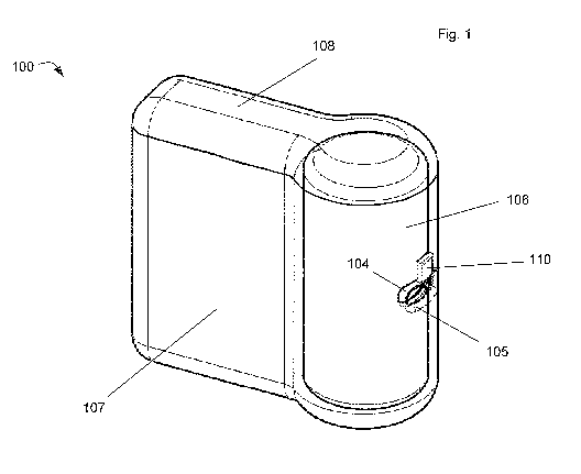
A blood glucose meter (BGM) 100 is shown in a perspective view in Figure 1.
Figure 2
shows a cross-sectional view through the front face of the BGM 100. The BGM
100 has
a generally flat base, is approximately as tall as it is long, and its width
is approximately
On one side face of the BGM may be provided with user inputs (not shown).
These may
take the form of push-switches or touch sensitive transducers, for instance. A
display
(not shown) may also be provided on the side of the BGM. This may take any
suitable
Located at a front face of the BGM 100 is an aperture 105. The aperture 105 is
located
CA 02845254 2014-02-13
WO 2013/041704
PCT/EP2012/068698
rectangular in shape. Its edges may be bevelled, so as to guide a user's digit
into a
specific location.
A corresponding aperture 104 is provided in the side of a cartridge 106. The
cartridge
5 has a generally cylindrical form, and is arranged vertically in the BGM
100.
In particular, the BGM includes an outer housing part 107. The outer housing
part 107
forms at least the base, front, rear and side faces of the BGM 100. A lid part
108 may
be attached to the first housing part 107. The lid part 108 may form the top
surface of
the BGM 100. The lid part 108 may be removed to allow access to the inside of
the
BGM 100. Alternatively, the lid part 108 may cover a smaller area above the
cartridge
106 at the front face of the BGM 100. In this arrangement the outer housing
107 may
extend to form the remainder of the upper surface and the lid part 108 may be
removed
to allow access to the cartridge 106 only.
A slidable or pivotable door (not shown) in the housing part 107 of the BGM
100 may
hide the aperture 105 when the BGM is not in use such as to prevent the
ingress of dirt
and other potential contaminants into the aperture 105. This door may also act
as or
include an humidity seal to prevent reaction of enzymes contained within the
testing
strips from reacting with moisture in the air.
The cartridge 106 has a generally cylindrical form, and is arranged
vertically. The
cartridge 106 has a length that is between 3 or 4 times its diameter.
The lid part 108 is configured such that when it is in place on the BGM the
cartridge 106
is retained by mechanical interaction between these components but is
removable by a
user. The exact way in which the lid part 108 is released from the BGM 100 is
not
critical and is not described in detail here.
The lid part 108 is configured such that when removed from the BGM 100 the
cartridge
106 is able to be extracted from the BGM by moving it vertically along its
axis. A
replacement cartridge can then be introduced into the BGM 100 in the opposite
manner
to which the old cartridge 106 was removed. Once located at the bottom of the
cavity in
the BGM, the new cartridge 106 is surrounded by the first housing part 107.
Once the lid
part 108 has been replaced, to the position shown in Figure 1, the cartridge
106 is
CA 02845254 2014-02-13
WO 2013/041704
PCT/EP2012/068698
6
retained in place by the action of the first housing part 107 and the lid part
108. The
cartridge 106 and the cavity which receives the cartridge may have a keying
feature,
such as a protrusion and a groove, a non circular diameter, or the like. Thus,
when the
cartridge 106 is fully inserted, the cartridge aperture 104 is aligned with
the aperture 105
in the outer housing 107.
A camera 110 is also shown in both of Figures 1 and 2. In the embodiment
depicted, the
camera 110 is disposed on the inner surface of the outer housing 107,
immediately
adjacent the aperture 105. The thickness of the wall of the outer housing 107
in this
region may be reduced in order to accommodate the camera 110. Alternatively,
the
camera 110 may be mounted within a recess in the outer housing 107. In another
alternative arrangement, the camera 110 may protrude from the outer housing
107. In
any case, the camera 110 is arranged such that some or all of the aperture 105
is within
the field of view of the camera 110.
In the embodiments depicted, then lens part of the camera 110 is immediately
adjacent
to the aperture 105. The camera may be focussed on a point substantially in
the centre
of the aperture 105, or it may have a fixed focus point directed to the centre
of the
aperture 105. The camera lens is positioned with respect to the aperture 105
in the
outer housing 107 such that the user's body part does not cover or otherwise
obscure
the lens when pressed against the aperture 105.
In some alternative embodiments, the camera 110 may be disposed on or within
the
body of the cartridge 106. The thickness of a wall of cartridge 106 in this
region may be
reduced in order to accommodate the camera 110. Alternatively, the camera 110
may
be mounted within a recess in the cartridge wall. If the camera 110 is mounted
on or
within the cartridge 106, the camera may be angled towards the aperture 105
however
the skilled person will be aware of other means of ensuring that the field of
view of the
camera 110 encompasses the aperture 105, such as by the use of lenses or
mirrors. If
the camera 110 is disposed on or within the cartridge 106, the cartridge also
has
contacts which communicate with corresponding contacts within the main body of
the
BGM 100 in order to supply power and signals to the camera 110 and to receive
image
data from the camera 110.
CA 02845254 2014-02-13
WO 2013/041704
PCT/EP2012/068698
7
The camera 110 may be in communication with a processor, described in more
detail
below with reference to Figure 3. The camera 110 is configured to capture
images and
relay them to the processor. The camera 110 may also comprise a light source
to
illuminate the aperture 105. Alternatively the light source may be a separate
module
provided within the body of the BGM 100.
Figure 3 shows a subsystem 200 of the blood glucose meter 100. The subsystem
200
includes the cartridge 106, a drive wheel 201 and a drive belt 202.
In Figure 3, the cartridge 106 is shown as having a hollow cylindrical housing
part 203.
An aperture 104 is formed in the hollow cylindrical housing part 203. Coaxial
with the
hollow cylindrical part 203 is an elongate shaft 204, only the top part of
which is
illustrated in Figure 3. The length of the shaft 204 is such that its
uppermost end is
slightly below the uppermost end of the hollow cylindrical housing part 203.
As will be
described below, the shaft 204 is mechanically coupled with the drive belt 202
so as to
be rotatable by rotation of the drive wheel 201.
Formed with the inner surface of the hollow cylindrical housing part 203 are
first and
second guide members 205, 206. In Figure 3, it can be seen that the first and
second
guide members 205, 206 have a generally triangular cross section. One side of
the
triangular cross section of the first and second guide members 205, 206 is
integral with
the inner surface of the hollow cylindrical housing part 203, with a point of
the triangular
cross section extending towards the centre of the cartridge 106. A part of the
length of
the first guide member 205 is visible in Figure 3, but only the uppermost
surface of the
second guide member 206 is visible in that figure.
Figure 4 shows some electronic components that form parts of the blood glucose
meter
100. These components are provided within the housing 107.
A bus 211 is arranged to connect a number of components including a
microprocessor
212, random access memory (RAM) 213, read-only memory (ROM) 214, a camera 110,
a light source 216, an analyte interface circuit 219 and a motor interface
217. All of
these components are powered by a battery 218, which may take any suitable
form.
CA 02845254 2014-02-13
WO 2013/041704
PCT/EP2012/068698
8
Stored in the ROM 214 is software and firmware that governs operation of the
blood
glucose meter 100. The software/firmware is executed by the microprocessor 212
using
the RAM 213. The software/firmware stored in the ROM 214 is operable to
operate the
blood glucose meter 100 such as to allow control by a user through keys or
input
devices (if present). The software/firmware is also operable to control
operation of the
camera 110 and light source 116, to receive image data from the camera 110 and
to
process the received image data. A blood glucose measurement and other
information
may be provided on a display (if present) at suitable times by operation of
the
software/firmware and the microprocessor 212. The BGM 100 may also contain a
display driver and user inputs interface (not shown).
The camera 110 may comprise any image sensing technology such as a charge-
coupled device (CCD) or an active pixel sensor such as a complementary metal
oxide
semiconductor (CMOS) device. The camera 100 may have a fixed focal point or
the
focussing of the camera 110 may be adjustable. The camera focus may be
adjusted
automatically under control of the microprocessor 212. The light source 216
may
comprise any suitable technology, such as an incandescent bulb, fluorescent
bulb or
LED. The light source may produce white light or coloured light. The light
source 216
may be integral with the camera 110 and may be located immediately adjacent to
the
camera lens. Alternatively, the light source 216 may be a separate component
located
adjacent to the camera lens. In other alternatives, the light source 216 may
be located
on the opposite side of the aperture 105 from the camera 110. In embodiments
where
the camera is disposed on or within the housing of the cartridge 106, the
light source
may also be disposed on the cartridge 106 or may be disposed on the outer
housing
107. In any case, the light source 216 is directed at or across the aperture
105 so as to
illuminate a user's body part placed against the aperture 105. As the
cartridge 106 is
disposable, the camera 110 and/or light source 216, if disposed on the
cartridge 106,
are also disposable.
In further embodiments the BGM 100 may comprise multiple light sources 216 in
order
to provide greater or more uniform illumination of the user's body part.
The motor interface 217 allows the microprocessor 212, according to the
software/firmware stored in the ROM 214, to control the motor that is coupled
to the
drive wheel 201, and any other motors that are included in the blood glucose
meter 100.
CA 02845254 2014-02-13
WO 2013/041704
PCT/EP2012/068698
9
The analyte interface circuit 219 is operable to provide electrical signals
with certain
voltages to electrical contact terminals 401 (described in more detail with
respect to
Figure 11), and thus via contact pads 318 to an analyte measuring part 316
(described
in more detail with respect to Figure 6). The analyte interface circuit 219 is
also
operable to measure parameters of signals such as to allow the microprocessor
212 to
measure a blood glucose level of a blood sample.
Referring now to Figures 5 to 10, an arrangement embodying aspects of the
invention is
shown.
As shown in Figure 5, the hollow cylindrical housing part 203 is provided with
the
aperture 104 and a slit aperture 400 (described in greater detail with respect
to Figure
11). The shaft 204 is supported centrally within the hollow cylindrical
housing part 203 of
the cartridge 106.
A plunger arrangement 500 comprising a plunging arm 501 and a plunging head
502 is
provided adjacent a plunging aperture (not shown) in the hollow cylindrical
housing part
203. The plunging aperture (not shown) is located next to the slit aperture
400. The
plunging aperture (not shown) is located directly opposite to the aperture
104. The
plunger aperture and the slit aperture 400 may be combined to form a single
aperture.
The plunger aperture is configured to allow the plunging head 502 to be forced
by the
plunging arm 501 to a position internal to the hollow cylindrical housing part
203.
Within the cartridge 106 are plural test disc members, one of which is shown
as 505 in
Figure 6. The test disc member 505 has a generally circular shape, although on
one
side a notch 301 is formed and on another side a cutaway portion 302 is
provided.
The test disc member 505 includes an uppermost surface 303, a lowermost
surface 304
and a disc edge 305. The diameter of the test disc member 505 is between 15
and 25
millimetres, for instance 20 millimetres. The thickness of the disc, which is
equal to the
height of the disc edge 305, is between 0.5 millimetres and 1 millimetre.
A hole 306 is formed at the centre of the test disc member 208. This hole 306
allows the
test disc member 505 to be mounted on the shaft 204.
CA 02845254 2014-02-13
WO 2013/041704
PCT/EP2012/068698
The underside of each test disc member 505 may be provided with a spacer
member.
The spacer member may comprise a slice of a hollow cylinder, for example. The
height
of the spacer member may be between 0.5 and 1 millimetre. When plural test
disc
members are stacked together, the spacer member provides separation between
the
5 upper surface 303 of one test disc member and the lower surface 304 of
the test disc
member that is directly above it. The separation is determined by the height
of the
spacer member.
A lancet 506 is provided extending from the disc edge 305 in the cutaway
portion 302. A
10 first end of the lancet 506 is embedded within the material of the test
disc member 505,
and a second end 506a is provided with a sharp point and extends outwardly. In
particular, the lancet 506 extends in a radial direction with respect to the
centre of the
test disc member 505. The second end 506a of the lancet 506 is located at or
just
outside a circumference 311 of the test disc member 505. The circumference 311
is
shown as a dotted line in Figure 6 because it is virtual, instead of tangible.
The lancet
506 extends from the disc edge 305 at a first position 312 on the disc edge.
The first
position 312 is close to a second position 313 at which the cutaway portion
302 starts.
The cutaway portion 302 ends at a third position 314. Between the second and
third
positions 313, 314 opposite to the cutaway portion 302, the disc edge 305
generally
takes the form of a circle, although the notch 301 interrupts that circle.
Located next to the third position 314 is a blood collection part 315. This
may take any
suitable form. For instance, it may comprise a laminated material. The blood
collection
part 315 has the function of drawing blood that is in contact with the disc
edge 305 at
the third position into the test disc member 505 to an blood analyte measuring
part 316,
that adjoins the blood collection part 315, for example a part containing an
enzyme for
blood glucose measuring, or the like. Blood may be drawn through capillary
action. The
analyte measuring part 316 includes an enzyme that reacts chemically with
blood in
such a way that blood glucose level can be measured. The analyte measuring
part 316
is connected to first to third contact pads 318 by first to third conductive
tracks 317. The
contact pads 318 and the conductive tracks 317 are formed on the upper surface
303 of
the test disc member 505. The analyte measuring part also is formed on the
upper
surface 303 of the test disc member 208. Some or all of the conductive tracks
317, the
contact pads 318 and the analyte measuring part 316 may be printed onto the
upper
surface 303 of the test disc member 208.
CA 02845254 2014-02-13
WO 2013/041704
PCT/EP2012/068698
11
Although in the figures three conductive tracks 317 and three conductive pads
318 are
shown, it will be appreciated that this is merely illustrative. There may
instead be only
two conductive tracks 317 and two conductive pads 318, or alternatively there
may be
more than three conductive tracks and conductive pads.
The majority of the test disc member 505 may be substantially rigid.
Alternatively, the
majority of the test disc member 505 may have some degree of compressibility.
However, an annular centre portion 508 is comprised of an elastically
deformable
material. In particular, the annular centre position 508 is deformable in the
presence of
an externally applied force. This means that the test disc member 505 can be
displaced
relative to the shaft 204, as will be described in more detail below. The
material used to
form the annular centre portion 508 may take any suitable form, and for
instance may
be a rubberised plastic.
The plural test disc members are biased in an upwards direction by bias means
(not
shown), which may be a spring. However, the test disc members are prevented
from
moving upwards within the cartridge 106 by virtue of the contact between the
upper
surface 303 of the uppermost test member and the lowermost end of the first
guide
member 205. Only when the notch 301 in the test disc member is aligned with
the
second guide member 206 is the test disc member free to move upwards.
In Figure 7, the hollow cylindrical housing part 203 is omitted from the
figure. In Figure 7,
the test disc member 505 is shown as having been rotated to a position at
which the
lancet 506 is coincident with the aperture 104. In use, a user places a body
part
(hereafter the part will be referred to as a user's digit, for the sake of
convenience)
against the aperture 105 in the outer housing 107. It can be seen that the
plunging head
502 is aligned with the test disc member 505 such that movement of the plunger
arrangement 500 along the longitudinal axis of the plunging arm 501 causes the
plunging head to contact the test disc member 505 and apply force to it. Since
the
longitudinal axis of the plunging arm 501 is radial with respect to the shaft
204, the force
applied by the plunger arrangement is directed towards the shaft 204.
In Figure 8, the arrangement is shown after a force has been applied to the
plunger
arrangement 500 so as to displace it by a predetermined amount. Here, the
plunging
CA 02845254 2014-02-13
WO 2013/041704
PCT/EP2012/068698
12
head 502 has contacted the test disc member 505 on the opposite side of the
test disc
member to the lancet 506. The annular centre portion 508 has become compressed
on
the side closest to the plunger arrangement 500 such as to allow the whole of
the test
disc member 505 to be displaced in the direction of the force supplied by the
plunger
arrangement 500. The test disc member 505 remains horizontal by virtue of the
spacer
members 308.
Displacement of the test disc member 505 in the direction of the force
supplied by the
plunger arrangement 500 has resulted in displacement of the lancet 506 in a
radial
direction away from the shaft 204. In this position, the lancet 506 extends
through the
cartridge aperture 104 and the aperture 105 in the outer housing 107 such that
the
lancet penetrates the skin of the user's digit. This produces a puncture in
the skin of the
digit, through which blood can escape. This position is shown in Figure 8.
Removal of
the force by the plunger arrangement 500 allows the annular centre portion 508
to
return to its original form, through elastic reformation. After the plunger
arrangement
500 has been fully retracted, the arrangement again has the form shown in
Figure 7.
Here, the test disc member 505 is in its original position and the lancet 506
is retracted
from the user's digit. It will be appreciated that it is the elasticity of the
annular centre
portion 508 of the test disc member 505 that allows the test disc member 505
to return
to this position once the force applied through the plunger arrangement 500 is
removed.
Referring again to Figure 4, once the lancing of the user's digit has been
performed, the
camera 110 and light source 216 may be activated under control of the
microprocessor
212. The field of view of the camera 110 encompasses the aperture 105 in the
outer
housing 107 in which the user has located their digit. The user's digit may
obscure the
majority of the light which would otherwise enter through the aperture 105.
The light
source 216 provides illumination of the user's digit to allow the camera 110
to capture
clear images.
The microprocessor 212 is configured to receive image data from the camera 110
and
to process the image data. The microprocessor 212 may process the image data
to
determine the quantity of blood which has been expelled from the puncture
wound
made by the lancing. The blood generally exits the puncture wound and forms a
substantially circular droplet on the surface of the user's digit. The
microprocessor 212
may use known information relating to the size of the aperture 105, distance
of the
CA 02845254 2014-02-13
WO 2013/041704
PCT/EP2012/068698
13
camera 110 from the centre of the aperture 105 and the focal properties of the
camera
110 to calculate the dimensions of the blood droplet from the received images
and
hence estimate the quantity of blood present. Alternatively or additionally,
the
microprocessor 212 may perform a colour analysis of the received images to
determine
when sufficient 'red blood' is present. It can be important that a sufficient
blood sample
is collected for analysis as the accuracy of the blood glucose measurement may
be
negatively affected if the blood sample collected is too small. The
microprocessor 212
may make regular (e.g. one per second or fraction of a second) calculations of
the
quantity of blood expressed from the wound. When the microprocessor 212
detects that
a predetermined quantity of blood has been expressed, it may control the motor
via
motor interface 217 to rotate the test disc member 505 to the blood collection
position.
The test disc member 505 can be rotated by the drive wheel 201 and the drive
belt 202
such that the blood collection part 315 is aligned with the cartridge aperture
104, which
position is shown in Figure 9. This may be done immediately after the lancing
or only
after the microprocessor 212 has determined that a predetermined quantity of
blood has
been expressed. The plunger arrangement 500 may then be activated again so as
to
displace the test disc member 505 radially. This causes the blood collecting
portion 315
to be moved into the cartridge aperture 104. Due to the elongate shape of the
cartridge
aperture 104, the test disc member 505 is able to protrude from the cartridge
106 such
that the blood collection part 315 enters the aperture 105 in the outer
housing 107. This
brings the blood collection part 315 into contact or close proximity with the
user's digit,
allowing a blood sample to be absorbed.
In some embodiments, a second cutaway portion may be provided on the other
side of
the blood collection part 315 from the first cutaway portion 302. The
resulting shape of
the test disc member 505 in this region allows the blood collection part 315
to protrude
further from the cartridge 106. Alternatively or in addition, the test disc
member 505 may
have a degree of compressibility and the plunger arrangement 500 may be
configured
to exert sufficient force to compress the test disc member 505 against the
inner wall of
the cartridge 106 in order to increase the degree of radial displacement of
the test disc
member 505.
In any case the plunger arrangement 500 is configured to displace the test
disc member
radially and to maintain it in this position for a predetermined length of
time (e.g. 5 to 20
CA 02845254 2014-02-13
WO 2013/041704
PCT/EP2012/068698
14
seconds) sufficient for a blood sample to be absorbed into the blood
collection part 315.
The blood sample is drawn through the blood collection part 315 into the blood
analyte
measuring part 316. After the predetermined length of time, force is removed
by the
plunger arrangement 500 allowing the annular centre portion 508 to return to
its original
form, through elastic reformation.
A measuring circuit connected to the analyte measuring part 316 by way of the
conductive tracks 317 and the contact pads 318 then is able to determine a
blood
glucose level of the user. After a measurement of blood glucose level is
taken, the test
disc member 505 is rotated further anticlockwise so that the second guide
member 206
is aligned with the notch 301. At this point the first guide member 205 is
coincident with
the cutaway portion 302 and thus the test disc member 505 is allowed to move
upwards
within the cartridge 106. As a result, the test disc member 509 that is
immediately below
the first test disc member 505 also moves upwards within the cartridge 106 and
is
provided to be coincident with the aperture 104, the slit aperture 400 and the
plunger
aperture (not shown). Subsequent application of a plunging force by the
plunger
arrangement 500 causes a lancet 506 of the second test disc member 509 to be
forced
out of the aperture 104, as is shown in Figure 10. The process can be repeated
for
other test disc members included in the cartridge 106.
By providing a stack of test disc members within the cartridge 106 and by
providing a
suitable physical arrangement, a cartridge 106 can be used for multiple tests.
When the
cartridge 106 is new, the test disc members are located in the bottom half of
the
cartridge 106, with the uppermost test disc member being aligned with the
aperture 104.
As test disc members are used, the stack of test disc members moves upwards in
the
cartridge. When the last test disc member is used, the cartridge can be said
to be spent.
At this stage, all of the test disc members are located in the uppermost
portion of the
cartridge 106.
It will be appreciated that the number of test disc members that can be
accommodated
within the cartridge 106, and thus the number of tests that can be provided by
a
cartridge 106, is a factor of the height of the cartridge 106 and the
separation between
corresponding parts (e.g. the upper surfaces) of adjacent test disc members. A
taller
cartridge and/or a reduced separation of test disc members increases the
number of
tests that can be performed using a single cartridge 106.
CA 02845254 2014-02-13
WO 2013/041704
PCT/EP2012/068698
An advantage of the arrangement shown in Figures 5 to 10 is that a rotational
arrangement can be used whilst allowing the lancet 506 to penetrate a user's
skin in a
longitudinal direction with respect to the lancet 506. Another advantage is
that puncture
5 can occur at any desired location, for instance on the end of the user's
digit.
The convenient size of the BGM 100 and the automation of the lancing and blood
collecting steps means that the device 100 can be operated with one hand by a
user.
10 Another advantage is that the arrangement can allow the penetration
depth of the lancet
506 to be easily predictable. Furthermore, it allows the penetration or
puncturing depth
to be adjustable. In particular, the adjustment of the penetration depth can
be achieved
by a mechanical arrangement that limits movement of the plunger arrangement
towards
the shaft 204. Alternatively, it can be achieved in an electro-mechanical
manner by
15 measuring the location or displacement of some part of the mechanism and
ceasing
applying an energising voltage to a solenoid or other transducer that is used
to affect
movement of the plunger arrangement 500. The penetration depth may be
specified by
a user. The depth may be specified by a user and may be achieved through
software or
firmware control of rotation of the shaft 204. The value defining the depth
may be stored
in memory. Penetration depth control is important to many users since lancet
penetration usually is painful and since penetration depth control allows
users some
control over their experience. The device may also allow the user to set and
adjust the
penetration speed. The speed of the lancing may also affect the amount of pain
felt by a
user.
In Figure lithe hollow cylindrical housing part 203 is shown with the aperture
104 and
the shaft 204 located as described above. A slit aperture 400 is provided in
the hollow
cylindrical housing part 203. The slit aperture 400 is located at
substantially the same
height as the aperture 104. However, the slit aperture 400 is located on a
side of the
hollow cylindrical housing part 203 that is substantially opposite the
aperture 104.
The slit aperture 400 is not visible when the cartridge 106 is in place within
the BGM
100.
CA 02845254 2014-02-13
WO 2013/041704
PCT/EP2012/068698
16
Adjacent to the slit aperture 400 is located a swing arm 401. The swing arm
401 is
rotatable about a spindle 402. The spindle 402 has an axis that is parallel to
the axis of
the shaft 204. The axis of the spindle 402 is located above the drive belt
202. A
connecting arm (not visible) connects the spindle 402 to the swing arm 401. In
this
example, the connecting arm is connected to the swing arm 401 by a vertical
connector
404. The vertical connector 404 allows the spindle 402 on which the connecting
arm is
mounted to be located at a different vertical position to the swing arm 401.
The spindle
402, the connecting arm and the vertical connector 404 are arranged such that
when
the connecting arm is rotated on the axis of the spindle 402 the swing arm 401
is moved
towards the shaft. The movement of the swing arm 401 is substantially radial
with
respect to the shaft 204.
Mounted on the swing arm 401 are first to third electrical contact terminals
405. Each
includes a generally horizontal arm and a depending contact head. The
electrical
contact terminals 405 are made of a resilient conductive material, for
instance metal.
The depending contact heads are angled at their ends furthest from the swing
arm 401.
In one position, shown in Figure 11, the electrical contact terminals 405 are
supported
by the swing arm 401 such that the dependent contact heads are located within
the slit
aperture 400 or alternatively outside of the hollow cylindrical housing part
203. After the
test disc member 505 has been rotated such that the blood collection part 315
is
coincident with the aperture 104, and the blood sample has been collected via
action of
the plunger arrangement 500, the contact pads 318 are coincident/aligned with
the slit
aperture 400. As the test disc member 505 is held in this position, the
connecting arm is
caused to rotate around the axis of the spindle 402 such that the swing arm
401 moves
towards the shaft 204. The arrangement is such that the depending contact
heads of
the electrical contact terminals 405, but not the horizontal arms, come into
contact with
the contact pads 318 as the electrical contact terminals 405 move into the
volume
above the upper surface 303 of the test disc member 505. The resilient
properties of the
electrical contact terminals 405 causes the electrical contact terminals to be
forced
against the contact pads 318. As such, an electrical connection is provided
between the
horizontal arms of the electrical contact terminals 405 and the analyte
measuring part
316. Electronic measuring means (not shown) connected to the electrical
contact
terminals 405 operate to pass a voltage through the contact terminals 405 and
the
analyte measuring part 316 and to take measurements of electrical parameters,
from
CA 02845254 2014-02-13
WO 2013/041704
PCT/EP2012/068698
17
which a measurement of an analyte concentration level, for example a blood
glucose
level, can be determined.
The connecting arm is controlled to remain in this position for a
predetermined time or
alternatively until it is detected that a blood glucose level measurement has
been made,
after which the connecting arm is caused to rotate around the shaft 402 so as
to remove
the electrical contact terminals 405 from the position above the upper surface
of the test
disc member 505. Once the electrical contact terminals 405 have been
retracted, the
test disc member 505 is rotated anticlockwise so as to allow the test disc
members to
move upwards on the shaft 204.
It will be appreciated that the maximum permissible height dimension of the
electrical
contact terminals 405 is determined by the height of the spacer member which
separates adjacent test disc members. A thicker spacer member allows larger
electrical
contact terminals 405 to be used. However, this is at the expense of an
increase in
separation between adjacent test disc members, and thus a reduced capacity for
the
cartridge 106. The use of electrical contact terminals 405 including a
horizontal arm and
a depending contact head allows the height dimension of the electrical contact
terminals
to be minimised whilst allowing good electrical contact between the electrical
contact
terminals and the contact pads 318 and also allowing the electrical contact
terminals
405 to operate correctly over a sufficient number of cycles.
Operation of the blood glucose meter 100 will now be described with reference
to the
flowchart of Figure 12.
Operation starts at step Ti. At step T2, the user locates their digit in or
against the
aperture 105. At step T3, the user initiates blood glucose measurement. This
may
involve the user operating an input key or switch (not shown) on the device
100. This is
detected by the microprocessor 212. The software/firmware stored in the ROM
214
uses the input to call a function or to execute a software module. The
software/firmware
stored in the ROM 214 then causes the microprocessor 212 to issue a command to
a
motor attached to the drive wheel 201 through the motor interface 217 to
rotate the
shaft 204 in a clockwise direction. The software/firmware controls the extent
of the
rotation.
CA 02845254 2014-02-13
WO 2013/041704
PCT/EP2012/068698
18
Following step T3, the microprocessor 212, under control of the
software/firmware
stored in the ROM 214, causes the shaft 204 to be rotated by a motor through
the motor
interface 217 and to cease rotation once the lancet 506 is aligned with the
apertures
104, 105, and thus is aligned with the user's digit, at step T4A. At step T4B,
the
microprocessor 212, under control of the software/firmware stored in the ROM
214,
causes actuation of the plunger arrangement 500, through the motor interface
217. The
control of the actuation of the plunger is such as to limit the extent of
movement of the
lancet 506 to a predetermined extent. The predetermined extent is set by a
user prior to
the blood glucose measurement. In effect, the user can set a lancing depth,
which is
stored in a suitable way in the ROM 214 by action of the microprocessor 212,
operating
under control of the software/firmware stored in the ROM 214.
When the maximum extent of plunger actuation has been reached at step T4B, at
step
T4C the plunger arrangement 500 is deactuated by the microprocessor 212, under
control of the software/firmware stored in the ROM 214, and lancing ceases. At
this step,
the test disc member returns to its original position by action of the
elasticity of the
annular centre portion 508 of the test disc member 508.
At step T5 the camera 110 and light source 216 are activated. The light source
216
illuminates the area of lancing. The camera 110 captures and relays images of
the area
of lancing back to the microprocessor 212. In some embodiments, one or both of
the
camera 110 and light source 216 may be activated prior to lancing. The
microprocessor
212 then performs determinations of the quantity of blood which has been
expressed
from the wound. At step T6, the microprocessor 212 determines that a
sufficient
quantity of blood is present of the surface of the user's digit for an
accurate blood
glucose analysis to be performed.
The software/firmware stored in the ROM 214 then causes the microprocessor 212
to
control the motor to rotate the shaft 204 in the opposite direction, at step
T7A.
At step T7B, the software/firmware causes the microprocessor 212 to control
the motor
to cease rotation when the shaft 214 is such that the blood collection part
315 is
coincident with the apertures 104, 105, and thus the user's digit.
At step T8A, the microprocessor 212, under control of the software/firmware
stored in
the ROM 214, causes actuation of the plunger arrangement 500, through the
motor
CA 02845254 2014-02-13
WO 2013/041704
PCT/EP2012/068698
19
interface 217. The plunger is maintained at this position for a predetermined
length of
time to allow the blood to be absorbed into the blood collection part 315. At
step T8B
the plunger arrangement 500 is deactuated by the microprocessor 212, under
control of
the software/firmware stored in the ROM 214. At this step, the test disc
member returns
to its original position by action of the elasticity of the annular centre
portion 508 of the
test disc member 508
At step T9, the software/firmware controls a motor such as to cause the swing
arm 401
to be rotated towards the shaft 204. The software/firmware stored in the ROM
214 is
such that the microprocessor 212 causes only the required amount of travel of
the
swing arm 401. At this point, the analyte interface circuit 219 is coupled
directly to the
blood analyte measuring part 316, which by action of the blood collection part
315 has
been provided with blood from the user's digit. At step T10, analyte
measurement is
performed. This involves the analyte interface circuit 219 providing voltages
to the
electrical connection contacts 318, and thus to the blood analyte measuring
part 316,
and measuring parameters of resulting signals. The measured parameters,
particularly
voltage parameters, are used by the software/firmware stored in the ROM 214,
as
executed by the processor 212, to calculate a blood glucose measurement level
of the
user. The blood glucose measurement may then be displayed on a display. At
step T11,
the swing arm is caused to be removed by action of the microprocessor 212,
under
control of the software stored in the ROM 214, the motor interface 217 and the
motor
(not shown).
At step T12, the software/firmware results in the microprocessor 212
controlling the
drive disc 201 to rotate anticlockwise. Rotation continues until the notch 301
on the test
disc member is coincident with the guide 206. At step T13, the test disc
member rises
up the cartridge 106. In the case where biasing of the test discs up the
cartridge 106 is
provided by a bias means, for instance a spring, step T13 requires no action
on part of
the software/firmware and microprocessor 212, although there may be a pause
before
the next step. In embodiments where movement of the test disc members along
the
shaft 204 occurs through driving action, step T13 involves the microprocessor
212,
under control of the software/firmware stored in the ROM 214, controlling a
motor
through the motor interface 217. Subsequently, at step T14, the microprocessor
212,
under control of the software/firmware stored in the ROM 214, causes the shaft
204 to
rotate again in a clockwise direction in order to engage with the next test
disc member
CA 02845254 2014-02-13
WO 2013/041704
PCT/EP2012/068698
in the cartridge 106. At this stage, the test disc members rise up the
cartridge 106
slightly.
The operation ends at step T15.
5
Various modifications and alternative features can be used in connection with
the
above-described embodiments. Some alternatives now follow.
Although the test disc member 505 has been described as having a lancet 506
which
10 protrudes radially from the disc, the lancet 506 may instead protrude at
an angle with
respect to a radial line. Additionally, although the lancing has been
described as
occurring radially, where the lancet is disposed at an angle, the lancing may
instead
occur by rotational movement of the test disc member 505. Although the lancet
506 has
been illustrated as straight, it may instead be curved for a portion or all of
its length.
In addition to analysing images received from the camera 110 to determine
whether a
sufficient quantity of blood is present, the microprocessor 212 may also be
configured to
analyse images of the user's digit when placed against the aperture 105. From
this
analysis, the microprocessor 212 is able to determine whether the user's digit
is present
in the aperture 105. This allows the process of lancing and blood collection
to begin
automatically. This analysis may also allow the microprocessor 212 to
determine
whether the user's digit is located in the optimal position for lancing. In
order to produce
a good contrast between the user's digit, which may have a one of a range of
skin tones
depending on the user, and the inner surfaces of the BGM 100, the inside of
the BGM
may be painted or otherwise coloured green. This contrast allows the
microprocessor
212 to determine the silhouette of the user's digit and/or to subtract the
background of
the image.
In some further embodiments, the BGM 100 may additionally comprise a speaker
and
may be configured to issue pre-recorded voice commands to a user to encourage
correct placement of their digit in the aperture 105. For example, when the
device 100 is
ready to perform a blood collection operation a command "present finger to
sampling
area" may be issued. Once presented, if the user's digit is not protruding far
enough into
the aperture 105, as determined by the microprocessor 212 from the received
images, a
command such as "press harder" may be issued. After lancing and when the blood
CA 02845254 2014-02-13
WO 2013/041704
PCT/EP2012/068698
21
collection part 315 is presented to the user's digit, a command such as
"please wait"
may be issued.
The camera 110 may additionally be used as a blood analysis tool. For
instance, the
colour of the blood expressed from the lancet puncture may be analysed to
determine
haemoglobin and/or oxygenation levels. In particular the brightness and hue of
the
blood are indicative of these properties. These properties may be measured by
the
microprocessor 212 directly from the images received from the camera 110.
It has been described that the penetration depth of the lancet 506 is settable
by the user.
However, the actual penetration depth may vary depending on the precise
position of
the user's digit. With a suitable field of view, the camera 110 is able to
capture images
of the lancet 506 and determine its position. Positional indicators may be
provided on
the lancet 506 and/or on the test disc member 505 to aid in this
determination. The
microprocessor 212 may use these "live" images of the position of the lancet
506 to
control and adjust the movement of the lancet (via the plunger arrangement
500) and
hence the resulting penetration depth.
Instead of the blood collection part 315 being located next to the third
position 314, i.e.
bounding only the part of the disc edge 305 that is purely circumferential,
the blood
collection part could instead be located on the disc edge 305 at the junction
between
the cutaway portion 302 and the circumferential portion. The blood collection
315 part in
this instance may extend for between 0.5 mm and 2 mm along the disc edge 305
at the
cutaway portion 302. The blood collection 315 part in this instance may also
extend for
between 0.5 mm and 2 mm along the disc edge 305 at the circumferential part.
Alternatively or additionally, the analyte measuring part 316 may be
sandwiched
between two layers of wicking material, the wicking material causing the blood
to be
drawn through the analyte measuring part 316.
Although in the above the shaft 204 is said to be driven by a drive wheel 201
that is
coupled to the shaft 204 by a drive belt 202, the drive may instead be direct
(i.e. the
drive mechanism is coupled directly to the shaft 204), or connection may be
made by a
notched belt, a vee belt, or by a direct gear mechanism. Instead of an
electric motor, a
clockwork drive could be used. A clockwork drive mechanism has a number of
CA 02845254 2014-02-13
WO 2013/041704
PCT/EP2012/068698
22
advantages, particularly where access to batteries or battery chargers or
electricity
supplies are limited. In the embodiments in which a clockwork mechanism is
used, the
user can be sure that the BGM 100 will not cease operating because of drained
batteries. A clockwork mechanism may be particularly suited to developing
countries
and emerging markets.
In embodiments in which an electrical motor is used to drive the shaft 204,
preferably
control is exerted over the motor by software. In this way, the speed of
rotation can
easily be controlled. Additionally, the extent of rotation can more easily be
controlled.
The motor may be a stepper motor.
Alternatively, a mechanical drive arrangement may be present, for instance
using a
lever or other device for manual actuation. A suitable mechanism may be one
similar to
those previously used in SLR cameras.
The swing arm 401 may be actuated in any suitable way. For instance, it may be
driven
by the same motor or mechanism as the shaft 204. Alternatively, it may be
driven by a
separate motor. In either case, the rotation of the swing arm 404 may be
affected by a
cam mechanism, or by a pin and slot (track path) mechanism. In the event of an
electric
motor being used, the motor preferably is software driven. The motor
preferably is a
stepper motor.
The mechanical arrangement may include a mechanism by which a bias means, for
instance a mechanical compression spring, is biased and then released in order
to push
the electrical contact terminals 405 into place. The terminals 405 can then be
retracted
by the swing arm 401 using a rotating motion. The overall mechanism can be
termed a
latch type trigger mechanism.
Instead of a swing arm 401 being used to rotate the electrical contact
terminals 405 into
place, the contact pads 318 may instead be located on the disc edge 305,
allowing the
use of fixed electrical contact terminals 405. The electrical contact
terminals may
include a brush or other deformable feature such that the test disc members
can move
whilst in contact with the electrical contact terminals without damage
occurring to any of
the components. Similar arrangements are used in brushed DC motors. In this
case the
CA 02845254 2014-02-13
WO 2013/041704
PCT/EP2012/068698
23
electrical contact terminals 405 could be flexible finger contacts that rest
on the
periphery of the test disc members in order to contact the contact pads 308.
Alternatively, instead of a swing arm 401, a mechanism may be used to affect
longitudinal movement of the electrical contact terminals 405 into place to
contact the
contact pads 318.
The conductive tracks 317 and the contact pads 318 may be formed by leadframe.
Alternatively, overmoulding may be used. Alternatively, printed circuit board
(PCB)
printing may be used.
Optionally, each of the test disc members is separated from adjacent test disc
members
by a membrane (not shown in the drawings). In this case, the membrane
preferably fits
closely to the internal surface of the hollow cylindrical housing part 203. An
effect of the
membrane is to reduce the possibility of disc cross-contamination. Use of a
membrane
may allow the test disc members to have a reduced separation than would be the
case
without the use of a membrane.
In the above, the test disc members 505 are said to be biased upwards by a
bias means,
for instance a compression spring. Alternative mechanisms for moving the test
disc
members 505 up the cartridge may be used. For instance, a threaded lifting cam
may
be provided on the shaft 204 or alternatively on the interior surface of the
hollow
cylindrical housing part 203.
Instead of the blood collection part 315 wicking blood towards the analyte
measuring
part 316, blood may be communicated to the analyte measuring part 316 instead
through gravity.
Additionally, the test disc members 505 may include a disinfecting or cleaning
portion
that contacts the digit before lancing. This can reduce risk of infection of
the wound and
also can increase accuracy in particular by removing any glucose from the skin
(as may
occur after eating fruit etc.). In addition, some blood glucose measuring
technologies
require the first drop of blood to be removed in order to produce an accurate
result.
Additionally or alternatively, the test disc members 505 may include a
cleaning portion
that is arranged to contact the digit subsequent to the blood collection part
305. This
CA 02845254 2014-02-13
WO 2013/041704 PCT/EP2012/068698
24
can remove additional blood from the finger, and may also serve to assist
closure of the
puncture.